CJMSCanadian Journal of Medical Sonography - Sonography Canada
←
→
Page content transcription
If your browser does not render page correctly, please read the page content below
CJMS
                                                                                  Volume 12, Issue 1 2021

                                                                     Sonography          Échographie®
                                                                       Canada              Canada

                                         Canadian Journal of Medical Sonography

                          Making a Difference: Blue Nevus versus Melanoma | Ray T. Yuen
                          Rationale for an Integrative/Interactive Approach to the Diagnostic
                          Ultrasound for Transitioning Patients with Juvenile Idiopathic
                          Arthritis | Robert Dima, Priya Appea, Theresa Semalulu,
                          Assessing the Severity of Aortic Regurgitation Using 3-D Colour
                          Doppler Echocardiography: A Pictorial Essay | Babitha Thampinathan,
                          Cindy Chow

                          Publications Agreement Number 40025049
                          ISSN: 1923-0931

CJMS_1_2021_WKBK.indd 1                                                                               3/22/21 12:27 PM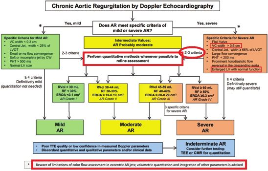
EDUCATION FOR THE
                                  MEDICAL PROFESSION
                                       SINCE 1985

                           “STUDY WHILE YOU WORK”
                                  HOME STUDY COURSES
                                           IN
                                  GENERAL ULTRASOUND
                                  BREAST SONOGRAPHY
                                    MUSCULOSKELETAL
                                   ECHOCARDIOGRAPHY
                                 VASCULAR TECHNOLOGY

                                *MOST COURSES ARE WORTH
                                 30 CME/CEU/CPD CREDITS*

                              www.burwin.com
                          1-800-322-0737 or 1-902-634-3238
                          (Atlantic Time)

                          Email: burwin@burwin.com

                                 @BurwinInstitute

CJMS_1_2021_WKBK.indd 2                                      3/22/21 12:28 PM
table of
                                                                              contents
  Sonography                           Échographie®
    Canada                               Canada
                                                                                                                            Volume 12, Issue 1 • 2021
       Vo l u me 12, I s sue 1 • 2021
            Publications Agreement Number 40025049

                       E DIT OR-I N-CHI EF
        Sheena Bhimji-Hewitt, MAppSc, DMS,
              CRGS, CRVS, RVT, RDMS
            Lori Arndt FSC, CRGS, CRVS,                              4       Message from the Editor-in-Chief
                     RDMS, RVT
                                                                     5       Message de la rédactrice en chef
                      E DIT OR I AL BOARD                                    Sheena Bhimji-Hewitt, MAppSc, DMS, CRGS, CRVS, RDMS, RVT
        The Editors: Marion Cairnduff, MASci
      Med Ultrasound, BASci Med Rad, CRGS;
      Kimberly Jozkow, MAppSc(MI)BSc, DMS,                           Peer Reviewed Case Report
       CRCS, CRVS, CRGS, RDCS, RDMS, RVS;
         Megan White, BMRSc, CRGS, CRVS
              RDMS, RVT CRGS, CRVS                                   6       Making a Difference: Blue Nevus versus Melanoma
                                                                             Ray T. Yuen, BMRSc, CRGS, RVT, RDMS, RMSKS
      The Reviewers: Ayesha Dost, BHSc, DMS,
      CRCS, RDCS; Tony Li, DMS, CRGS, CRVS;
        Wasiu Raimi dms, CRGS, CRVS, RVS;
       Anand Rattansingh, MSc, DMS, CRGS,
                                                                     Peer Reviewed Literature Review
      CRVT, RDMS, RVT; Cathy Ridsdale, CRGS,
       CRVS, RDMS, RVT; Bernie Rittau, DMS,                          12      Rationale for an Integrative/Interactive Approach to the Diagnostic
       MRT (R) CRGS, CRCS-AE, CRVS; Silvia                                   Ultrasound for Transitioning Patients with Juvenile Idiopathic Arthritis
        Straus, CRGS, RDMS; Justyna Tchorni
      CRGS, RDMS; Laura Thomas, MMedUlt,                                     Robert Dima, BMRSc, CRGS, DMS, PhD(c), Priya Appea, BMRSC, CRGS, Theresa
                    DMS, CRGS                                                Semalulu, MD
                     M A NA GI NG EDI TOR
                           Scott Bryant                              Peer Reviewed Pictorial Essay
                         AR T D I RECTOR
               S4Carlisle Publishing Services                        18      Assessing the Severity of Aortic Regurgitation Using 3-D Colour
                          ADVERTI SI NG                                      Doppler Echocardiography: A Pictorial Essay
                        John Birkby                                          Babitha Thampinathan, HBSc, RDCS, CRCS (AE), Cindy Chow, BA, RDCS, CRCS (AE)
                      (289) 238-7917
               jbirkby@dougmargroup.com
                                                                     29      Sonographers: Worthy of Recognition
              C IR C ULAT ION COORDI NATOR
                      Kelly Brooks
              kbrooks@dougmargroup.com                               30      Sonography Canada: Key Events for 2021
                          A C C O UNTI NG
                        Susan McClung                                Advertisers
                      GR OUP PUBLI SHER
                         John D. Birkby
                                                                     The Burwin Institute

        For Instructions to Authors, please visit
       https://sonographycanada.ca/about-us/
                      publications

            Return undeliverable Canadian Addresses to:

                                  The Dougmar
                                  Publishing Group Inc.
         115 King St W., Suite 220, Dundas, ON L9H 1V1

      Canadian Journal of Medical Sonography is published
      four times a year by Dougmar Publishing Group Inc., with
      offices located at 115 King Street West,Suite 220, Dundas,
      ON L9H 1V1.
                               •••••
      We welcome editorial submissions but cannot assume
      responsibility or commitment for unsolicited material. Any
      editorial material, including photographs that are accepted
      from an unsolicited contributor, will become the property of
      Dougmar Publishing Group Inc.
                                                                     About the Cover
      The publisher and Sonography Canada shall not be liable
                                                                     This image is Figure 4B from Babitha Thampinathan and Cindy Chow’s article. Pressure
      for any of the views expressed by the authors published in
      Canadian Journal of Medical Sonography, nor shall these        half time was 371 ms but likely inaccurate due to poor Doppler alignment of the eccentric
      opinions necessarily reflect those of the publisher.           jet as seen in Figure 4A.

CJMS_1_2021_WKBK.indd 3                                                                                                                                 3/22/21 12:28 PM
Message from the Editor-in-Chief
Message from the Editor hope this is a sign of the next step in patient care and the
Welcome to our first issue of 2021. I reviewed the editorial growth and progression of the Canadian sonographer.
from last year’s first issue and reflected on the ‘normalcy’ And finally, we feature an article by Ray T. Yuen, an internal
of the message. I chatted about regulation and expanding medicine clerk at the Detroit Medical Center in Detroit,
the sonographer’s scope of practice and participation Michigan, a credentialed Canadian generalist sonographer.
in research. Today, the words’ unprecedented times’ are He shares a case report on the blue nevus and how to
top of mind as Canadians and our profession have had differentiate it from melanoma on sonographic imaging.
to adapt to the evolution of the COVID-19 pandemic and As one-year ends and another starts, we say goodbye and
its impact on every person, especially front-line workers welcome our editors and reviewers. First, I would like to
and our elderly in long-term care facilities. We are awaiting thank Leonardo Faundez for his volunteer contributions.
the arrival of more vaccines in Canada to ensure some He has evaluated many articles and given vast amounts
protection for the most vulnerable and at-risk members of feedback that have contributed to our articles and
of our population. Sonography Canada has taken a stand journal’s quality as a whole. We will miss him. With
with the federal and provincial governments to ensure much enthusiasm, we welcome three new reviewers,
that sonographers receive the recognition and resources Lori Arndt from Alberta, who has graciously accepted
(e.g., PPE, vaccines) they deserve, as specialized healthcare to review generalist and vascular articles, Tony Li from
professionals working on the front line. Toronto, who will be our generalist & MSK reviewer, and
This issue of the CJMS includes a case report, a pictorial Laura Thomas from Mohawk/McMaster in Hamilton, who
essay, and a narrative review. The authors are from will serve as our first resident reviewer for research and
Canada and the United States. Our resident author educational manuscripts.
Babitha Thampinathan, who has published extensively Now, all we need are some articles from you for them to
on cardiac topics in past issues of the CJMS, has partnered review! The publishers and I are at your service to answer
with fellow UNH cardiac sonographer Cindy Chow to questions or to provide mentoring to help you publish
produce a pictorial essay on a post-partum patient and your manuscripts. The following link will take you to the
the assessment of aortic regurgitation using 3-D Color CJMS manuscript submissions page: https://jsonocan.com/
Doppler echocardiography. index.php/CJMS/user/register; you will need to register
Robert Dima and Priya Appea, generalist sonographers and create a password for submission. You can also reach
from Hamilton Health Sciences & McMaster University/ me directly at editorCJMS@sonographycanada.ca.
Mohawk College, have teamed up with Dr. Theresa This issue is in honour of all sonographers globally for
Semalulu to create a narrative review discussing enhanced what they do daily in their patient care practice.
patient care practice and an expanded scope of practice
for sonographers in the care of patients with juvenile Pushing the boundaries
idiopathic arthritis. I take this article to heart because I Sheena Bhimji-Hewitt
*The opinion in this editorial is that of the Editor-in-Chief and not that of Sonography Canada or the Sonography Board of Directors.
4 The Canadian Journal of Medical Sonography
CJMS_1_2021_WKBK.indd 4 3/22/21 12:28 PMMessage from the Editor-in-Chief
Message de la rédactrice en chef et de la croissance et de la progression de l’échographiste
Bienvenue à notre premier numéro de 2021. J’ai passé en canadien.
revue l’éditorial du premier numéro de l’année dernière Enfin, nous présentons un article de Ray T. Yuen, interne au
et j’ai réfléchi à la “normalité” du message. J’ai discuté de Detroit Medical Center à Detroit, Michigan, échographiste
la réglementation, de l’élargissement du champ d’activité généraliste canadien diplômé. Il nous fait part d’une
de l’échographiste et de sa participation à la recherche. étude de cas sur le nævus bleu et sur la manière de le
Aujourd’hui, les mots “une époque sans précédent” sont différencier du mélanome par imagerie échographique.
en tête de l’actualité, car les Canadiens et notre profession Alors qu’une année se termine et qu’une autre commence,
ont dû s’adapter à l’évolution de la pandémie COVID-19 nous disons au revoir et souhaitons la bienvenue à nos
et à son impact sur chaque personne, en particulier les rédacteurs et à nos réviseurs. Tout d’abord, je voudrais
travailleurs de première ligne et nos personnes âgées remercier Leonardo Faundez pour sa contribution
dans les établissements de soins de longue durée. Nous bénévole. Il a évalué de nombreux articles et donné de
attendons l’arrivée de nouveaux vaccins au Canada pour nombreux commentaires qui ont contribué à la qualité
assurer une certaine protection aux membres les plus de nos articles et de notre journal dans son ensemble. Il
vulnérables et à risque de notre population. Sonography va nous manquer. Avec beaucoup d’enthousiasme, nous
Canada a pris position auprès des gouvernements fédéral accueillons trois nouveaux examinateurs, Lori Arndt de
et provinciaux pour s’assurer que les échographistes l’Alberta, qui a gracieusement accepté de revoir les articles
reçoivent la reconnaissance et les ressources (p. ex., EPI, généralistes et vasculaires, Tony Li de Toronto, qui sera
vaccins) qu’ils méritent, en tant que professionnels de la notre examinateur généraliste et MSK, et Laura Thomas
santé spécialisés travaillant en première ligne. de Mohawk/McMaster à Hamilton, qui sera notre premier
Ce numéro du CJMS comprend un rapport de cas, examinateur résident pour les manuscrits de recherche
un essai en images et un compte rendu narratif. Les et d’éducation.
auteurs sont originaires du Canada et des États-Unis. Il ne nous manque plus que quelques articles de votre
Notre auteur résident Babitha Thampinathan, qui a part pour qu’ils puissent les examiner ! Les éditeurs et
publié de nombreux articles sur des sujets cardiaques moi-même sommes à votre service pour répondre à vos
dans les précédents numéros de la RCMS, s’est associée questions ou pour vous fournir un encadrement afin
à Cindy Chow, échographiste cardiaque de l’ONU, pour de vous aider à publier vos manuscrits. Le lien suivant
produire un essai en images sur une patiente post- vous mènera à la page de soumission des manuscrits de
partum et l’évaluation de la régurgitation aortique par la CJMS : https://jsonocan.com/index.php/CJMS/user/
échocardiographie Doppler couleur en 3-D. register ; vous devrez vous inscrire et créer un mot de
Robert Dima et Priya Appea, échographistes généralistes passe pour la soumission. Vous pouvez également me
de Hamilton Health Sciences & McMaster University/ contacter directement à l’adresse suivante : editorCJMS@
Mohawk College, ont fait équipe avec le Dr Theresa sonographycanada.ca.
Semalulu pour créer une revue narrative discutant de Ce numéro rend hommage à tous les échographistes du
l’amélioration des pratiques de soins aux patients et de monde entier pour ce qu’ils font quotidiennement dans
l’élargissement du champ d’activité des échographistes leur pratique des soins aux patients.
dans les soins aux patients atteints d’arthrite idiopathique
juvénile. Je prends cet article à cœur car j’espère qu’il est Repousser les limites
le signe de la prochaine étape dans les soins aux patients Sheena Bhimji-Hewitt
*L’opinion exprimée dans cet éditorial est celle du rédacteur en chef et non celle de Sonography Canada ou du conseil d’administration de
Sonography.
www.sonographycanada.ca 5
CJMS_1_2021_WKBK.indd 5 3/22/21 12:28 PMCase Report Ray T. Yuen, BMRSc, CRGS, RVT,
RDMS, RMSKS
Making a Difference: Blue Nevus versus Melanoma
About the Author
Ray T. Yuen is an Internal Medicine Clerk MS3 at the Detroit Medical Center in Detroit, Michigan
ABSTRACT
The blue nevus is an uncommon lesion that may appear worrisome due to its similar appearance
to the sinister melanosarcoma. The blue nevus is a subset of dendritic melanocytic proliferations
that is commonly believed to be remnant embryonal neural crest cells that failed to migrate
from the dermis.
This is a case of a 58-year-old male who presented with a bluish lump on his right third finger
for 6 months complaining of dull ache, swelling and decreased range of motion (ROM). The
impression of the lesion was of a blue nevus, a benign tumour that required investigation and
follow up due to concern for a melanoma. His pain and decreased ROM can be attributed to
repeated use due to his trade and were later deemed as a red herring presentation.
Although histopathology continues to be the gold standard in the diagnosis and classification
of melanocytic nevi lesions, ultrasound imaging can be considered as a quick tool to
characterise and quantify such lesions as a preliminary exam to rule out more concerning
features of melanomas.
Keywords: blue nevus; dendritic; melanoma; neural crest
Introduction and familial associations are rare. The commonly
A blue nevus is usually a solitary blue mole that accepted theory about the aetiology of blue nevi
can take on a range of appearances. It may have an suggests they were dendritic melanocytes arrested
elevated profile like a plaque or papule or a flat profile in their migration from the neural crest to the
like a macule. Its blue hue can take on a greyish or epidermis.1 A reputable online source states that
even blackish hue. It was first described as a subset in the United States, the prevalence for the blue
of melanoma but in the 1900s pathologists learned nevi is 3–5% amongst Asian populations, 1–2% in
that the true blue nevus was a benign lesion unlike Caucasians and rarely found in blacks. Congenital
the melanoma. Blue nevi are mostly discovered in blue nevi are rare at less than one case per 1000
the second decade or later. Congenital blue nevi population.2
6 The Canadian Journal of Medical Sonography
CJMS_1_2021_WKBK.indd 6 3/22/21 12:28 PMCase Description
A 58-year-old male from a rural area presented with
a very mild dull ache, swelling and difficulty when
extending his right third finger. He declared no
alleviating or aggravating factors, nor did it radiate
anywhere. He would notice the onset of pain when
he was using his hammer. He was a carpenter and
worked with his hands. The patient was not in acute
distress and appeared well oriented to time and place.
In physical examination, there was a bluish induration
on his dorsal right proximal interphalangeal joint
that he claimed was the source of his pain. It was
not tender when palpated. There was no discharge
or bleeding.
A 15–18 MHz linear transducer was employed for the
scan. The third finger was interrogated in short and
long axis, with and without a standoff pad. Routine
calliper and Doppler documentation were included
in the scan. The patient was asked to extend and
flex his finger to interrogate the lesion dynamically
as well. The radiologist was invited to the room to
inspect the lesion during the scan.
At the extensor surface of the third digit near the
proximal interphalangeal joint was a 4 x 5 x 2 mm
subcutaneous, well-circumscribed nodule with no
internal vascularity (Figure 1). This nodule moved
independently from the underlying extensor tendons.
Dynamically, there were no impingement or extensor
hood abnormalities. There were no concerning
sonographic features identified. It was dictated as a
tiny, benign-appearing subcutaneous nodule. The
constellation of sonographic findings and clinical
presentation were consistent with a blue nevus. Patient
stated a dull ache at the same area of question but
it was likely due to osteopathic disease processes
that can be attributed to his work as a carpenter for Figure 1. (A) Long axis right third proximal interphalangeal
many years. joint (PIPJ) demonstrating a 4mm long hypoechoic lesion with
a nonspecific central echogenicity. (B) Short-axis of the PIPJ.
Discussion (C) Colour-Doppler with adequate gain and low PRF exhibit
The blue nevus is a type of melanocytic tumour no flow. (D) Power-Doppler exhibiting an avascular lesion in
short axis.
that is classed under dermal dendritic melanocytic
proliferations. It falls under the same family as its
more, well-known cousins such as the Mongolian
spot and Nevus of Ota and Ito common in Asian
www.sonographycanada.ca 7
CJMS_1_2021_WKBK.indd 7 3/22/21 12:28 PMpopulations. Current literature describes two main
subtypes of blue nevi: the dendritic blue nevus (DBN)
(Jadassohn–Tièche Type) and cellular blue nevus (CBN)
(Allen type). The pathologists believed there was a
need to differentiate these benign lesions because
they were often mistaken for the much more sinister
melanosarcomas.3 In one study, the DBNs constituted
the majority cases at 74%, CBNs accounted for 1.5%,
and combined featured blue nevi accounted for
24.5% of all cases.4
DBN usually presents during young adulthood (clinical criteria “ABCDE”: Asymmetry, Border Irregularity, minimum is warranted for these nodules until a trend
Colour variegation, Diameter >6mm and Evolvement. is established. Risk of complication from true blue nevi
The blue nevus in this case was the classic round, is low and they rarely evolve from CBNs to BNLMs.
well-demarcated, greyish-blue, less than 6mm. Its BNLMs can recur after excision. Differentials of the
evolving status in regards to its evolution in size, shape, blue nevus include the thrombosed wart, tattoo effect,
colour and symptoms (e.g.: discharge, tenderness, dermatofibroma, desmoplastic melanoma, dermal
etc…) could not be ascertained given this was the spindle cell proliferation, amelanotic melanoma and
patient’s initial and final visit.8, 9 the aforementioned BNLM.1
Sonographic features of this blue nevus was an avascular, Conclusion
5mm, ovoid, well-circumscribed, hypoechoic and solid Ultrasound is important in this setting of a rural
solitary lesion. It rests firmly within the dermis layer and patient who may have limited time or resources to
has an unexplained central punctate echogenicity seek primary and specialist care for his now subacute
with no shadowing. Conversely, descriptors such as 6 month lesion. Through clinical experience and
vascular, elliptical, spindle-shaped, potato-shaped sonography, it is possible to arrive at a satisfactory
(indication of depth), invasion into the hypodermis preliminary conclusion without the need for punch
and heterogeneous are more consistent with biopsy or excision. High frequency ultrasound with
melanosarcomas10, 11 (Figure 4). A study by Bessoud Doppler demonstrating lack of internal flow continues
et al. boasted an impressive 100% specificity and 34% to be a simple and power tool in characterising blue
sensitivity in differentiating melanomas from other nevi and other benign melanocytic lesions.
benign pigmented lesions if internal Doppler flow
is detected.12 The low sensitivity can be attributed Acknowledgements
to technical limitations and the superficial nature of The author conveys his/her deepest thanks to Dr.
most melanocytic lesions. Jan and R. Mekkes for allowing the use of the clinical
images; and also Dr. Elisa Barcaui for allowing the
The prognosis of a true blue nevus is good, and its use of an image from her journal. Special thanks
definitive treatment is surgical excision. If the level also to Kathy Quenneville, Laura Bubar and Diane
of suspicion is low, then yearly surveillance at the Kesti, past and current chairpersons of the ARDMS
MSK examination board with whom I’ve worked.
Lastly, the author’s fondest gratitude goes to Rola
Sleiman who was a supervisor, mentor and friend
to the author for 6 years. Thank you.
Funding
This work received no external funding or support.
References
1. Zembowicz A, Phadke PA. Blue nevi and variants: An
update. Arch Pathol Lab Med. 2011;135(3):327–36.
2. Roth RR, Acker S. Blue nevi: Background,
pathophysiology, etiology [Internet]. Emedicine.
medscape.com; 2018 [cited 2020 Jul 20].
Available from: https://emedicine.medscape.
Figure 4. Metastatic melanoma. (A) HFUS, longitudinal
com/article/1056397-overview
view. Epidermis and dermis with normal appearance. In the
subcutaneous tissue, irregular lesion with variable echogenicity. 3. Murali R, McCarthy SW, Scolyer RA. Blue nevi and
(B) Colour Doppler. Intralesional vascularity. Reproduced with related lesions: A review highlighting atypical and
permission from author.10 newly described variants, distinguishing features
www.sonographycanada.ca 9
CJMS_1_2021_WKBK.indd 9 3/22/21 12:28 PMand diagnostic pitfalls. Adv Anat Pathol. 2009 examination and self-examination of the skin.
Nov;16(6):365–82. http://dx.doi.org/10.1097/ CA Cancer J Clin. 1985;35:130–51. http://dx.doi.
PAP.0b013e3181bb6b53 org/10.3322/canjclin.35.3.130
4. Cabral ES, Chen FW, Egbert BM, Swetter SM. Acquired 9. Abbasi NR, Shaw HM, Rigel DS, et al. Early diagnosis
blue nevi in older individuals: Retrospective case of cutaneous melanoma: Revisiting the ABCD
series from a veterans affairs population, 1991 to criteria. JAMA. 2004;292:2771–6. http://dx.doi.
2013. JAMA Dermatol. 2014;150(8):873–6. http:// org/10.1001/jama.292.22.2771
dx.doi.org/10.1001/jamadermatol.2013.7366 10. Barcaui Ede O, Carvalho AC, Lopes FP,
5. Mekkes JR. Naevus coeruleus/blue nevus (naevus Piñeiro-Maceira J, Barcaui CB. High frequency
van tieche, allen en masson) [Internet]. Huidziekten. ultrasound with color Doppler in dermatology.
nl.; 2017 [cited 2020 Jul 21]. Available from: https:// An Bras Dermatol. 2016 May-Jun;91(3):262–73.
www.huidziekten.nl/zakboek/dermatosen/ntxt/ http://dx.doi.org/10.1590/abd1806-4841
NaevusCoeruleus.htm .20164446
6. Martin RC, Murali R, Scolyer RA, Fitzgerald P, 11. Samimi M, Perrinaud A, Naouri M, Maruani A,
Colman MH, Thompson JF. So-called “malignant Perrodeau E, Vaillant L, et al. High-resolution
blue nevus”: A clinicopathologic study of 23 ultrasonography assists the differential diagnosis
patients. Cancer. 2009 Jul 1;115(13):2949–55. of blue naevi and cutaneous metastases of
http://dx.doi.org/10.1002/cncr.24319 melanoma. Br J Dermatol. 2010 Sep;163(3):550–6.
7. Mahmood MN, Lee MW, Linden MD, Nathanson http://dx.doi.org/10.1111/j.1365-2133.2010
SD, Hornyak TJ, Zarbo RJ. Diagnostic value of .09903.x
HMB-45 and anti-Melan A staining of sentinel 12. Bessoud B, Lassau N, Koscielny S, et al. High-
lymph nodes with isolated positive cells. Mod frequency sonography and color Doppler in
Pathol. 2002 Dec;15(12):1288–93. http://dx.doi. the management of pigmented skin lesions.
org/10.1097/01.MP.0000037313.33138.DF Ultrasound Med Biol. 2003;29(6):875–9.
8. Friedman RJ, Rigel DS, Kopf AW. Early detection http://dx.doi.org/10.1016/s0301-5629(03)
of malignant melanoma: The role of physician 00035-8
10 The Canadian Journal of Medical Sonography
CJMS_1_2021_WKBK.indd 10 3/22/21 12:28 PMCJMS Article: Sonography Canada CPD Credit
Sonography Canada members can earn 1 Free CPD credit by reading this article and successfully
completing the online quiz. Visit Sonography Canada member’s site at https://sonographycanada.ca/
members/canadian-journal-medical-sonography
Article Name: Making a Difference: Blue Nevus versus Melanoma
Author: Ray T. Yuen, BMRSc, CRGS, RVT, RDMS, RMSKS
1. Melanocytes are derived from which of the following 4. What is the preferred management for an asymptomatic
germ layer? low suspicion mole?
a) Ectoderm a) Radiation
b) Mesoderm b) Chemotherapy
c) Endoderm c) Excision with wide margin
d) Trophoblast d) Surveillance
e) Mohs surgery
2. Which of the following characteristic is not a criterion
in the examination of a melanocytic tumor? 5. Which of the following is a risk factor in developing
a melanoma?
a) Asymmetry
b) Border a) Repeated use
c) Color b) Cocaine-use
d) Diameter c) UV radiation
e) Elevation d) Medication side-effect
3. Which immunohistochemical marker is sensitive in
testing for tumors of neural crest origins?
a) HER2+
b) HLA-DQ8+
c) S100+
d) HLA-B27
e) CFTR
www.sonographycanada.ca 11
CJMS_1_2021_WKBK.indd 11 3/22/21 12:28 PMLiterature Review Robert Dima, BMRSc, CRGS, DMS,
PhD(c), Priya Appea, BMRSC,
CRGS, Theresa Semalulu, MD
Rationale for an Integrative/Interactive Approach to
the Diagnostic Ultrasound for Transitioning Patients
with Juvenile Idiopathic Arthritis
About the Authors
Robert S Dima is a registered Canadian sonographer at Hamilton Health Sciences. Robert’s
research interests involve all aspects of medical imaging, professional development research and
interprofessional research in healthcare.
Priya Appea is with the Department of Diagnostic Imaging, McMaster University Medical Centre,
Hamilton, ON, Canada, and the Department of Diagnostic Imaging, The Hospital for Sick Children,
Toronto, ON, Canada
Theresa Semalulu is with the Department of Medicine, Internal Medicine Training Program at
McMaster University Medical Centre, Hamilton, ON, Canada
Correspondence may be directed to: robdima96@gmail.com
ABSTRACT
Juvenile idiopathic arthritis (JIA) is a group of debilitating childhood rheumatic diseases. More
than 50% of patients have persistent disease into adulthood. As a result, smooth transition
from paediatric to adult care is essential for these patients, especially as disruption in care
has been linked to poor outcomes. In the following manuscript, we provide a review of the
literature on JIA transition, and a commentary on Diagnostic Medical Sonographer (DMS)
role development and the use of ultrasound as an educational tool. In the context of the
literature surrounding these topics, we present an argument for a potential new role of the
DMS in the education and empowerment of children suffering from JIA transitioning to the
adult care model. Barriers to, and advantages of this new role are discussed briefly, and an
example framework of implementation is proposed.
12 The Canadian Journal of Medical Sonography
CJMS_1_2021_WKBK.indd 12 3/22/21 12:28 PMIntroduction not currently play a role in JIA patients’ transition. We
Juvenile idiopathic arthritis (JIA) encompasses a propose that the diagnostic ultrasound (US) exam may
heterogeneous group of idiopathic inflammatory be an excellent tool which can serve a central role in the
arthritides with an onset before the age of 16.1 With successful transition of children with JIA by enhancing
the potential for significant disability, up to 60% of disease-related knowledge, facilitating the development of
patients with JIA may have persistently active disease better self-management skills and ultimately establishing
into adulthood.1–3 Management of these patients is a greater investment in one’s health.
complex and often challenging, particularly as these
patients will inevitably require a transition from Methods
paediatric care to adult care. This transition has been This article presents a narrative review of the literature
associated with poor outcomes such as increased disease on child to adult care transition in JIA patients and
activity, morbidity and mortality.4, 5 There has been a role advancement of the Canadian DMS. Our search
push to view transition as more than just a transfer of strategy involved a systematic query of multiple
care; rather as “the purposeful, planned movement of databases (Google Scholar, ScienceDirect, MEDLine)
adolescents and young adults with chronic physical restricted by Medical Subject Headings (MeSH):
and medical conditions from child-centred to adult- “juvenile idiopathic arthritis,”“ultrasound,”“advanced
oriented healthcare systems6.” To this end, national practice,”“sonographer” and “transition.” We restricted
policy in the UK has emphasized the importance our search results to English-language peer-reviewed
of collaborative working to improve transition, and journal articles from 1990 to 2020.
that the transition should stand by a holistic and a
multi-agency approach.7 In support of this concept, Step 1. Review of abstracts. Reference list of articles
a recent scoping review8 has found that for children identified by search engines were triaged into relevant
with medical complexities, there are improvements publications on the basis of scope and content by
in quality of life and emotional health post-transition reading of abstracts.
when collaborative models of care are available.
Step 2. Reading in full. Selected articles were reviewed
The adult care setting is dependent on shared by the authors and summarised into a table of
decision making as opposed to the paternalistic
model of paediatric care and requires a certain level of
understanding to enable competent decision making.9
It was identified by 24% paediatric rheumatologists
that the patients’ “lack of knowledge about their
own condition” as a barrier to support transition.10
Also, lack of self-management skills is one of the
most commonly cited barriers to a better-care
transition.11 Thus, the ability to build one’s self-
capacity and enhance self-management skills have
been highlighted as key components necessary to
enable young patients to independently navigate
the adult care setting.12 Interventions to enhance
disease-related knowledge are likely to facilitate a
successful seamless transition,13–15 emphasizing the
importance of patient education in this population.
Although we know that collaborative models of care
improve quality of life and emotional health for these
patients, Diagnostic Medical Sonographers (DMS) do
www.sonographycanada.ca 13
CJMS_1_2021_WKBK.indd 13 3/22/21 12:28 PMreferences. Articles were included or excluded after cause of symptoms of the individual in circumstances
reading in full based on content relevance to JIA in which it is reasonably foreseeable that the individual
transition and sonographer role development. or his or her personal representative will rely on the
diagnosis.”17 This controlled act is not within the
Step 3. Summary and writing. The authors reviewed scope of practice of the DMS, and as a result, limits
the articles and summarised the information into the discussions between the sonographer and the patient.
present article, relevant to the topic of JIA transition By the same act however, controlled acts may be
and DMS role development. performed by the DMS if delegated by a member of
the College of Physicians and Surgeons of Ontario,
Discussion providing a legal framework to enable sonographer-
There has been growing interest in the use of led discussions.
diagnostic or point-of-care (POC) US as a tool to
assist and enhance patient education. It has been Currently, the DMS is part of the circle of care of
proposed that direct visualisation of the affected JIA patients, but exists in isolation from a transition
anatomical structures and dynamic motion can equip perspective. In view of the benefits of patient education
patients with knowledge regarding their pathology for transitioning JIA patients, and the uniquely
and the underlying factors that contribute to their interactive and engaging nature of the US exam, the
symptoms.16 This unique process has the potential routine ultrasound check-up for JIA patients is likely
to empower patients to take an active role in their an excellent opportunity for patient education and
illness by facilitating a deeper understanding of their empowerment.
condition and how it affects them.
Framework for the Interactive Ultrasound
JIA patients already regularly undergo diagnostic Here, we propose a broad conceptual framework to
ultrasound to assess disease progression, and this inform and support an interactive and educational
routine evaluation is only expected to increase ultrasound routine.
in frequency given advancements in ultrasound
technology and growing interest in the use of 1. Room Set-up. It is important to set up the ultrasound room
musculoskeletal (MSK) US. As a result, the routine prior to the patient’s arrival. The appropriate use of stand-off
diagnostic ultrasound is an excellent opportunity for pads, towels, visual distractions such as TV shows, cartoons
patients with JIA to learn more about their disease and so on, warm ultrasound gel and height-adjustable
process, visualise the manifestation and the causes stretchers (for wheelchair patients) will help welcome the
of their symptoms and ultimately establish a greater patient in a comforting way.
investment in their own health. Transformation of 2. Introduction of sonographer and role. By verbally communicating
the US exam from a check-up in which the patient the role of the sonographer and introducing oneself, we
themselves are passive observers, to an interactive simultaneously engage the patient, encourage them that
and discussion-based activity, may help develop we are invested in their care and create an environment
patient autonomy and correspondingly improve that is conducive to engagement and explicit discussion.
transition outcomes. 3. Explanation of the procedure. To enable a patient’s investment
in the procedure and to establish the sonographer-patient
Barriers to an interactive approach to the US exam relationship, the patient should understand what the
include issues surrounding cost, time, education procedure is and why it is being performed. This crucial
and, not least of all, the fact that the Canadian DMS step is too often missed.
is limited by their Scope of Practice. The Regulated 4. Dynamic interactive imaging. Throughout the procedure, the
Health Professions Act (RHPA), 1991 defines the sonographer can demonstrate the anatomy being visualised,
following as a controlled act: “Communicating to describe (where appropriate) the relevant imaging findings
the individual or his or her personal representative and correlate this with patient movements and anatomy. This
a diagnosis identifying a disease or disorder as the process can facilitate joint decision-making and encourage
14 The Canadian Journal of Medical Sonography
CJMS_1_2021_WKBK.indd 14 3/22/21 12:28 PMpatients to follow recommendations by creating a link 2. Bingham CA, Scalzi L, Groh B, et al. An assessment of
between the patient and the disease process. variables affecting transition readiness in pediatric
5. Involve the family. If the family is present, this can be used rheumatology patients. Pediatr Rheumatol Online
as time to encourage and engage parents or other relations J [Internet]. 2015 Oct 13 [cited 2020 Jan 30];13.
to learn about the disease process, helping to develop an Available from: https://www.ncbi.nlm.nih.gov/
environment of support for the patient. pmc/articles/PMC4604737/
3. Bertilsson L, Andersson-Gäre B, Fasth A, et al.
A calm, positive environment coupled with visual Disease course, outcome, and predictors of outcome
biofeedback with real-time ultrasound imaging in a population-based juvenile chronic arthritis
may play a crucial role in the rehabilitative process cohort followed for 17 years. J Rheumatol. 2013
of patients by providing an opportunity for patient May 1;40(5):715–24. http://dx.doi.org/10.3899/
learning and empowerment. We feel that the DMS jrheum.120602
has a great opportunity to take on a supportive role 4. Sabbagh S, Ronis T, White PH. Pediatric rheumatology:
for the patient in their vulnerable transition phase as Addressing the transition to adult-orientated
well as provide excellent diagnostic services. health care. Open Access Rheumatol Res Rev.
2018 Jul 3;10:83–95.
Conclusion 5. Clemente D, Leon L, Foster H, et al. Transitional
Better transition leads to better health outcomes for care for rheumatic conditions in Europe: Current
children with JIA, and collaborative healthcare models clinical practice and available resources. Pediatr
have been shown to improve their quality of life. The Rheumatol Online J [Internet]. 2017 Jun 9 [cited
diagnostic ultrasound represents an opportunity for 2020 Jan 30];15. Available from: https://www.
patient education and the development of patient ncbi.nlm.nih.gov/pmc/articles/PMC5466791/
autonomy, presenting a potential role for the DMS 6. Blum R, Garell D, Hodgman C, et al. Transition from
to facilitate the transition to adult care, which is child-centered to adult health-care systems for
a vulnerable time in the course of JIA. Enlisting adolescents with chronic conditions: A position
sonographers as an educational resource to facilitate paper of the Society for Adolescent Medicine. J
successful transition is a novel idea that has the potential Adolesc Health. 1993 Nov 1;14(7):570–6. http://
to improve patient outcomes in a substantial way. dx.doi.org/10.1016/1054-139X(93)90143-D
Future initiatives in role development for the DMS 7. GBD of Health. Our health, our care, our say: A new
should emphasize the educational value of the routine direction for community services. The Stationery
diagnostic ultrasound in these patients. Research Office; 2006. 240 p.
initiatives, enabled by delegation of controlled act 1 8. Mantler T, Jackson KT, Baer J, et al. Changes in care
under the RHPA 1991, should prospectively evaluate – A systematic scoping review of transitions for
the efficacy of an educational ultrasound exam children with medical complexities. Curr Pediatr
intervention in JIA patients undergoing transition. Rev. 2019 Dec 17.
9. Robertson L. When should young people with
All healthcare professionals should endeavour to chronic rheumatic disease move from paediatric to
raise their awareness of transition and the potential adult-centred care? Best Pract Res Clin Rheumatol.
positive or negative implications that the process 2006 Apr 1;20(2):387–97. http://dx.doi.org/10.1016/j.
can have for individuals to ensure that they provide berh.2005.11.005
efficient and effective care.18 10. Chira P, Ronis T, Ardoin S, et al. Transitioning youth
with rheumatic conditions: Perspectives of pediatric
References rheumatology providers in the United States and
1. Conti F, Pontikaki I, D’Andrea M, et al. Patients Canada. J Rheumatol. 2014 Apr 1;41(4):768–79.
with juvenile idiopathic arthritis become adults: http://dx.doi.org/10.3899/jrheum.130615
The role of transitional care. Clin Exp Rheumatol. 11. Gray WN, Schaefer MR, Resmini-Rawlinson A, et
2018;36(6):1086–94. al. Barriers to transition from pediatric to adult
www.sonographycanada.ca 15
CJMS_1_2021_WKBK.indd 15 3/22/21 12:28 PMcare: A systematic review. J Pediatr Psychol. 2018 15. McDonagh JE. Young people first, juvenile idiopathic
Jun 1;43(5):488–502. http://dx.doi.org/10.1093/ arthritis second: Transitional care in rheumatology.
jpepsy/jsx142 Arthritis Care Res. 2008;59(8):1162–70. http://
12. Stinson J, White M, Isaac L, et al. Understanding dx.doi.org/10.1002/art.23928
the information and service needs of young adults 16. Takata SC. Broadening the perceptions of sonographic
with chronic pain: Perspectives of young adults and applications to promote client-centered care,
their providers. Clin J Pain. 2013 Jul;29(7):600–12. precision medicine, and holistic practices. J Diagn
http://dx.doi.org/10.1097/AJP.0b013e31826dce65 Med Sonogr. 2020 Jan 1;36(1):1–2. http://dx.doi.
13. Clemente D, Leon L, Foster H, et al. Systematic org/10.1177/8756479319882624
review and critical appraisal of transitional care 17. 17. Regulated Health Professions Act, S.O. CHAPTER
programmes in rheumatology. Semin Arthritis 18. Retrieved from the Ontario Laws and Statutes
Rheum. 2016 Dec 1;46(3):372–9. http://dx.doi. [Internet]. 1991. Available from: https://www.
org/10.1016/j.semarthrit.2016.06.003 ontario.ca/laws/statute/91r18#BK26
14. Hilderson D, Moons P, Van der Elst K, et al. The 18. Baines JM. Promoting better care: Transition
clinical impact of a brief transition programme from child, to adult services. Nurs Stand. 2009
for young people with juvenile idiopathic Jan 14;23(19):35. http://dx.doi.org/10.7748/
arthritis: Results of the DON’T RETARD project. ns2009.01.23.19.35.c6742
Rheumatology. 2016 Jan 1;55(1):133–42. http://
dx.doi.org/10.1093/rheumatology/kev284
16 The Canadian Journal of Medical Sonography
CJMS_1_2021_WKBK.indd 16 3/22/21 12:28 PMCJMS Article: Sonography Canada CPD Credit
Sonography Canada members can earn 1 Free CPD credit by reading this article and successfully
completing the online quiz. Visit Sonography Canada member’s site at https://sonographycanada.ca/
members/canadian-journal-medical-sonography
Article title: Rationale for an Integrative/Interactive Approach to the Diagnostic Ultrasound for
Transitioning Patients with Juvenile Idiopathic Arthritis
Author’s Names: Robert Dima, BMRSc, CRGS, DMS, PhD(c), Priya Appea, BMRSC, CRGS, Theresa
Semalulu, MD
1. What model of care improves quality of life and 4. In this article identify the framework described to
emotional health post-transition from a paediatric support an interactive and educational ultrasound
to adult health care setting? routine
a) Collaborative model 1) Room set-up
b) Transactional model 2) Sonographers education
c) Independence model 3) Explanation of the procedure
d) Compassionate model 4) Introduction of sonographer and role
5) Dynamic interactive imaging
6) Involve the family
2. In adult patients with Juvenile idiopathic arthritis
(JIA), direct visualisation of the affected anatomical
structures and dynamic motion can equip patients a) 1,2,3,4
with knowledge regarding their pathology and the b) 1,2,3,4,5
underlying factors that contribute to their symptoms c) 1,3,4,5,6
d) 1,2,3,4,5,6
a) True
b) False
3. According to this article a crucial barrier to an
interactive approach between the sonographer and
the patient with JIA during the ultrasound exam is
a) Regulation
b) Professional Practice
c) Evidence based practice
d) Sonographers Scope of Practice
www.sonographycanada.ca 17
CJMS_1_2021_WKBK.indd 17 3/22/21 12:28 PMPictorial Essay Babitha Thampinathan, HBSc,
RDCS, CRCS (AE), Cindy Chow, BA,
RDCS, CRCS (AE)
Assessing the Severity of Aortic Regurgitation Using
3-D Color Doppler Echocardiography: A Pictorial Essay
About the Authors
Babitha Thampinathan is affiliated with Toronto General Hospital, Ted Rogers Center for Heart Research
and Mohawk-McMaster University Institute for Applied Health Sciences.
Cindy Chow is a cardiac sonographer at University Health Network in Toronto.
ABSTRACT
Transthoracic echocardiography (TTE) is the most common non-invasive diagnostic test that is
being used all around the world to assess the heart. With the development of three-dimensional
echocardiography over the last decade, accurate, and detailed imaging has allowed for
significant improvements in the capability of assessing and understanding cardiac pathology.
Introduction This pictorial essay demonstrates the limitations
A 30-year-old woman; 2 months post-partum had of traditional 2-D echocardiography assessments
a previous history of bicuspid valve with moderate of eccentric AR as the acquisition of 2-D Doppler
aortic regurgitation (AR) and normal left ventricle measurements are operator dependent and may not
size and systolic function. She has no history of be reliably obtained with eccentric jets. By acquiring
intervention on her valve. Her pregnancy and delivery and correctly post processing 3-D transthoracic echo
were uncomplicated but she reported shortness datasets, cardiac sonographers can provide additional
of breath on exertion and when laying down. The information such as 3-D derived EROA which can be
transthoracic echocardiography (TTE) was performed useful when determining the severity of AR. Particularly
as part of the routine clinical follow-up post-partum in the setting of eccentric AR, 3-D TTE has the advantage
and was conducted on the Philips EPIQ 7 ultrasound of unlimited plane orientation, which allows the exact
system. shape and size of the true regurgitant orifice to be
measured accurately especially in situations where
transesophageal echocardiography is not an option.
18 The Canadian Journal of Medical Sonography
CJMS_1_2021_WKBK.indd 18 3/22/21 12:28 PMFindings
2-D and Doppler Indicators of
Transthoracic 2-D and Doppler Images Acquired Severity
• Figure 1A, 1B: TTE demonstrated a
posteriorly-directed eccentric jet of
aortic regurgitation (AR).
Figure 1A Figure 1B
• Figure 2A, B: 2-D showed a bicuspid
aortic valve with an incomplete raphe,
partially fused RCC-LCC.
Figure 2A Figure 2B
• Figure 3A: Color Doppler of vena
contracta. The vena contracta width
(VCW) was measured at 6.5 mm.
• Figure 3B: Color Doppler jet width.
Measurement of the jet width/
LVOT diameter ratio was felt to be
unreliable as the eccentricity would
cause underestimation of AR severity.
Figure 3A Figure 3B
• Figure 4B: Pressure half time was 371
ms but likely inaccurate due to poor
Doppler alignment of the eccentric
jet as seen in Figure 4A.
Figure 4A Figure 4B
www.sonographycanada.ca 19
CJMS_1_2021_WKBK.indd 19 3/22/21 12:28 PM• Figure 5A, 5B, 5C, 5D: LV enlargement.
Figure 5A Figure 5B There is severe left ventricular dilatation
(LVEDVi 150 ml/m2; LVESVi 85
ml/m2) and mild global systolic
dysfunction (LVEF 43%).
Figure 5C Figure 5D
• Figure 6A, 6B, 6C, 6D: There was
Figure 6A Figure 6B
no holodiastolic flow reversal in the
thoracic or abdominal aorta.
Figure 6C Figure 6D
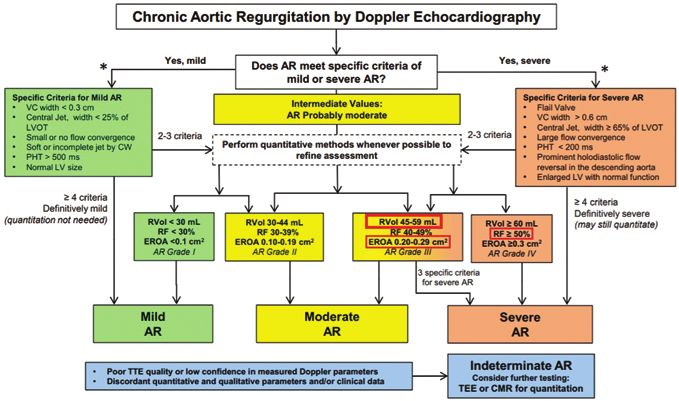
How Severe Is the AR? severe AR, vena contracta width, and LV enlargement.
Quantitative measures were performed to clarify the
According to the algorithm in Table 1 and based on severity of the AR jet as there was concern that it may
the images acquired, two specific criteria were met for be underestimated.
20 The Canadian Journal of Medical Sonography
CJMS_1_2021_WKBK.indd 20 3/22/21 12:28 PMTable 1. Quantitative Doppler: Effective Regurgitant Orifice Area (EROA), regurgitation volume (RVol), and regurgitant fraction (RF) formulas
and calculations1, 2
Quantitative 2-D and Doppler Images Acquired Volumetric Assessment
1) STROKE VOLUME METHOD: SV = CSA X VTI LVOT DIAMETER= 2.2 cm
LVOT Diameter LVOT Pulsed Doppler
VTI LVOT= 22 cm
CSA LVOT = π (LVOT DIAMETER /2)2
= 3.14 (2.2/2)2
= 3.799 cm2
SV LVOT = (CSA LVOT X VTI LVOT)
= 3.799 cm2 x 22 cm
= 83.58 ml
Figure 7A Figure 7B
www.sonographycanada.ca 21
CJMS_1_2021_WKBK.indd 21 3/22/21 12:28 PMMitral Annulus Diameter Mitral Inflow Pulsed Doppler MITRAL ANNULAR (MA) DIAMETER
= 2.31 cm
VTI MITRAL INFLOW = 16 cm
CSA MA = π (MA DIAMETER /2)2
= 3.14 (2.31/2)2
= 4.188 cm2
SV MA = (CSA MITRAL ANNULUS X VTI
MITRAL INFLOW)
2
= 4.188 cm x 16 cm
Figure 8A Figure 8B = 67.02 ml (without MR)
RVol= SV LVOT − SV MITRAL ANNULUS
= 83.58 ml − 67.02 ml
= 16.56 ml
RF= RVol/ SV LVOT
= 16.56 ml/ 83.58 ml
= 0.198 or 19.8%
RVOT Diameter RVOT Pulsed Doppler RVOT DIAMETER= 2.13 cm
VTI RVOT= 14.6 cm
CSA RVOT = π (RVOT DIAMETER /2)2
= 3.14 (2.13/2)2
= 3.561 cm2
SV RVOT = (CSA RVOT X VTI RVOT)
= 3.561 cm2 x 14.6 cm
= 51.99 ml
RVol= SV LVOT − SV RVOT
Figure 9A Figure 9B
= 83.58 ml − 51.99 ml
= 31.59 ml
RF= RVol/ SV LVOT
= 31.59 ml/ 83.58 ml
= 0.377 or 37.7%
22 The Canadian Journal of Medical Sonography
CJMS_1_2021_WKBK.indd 22 3/22/21 12:28 PM2) Proximal Isovelocity Surface Area (PISA) METHOD Regurgitant Flow Rate (RFR) =
PISA (2πr2) x VAlias
= 2 x 3.14 x (0.72 cm)2 x 31.3 cm/s
= 101.89 ml/s
EROA = Regurgitant Flow Rate /
Vmax AORTIC REGURGITATION
= 101.89 ml/s / 4.5 m/s
= 22.64 mm2
RVol = EROA x VTI AORTIC
REGURGITATION
Figure 10A Figure 10B 2
= 22.64 mm x 2.336 m
= 52.89 ml
Aliasing Velocity (VAlias) = 31.3 cm/s Vmax AORTIC REGURGITATION = 4.5 m/s
PISA Radius = 0.72 cm VTI AORTIC REGURGITATION = 2.336 m
RF = RVol/ SV LVOT
= 52.89 ml / 83.58 ml
= 0.63 or 63%
Zoghbi WA et al.1
Two different quantitative techniques were used. Using RVOT and LVOT stroke volumes, the calculated
regurgitant volume was 31.59 ml, and regurgitant fraction was 37.7% (Figure 9) which is in the moderate
range. The regurgitant volume calculated from 2D PISA was 52.89 ml/beat (see Figure 10), which is more
in the moderate to severe range. The calculated regurgitant fraction was 63% (see Figure 10) which is in
the severe range. The 2-D PISA radius measured 0.72 cm at an aliasing velocity of 31.3 cm/s. The calculated
EROA was 22.64 mm2 (Figure 10) which is more in the moderate to severe range. The quantitative methods
resulted in different estimations of regurgitant volumes and regurgitant fractions.
Based on the algorithm, 2-D PISA technique was suggestive of moderate to severe AR. However, there was
concern about the PISA technique due to challenges with Doppler alignment which results in errors when
calculating the AR VTI and peak velocity. 3-D TTE full volume color Doppler datasets of the aortic valve
were obtained and post processed.
www.sonographycanada.ca 23
CJMS_1_2021_WKBK.indd 23 3/22/21 12:28 PMTable 2. Recommendations for Noninvasive Evaluation of Native Valvular Regurgitation: A Report from the American Society of Echocardiography Developed In Collaboration
CJMS_1_2021_WKBK.indd 24
with the Society of Cardiovascular Magnetic Resonance
24 The Canadian Journal of Medical Sonography
3/22/21 12:28 PM3D Post-Processed Color Doppler Dataset Images 3-D Assessment
• Figure 11A, 11B: Post processing the
3D color doppler datasets involved
using multiplanar reconstruction by
aligning the AR jet in the orthogonal
planes (green, red, and blue) during
mid-diastole.1
• The aliasing velocity was set between
50 and 60 cm/s.
• The cross-sectional plane was placed
through the narrowest portion of the
Figure 11A Figure 11B jet, the vena contracta, perpendicular
to the direction of the jet.
• Figure 12A, 12B: From the en-face view
of the vena contracta, the 3D-EROA
of the narrowest cross-sectional area
of the regurgitant jet was measured
Figure 12A
by manual planimetry obtaining a
value of 0.36 cm2.
• The RVol was calculated by multiplying
the 3D-EROA with the velocity-time
integral of the AR jet.
RVol = 3D EROA x VTI AORTIC
REGURGITATION
2
= 36 mm x 2.336 m
= 84.10 ml
Figure 12B
Zoghbi WA, et al.1
The 3-D regurgitant volume was calculated as 84.10 ml/beat, consistent with severe aortic regurgitation.
However, the CW Doppler that was used for the VTI to calculate the 3-D regurgitant volume was felt to be
unreliable due to the eccentricity of the AR jet.
www.sonographycanada.ca 25
CJMS_1_2021_WKBK.indd 25 3/22/21 12:28 PMCardiac Magnetic Resonance Images Acquired CMR Assessment
• Figure 13A, 13B: CMR imaging shows
the aortic regurgitation appearing as
a black stream (red arrow) in the light
grey LV chamber during diastole in
the three-chamber cine.
Figure 13A Figure 13B
• Figure 14: The short axis cine shows
restriction of the anterior mitral valve
leaflet motion during diastole caused
by the eccentric AR jet. This is also
be seen in the above three-chamber
cine. This restricted orifice could cause
an inaccurate PW Doppler tracing
of the mitral inflow on echo. The
inaccurate tracing and measurement
could explain the lower regurgitant
volume that was calculated noting that
the forward flow by 2D echo (83.58
ml) was similar to the forward flow
by CMR (82 ml).
Figure 14.
• Figure 15: Phase contrast imaging
for the volumetric assessment of AR
found a forward flow during systole
of 82 ml/beat, reverse (regurgitation)
flow of 52 ml/beat during diastole,
and net forward flow of 30 ml/beat
(1.8 L/min) resulting in a regurgitant
fraction of 63%.
Figure 15.
26 The Canadian Journal of Medical Sonography
CJMS_1_2021_WKBK.indd 26 3/22/21 12:28 PM• Figure 16: CMR demonstrated severe
LV dilatation (LVEDV 299 ml, indexed
177 ml/m2, LVESV 161 ml, indexed 96
ml/m2) with mildly reduced systolic
function (LVEF 46%).
Figure 16.
Given the recent pregnancy and reduction in LVEF, there valuable information such as 3-D derived EROA to
was a concern regarding whether it was related to a accurately determine the severity of AR. If highly
postpartum cardiomyopathy or significant worsening trained cardiac sonographers are able to acquire, post-
of the AR, potentially being underestimated by 2-D process, and provide these measurements, it can have
echo assessments. The patient refused to undergo significant impact how clinical decisions are made in
a transesophageal echocardiography procedure. managing a patient. 3-D TTE has the advantage of
unlimited plane orientation, which allows the exact
In cases where 2-D and 3-D TTE acquisition is limited shape and size of the true regurgitant orifice to be
due to poor imaging windows, cardiac magnetic measured accurately especially in situations where
resonance (CMR) imaging can provide additive clinical transesophageal echocardiography and CMR is not
decision-making information about regurgitant an available option.
volumes and regurgitant fractions.2 As quantitation
of AR is more reproducible with CMR imaging than References
echocardiography,2 CMR was performed for accurate 1. Zoghbi WA, et al. Recommendations for
assessment of LVEF, assessment of AR severity, and noninvasive evaluation of native valvular
identification of other potential causes for reduction regurgitation: A report from the American Society
in LV function. of Echocardiography Developed in Collaboration
with the Society of Cardiovascular Magnetic
Conclusion Resonance [Internet]. J Am Soc Echocardiogr. 2017
The final conclusion was that there was severe AR, April [cited 2020 May 11];30(4):303–71. Available
severely dilated LV with mildly reduced LVEF 46%. from: https://www.asecho.org/wp-content/
The patient underwent aortic valve replacement. 2-D uploads/2017/04/2017VavularRegurgitationGuideline.
echocardiography can provide important information pdf
about cardiac structures; however, there are limitations 2. Otto CM. Valvular regurgitation, approaches to
such as operator variability or suboptimal acoustic quantitation of regurgitant severity. In: Textbook
imaging window that may cause a diagnosis to be of clinical echocardiography. 6th ed. Philadelphia,
missed. PA: Elsevier/Saunders; p. 337.
In cases where the valvular regurgitation appears
eccentric, careful evaluation using 3-D datasets provide
www.sonographycanada.ca 27
CJMS_1_2021_WKBK.indd 27 3/22/21 12:28 PMCJMS Article: Sonography Canada CPD Credit
Sonography Canada members can earn 1 Free CPD credit by reading this article and successfully
completing the online quiz. Visit Sonography Canada member’s site at
https://sonographycanada.ca/members/canadian-journal-medical-sonography
Article title: A
ssessing the Severity of Aortic Regurgitation Using 3-D Color Doppler Echocardiography:
A Pictorial Essay
Author’s Names: Babitha Thampinathan, HBSc, RDCS, CRCS (AE), Cindy Chow, BA, RDCS, CRCS (AE)
1. The authors contend that in cases where valvular 4. In the stroke volume method, the CSA stands for:
regurgitation is eccentric, 3D datasets add
information to help assist the degree of severity a) Cardiac stroke area
of the regurgitation. b) Calculated stroke area
c) Canadian space agency
a) True d) Circumferential Surface Area
b) False
5. Where 2-D and 3-D image acquisition is challenging
2. As per ASE guidelines, mild aortic regurgitation the following modalities may assist in clinical
demonstrates all the following factors except: decision making:
a) EROAYou can also read