Dark Circles Etiology and Management Options - Clínica de Felipe
←
→
Page content transcription
If your browser does not render page correctly, please read the page content below

Dark Circ les
Etiology and Management Options
Daniel P. Friedmann, MDa,*, Mitchel P. Goldman, MDb,c

 KEYWORDS
  Under-eye circles  Infraorbital hyperpigmentation  Tear trough  Nasojugal groove
  Suborbicularis oculi fibroadipose tissue  Intense pulsed light  Fractionated resurfacing
  Soft-tissue augmentation

 KEY POINTS
  Under-eye dark circles are an unsurprising source of aesthetic concern of patients owing to the
   fatigued and less youthful appearance that they can impart.
  The etiology of under-eye circles is multifactorial and includes periorbital volume loss and skin
   laxity, orbital fat prolapse, increased prominence and density of subcutaneous vasculature, and
   excessive pigmentation.
  The ease of use, minimal incidence of complications, and lack of downtime associated with hyal-
   uronic acid fillers make these products nearly ideal for treating infraorbital volume loss.
  Long-pulsed lasers target lower eyelid vasculature; Q-switched lasers and fractionated resurfacing
   treat cutaneous pigmentation. Skin laxity can be improved with fractionated resurfacing and micro-
   focused ultrasound.
  Standardized pretreatment and posttreatment digital photography is essential. Variations in lighting
   alone may mask or worsen lower eyelid appearance.

INTRODUCTION                                               eyelid begins at the free palpebral margin and
                                                           extends caudal to the inferior orbital rim, merging
Periorbital cutaneous and structural changes play          into the superior aspect of the cheek. It is bor-
a significant role in the perceived age of individuals     dered laterally by the lateral canthus and malar
of all ages and races.1–3 The relative darkening of        eminence and medially by the medial canthus
lower eyelid skin, commonly referred to as                 and lateral nasal sidewall. The tear trough is an
under-eye (infraorbital) dark circles or periorbital       anatomic depression found in all age groups
hyperpigmentation, can impart a fatigued and               that extends obliquely from the medial canthus
less youthful appearance to the face. Dark circles         along the medial third of the lower eyelid.6 It is
are therefore an unsurprising source of significant        bound medially by the anterior lacrimal crest
aesthetic concern for a number of patients.4,5             and inferiorly by the inferior orbital rim, lying within
                                                           the limits of the orbicularis oculi muscle and cor-
ANATOMY
                                                           responding to the anatomic location of the
Any evaluation of lower eyelid dark circles must           lacrimal sac.6,7 The tear trough forms the superior
begin with an appropriate understanding of the             anatomic aspect of the nasojugal groove, which
underlying periorbital anatomy (Fig. 1). The lower         extends below the orbital rim.7–9
                                                                                                                      plasticsurgery.theclinics.com

 Funding Sources: None.
 Conflict of Interest: Consultant for SkinMedica, Inc and speaker for Lumenis Ltd (D.P. Friedmann); Consultant
 for SkinMedica, Inc, Consultant and speaker for Lumenis Ltd (M.P. Goldman).
 a
   Westlake Dermatology Clinical Research Center, Westlake Dermatology & Cosmetic Surgery, 8825 Bee Cave
 Road, Suite 100, Austin, TX 78746 USA; b Cosmetic Laser Dermatology, 9339 Genesee Avenue, Suite 300, San
 Diego, CA 92121, USA; c Division of Dermatology, University of California, San Diego, 9500 Gilman Drive,
 San Diego, CA 92093, USA
 * Corresponding author.
 E-mail address: daniel@westlakedermatology.com

 Clin Plastic Surg 42 (2015) 33–50
 http://dx.doi.org/10.1016/j.cps.2014.08.007
 0094-1298/15/$ – see front matter Ó 2015 Elsevier Inc. All rights reserved.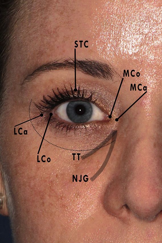
34        Friedmann & Goldman

                                                               Excessive Pigmentation
                                                               Excessive pigmentation of the lower eyelids owing
                                                               to a number of underlying causes can also lead to
                                                               under-eye circle formation.12–16 Melasma is an
                                                               acquired facial hypermelanosis common in South-
                                                               east Asian and Hispanic populations with Fitzpa-
                                                               trick skin types III-IV that may predominate in the
                                                               infraorbital areas. UV light exposure, pregnancy,
                                                               exogenous hormones (including oral contracep-
                                                               tives), and genetic predisposition all likely play a
                                                               role.17 Nevi of Ota in Asian populations are either
                                                               congenital or develop during childhood and are
                                                               thereby easily differentiated.18

                                                               Orbital Lipodystrophy from Prostaglandin F2a
                                                               Periorbital changes have also been reported with
                                                               ophthalmic and topical use of the prostaglandin
                                                               F2a analogs, including bimatoprost 0.03% (Lumi-
                                                               gan or Latisse; Allergan, Inc, Irvine, CA), travoprost,
                                                               or latanoprost.19 An acquired orbital lipodysotrophy
                                                               characterized by hollowing of lid sulci may rarely
                                                               develop from local adipocyte atrophy owing to the
                                                               potent anti-adipogenic effects of prostaglandin
                                                               F2a. Improvement is typically noted after cessation
                                                               of therapy or change to an alternative prostaglandin
                                                               analog.20 Ophthalmic use of prostaglandin F2a
                                                               analogs can also lead to reversible periocular hyper-
     Fig. 1. Periorbital landmarks. Dotted line overlies the
     orbital rim. LCa, lateral canthus; LCo, lateral commis-
                                                               pigmentation owing to increased melanogen-
     sure; MCa, medial canthus; MCo, medial commissure;        esis.21–23 The risk of pigmentation seems to be
     NJG, nasojugal groove; STC, superior tarsal crease;       dramatically lower with topical bimatoprost for
     TT, tear trough.                                          eyelash hypotrichosis because of minimal cuta-
                                                               neous contact via brush application.24,25

                                                               Subcutaneous Fat and Veins
     ETIOLOGY
                                                               The minimal infraorbital subcutaneous fat, superfi-
     The formation of dark circles is often multifactorial,    cial location of the orbicularis oculi muscle, and
     with a number of factors reported to play a role          thin, translucent skin of the lower eyelid can impart
     (Table 1). A retrospective evaluation of periorbital      a violaceous appearance to the entire area as a
     hyperpigmentation in Southeast Asian patients re-         result of prominent underlying intramuscular
     vealed a predominantly vascular etiology, followed        vasculature. Excess subcutaneous telangiectatic
     by constitutional (ie, periorbital melanosis), postin-    and reticular veins may also play a role.5,26 Greater
     flammatory hyperpigmentation (PIH), and shadow-           dermal vessel congestion and stasis-related
     ing types.11                                              extravasation during episodes of physical and
                                                               mental stress, including menstrual periods and
     Shadowing Effect                                          pregnancy, may also worsen dark under-eye
                                                               circles.
     Infraorbital skin laxity and volume loss with sub-
     cutaneous fat atrophy result from a combination           EVALUATION
     of advancing age and chronic photodamage.
     These factors, along with hypertrophy of orbicula-        A thorough history and clinical assessment of the
     ris oculi muscles, pseudoherniation of suborbicu-         lower eyelids and cheeks is necessary to determine
     laris oculi fibroadipose tissue, and/or volume loss       the underlying cause of a patient’s dark circles,
     of the malar cheek, create a shadowing effect on          choose the most appropriate course of treatment,
     the tear trough.8 This shadowing is lighting depen-       and avoid complications. A history of ocular proce-
     dent, often masked with the use of direct flash           dures, trauma, or allergies and the presence of
     photography.                                              autoimmune and neuromuscular disorders should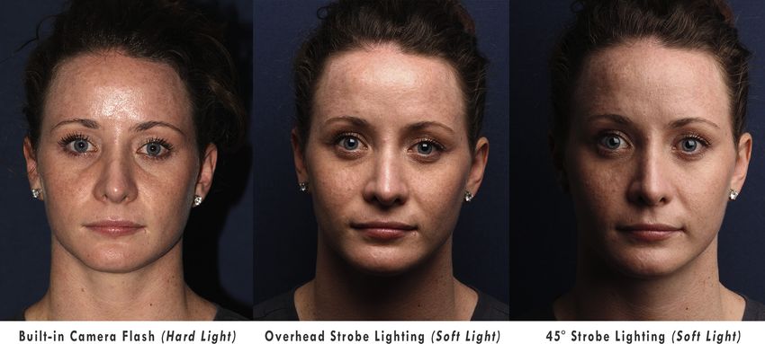
Dark Circles 35
Table 1
Etiology of dark under-eye circles
Type Mechanism Treatment Option
Hollowing/ Age-related infraorbital skin laxity and volume loss Hyaluronic acid filler
shadowing SOOF pseudoherniation Fractional ablative CO2
Orbicularis oculi muscle hypertrophy laser resurfacing
Excessive Periorbital melanosis (“constitutional type”, may be an IPL
pigmentation extension of pigmentary demarcation lines)10 Q-switched laser
Postinflammatory hyperpigmentation (allergic contact Nonablative fractionated
dermatitis, atopic dermatitis) resurfacing
Melasma
Oculodermal melanocytoses (bilateral nevus of Ota-like
macules)
Rare: Acanthosis nigricans, fixed drug eruptions, and
erythema dyschromium perstans
Prominent Thin, translucent skin Long-pulsed laser
vasculature Excess subcutaneous vascularity IPL
Venous stasis Hyaluronic acid filler
Fractional ablative CO2
laser resurfacing
Exogenous Penicillamine-induced periorbital pigmentation Hyaluronic acid filler
Bimatoprost-induced periorbital hollowing and Fractional ablative CO2
hyperpigmentation laser resurfacing
IPL
Q-switched laser
Abbreviations: IPL, intense pulsed light; SOOF, suborbicularis oculi fibroadipose tissue.
first be elicited. Genetic or acquired diseases that Evaluating and documenting the presence of a
may predispose patients to bleeding and infections prominent tear trough deformity is essential. The
must also be ruled out. Depending on individual medial and central aspects of the tear trough may
underlying factors, a baseline comprehensive be accentuated with an upward gaze, whereas
ocular examination may be warranted. the lateral border may be accentuated with an up-
The cutaneous lower eyelids should be evalu- ward outward gaze contralaterally.7 Moreover, the
ated systematically to maximize consultation importance of standardized, high-quality pretreat-
time and prevent oversights. Both epidermal and ment and posttreatment digital photography with
dermal features (textural irregularities, dyspigmen- appropriate lighting cannot be overstated, given
tation, photodamage, atrophy, or vessel promi- that different lighting conditions may mask or
nence) and subcutaneous findings (atrophy, accentuate tear troughs and other aspects of the
hollowing, or prolapse) ought to be assessed. lower eyelids (Fig. 3). Manual stretching of lower
The presence of bilateral asymmetry should be eyelid skin can help to differentiate between true
noted. A detailed history and review of prior photo- pigmentation and shadowing effect. Although the
graphs can help to distinguish normal anatomic former retains its appearance with stretching, the
variation from age-related changes.27 Imaging latter improves or resolves entirely. An increase in
with the VISIA system (Canfield Scientific, Inc, violaceous discoloration on manual stretching of
Fairfield, NJ) can highlight blood vessels and the lower eyelids, on the other hand, is consistent
pigmentation with UV light and cross-polarized with a translucent skin and/or hypervascular
flash photography (Fig. 2). etiology.
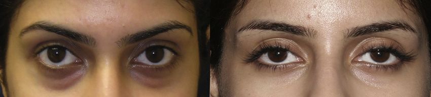
Fig. 2. (A) Clinically evident dark circles due to volume loss combined with mild underlying (B) erythema and (C)
hyperpigmentation visualized by the VISIA multispectral imaging system. (Canfield Scientific, Inc, Fairfield, NJ.)
36        Friedmann & Goldman

     Fig. 3. The effect of lighting conditions on tear trough appearance. Note its accentuation with hard light from
     direct flash photography versus masking with 45 soft light from a strobe light source.

        Eyelid skin laxity and tone should also be prop-      treatments, respectively. IPL glasses or laser
     erly assessed before any procedure using distrac-        eyewear with an optical density of 51 are also
     tion or snap tests, respectively.6 Extensive lower       mandatory for device operators and ancillary staff.
     eyelid laxity and/or orbital fat herniation on evalua-
     tion precludes the benefits of noninvasive treat-        Intense Pulsed Light
     ment options, warranting operative intervention.
                                                              Lumenis IPLs (M22 or Lumenis One, Lumenis Ltd,
                                                              Yokneam, Israel) emit 515- to 1200-nm wave-
     PROCEDURES
                                                              lengths via interchangeable cutoff filters ranging
     Preoperative
                                                              from 560 to 755 nm. An integrated chilled sapphire
     A history of keloids, conditions that may impair         crystal tip, a thin layer of cold gel, and periproce-
     wound healing, recent oral retinoid use, preg-           dural cold air cooling (Cryo 5, Cynosure, Westford,
     nancy, breastfeeding, photosensitivity, and/or           MA) combine to guarantee proper epidermal
     abnormalities localized to the treatment area            protection and minimal patient discomfort during
     (active infections, malignant lesions, scarring, or      treatment. The cold, water-based gel also en-
     burns) should be ruled out before undertaking            hances optical coupling between the crystal and
     any procedure. Any nonessential medications or           treatment area, decreasing the index of refraction
     supplements that may predispose patients to              of light and leading to deeper energy delivery.28
     bleeding should be stopped if possible 2 weeks           Treatment parameters with a 35-  15-mm crystal,
     before any injectable treatment. Given the area,         individualized for each patient based on skin type,
     prophylactic antiviral therapy for herpes simplex        are described in Table 2. Darker skin types should
     virus is not routinely performed.                        be treated with greater caution using higher cutoff
        All patients should have photographs and written      filter, lower fluences, and/or double to triple puls-
     informed consent obtained upon arrival. The treat-       ing with longer interpulse delays to help spare
     ment area is then washed with a neutral cleanser to      epidermal melanin.
     remove any makeup or other impurities. Topical               For lower eyelid hyperpigmentation, 3.0-ms
     anesthesia is generally unnecessary given the            double pulsing is performed, with mild darkening
     limited treatment area. Moreover, tetracaine-            of pigmented areas noted almost immediately
     based topical anesthetics may cause transient            posttreatment. Sequential 4.0-ms pulses are used
     erythema and should be avoided before intense            for telangiectasias or diffuse erythema; transient
     pulsed light (IPL) or vascular lasers.                   vessel spasm is the treatment endpoint for discrete
        Any laser or IPL treatment of lower eyelid skin       vascular lesions. Large-caliber telangiectasias or
     within the borders of the bony orbital rim requires      reticular veins (1 mm) can be safely double pulsed
     intraocular metal eye shields. Otherwise, disposable     with higher fluences (19–26 J/cm2) and longer
     adhesive and stainless steel or polymer-based            pulse durations (4–12 ms) using a smaller 15-
     ocular shields of an appropriate, wavelength-            8-mm crystal. Minimal pressure should be applied
     specific optical density are used for IPL and laser      against the skin with the hand piece to avoid
Dark Circles          37

 Table 2
 Intense pulsed light parameters

                                                        Settings Used
 Fitzpatrick
 Skin Types      Cutoff Filter (nm)    Delay (ms)         Initial Fluences (J/cm2)
 I-II            560                   10–15              15–19 (increased by 10%–20% with subsequent
                                                            treatments, based on clinical response)
 III-IV          560 or 590            20–50              14–16
 V-VI            695 or 755            Triple pulsing     14–16

compression of target vessels. A typical patient         settings because they have different pulse charac-
requires 1 to 3 sessions to achieve significant          teristics (eg, wavelength range, pulse duration, and
improvement, with subsequent semiannual main-            energy distribution).
tenance treatments.
   Twelve subjects treated with 2 to 4 sessions with
                                                         Q-Switched Lasers
an older version IPL (Quantum, Lumenis Ltd) with
2.6/4.0 ms double-pulsing (20 ms delay) and fluen-       Cutaneous melanin has a broad, polychromatic
ces of 36 to 37 J/cm2 demonstrated significant           absorption spectrum that peaks in the UV range
lightening (P 5 .024) of lower eyelid idiopathic         and declines steadily as a function of increasing
hyperpigmentation, as rated by 7 independent             wavelength (Fig. 4). Although significantly attenu-
observers.29 Pigment recurrence was not seen at          ated past 755 nm, energy absorption is still likely
the 1-year follow-up. All subjects experienced           with wavelengths up to 1064 nm, allowing for
posttreatment PIH (mean, 6.7 months). A prior            treatment of deeper pigment and darker Fitzpa-
study by the same group30 found greater impro-           trick skin types. Given that melanosomes have
vement in under-eye hyperpigmentation with fewer         thermal relaxation times of less than 1 ms, ultra-
adverse events using IPL compared with Q-                short pulse durations are required to selectively
switched ruby laser (QSRL).                              confine photothermal and photoacoustic effects
   Other IPL systems may also be used, but treat-        to these structures.31 Multiple Q-switched lasers
ment should proceed with more conservative               with nanosecond (and recently picosecond) pulse

Fig. 4. Absorption spectra for water, oxyhemoglobin, deoxyhemoglobin, and melanin in the visible light and
near-infrared wavelength range.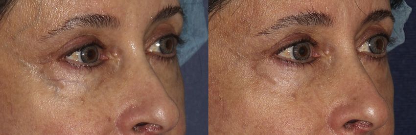
38        Friedmann & Goldman

     durations and wavelengths within the absorption          spot sizes and 4 to 8 J/cm2. Lower fluences may
     range of melanin are currently available. The            lead to equal efficacy with decreased PIH.38
     typical clinical endpoint of these treatments is im-       A novel QSAL (PicoSure, Cynosure, Inc) with en-
     mediate lesion whitening without pinpoint                ergy delivered in picoseconds (as low as 550 ps)
     bleeding. Lower energy settings should be used           may produce greater tensile stress on melano-
     initially to minimize the occurrence of PIH.             somes than nanosecond pulse durations,
                                                              enhancing their photomechanical and photother-
     Q-switched ruby lasers                                   mal destruction. Collateral tissue heating and
     The 694-nm wavelength of QSRLs is moderately             associated adverse events are minimized owing
     absorbed by melanin, yet poorly absorbed by              to the lower fluences required.39 As a result,
     competing chromophores such as hemoglobin.32             potentially all skin types may be treated with this
     Rapid delivery of high-intensity energy at this          device. A 3- to 5-mm spot size and 1.5 to 2.83 J/
     wavelength disrupts melanosomes within kerati-           cm2 fixed fluence are favored.
     nocytes, melanocytes, and melanophages, mak-
     ing them ideal for pigmented epidermal and               Q-switched Nd:YAG lasers
     superficial dermal lesions in Fitzpatrick skin types     With a wavelength of 1064 nm, these devices allow
     I-II.33                                                  for much deeper energy penetration and minimal
        QRSL treatment is performed with 2 to 4 J/cm2         melanin absorption compared with QSRL or
     using a 5-mm spot size (or varied accordingly) at        QSAL. Fitzpatrick skin types V and VI can thereby
     1.5 Hz (Fig. 5). The clinical endpoint with this de-     be treated with minimal risk of posttreatment dys-
     vice is immediate lesion whitening that resolves         pigmentation. The Spectra (Lutronic, Inc, Fremont,
     over 20 minutes, followed by erythema and                CA) 1064-nm Q-switched neodymium-doped
     edema. Lowe and colleagues34 showed greater              Nd:YAG laser uses a collimated hand piece to
     than 50% improvement in infraorbital hyperpig-           deliver a high peak power over very short pulse du-
     mentation in 23.5% and 88.9% of 17 subjects after        rations (10 ns), maximizing selective photother-
     1 and 2 sessions, respectively. Another study of         molysis of cutaneous melanosomes. As
     QSRL (6 to 7 J/cm2) for under-eye dermal melano-         demonstrated in treating melasma, repeat ses-
     cytoses showed greater than 40% improvement in           sions with low-fluence, Q-switched Nd:YAG treat-
     4 subjects after 1 to 5 sessions.35 Combining            ments can decrease stage IV melanosomes,
     QSRL with topical hydroquinone and tretinoin             damage melanocytes, and reduce expression of
     before and after treatment has also led to signifi-      melanogenesis-associated proteins.40 Greater flu-
     cant improvement in this location.36                     ences (4 to 5 J) can be used with a 3-mm spot size
                                                              for other types of lower eyelid hyperpigmentation.
     Q-switched alexandrite lasers                               Thirty female Chinese subjects with under-eye
     The more deeply penetrating 755-nm wavelength            circles owing to dermal melanin deposition
     of the Q-switched alexandrite laser (QSAL) has a         received 8 low-fluence treatments (3.5-mm spot
     lower absorption coefficient for melanin and is          size, 4.2 J/cm2, 2 passes) at 3- to 4-day intervals.
     emitted over a longer pulse duration (50–70 ns)          Blinded evaluators rated a mean global improve-
     than that of QSRL, which may serve to decrease           ment of 50% to 75% at 3 and 6 months, and
     adverse events (eg, PIH) in dark-skinned patients        93.3% subjects reported good to excellent results
     as a result of gentler melanosomal heating.37            without significant adverse events.41
     QSAL treatments of Fitzpatrick skin types of IV or          Frequency-doubled Nd:YAG or potassium-
     lower are typically performed with 3- to 5-mm            titanyl-phosphate lasers (532 nm) can also

     Fig. 5. Significant improvement in infraorbital hyperpigmentation after a single treatment with combination Q-
     switched 694 nm ruby laser (SINON, Alma Lasers Ltd, Buffalo Grove, IL) for hypermelanosis and long-pulsed
     1064 nm Nd:YAG laser (CoolTouch VARIA, CoolTouch Inc, Roseville, CA) for reticular veins.
Dark Circles           39

effectively target pigmentation with nanosecond         (greater than 70 J/cm2) for their thermocoagulation.
pulse durations. A split-face study of 10 female sub-   Nevertheless, long-pulsed 1064 nm Nd:YAG de-
jects with bilateral acquired nevus of Ota-like mac-    vices are ideal for the treatment of larger, deeply sit-
ules compared 532 nm with a combination of 532/         uated facial vessels (eg, reticular veins) owing to the
1064 nm.42 Parameters included a 4-mm spot size         superior penetration of laser energy at this wave-
with 1.2 J/cm2 (532 nm) and 4 mm with 6.5 J/cm2         length. Fitzpatrick skin types V and VI can be
(1064 nm). Objective measures of pigmentation           treated with low risk of epidermal injury, given the
and subject- and blinded physician-graded               low absorption coefficient for melanin at 1064 nm.
improvement were significantly better with combi-          The CoolTouch VARIA (CoolTouch, Inc, Ro-
nation treatment at 6 months after a single session.    seville, CA) dispenses cryogen cooling before
                                                        and/or after laser pulse delivery, maximizing
Pulsed-Dye Lasers                                       treatment-related safety and reducing procedural
                                                        discomfort.46 Treatment parameters for periorbital
Unlike prior flashlamp-pumped pulsed-dye lasers
                                                        veins are based directly on vessel size and a
with short pulse durations (0.1 to 0.45 ms) and
                                                        3.5-mm (range, 2 to 10) spot size; 1-mm reticular
577-nm light emission, current pulsed-dye lasers
                                                        veins are treated with a 25-ms pulse duration
deliver 585 and 595 nm wavelengths over
                                                        and fluences of 160 to 190 J/cm2, whereas 1- to
extended pulse widths (less than or equal to
                                                        3-mm veins require up to 50 ms and 190 to
40 ms) that allow for selective photothermolysis
                                                        210 J/cm2 (Fig. 6). Both have cryogen cooling of
of larger, deeper ectatic vessels and a far greater
                                                        20 to 30 ms delivered immediately after the laser
purpuric threshold.43,44 Although pulse stacking
                                                        pulse to quench transmission of heat from thermo-
and multiple passes at subpurpuric fluences with
                                                        coagulated blood vessels to the epidermis.
adequate epidermal protection (cryogen or con-
                                                        A distal (superior) to proximal (inferior) technique
vection cooling) lead to significant improvement
                                                        ensures sufficient chromophore (hemoglobin)
in vessel clearance without added adverse events,
                                                        within subsequent areas of treatment. Vessel
multiple treatment sessions may still be needed.45
                                                        spasm or thrombosis is the endpoint of treatment,
Superficial telangiectasias are treated with pulse
                                                        evidenced by immediate vessel blanching or dark-
durations and fluences of 6 ms and 7 to 9 J/cm2
                                                        ening. Although pulse stacking or overlapping
(less than 0.6 mm) or 10 ms and 8 to 12 J/cm2
                                                        should be avoided to prevent bulk heating of
(greater than 0.6 mm) using a 7-mm spot size,
                                                        treated areas, a second pass can be attempted
with marginally overlapping pulses. Thicker facial
                                                        after an interim of several minutes. One to 2
vessels (w1 mm) require 20 to 40 ms pulse widths
                                                        monthly sessions are required.
and subpurpuric fluences as high as 13 to 15 J/
                                                           Twenty-six Chinese subjects with under-eye
cm2. One to 3 sessions at 4- to 8-week intervals
                                                        dark circles owing to prominent reticular veins
are often needed. Diffuse erythema can be tar-
                                                        (1.0 to 2.5 mm) were treated with a mean of 1.6
geted with a 10-mm spot size and 6 or 10 ms at
                                                        (range, 1 to 3) monthly sessions using a contact-
5 to 8 J/cm2 or 20 ms at 7.5 to 9 J/cm2. Dark-
                                                        cooled long-pulsed Nd:YAG laser.47 Parameters
skinned patients should be treated with longer
                                                        included a 6-mm spot size, 120 to 140 J/cm2
pulse durations and lower fluences. Treatment
                                                        fluence, and 6- to 10-ms double-pulsing with a
endpoint is immediate vessel spasm and transient
                                                        20-ms delay. At 12-month follow-up, all subjects
purpura indicative of intravascular coagulation.
                                                        were found to have complete vessel resolution. A
Care should be taken when using cryogen cooling,
                                                        retrospective study confirmed nearly 100% sub-
because the cryogen is likely to enhance PIH.
                                                        jective and objective improvement after 1 to 2 ses-
   Given that pulsed-dye laser wavelengths lie
                                                        sions with appropriate settings.46
within the absorption coefficient for melanin,
hyperpigmentation can be targeted with a 7-mm           532 nm frequency-doubled Nd:YAG
spot size using a single 10-ms pulse, low fluences      KTP lasers emit energy across millisecond pulse
(7 to 8 J/cm2), and no epidermal cooling. Unlike the    durations, leading to purpura-free treatment of
treatment of cutaneous vessels, pulse stacking or       superficial cutaneous vessels. The high absorption
multiple passes should be avoided.                      coefficient for melanin at 532 nm restricts their use
                                                        to light-skinned individuals, yet renders them
Long-Pulsed Nd:YAG Lasers                               effective for hyperpigmentation, despite their
1064 nm Nd:YAG                                          long-pulsed energy delivery.48
Although dermal vessels strongly absorb 532 and
                                                        Traditional Ablative Lasers
595 nm, approximating the absorption peaks of
oxyhemoglobin, 1064 nm energy is poorly ab-             Pulsed CO2 traditional ablative lasers emit
sorbed and requires significantly greater fluences      10,600 nm energy preferentially absorbed by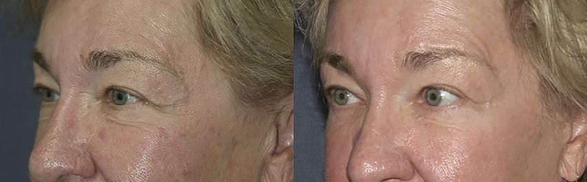
40        Friedmann & Goldman

     Fig. 6. Lower eyelid reticular veins before (left) and 2 months after (right) a single treatment with a cryogen-
     cooled, long-pulsed 1064 nm Nd:YAG laser. (Courtesy of Richard E. Fitzpatrick, MD, San Diego, CA.)

     water, leading to confluent epidermal vaporization        resurfacing (AFR) using a CO2 laser (25% coverage,
     and thermal damage of the superficial dermis.             30 W, 1-ms dwell time) by Tierney and col-
     Erbium:YAG lasers, on the other hand, allow for           leagues.58 At 6-month follow-up, 2 blinded physi-
     more precise epidermal vaporization as a result           cians found a mean improvement of 65.3% and
     of the larger absorption coefficient for water at         62.1% in laxity and rhytides, respectively. Another
     2940 nm. The amount of nonspecific thermal dam-           study found 50% to 100% tightening in 68% of sub-
     age produced by erbium:YAG devices is directly            jects after a single session with 1 to 3 passes (25 to
     contingent on dwell time, with only longer (milli-        35 J, 1-ms dwell time, 100 spots/cm2).
     second) pulse widths producing significant ther-             We prefer the Encore UltraPulse (Lumenis Ltd)
     mal contraction and denaturation of collagen.             with single, nonoverlapping passes of Deep FX for
     Shorter (microsecond) pulse widths thereby allow          deep wrinkles, followed by Active FX to target fine
     treatment of darker skin types with minimal risk          lines and irregular pigmentation (Fig. 8). Technique
     of dyspigmentation or scarring.49                         video on lower eyelid ablative fractionated re-
        Pulsed CO2 and erbium:YAG lasers have both             surfacing with appropriate treatment parameters
     demonstrated significant improvement in eyelid            and aftercare instructions can be accessed on-
     wrinkling and laxity after a single treatment             line at: http://www.plasticsurgery.theclinics.com.
     (Fig. 7).50,51 CO2 laser resurfacing has also been        Although treatment of pretarsal skin within the
     shown to improve infraorbital hyperpigmentation           margin of the superior tarsal crease is avoided, lower
     by up to 50%.52,53 Lower eyelid rhytides and PIH          eyelid skin up to 1 to 2 mm from the ciliary margin
     may likewise be improved with a 2790-nm                   can be treated without adverse events. Manual
     erbium:yittrium-scandium-galium-garnet                    stretching of eyelid skin during treatment avoids
     laser.54,55                                               overlap or skip areas and ensures proper, per-
                                                               pendicular delivery of laser energy. Eyelid rhytides
     Ablative Fractional Lasers                                and redundancy improved by 53.1% and 42.0%,
                                                               respectively, in a study of 15 subjects treated with
     Fractionated lasers are becoming increasingly             a single session of Deep and Active FX.59
     popular owing to the prolonged and meticulous
     postoperative care, need for general anesthesia,
                                                               Nonablative Fractional Lasers
     and potential adverse effects associated with
     traditional resurfacing, all of which are mitigated       Despite the advances of fractionated ablative
     with fractionated resurfacing. Instead of producing       lasers on their traditional counterparts, a subset
     confluent thermal damage, fractionated lasers             of patients refuse to undergo a procedure that is
     create columnar microthermal treatment zones              associated with greater discomfort, side effects,
     that leave up to 95% of the cutaneous surface             a weeklong downtime, and an intense postope-
     intact, providing an endogenous reservoir for rapid       rative regimen. Although both cause dermal coag-
     healing and barrier to infection.56,57 Significant        ulation necrosis limited to microthermal treatment
     improvement in deep wrinkles, fine lines, texture ir-     zones eventuating in collagen remodeling, non-
     regularity, laxity, and dyschromia can be achieved        AFR (NAFR) spares the overlying epidermis,
     with a single treatment.                                  leading to rapid recovery and reduced adverse
        Twenty-five subjects with lower eyelid laxity were     events after the procedure.57 Greater procedural
     treated with 2 to 3 sessions of ablative fractional       and postoperative tolerability must, however, be
Dark Circles            41

Fig. 7. Periorbital resurfacing with a traditional ablative CO2 laser, before (left) and 3 months after (right) a single
treatment session.

weighed against the need for a minimum of 4 to 6              to their M22 system with spot sizes of 5 to 18 mm,
monthly treatments to appreciate substantial                  pulse energies of 10 to 70 mJ, and densities from
improvement.                                                  50 to 500 spots/cm2 (Fig. 10).
   The Fraxel DUAL re:store (Solta Medical, Inc,
Hayward, CA) houses both erbium-doped                         Soft-Tissue Augmentation
(1550 nm) and thulium (1927 nm) laser fibers in a             Under-eye dark circles resulting from infraorbital
single device. Although the absorption coefficient            volume loss and shadowing can be addressed
for water is far greater at 1927 nm, making it ideal          with a number of commercially available fillers or
for superficial resurfacing and hyperpigmentation             autologous fat transplantation. Both techniques
(Fig. 9), the 1550-nm wavelength is able to target            can effectively restore volume to aging tear
laxity and fine lines owing to its greater depth of           troughs and infraorbital hollows, rebuilding the
penetration. A study by Sukal and colleagues60                natural convexity of the lower eyelid–midface tran-
found 50% to 100% improvement in eyelid skin                  sition zone.62
tightening in 55% of subjects after 3 to 7 sessions
(17 to 20 mJ, 500 to 750 microthermal treatment               Fat autologous muscle injection
zones per cm2) with a 1550-nm NAFR. Standard                  The placement of small volumes of fat within or
treatment parameters for lower eyelid treatment               adjoining muscles of facial expression for facial
with this device are listed in Table 3. A number              rejuvenation is a technique known as fat autograft
of other erbium, diode, and Nd:YAG devices are                muscle injection (FAMI).63 Prolonged cosmetic
also currently available. Lumenis recently launched           results after fat transplantation are likely a combi-
a contact-cooled 1565-nm fiber upgrade (ResurFX)              nation of adipocyte survival within these highly

Fig. 8. Fractional ablative CO2 resurfacing demonstrating marked improvement in lower eyelid wrinkling and laxity.
42        Friedmann & Goldman

     Fig. 9. Expected posttreatment erythema and crusting after 1927-nm nonablative fractional resurfacing (Fraxel
     DUAL re:store, Solta Medical, Inc, Hayward, CA). Note the significant improvement in lower eyelid and malar
     hyperpigmentation at 2 months posttreatment.

     vascular recipient sites and subcutaneous fibrosis      Approximately 1 to 1.5 mL of centrifuged fat is
     in response to grafted adipose tissue.64 A study of     typically required for each infraorbital area.
     fat transfer in 10 subjects with under-eye circles        Autologous fat transfer, although effective, may
     owing to thin, translucent skin showed a mean           have the adverse effect of variable “take” of the in-
     78% improvement in lower eyelid discoloration           jected fat. This may lead to areas of excessive fat,
     and contour.65 Fig. 11 presents the FAMI proce-         presenting clinically as palpable and/or visible
     dure for infraorbital volume augmentation.66            lumps. In addition, transferred fat may calcify.

      Table 3
      Nonablative fractionated resurfacing of lower eyelids with the Fraxel DUAL

      Fiber          Settings                                                Purpose
      1550 nm        Pulse energies: 30–50 mJ (with 8–20 mJ for              Skin laxity, fine lines, reticular
                       pigmentation only, despite reports61 of 70 mJ)         dermal pigmentation
                     Treatment levels based on skin type: I–III: 7–11
                       (20%–32% coverage); IV–VI: 4–7 (11%–20%
                       coverage)
      1927 nm        Pulse energies: 5–20 mJ with treatment levels 1–4       Skin texture and tone, epidermal
                       (20%–35% coverage). Melasma: Pulse energy of            and superficial dermal
                       5–10 mJ and treatment level 5 (40% coverage)            pigmentation
      1550 1         Same pulse energies, but total combined coverage
        1927 nm        should not exceed 70%
Dark Circles 43 Fig. 10. (A) Variable pattern shapes and (B) densities ranging from 50 to 500 spots/cm2 with the ResurFX (Lumenis Ltd, Yokneam, Israel), a novel contact-cooled, 1565-nm erbium doped fiber laser with nonsequential delivery of laser energy. (C) Single-pass with ResurFX (above) compared with 5 passes with the 1550-nm Fraxel DUAL re:store (below), both leading to 500 spots/cm2 at 20-mJ pulse energy.
44        Friedmann & Goldman

     Fig. 11. (A) Fat is harvested from fully tumesced donor fat with a 12-gauge Klein finesse cannula attached to a 10-mL
     syringe; negative pressure is achieved manually by pulling back on the plunger. (B) Syringes of fat are inverted, al-
     lowing separation of supranatant fat from infranatant and oil fractions, which are discarded. (C) Fat is then further
     concentrated via centrifuge (3 min at 3600 rpm), and the new infranatant is discarded. (D) Concentrated fat is trans-
     ferred to 1-mL syringes. (E) An entry site (via an awl or noncoring needle) in the lateral mid cheek at the level of the
     bony orbital rim is preferred for targeting the infraorbital portion of the orbicularis oculi muscle and the overlying
     subcutaneous plane. Pretreatment nerve blocks (lidocaine 1% with epinephrine 1:100,000) are performed at level of
     the infraorbital foramen. (F) Aliquots of fat are injected in a retrograde fashion with low injection pressure by means
     of a curved, blunt-tipped cannula. (Courtesy of Kimberly J. Butterwick, MD, San Diego, CA.)

     Therefore, we now favor the use of other fillers              (24 mg/mL) homogeneous nonparticulate gel.70
     described.                                                    Given that Juvederm is 6 times more hydrophilic
                                                                   than Restylane, significantly greater posttreatment
     Hyaluronic acid soft-tissue fillers                           edema is expected and overcorrection should be
     The market for injectable hyaluronic acid (HA)                avoided.71 Both contain 0.3% lidocaine to
     dermal fillers continues to expand rapidly. Accord-
     ing to the American Society for Dermatologic
     Surgery, nearly 1 million filler (primarily HA) proce-          Box 1
                                                                     Desirable characteristics of dermal fillers
     dures were performed in 2012 by dermatologic
     surgeons, a 10.4% increase over the previous                    Safety
     year.67 The ease of use, minimal incidence of com-
                                                                     Biocompatible
     plications, and lack of downtime associated with
     these products make them nearly ideal for treating              Lack of hypersensitivity
     infraorbital volume loss (Box 1). The biocom-                   Lack of clinical inflammatory response
     patibility of HA across all species also safeguards             Nonmigratory, nonclumping
     against hypersensitivity reactions. Retrospec-
     tive studies of periorbital HA have demonstrated                Negligible pain
     85% to 89% patient satisfaction after 1 to 3                    Efficacy
     sessions.68,69
                                                                     FDA-approved
        Despite being approved by the US Food and
     Drug Administration for correction of moderate to               Reproducible technique/result
     severe nasolabial wrinkles and folds, the use of                Ease of use (room temperature storage; re-
     HA products in the under-eye area is off-label.                 quires minimal to no preparation; effortless to
     We prefer to use the non-animal, bacterium-                     inject)
     derived stabilized HA fillers Restylane-L (Medicis,             Results (long-duration; good contouring ability;
     a division of Valeant Pharmaceuticals, Inc, Scotts-             collagen-stimulating)
     dale, AZ) or Juvederm Ultra XC (Allergan, Inc) for              Cost effective
     infraorbital volumization. Restylane is a medium-
     sized, particle-based HA with a concentration of                FDA, Food and Drug Administration.
                                                                        Adapted from Carruthers J, Cohen SR, Joseph JH,
     20 mg/mL and the highest G0 (measure of stiffness)              et al. The science and art of dermal fillers for soft-
     of any currently available HA filler. On the other              tissue augmentation. J Drugs Dermatol 2009;8:335–50.
     hand, Juvederm is a higher concentration
Dark Circles 45
minimize procedural discomfort when injecting Topicals
with prepackaged 27-G needles and obviate the
A number of topical products have been reported to
need for topical anesthesia.
improve lower eyelid pigmentation, laxity, and rhy-
Technique video on periocular rejuvenation with
tides. A study by Mitsuishi and colleagues77 found
an HA filler can be accessed online at http://www.
subjective improvement in under-eye hemostasis
plasticsurgery.theclinics.com. Although a number
and wrinkling with use of a gel containing 0.1%
of techniques have been described in the litera-
retinol and vitamins C, E, and K for 8 weeks. Other
ture,62,72–74 we have found that injecting a sup-
uncontrolled studies of creams with vitamins C, E,
raperiosteal bolus into the area underlying the
and K or vitamin K and retinol have demonstrated
tear trough and nasojugal groove using minimal
improvement in infraorbital pigmentation in 93%
injection points leads to excellent results with min-
to 100% of subjects, albeit with variable results.78,79
imal risk of bruising. Volumes of 0.4 to 1.0 mL per
Vitamin C in particular may serve to thicken eyelid
side are used, depending on the product and
dermal tissue, masking hemostasis-related dis-
extent of volume loss. Gentle digital massage of
coloration.80 Although hydroquinone has been the
the area helps to smooth the product into place.
gold standard for hyperpigmentation, a recently
Results typically last up to 1 year (Figs. 12 and
released hydroquinone-free topical product target-
13). We have not found any added benefit from
ing all major biochemical pathways of cutaneous
the infraorbital use of blunt-tipped cannulas
pigmentation may lead to equivalent results without
compared with needles.
safety concerns associated with long-term use.81,82
Topical application of growth factors may also help
Microfocused Ultrasound to reverse under-eye photodamage and laxity.83,84
Microfocused (or intense focused) ultrasound is a
novel technology for the correction of mild to mod- COMPLICATIONS AND AFTER CARE
erate skin and soft tissue laxity introduced in 2009
The risk of complications after laser and injectable
(Ultherapy, Ulthera, Inc, Mesa, AZ). Short duration
procedures is low when experienced practitioners
(25 to 50 ms) pulses of transcutaneous ultrasound
utilize appropriate treatment settings and/or
energy with frequencies in the megahertz (MHz)
proper technique. If they occur, adverse events
range create precise areas of spatially focused,
are typically mild and transient. Apart from frac-
chromophore-independent thermal coagulative
tionated resurfacing, little to no after care regimen
damage, sparing intervening tissues or overlying
is required for these procedures.
skin.75 A single treatment session has been shown
to increase reticular dermal collagen and
Intense Pulsed Light
thickness.76 Suh and colleagues76 showed objec-
tive and subjective improvement in infraorbital skin Mild-to-moderate erythema lasts between hours
laxity at 6 months after 1 to 2 sessions, each with and 3 days. Postprocedure topical steroid use,
110 to 280 lines. We use 7-MHz, 3.0-mm (0.25 to however, is rarely necessary. Areas of increased
0.45 J) and 10-MHz, 1.5-mm (0.15 to 0.25 J) trans- pigmentation may develop scattered crusting
ducers with a combined 25 to 50 parallel lines per within days of treatment.85 Employing a 10%
eyelid (Fig. 14). overlap between pulses minimizes reticular
Fig. 12. Lower eyelid rejuvenation with a single session of a non-animal–stabilized hyaluronic acid filler.
(Restylane-L, Medicis, a division of Valeant Pharmaceuticals, Inc, Scottsdale, AZ.)46 Friedmann & Goldman
Fig. 13. Treatment of tear trough deformity with a double-crosslinked, nonparticulate hyaluronic acid filler. (Be-
lotero Balance, Merz Aesthetics, Inc, Greensboro, NC.)
footprinting. Subsequent treatment with the IPL purpura that resolves over 1 to 2 weeks. More effi-
should be performed with the crystal rotated 90 . cient cooling mechanisms help to limit epidermal
Purpura formation is uncommon with appropriate thermal injury (eg, dyspigmentation, crusting, blis-
settings. tering, or scarring) in dark skin types.87
Blistering and persistent dyspigmentation are un-
common and likely to be a direct result of excessive Long-Pulsed Nd:YAG Lasers
overlapping, exorbitant fluences, poor epidermal
cooling, and/or insufficient delay between sequen- Treatment of veins within the orbital rim with a
tial pulses, particularly in dark-skinned or tanned 1064-nm laser places patients at significant risk
individuals.86 Nevertheless, scarring is exceedingly of macular holes, uveitis, and pupillary abnormal-
rare. Given the ability of IPL to permanently damage ities. The combination of metal corneal shields,
dark-colored terminal hairs, IPL treatments are pulling infraorbital skin away from the eye, and
generally avoided in hair-bearing skin of the male aiming the laser away from the orbit prevent intra-
beard area. ocular injury during treatment.88 When treating the
lower eyelid, veins at or medial to the mid pupillary
line should likely be avoided owing to their
Q-Switched Lasers
drainage into ophthalmic veins and cavernous
Erythema and edema resolve over 24 to 48 hours. sinus.89
Mild transient crusting is expected after higher
fluence treatment of pigmented areas. Blistering Fractionated Resurfacing
or bleeding may result if excessive fluences are
used. Overly aggressive treatment in darker Cold, sterile saline compresses are applied imme-
skin types increases the risk of PIH or diately post-AFR. An occlusive ointment is then
hypopigmentation. applied every 4 hours for 4 to 7 days until reepithe-
lialization is complete, at which point the use of a
noncomedogenic, ceramide-based moisturizer
Pulsed-Dye Lasers
and mineral-based (zinc oxide or titanium dioxide)
Posttreatment erythema usually resolves within sunscreen is advised. A mid potency topical
hours, but can persist for up to 2 days. Short pulse steroid (eg, fluocinolone 0.025% ointment BID)
durations with excessive fluences may induce is started at day 3 or 4 to decrease erythema,
Fig. 14. Improvement in lower eyelid skin laxity after microfocused ultrasound. (Ultherapy, Ulthera, Inc,
Mesa, AZ.)Dark Circles 47
pruritus, and PIH. After NAFR, a ceramide-based 4. Freitag FM, Cestari TF. What causes dark circles
moisturizer and mineral-based sunscreen are under the eyes? J Cosmet Dermatol 2007;6:211–5.
applied every 6 to 8 hours for 3 to 5 days until 5. Roh MR, Chung KY. Infraorbital dark circles: defini-
erythema and/or crusting have resolved. tion, causes, and treatment options. Dermatol Surg
The risk of adverse events is low with NAFR. 2009;35:1163–71.
Although blistering, scarring, infection, pigmentary 6. Stutman RL, Codner MA. Tear trough deformity:
changes, herpes simplex virus reactivation, and review of anatomy and treatment options. Aesthet
acneiform eruptions (in acne-prone patients) are Surg J 2012;32:426–40.
still possible, they are far more likely with AFR. 7. Hirmand H. Anatomy and nonsurgical correction of
the tear trough deformity. Plast Reconstr Surg
Soft-Tissue Augmentation 2010;125:699–708.
8. Sadick NS, Bosniak SL, Cantisano-Zilkha M,
Injection-related adverse events including bruis-
et al. Definition of the tear trough and the tear
ing, erythema, tenderness, and edema are rela-
trough rating scale. J Cosmet Dermatol 2007;6:
tively common. Not unexpectedly, all are more
218–22.
severe with FAMI. Papules, nodules, or bluish
9. Haddock NT, Saadeh PB, Boutros S, et al. The tear
discoloration secondary to Tyndall effect are
trough and lid/cheek junction: anatomy and impli-
possible if HA fillers are injected too superficially,
cations for surgical correction. Plast Reconstr
but are readily reversed with hyaluronidase
Surg 2009;123:1332–40.
(10–30 U). However, a novel double-crosslinked,
10. Malakar S, Lahiri K, Banerjee U, et al. Periorbital
nonparticulate HA filler, Belotero Balance (Merz
melanosis is an extension of pigmentary demarca-
Aesthetics, Inc, Greensboro, NC), now allows for
tion line-F on face. Indian J Dermatol Venereol Leprol
superficial product placement without these side
2007;73:323–5.
effects. Irregularities after superficial placement
11. Ranu H, Thng S, Goh BK, et al. Periorbital hyper-
of fat are more difficult to treat and may require
pigmentation in Asians: an epidemiologic study
intralesional corticosteroids or excision. Arterial
and a proposed classification. Dermatol Surg
occlusion with embolization and/or injection site
2011;37:1297–303.
necrosis have not reported with under-eye place-
12. Ortonne JP, Sharma V, Verschoore M, et al. Deter-
ment of HA fillers or FAMI.90,91
mination of melanin and haemoglobin in the skin
of idiopathic cutaneous hyperchromia of the orbital
SUMMARY region (ICHOR): a study of Indian patients. J Cutan
Given their multifactorial nature and the fact that Aesthet Surg 2012;5:176.
individual patients may have more than a single 13. Cho S, Lee SJ, Chung WS, et al. Acquired bilateral
underlying cause, cosmetic practitioners should nevus of Ota-like macules mimicking dark circles
be well versed in a number of potential treatment under the eyes. J Cosmet Laser Ther 2010;12:
options encompassing all facets of under-eye 143–4.
dark circles. New therapeutic options are also 14. Marks MB. Allergic shiners: dark circles under the
forthcoming. Longer-lasting HA fillers, wavelength eyes in children. Clin Pediatr 1966;5:655–8.
tunable laser devices, and topicals speeding up 15. Safoury El OS, Fatah El DS, Ibrahim M. Treatment
healing and enhancing results after fractionated of periocular hyperpigmentation due to lead of
laser therapy will all serve to make the future of kohl (surma) by penicillamine: a single group
dark circle treatment unabatedly bright. non-randomized clinical trial. Indian J Dermatol
2009;54:361–3.
16. Peters NT, Conn H, Côté MA. Extensive lower
REFERENCES
eyelid pigment spread after blepharopigmentation.
1. Nkengne A, Bertin C, Stamatas GN, et al. Influence Ophthal Plast Reconstr Surg 1999;15:445–7.
of facial skin attributes on the perceived age of 17. Sheth VM, Pandya AG. Melasma: a comprehensive
Caucasian women. J Eur Acad Dermatol Venereol update: part I. J Am Acad Dermatol 2011;65:689–97.
2008;22:982–91. 18. Kannan SK. Oculodermal melanocytosis–nevus of
2. Camp MC, Wong WW, Filip Z, et al. A quantitative Ota (with palatal pigmentation). Indian J Dent Res
analysis of periorbital aging with three-dimensional 2003;14:230–3.
surface imaging. J Plast Reconstr Aesthet Surg 19. Sira M, Verity DH, Malhotra R. Topical bimatoprost
2011;64:148–54. 0.03% and iatrogenic eyelid and orbital lipody-
3. Mayes AE, Murray PG, Gunn DA, et al. Ageing strophy. Aesthet Surg J 2012;32:822–4.
appearance in China: biophysical profile of facial 20. Filippopoulos T, Paula JS, Torun N, et al. Periorbital
skin and its relationship to perceived age. J Eur changes associated with topical bimatoprost.
Acad Dermatol Venereol 2010;24:341–8. Ophthal Plast Reconstr Surg 2008;24:302–7.48 Friedmann & Goldman
21. Doshi M, Edward DP, Osmanovic S. Clinical course 36. Momosawa A, Kurita M, Ozaki M, et al. Combined
of bimatoprost-induced periocular skin changes in therapy using Q-switched ruby laser and bleaching
Caucasians. Ophthalmology 2006;113:1961–7. treatment with tretinoin and hydroquinone for peri-
22. Kapur R, Osmanovic S, Toyran S, et al. Bimato- orbital skin hyperpigmentation in Asians. Plast Re-
prost-induced periocular skin hyperpigmentation: constr Surg 2008;121:282–8.
histopathological study. Arch Ophthalmol 2005; 37. Jang KA, Chung EC, Choi JH, et al. Successful
123:1541–6. removal of freckles in Asian skin with a Q-switched
23. Sharpe ED, Reynolds AC, Skuta GL, et al. The alexandrite laser. Dermatol Surg 2000;26(3):231–4.
clinical impact and incidence of periocular 38. Wang CC, Chen CK. Effect of spot size and fluence
pigmentation associated with either latanoprost or on Q-switched alexandrite laser treatment for
bimatoprost therapy. Curr Eye Res 2007;32(12): pigmentation in Asians: a randomized, double-
1037–43. blinded, split-face comparative trial. J Dermatolog
24. Centofanti M, Oddone F, Chimenti S, et al. Preven- Treat 2012;23:333–8.
tion of dermatologic side effects of bimatoprost 39. Dover J, Arndt K, Metelitsa A, et al. Picosecond
0.03% topical therapy. Am J Ophthalmol 2006; 755 nm alexandrite laser for treatment of tattoos
142:1059–60. and benign pigmented lesions: a prospective trial.
25. Priluck JC, Fu S. Latisse-induced periocular skin hy- Lasers Surg Med 2012;44:6.
perpigmentation. Arch Ophthalmol 2010;128:792–3. 40. Kim HS, Kim EK, Jung KE, et al. A split-face
26. Epstein JS. Management of infraorbital dark cir- comparison of low-fluence Q-switched Nd:YAG
cles. A significant cosmetic concern. Arch Facial laser plus 1550 nm fractional photothermolysis vs.
Plast Surg 1999;1:303–7. Q-switched Nd:YAG monotherapy for facial mel-
27. Buckingham ED, Bader B, Smith SP. Autologous fat asma in Asian skin. J Cosmet Laser Ther 2013;
and fillers in periocular rejuvenation. Facial Plast 15:143–9.
Surg Clin North Am 2010;18:385–98. 41. Xu TH, Yang ZH, Li YH, et al. Treatment of in-
28. Weiss RA, Weiss MA, Goldman MP. Intense pulsed fraorbital dark circles using a low-fluence Q-
light and nonablative approaches to photoaging. switched 1,064-nm laser. Dermatol Surg 2011;
In: Goldman MP, Weiss RA, editors. Advanced 37:797–803.
techniques in dermatologic surgery. New York: Tay- 42. Ee HL, Goh CL, Khoo LS, et al. Treatment of
lor & Francis Group; 2006. p. 295–315. acquired bilateral nevus of Ota-like macules (Hori’s
29. Cymbalista NC, Prado de Oliveira ZN. Treatment of nevus) with a combination of the 532 nm Q-
idiopathic cutaneous hyperchromia of the orbital Switched Nd:YAG laser followed by the 1,064 nm
region (ICHOR) with intense pulsed light. Dermatol Q-switched Nd:YAG is more effective: prospective
Surg 2006;32:773–84. study. Dermatol Surg 2006;32:34–40.
30. Cymbalista NC, Osorio N, Torezan L, et al. Treatment 43. Goldman MP. Optimal management of facial telan-
of eyelid hyperpigmentation with QS ruby laser and giectasia. Am J Clin Dermatol 2004;5:423–34.
intense pulsed light device. Lasers Surg Med 2002; 44. Tanghetti E, Sierra RA, Sherr EA, et al. Evaluation
30:66. of pulse-duration on purpuric threshold using
31. Anderson RR, Margolis RJ, Watenabe S, et al. Selec- extended pulse pulsed dye laser (Cynosure V-
tive photothermolysis of cutaneous pigmentation by star). Lasers Surg Med 2002;31:363–6.
Q-switched Nd:YAG laser pulses at 1064, 532, and 45. Iyer S, Fitzpatrick RE. Long-pulsed dye laser
355 nm. J Invest Dermatol 1989;93:28–32. treatment for facial telangiectasias and erythema:
32. Taylor CR, Anderson RR. Treatment of benign pig- evaluation of a single purpuric pass versus multi-
mented epidermal lesions by Q-switched ruby ple subpurpuric passes. Dermatol Surg 2005;31:
laser. Int J Dermatol 1993;32:908–12. 898–903.
33. Kopera D, Hohenleutner U, Landthaler M. Quality- 46. Lai SW, Goldman MP. Treatment of facial reticular
switched ruby laser treatment of solar lentigines veins with dynamically cooled, variable spot-sized
and Becker’s nevus: a histopathological and immu- 1064 nm Nd:YAG laser. J Cosmet Dermatol 2007;
nohistochemical study. Dermatology 1997;194: 6:6–8.
338–43. 47. Ma G, Lin XX, Hu XJ, et al. Treatment of venous
34. Lowe NJ, Wieder JM, Shorr N, et al. Infraorbital pig- infraorbital dark circles using a long-pulsed
mented skin. Preliminary observations of laser ther- 1,064-nm neodymium-doped yttrium aluminum
apy. Dermatol Surg 1995;21:767–70. garnet laser. Dermatol Surg 2012;38:1277–82.
35. Watanabe S, Nakai K, Ohnishi T. Condition known 48. Kilmer SL, Wheeland RG, Goldberg DJ, et al. Treat-
as “dark rings under the eyes” in the Japanese ment of epidermal pigmented lesions with the
population is a kind of dermal melanocytosis which frequency-doubled Q-switched Nd:YAG laser. A
can be successfully treated by Q-switched ruby controlled, single-impact, dose-response, multi-
laser. Dermatol Surg 2006;32:785–9. center trial. Arch Dermatol 1994;130:1515–9.Dark Circles 49
49. Manaloto RM, Alster TS. Periorbital rejuvenation: a 66. Donofrio L. Technique of periorbital lipoaugmenta-
review of dermatologic treatments. Dermatol Surg tion. Dermatol Surg 2003;29:92–8.
1999;25:1–9. 67. The American Society for Dermatologic Surgery.
50. Alster TS, Bellew SG. Improvement of dermatocha- 2012 ASDS Survey on Dermatologic Procedures.
lasis and periorbital rhytides with a high-energy Available at: http://www.asds.net/WorkArea/linkit.
pulsed CO2 laser: a retrospective study. Dermatol aspx?LinkIdentifier5id&ItemID56607&libID56583.
Surg 2004;30:483–7. Accessed August 1, 2013.
51. Manuskiatti W, Siriphukpong S, Varothai S, et al. Effect 68. Goldberg RA, Fiaschetti D. Filling the periorbital
of pulse width of a variable square pulse (VSP) erbiu- hollows with hyaluronic acid gel: initial experience
m:YAG laser on the treatment outcome of periorbital with 244 injections. Ophthal Plast Reconstr Surg
wrinkles in Asians. Int J Dermatol 2010;49:200–6. 2006;22:335–41.
52. West TB, Alster TS. Improvement of infraorbital 69. Morley AM, Malhotra R. Use of hyaluronic acid filler
hyperpigmentation following carbon dioxide laser for tear-trough rejuvenation as an alternative to
resurfacing. Dermatol Surg 1998;24:615–6. lower eyelid surgery. Ophthal Plast Reconstr Surg
53. Lupton JR, Alster TS. Evaluation of one-pass CO2 2011;27:69–73.
laser resurfacing for infraorbital hyperpigmenta- 70. Bogdan Allemann I, Baumann L. Hyaluronic acid gel
tion. Lasers Surg Med 2002;30:21. (Juvéderm) preparations in the treatment of facial
54. Lee WL, Kim BJ, Kim MN, et al. Treatment of periorbi- wrinkles and folds. Clin Interv Aging 2008;3:629–34.
tal wrinkles using a 2,790-nm yttrium scandium gal- 71. Dayan SH, Arkins JP, Somenek M. Restylane
lium garnet laser. Dermatol Surg 2010;36:1382–9. persisting in lower eyelids for 5 years. J Cosmet
55. Park KY, Oh IY, Moon NJ, et al. Treatment of infraor- Dermatol 2012;11:237–8.
bital dark circles in atopic dermatitis with a 2790- 72. Bosniak S, Sadick NS, Cantisano-Zilkha M, et al.
nm erbium:yttrium scandium gallium garnet laser: The hyaluronic acid push technique for the nasoju-
a pilot study. J Cosmet Laser Ther 2013;15:102–6. gal groove. Dermatol Surg 2007;34:127–31.
56. Fisher GH, Geronemus RG. Short-term side effects 73. Klein AW. Technique issues in nonsurgical filling of the
of fractional photothermolysis. Dermatol Surg 2005; periorbital hollows. Aesthet Surg J 2007;27:294–5.
31:1245–9. 74. Viana GA, Osaki MH, Cariello AJ, et al. Treatment of
57. Bogle MA. Fractionated mid-infrared resurfacing. the tear trough deformity with hyaluronic acid.
Semin Cutan Med Surg 2008;27:252–8. Aesthet Surg J 2011;31:225–31.
58. Tierney EP, Hanke CW, Watkins L. Treatment of 75. Laubach HJ, Makin IR, Barthe PG, et al. Intense
lower eyelid rhytids and laxity with ablative fraction- focused ultrasound: evaluation of a new treatment
ated carbon-dioxide laser resurfacing: case series modality for precise microcoagulation within the
and review of the literature. J Am Acad Dermatol skin. Dermatol Surg 2008;34:727–34.
2011;64:730–40. 76. Suh DH, Oh YJ, Lee SJ, et al. A intense-focused
59. Kotlus BS. Dual-depth fractional carbon dioxide ultrasound tightening for the treatment of infraorbi-
laser resurfacing for periocular rhytidosis. Dermatol tal laxity. J Cosmet Laser Ther 2012;14:290–5.
Surg 2010;36:623–8. 77. Mitsuishi T, Shimoda T, Mitsui Y, et al. The effects of
60. Sukal SA, Chapas AM, Bernstein LJ, et al. Eyelid topical application of phytonadione, retinol and
tightening and improved eyelid aperture through vitamins C and E on infraorbital dark circles and
nonablative fractional resurfacing. Dermatol Surg wrinkles of the lower eyelids. J Cosmet Dermatol
2008;34:1454–8. 2004;3:73–5.
61. Moody MN, Landau JM, Goldberg LH, et al. Frac- 78. Enshaieh S, Jooya A, Iraji F, et al. The efficacy of
tionated 1550-nm erbium-doped fiber laser for topical cream composed of vitamins K, C, E, and
the treatment of periorbital hyperpigmentation. Der- CoQ10 in the treatment of infraorbital melanosis
matol Surg 2011;38:139–42. and wrinkling: an open-label, self-controlled study.
62. Finn JC, Cox S. Fillers in the periorbital complex. Cell Tissue Res 2008;8:1645–8.
Facial Plast Surg Clin North Am 2007;15:123–32. 79. Elson ML, Nacht S. Treatment of periorbital hyper-
63. Butterwick KJ. Fat autograft muscle injection pigmentation with topical vitamin K/vitamin A.
(FAMI): new technique for facial volume restoration. Cosmet Dermatol 1999;12:32–4.
Dermatol Surg 2005;31:1487–95. 80. Ohshima H, Mizukoshi K, Oyobikawa M, et al. Ef-
64. Butterwick KJ, Nootheti PK, Hsu JW, et al. Autolo- fects of vitamin C on dark circles of the lower eye-
gous fat transfer: an in-depth look at varying con- lids: quantitative evaluation using image analysis
cepts and techniques. Facial Plast Surg Clin and echogram. Skin Res Technol 2009;15:214–7.
North Am 2007;15:99–111. 81. Makino ET, Herndon JH, Sigler ML, et al. Clinical
65. Roh MR, Kim TK, Chung KY. Treatment of infraorbi- efficacy and safety of a multimodality skin bright-
tal dark circles by autologous fat transplantation: a ener composition compared with 4% hydroqui-
pilot study. Br J Dermatol 2009;160:1022–5. none. J Drugs Dermatol 2012;11:1478–82.You can also read