Electron microscopy of high pressure frozen samples: bridging the gap between cellular ultrastructure and atomic resolution
←
→
Page content transcription
If your browser does not render page correctly, please read the page content below
Histochem Cell Biol (2008) 130:877–889
DOI 10.1007/s00418-008-0500-1

 R EV IE W

Electron microscopy of high pressure frozen samples: bridging
the gap between cellular ultrastructure and atomic resolution
Daniel Studer · Bruno M. Humbel · Matthias Chiquet

Accepted: 22 August 2008 / Published online: 16 September 2008
© Springer-Verlag 2008

Abstract Transmission electron microscopy has provided           Moreover, recent Wndings will be discussed showing that
most of what is known about the ultrastructural organiza-        molecular models of proteins can be Wtted into depicted
tion of tissues, cells, and organelles. Due to tremendous        organellar ultrastructure of images of frozen hydrated sec-
advances in crystallography and magnetic resonance imag-         tions. High pressure freezing of tissue is the base which
ing, almost any protein can now be modeled at atomic reso-       may lead to precise models of macromolecular assemblies
lution. To fully understand the workings of biological           in situ, and thus to a better understanding of the function of
“nanomachines” it is necessary to obtain images of intact        complex cellular structures.
macromolecular assemblies in situ. Although the resolution
power of electron microscopes is on the atomic scale, in         Keywords High pressure freezing · CryoWxation ·
biological samples artifacts introduced by aldehyde Wxa-         Electron microscopy · Electron tomography ·
tion, dehydration and staining, but also section thickness       Freeze substitution · Frozen hydrated sections ·
reduces it to some nanometers. CryoWxation by high pres-         Immunolabeling
sure freezing circumvents many of the artifacts since it
allows vitrifying biological samples of about 200 m in
thickness and immobilizes complex macromolecular                 Introduction
assemblies in their native state in situ. To exploit the per-
fect structural preservation of frozen hydrated sections,        Transmission electron microscopy (TEM) has proven cru-
sophisticated instruments are needed, e.g., high voltage         cial to the advancement of modern cell biology. Thanks to
electron microscopes equipped with precise goniometers           the much higher resolution than that achieved with a light
that work at low temperature and digital cameras of high         microscope, many cellular organelles and substructures
sensitivity and pixel number. With them, it is possible to       were Wrst discovered by TEM (Palade and Porter 1954);
generate high resolution tomograms, i.e., 3D views of sub-       however, it is a drawback of electron microscopy that sam-
cellular structures. This review describes theory and appli-     ples containing liquid water cannot be imaged directly at
cations of the high pressure cryoWxation methodology and         room temperature. Accelerated electrons propagate only
compares its results with those of conventional procedures.      under high vacuum conditions where water is evaporating.
                                                                 Further, only thin layers or sections of solid matter can be
                                                                 viewed (Hayat 2000). In the standard procedure, biological
D. Studer (&) · M. Chiquet                                       samples are aldehyde and osmium tetroxide Wxed, dehy-
Institute for Anatomy, University of Bern,                       drated, embedded into a resin, and ultra-thin sections are
Baltzerstr. 2, 3000 Bern 9, Switzerland
e-mail: studer@ana.unibe.ch
                                                                 prepared that are stained with heavy metal ions (Luft 1961;
                                                                 Pease and Porter 1981). All preparation steps can introduce
B. M. Humbel                                                     artifacts; Wxation with glutaraldehyde and dehydration with
Electron Microscopy and Structure Analysis,                      organic solvents lead, e.g., to aggregation of proteins, col-
Cellular Architecture and Dynamics, Faculty of Sciences,
Utrecht University, Padualaan 8,
                                                                 lapse of highly hydrated glycans, and loss of lipids (Cope
3584 CH Utrecht, The Netherlands                                 and Williams 1968, 1969a, b; Kellenberger et al. 1992).

                                                                                                                    123
878                                                                                         Histochem Cell Biol (2008) 130:877–889

Moreover, the contrast seen in classical EM micrographs is         consequence, water becomes a very viscous Xuid, so-called
based on diVerential adsorption of heavy metal cations to          amorphous (vitreous) ice.
various sample components (anion formation depends on                 The simplest way to obtain the cooling rates required for
sample Wxation and embedding conditions), rather than to           water vitriWcation is implemented in the so-called “bare-
the biological structures themselves (Hayat 2000; North            grid” method (Adrian et al. 1984). In this approach a very
et al. 1999). Thus, although atomic resolution is possible         thin sample (
Histochem Cell Biol (2008) 130:877–889                                                                                               879

                                                                             Technology of high pressure freezing and follow up
                                                                             procedures

                                                                             High pressure freezing (HPF) devices are currently avail-
                                                                             able from three diVerent manufacturers (Leica EMPACT
                                                                             and Bal-Tec HPM 100, Leica-Microystems, Vienna, Aus-
                                                                             tria; Bal-Tec HPM 010, ABRA-Fluid AG, Widnau, Swit-
                                                                             zerland available from Boeckeler Instruments, Inc.,
                                                                             Tucson, Arizona, USA; Compact 01, Wohlwend, Switzer-
                                                                             land available from Technotrade International, Inc., Man-
                                                                             chester, NH, USA) and two distinct designs are being used.
                                                                             One (Leica, EMPACT) is based on the machine built by
                                                                             Riehle (Riehle and Hoechli, 1973). The machine builds up
Fig. 1 This scheme shows how calculated cooling rates (shaded ar-            pressure on the sample and then cools it by an independent
eas) and temperatures (curves) depend on the distance from surface (z)       mechanism. The specimen is introduced into a small closed
and on time (t) during freezing of an aqueous sample in an HPF device.       chamber that is Wrst pressurized to 210 MPa, and then
The basics of the calculations resulting in the data presented are de-
scribed in the appendix of Studer et al. (1995). The assumptions are: A      immediately cooled from outside to ¡196°C by a double jet
slab with inWnite x, y dimensions and a thickness z of 200 m is uni-        of liquid nitrogen. The other machines (Bal-Tec, Wohl-
formly cooled from both surfaces with a jet of an eYcient cryogen            wend), developed according to Moor et al. (1980), pressur-
starting at time 0, generating a superWcial cooling rate of at least         ize liquid nitrogen to 210 MPa and then “shoot” it onto the
10,000 K/s. In the graph, the sample surface is at left, and its center at
right. On the sample surface (z = 0), the cooling rate is high               sample holder to freeze the specimen. Thus, in this design
(>10,000 K/s) at the very beginning of cooling, and it decreases when        the cryogen also acts as pressurizing agent. Pressurization
the surface temperature approaches that of the coolant. In the center of     and cooling of the sample are synchronized to occur within
the sample (z = 100 m), the calculations show that at the start the         20 ms in both systems (Fig. 2). All commercially available
cooling rates are quite small; a maximal cooling rate of 6,000–8,000 K/s
is achieved after 10–20 ms that decreases again later. When following        devices do their job reproducibly well. There are diVer-
the light green ¡20°C isotherm (corresponding to the melting point of        ences in size of machine and sample holder, liquid nitrogen
water at 210 MPa), one observes that, at any location having this tem-       consumption and speciWc sample preparation tools. It is a
perature, the slab is cooled as rapidly as theoretically possible (cooling   big advantage of the EMPACT device (Studer et al. 2001)
rates >6,000 K/s). Further, one can see that 20 ms after the start of
cooling, the slab has reached a temperature of
880 Histochem Cell Biol (2008) 130:877–889
Freeze fracturing
Originally, high pressure freezing (Moor 1987) was intro-
duced as a cryoWxation method for freeze fracturing (Moor
and Mühletaler 1963). In the latter procedure, a frozen bio-
logical sample is broken open in the vacuum, and a plati-
num–carbon replica is prepared from the fractured sample
surface that can be investigated in the electron microscope.
In contrast to thin sections, freeze fracture replicas show
mostly surface views of organelles and cellular membranes
since the two lipid layers of a membrane tend to separate
during fracturing. Thus, the method is especially suited for
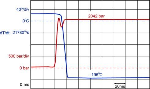
Fig. 2 Record of temperature (blue line) and pressure (red line) membrane studies, for e.g., it allows estimating the density
changes during freezing (total time = 200 ms) in the EMPACT high of transmembrane protein molecules that appear as parti-
pressure freezing machine. The temperatures and as a result the cooling
rates are measured with a thermocouple located just below the speci- cles on a smooth lipid leaXet (Kopp 1973; Shotton et al.
men carriers. The pressure is measured in the pressure system that is 1978). In the original freeze-fracturing procedure, samples
connected to the sample. As a consequence, the pressure measured is were Wrst Wxed with aldehydes and soaked in cryoprotec-
the one that really acts on the sample, whereas the temperature record tants in order to avoid ice crystal formation during freezing,
just tells us that pressure and temperature have been synchronized cor-
rectly and that the freezing cycle was technically working and high pressure freezing was developed to make these
pretreatments obsolete. Specialized sample holders are
available that allow freeze fracturing immediately after
Since the aim of vitrifying is to allow imaging of cellular high pressure freezing (Craig et al. 1987; Walther 2003).
ultrastructure close to the living state, it is of course impor-
tant to keep the time between sample collection and freezing Freeze substitution
as short as possible. Concentrated cell suspensions and small
organisms are easily introduced into sample holders and fro- Freeze substitution is a hybrid method that combines the
zen in a HPF machine within considerably less than a min- improved structural preservation of cryoWxation with resin
ute. Cells cultured on sapphire disks can be frozen within 5 s embedding (Humbel and Schwarz 1989; Van Harreveld
with the help of the so-called rapid transfer system (RTS; and Crowell 1964). After cryoWxation the sample is dehy-
Verkade 2008) of the EMPACT2 (Leica-Microsystems). drated with solvents (acetone/methanol/ethanol/) at around
Tissue samples are far more critical, because they often have ¡90°C, then in most applications chemically Wxed (e.g.,
to be dissected and trimmed before they can be Wtted into the with uranyl actetate, osmium tetroxide, and aldehydes) dur-
specimen holders. This not only takes time, but also causes ing the warming-up period. Osmium tetroxide starts to Wx
cell damage that might aVect the integrity of the entire tissue (i.e., to crosslink carbon double bonds) at ¡70°C (White
sample. Here, the biopsy system described above is useful by et al. 1976) and glutaraldehyde starts crosslinking at ¡40°C
allowing sample collection and freezing within 30 s. (Humbel et al. 1983). The dehydrated sample is Wnally
After sample collection, high pressure freezing is the embedded in resins at temperatures between ¡50°C and
second step of sample preparation. A sample of 200 m room temperature. At a Wrst glance the eVort to high pres-
thickness is cryoimmobilized within 50 ms (Fig. 2; Studer sure freeze and then chemically crosslink the sample may
et al. 1995). The next steps are required to process the vitre- confuse. The big diVerence is that at room temperature the
ous sample into sections or replicas suitable for electron cell is Wxed from the liquid phase. In freeze substitution,
microscopy. Four principally diVerent follow-up proce- however, a stabilized (frozen, solid) framework is cross-
dures are used that are described below. Freeze fracturing linked. One important feature has to be noted that no
(Moor and Mühletaler 1963) generates a replica of a frac- osmotic eVects can take place (Studer et al. 1992). Epoxy
tured surface through the sample. Freeze substitution (Van resins (Matsko and Müller 2005) are preferentially used for
Harreveld and Crowell 1964) and freeze drying (Edelmann morphological analysis, and methacrylates for immunola-
1978), respectively, result in resin-embedded samples that beling (Acetarin et al. 1986; Carlemalm et al. 1982; New-
are suitable for conventional ultrathin sectioning. Last but man and Hobot 1987; Newman et al. 1983; Roth et al.
not least, after mounting and trimming in a cryo-ultra- 1981a; Scala et al. 1992). Important to know: the freeze-
microtome the sample can be cryosectioned and the resulting substitution protocol (and there may exist about 100 diVer-
frozen hydrated sections investigated on a cold stage in the ent ones) determines contrast formation. In our experience
electron microscope at ¡170°C (Al-Amoudi et al. 2004; this can result in very diVerent patterns for initially identi-
Dubochet et al. 1983). cally well preserved samples (membranes with and without
123Histochem Cell Biol (2008) 130:877–889 881
contrast; Walther and Ziegler 2002; collagen Wbrils with some (not all) distinct lipid mixtures change structure when
and without water-rich “halos”; Studer et al. 1996). high pressure frozen. These two described artifacts are an
indication that chromatin and membranes may show pres-
Freeze drying sure-induced changes in their original molecular structure.
Neither the limitations in sample size nor the chance of
En bloc freeze drying of cryoWxed samples before resin pressure-induced artifacts questions the fact that, for most
embedding was shown to preserve the location of diVusible applications, high pressure freezing leads to improved
ions signiWcantly better than other methods (Edelmann ultrastructural preservation. Nowadays, many objects under
1986; Linner et al. 1986). However, the structural preserva- investigation that are of high interest, as for example cell
tion is mostly not as good as after freeze substitution (e.g., cultures, can be Wxed as complete entity. In all cases so far,
there is shrinkage of mitochondria). However, for SIMS well cryoWxed samples show a better morphology when
(secondary ion mass spectroscopy) applications the method compared to chemically (aldehydes and/or osmium tetrox-
is still used (Guerquin-Kern et al. 2005). ide) preWxed ones. The criteria for “better” are not easy to
determine and are often based on theoretical or esthetical
CEMOVIS considerations. However, a few reports show ample evi-
dence that cryoWxation leads to an ultrastructure closer to
Cryo-electron microscopy of vitreous sections (CEMOVIS; the living state than immediate chemical Wxation. During
Al-Amoudi et al. 2004) is the only one method in tissue aldehyde and osmium tetroxide preWxation membranous
electron microscopy where the “real” in situ structure is structures are reorganized resulting in the formation of
imaged directly. However, even such images need to be mesosomes (Dubochet et al. 1983; Ebersold et al. 1981),
interpreted critically. In this case, the “real” structure is collapse of early endosomes (Murk et al. 2003), rearrange-
what remains from a sample after high pressure freezing, ment of membrane discs of photoreceptor cells (Szczesny
ultrathin sectioning and exposure to the electron beam at et al. 1996), and degradation and loss of proteins (Behrman
low temperature (¡170°C). In contrast to freeze-substitu- 1984; Maupin and Pollard 1983). DiVerent osmotic pres-
tion or freeze-drying, in CEMOVIS only purely physical sures (solute concentrations) in the individual cell compart-
treatments are applied to the sample and no chemical inter- ments are not maintained (Studer et al. 1992). The
actions take place. Thus solvents, Wxatives, resins, and extracellular matrix of cartilage and soybeans nodules is
stains cannot dehydrate crosslink and precipitate sample preserved on the ultrastructural level only after cryoWxation
molecules; the main concern is damage by the electron (Studer et al. 1995, 1996). Last but not least, specimens
beam. It was shown that by this method it is possible to with an almost impermeable coat such as C. elegans or
depict the structures of distinct macromolecular assemblies plant pollen are practically impossible to preserve by direct
(Lucic et al. 2005; Lucic et al. 2008). The correct arrange- aldehyde Wxation. After high pressure freezing and freeze
ment of cadherins in the desmosome structure (Al-Amoudi substitution, however, their ultrastructure is beautifully pre-
et al. 2007) will be discussed in detail below. served (Hess 1993; Hohenberg et al. 1994; McDonald and
Morphew 1993).
Synapses in the rat hippocampus are well preserved after
Structural preservation of high pressure frozen high pressure freezing and freeze substitution (Fig. 3a, c;
versus chemically preWxed tissues Frotscher et al. 2007). To judge the quality of preservation
in comparison to aldehyde Wxation we took as a reference
Despite of the clear advantage of cryoWxation over immedi- the structural features of frozen hydrated brain sections
ate aldehyde Wxation in aqueous solution, there are limita- shown by Zuber et al. (2005). After high pressure freezing
tions. Because of the inherent physical limits of high pressure membranes are smooth, the structures are not shrunken and
freezing described above, only small samples can be pre- the cytoplasm appears denser, demonstrating that there is
pared (thickness 200 m, diameter 1.3–3 mm). This is in fact less precipitation and loss of solid matter. The tissue is uni-
a severe handicap because only very small intact organisms, formly stained when high pressure frozen whereas wrinkles
organs or tissue can be Wxed without preceding dissection. and empty spaces dominate the aldehyde and osmium
Possible alterations of distinct structures due to the high pres- tetroxide Wxed brain tissue (Fig. 3b, d).
sure are another concern. The two reports that we consider to
be important in this respect are on the one hand the work of
Leforestier et al. (1996) who showed that a liquid crystal Immunolabeling of high pressure frozen samples
phase of DNA did not persist during high pressure freezing,
whereas slam freezing preserved the structure perfectly. On In situ localization of biomolecules by means of speciWc
the other hand, Semmler et al. (1998) demonstrated that antibodies is one of the most powerful and popular
123882 Histochem Cell Biol (2008) 130:877–889 Fig. 3 Ultrastructural diVerences between brain tissue that was either tics of high pressure frozen tissue: (1) the membranes are clearly delin- high pressure frozen or chemically preWxed, respectively. Organotypic eated, (2) synaptic vesicles (S) are of uniform size and perfectly slice cultures were prepared from P6 rat hippocampus and maintained circular, (3) fusion (F) of vesicles with the bouton membrane is fre- for 7 days in vitro before being processed for EM. a, c The tissue was quently seen (catching this event may be facilitated by freezing), (4) high pressure frozen and freeze-substituted in acetone containing 2% the nuclei (N) appear as “turgescent” ellipsoids without concavities osmium tetroxide. b, d The tissue was directly chemically Wxed with indicating shrinking, (5) mass distribution appears more regular and glutaraldehyde and osmium tetroxide. Note the following characteris- therefore the structures are more evenly stained techniques in cell biology. On the ultrastructural level, this (Carlemalm et al. 1982; Causton 1986). Best eYciency of method has been boosted by the development of colloidal labeling is usually achieved with the so-called Tokuaysu gold-coupled protein A or secondary antibodies (Romano technique (Tokuyasu 1973, 1980): aldehyde preWxed, and Romano 1977; Romano et al. 1974; Roth et al. 1978), sucrose impregnated samples are frozen and cryosectioned. which allow to localize a speciWc antigenic site (for review The sections are thawed and labeled. see Roth 1983). Labeling eYciency depends on the speciWc To improve structural integrity and to try to maintain antigen–antibody combination, the Wxation and embedding labeling sensitivity, high pressure frozen samples are sub- protocol (pre-embedding approaches are not considered). jected to immunolabeling. In the following, four represen- As a rule of thumb the better the sample is Wxed (and pre- tative examples are described in more detail where this served) the worse is the labeling eYciency (Schwarz and approach has produced superior results compared to con- Humbel 1989). In general, Lowicryl resins (Acetarin et al. ventional methods. In one case, the ultrastructure was pre- 1986; Carlemalm et al. 1982; Humbel et al. 1983; Roth served well enough to determine the exact arrangement of a et al. 1981a) are better suited for successful labeling than motor protein within a microtubular network (Sharp et al. epoxy resins because they do not crosslink epitopes 1999). By pairing excellent ultrastructure with high labeling 123
Histochem Cell Biol (2008) 130:877–889                                                                                    883

eYciency, another paper demonstrates that traYcking of           freeze substituted, and embedded into Lowicryl or Epon.
membrane proteins can be assessed ultrastructurally at least     On Epon sections, the ultrastructure of various intracellular
in a semi-quantitative way by immuno-EM (Sawaguchi               compartments in gastric parietal cells was excellently pre-
et al. 2004). To label very small molecules like small hor-      served and incubation with an antibody to H+/K+-ATPase
mones the chemical crosslinking of epoxy resins can be           produced very speciWc and eYcient labeling on various
advantageous (Wang et al. 2005). COPII labeling by com-          intracellular membranes. By image analysis, the membrane
bining high pressure freezing with the Tokuyasu approach         length of diVerent compartments was measured, and all col-
is the forth example discussed (Van Donselaar et al. 2007).      loidal gold particles within 20 nm distance from mem-
    Sharp et al. (1999) investigated the role of bipolar kine-   branes were counted. Importantly, the labeling intensity of
sin KPL61F for microtuble dynamics in mitotic spindles of        the (Golgi-derived) tubulovesicles was found to be the
Drosophila embryo blastoderm. They prepared antibodies           same in non-stimulated and stimulated parietal cells,
against a terminal peptide of KPL61F both in its non- and        whereas after stimulation the labeling for H+/K+-ATPase
serine-phosphorylated form and found by immunocyto-              was increased fourfold on the intracellular canaliculi (with
chemistry that phosphorylated KPL61F speciWcally associ-         which the tubulovesicles fuse when stimulated). Thus, in
ates with spindle microtubules. For EM localization of           principle, optimized cryoWxation and embedding allows to
KPL61F, Lowicryl or Epon sections from high pressure-            follow membrane protein redistribution during cellular pro-
frozen and freeze-substituted embryos were labeled with          cesses by immuno-EM in a semi-quantitative way. Due to
the antibodies. The phospho-peptide antibody detected            the excellent ultrastructure, it could also be shown for the
KPL61F on the surface of spindle microtubules with               Wrst time that H+/K+-ATPase localizes to speciWc endocytic
extremely high eYciency in Lowicryl-embedded samples.            compartments of parietal cells. In the same study, an anti-
On Epon sections labeling eYciency was much reduced,             body to ezrin was used to localize this actin-associated pro-
but because of better contrast of the microtubules they were     tein to the microvilli of intracellular canaliculi; however,
better for statistical analysis. The KPL61F phospho-peptide      this antibody gave satisfactory labeling only on Lowicryl
epitope was localized within 10 nm from the microtubule          sections with the disadvantage that they exhibited reduced
surface. Moreover, a quantitative evaluation of the Lowi-        contrast and structural preservation.
cryl sections revealed that the observed rows of gold parti-        Small molecules, such as hormones and neurotransmit-
cles on the surface of adjacent microtubules were spaced         ters can pose diYculties for immunolabeling because they
laterally by 60 nm in average, corresponding to the pre-         can be lost during the tissue processing, although this
dicted distance between antibody epitopes on the tetra-          depends on the molecules studied (Ravazzola et al. 1981;
meric, bipolar kinesin molecule. These data strongly             Roth et al. 1981b) The hormone mesotocin is an extremely
supported a sliding Wlament model, where KPL61F cross-           small molecule of nine amino acids only (Cys-Tyr-Ile-Gln-
bridges microtubules and moves parallel to them. The             Asn-Cys-Pro-Ile-Gly-NH2) with only one primary amine
results also clearly demonstrated the value of the HPF           group that can be Wxed by aldehydes. Wang et al. (2005)
approach. In this study it is interesting that the antibody      described that after HPF, freeze substitution and Lowicryl
against the unmodiWed KPL61F peptide did not label on            embedding, the vesicles containing the mesotocin were
EM sections, although it worked as well as the phospho-          electron-translucent and showed no immunoreactivity.
peptide antibody for immunoXuorescence on permeabilized          When the glands, however, were embedded in Epon the
cells and for Western blotting. Because the two described        secretory vesicles appeared dark and mesotocin could
antibody epitopes are identical except for the modiWcation,      clearly be immunolabeled. Here, the epoxy resin acted
there is no obvious reason why their accessibility or stabil-    obviously as an additional chemical crosslinker (Causton
ity should diVer so drastically on identically treated EM        1986; Matsko and Müller 2005; Sung et al. 1996) and was
sections. Despite of the excellent results with one of the       essential for a successful localization. This example clearly
two antibodies, this example proves that it is impossible to     shows that there should be no dogmas in immunolabeling.
predict whether a certain antigen–antibody combination           After all, the Wrst protein A-gold immunolabeling was per-
will yield satisfying results by immuno-EM.                      formed on thin sections of epoxy resin-embedded tissue
    The work by Sawaguchi et al. (2004) shows that HPF in        (Roth et al. 1978).
combination with freeze substitution and post-embedding             A new method has recently been developed in which
immunolabeling can be used to study the traYcking of a           HPF-Wxed and freeze-substituted samples are rehydrated,
membrane antigen between intracellular compartments              sucrose/ice-embedded and cut, thus combining the advanta-
after cell stimulation. These authors isolated gastric glands    ges of rapid cryoWxation with the high labeling eYciency of
from rabbits and kept them at 37°C for 30 min in medium          the Tokuyasu technique (Ripper et al. 2008; Van Donselaar
containing histamine for stimulation of secretion. There-        et al. 2007). The Tokuyasu technique excels in visualizing
after the glands were immediately high pressure frozen,          membranes and in general in high labeling eYciency. The

                                                                                                                   123
884 Histochem Cell Biol (2008) 130:877–889
new method proves its value when delicate membrane paragraphs, i.e., without labeling, will for a long time only
interactions need to be studied, or when the object cannot be possible for large molecular assemblies with a character-
adequately be Wxed chemically. In combination with cryo- istic shape.
Wxation, small changes or interactions between membranes
can be interpreted with increased conWdence (Zeuschner
et al. 2006). In general, the labeling eYciency seems indeed Electron tomography of high pressure-frozen sections:
higher with the van Donselaar approach than on resin sec- from tissues to molecular assemblies in situ
tions after freeze substitution. It seems, however, to be
lower than on samples prepared by chemical Wxation and CryoWxation and imaging by cryo-electron microscopy is
processed by the original Tokuyasu technique. An ultra- the only method to preserve at the same time not only the
structural comparison between chemically Wxed (Tokuyasu molecular structure of macromolecules but also their con-
1980) and cryoWxed (Van Donselaar et al. 2007) sections text within the cell, and novel techniques are required to
shows that in the latter not only membranes seem to be exploit the full power of this approach. For example, a new
straighter, but also the cytoplasm appears much denser ‘virtual labeling’ method has been developed to identify
(Fig. 4). A “disadvantage” of better preservation of cyto- distinct protein species within cells or tissue sections
plasmic content is the fact that ribosomes cannot be dis- (Böhm et al. 2000; Frangakis et al. 2002). From thin layers
cerned in the dense, heavily stained cytoplasm. of cells grown and cryoWxed on electron microscopy grids,
Immunolabeling for ultrastructural localization has still a or alternatively from cryo-sections, cryo-electron tomo-
big potential for improvement. It appears that the superior grams are taken. Knowing the number of images and tilt
structural preservation achieved with cryoWxation, espe- angles, the spatial resolution for a protein of interest can be
cially high pressure freezing, may be combined with estimated. The molecular structure of the protein available
sophisticated immunolabeling protocols, such as genetic from a protein data bank is reduced to the expected resolu-
engineering of antigen–antibody binding sites to improve tion, and a virtual template is made. Then the whole tomo-
docking. This may result in almost perfect localization of gram is searched with the template in all possible
small and/or globular proteins in optimally preserved sam- orientations. The place where a molecule within the tomo-
ples, which so far cannot be detected by other means. Iden- gram Wts the template is marked and thus the protein of
tiWcation of proteins by tomography as shown in the next interest can be located in all three dimensions in the tomo-
gram (Baumeister 2002, 2005; Frangakis et al. 2002; Lucic
et al. 2005; Medalia et al. 2002; Nickell et al. 2006). The
limitation of this method is that the proteins must be large
(>2 £ 105 Da) and have a characteristic, unique signature
in terms of shape. With increasing resolution of the tomo-
grams, smaller proteins could be localized by the template
matching method.
A recent review in this journal is entirely devoted to
cryo-electron tomography in general (Lucic et al. 2008).
Here, one line of research shall be described in more detail
that demonstrates the power of combining high pressure
freezing with electron tomography of frozen sections and
molecular modeling. Among the organelles originally dis-
covered by electron microscopy are various junctions that
connect lateral surfaces of neighboring epithelial cells. Des-
mosomes are easily recognized as plaque-like structures
between adjacent cells with bundles of intermediate (cyto-
keratin) Wlaments emanating from their electron-dense
cytoplasmic surface (Drochmans et al. 1978; Waschke
Fig. 4 COPII antigen immunolabeling shown in a mouse chondro-
2008). In electron micrographs of chemically Wxed and
cyte. Following van Donselaar et al. (2007) the sample was high pres- stained epidermis, desmosomes are characterized by a gap
sure frozen, freeze-substituted, rehydrated and subjected to of a constant width of 35 nm between adjacent plasma
cryosectioning and immunolabeling according to Tokuyasu. Vesicles membranes; the only additional structure visible in classical
in the cis-Golgi region show speciWc labeling with immuno-gold
spheres. Note the clear membrane contrast and their straight appear-
electron micrographs is an electron-dense “midline” in the
ance. N nucleus, G Golgi apparatus, E endosomes. Scale bar represents gap between the two membranes (Drochmans et al. 1978).
200 nm. Reproduced with permission of Wiley Inc. To reveal more detail, skin samples were high pressure-fro-
123Histochem Cell Biol (2008) 130:877–889 885
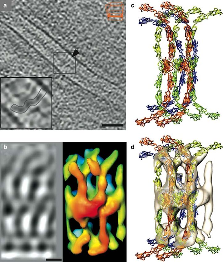
zen, freeze-substituted, embedded in Epon, and contrasted opposing plasma membranes, with a lateral periodicity of
by conventional methods (He et al. 2003). After sectioning, 7 nm. The regularity of desmosomal structure became even
electron tomograms of desmosomes revealed tiny Wlaments more obvious when sub-tomograms extracted from small
in the gap region that extended from the cell membranes to areas were rotationally aligned and averaged in the com-
the midline. Their ends appeared to join at the midline in puter. In averaged images, the rods appeared to be curved
knot-like structures, forming an irregular network. This and to interlock, in a stagger, at the midline with similar
suggested that in desmosomes, the cell adhesion trans- rods emanating from the opposite cell membrane (Fig. 5;
membrane proteins of the cadherin family (desmogleins Al-Amoudi et al. 2007).
and desmocollins) were arranged more or less stochasti- A priori, the ultrastructural model described above was
cally (He et al. 2003). The picture changed when desmo- obtained in an unbiased way that did not depend on previous
somes were viewed on high pressure-frozen but unstained, assumptions about cadherin or desmosome structure. There-
frozen hydrated sections. Now, complex regular patterns of fore, it could be used for evaluating published molecular
varying electron density became visible in the desmosomal models, by Wtting them into the averaged electron tomo-
gap region (Al-Amoudi et al. 2005a). In a follow-up study, grams (Al-Amoudi et al. 2007). Atomic models of desm-
electron tomography was applied to ultrathin vitriWed sec- ogleins and desmocollins are not available, but the crystal
tions through desmosomes (Al-Amoudi et al. 2007). These structure of the closely related and highly homologous
revealed electron-dense rod-like structures perpendicular to C-cadherin ectodomain has been solved (Boggon et al. 2002).
Fig. 5 From electron tomo-
graphs of frozen hydrated sec-
tion to a molecular model of
desmosome structure. a Electron
micrograph of a frozen hydrated
section of human skin, depicting
a desmosome. Note the regularly
spaced W-shape Wlaments (in-
sert in a) crossing the desmo-
some gap. b Averaged sub-
tomogram of desmosome sec-
tion (left), and calculated density
map (right). c Molecular model
of two cadherin tetramers. d
Molecular cadherin model Wtted
into the density map. Scale bar
in a represents 35 nm, in b 7 nm.
Reproduced from Al-Amoudi
et al. (2007) with permission
123886 Histochem Cell Biol (2008) 130:877–889
Like desmosomal cadherins, C-cadherin possesses a large tion with molecular modeling, tomography of such sections
ectodomain built from Wve consecutive immunoglobulin- allows the identiWcation and localization of macromolecu-
like (Ig) modules. The crystal structure of complete C-cad- lar complexes in cells and tissues. Also for CEMOVIS, we
herin ectodomains suggested that they arrange as parallel have to improve and facilitate the steps to let it become a
dimers interacting in cis, i.e., within the plane of the cell routine method. This is a big technical challenge for the
membrane (Boggon et al. 2002). On the other hand, a crystal next years. In our opinion, there is at the moment no
structure of the N-terminal Ig domain (EC1) of N-cadherin equivalent alternative available to describe the molecular
indicated that it is able to form parallel cis- as well as anti- architecture of cells.
parallel trans-interactions with its neighbors in a zipper-like X-ray microscopes (Williams et al. 2008) and sophisti-
manner (Shapiro et al. 1995). The EC1 had been proven cated STED-light microscopes (Schmidt et al. 2008) are
essential for homophilic interactions between cadherin mole- able to depict living cells. The resolution achieved with
cules by mutational studies, but there was disagreement both systems is nowadays in the range of 40 nm. In the far
about whether cadherin ectodomains would zipper rather future X-ray microscopes may depict ultrastructural details
than interact head-on (Koch et al. 1999; Pertz et al. 1999). In in the range of a few nm in a living cell, as it should be theo-
parallel, a model for cadherin-mediated cell–cell adhesion retically possible. Until then, high pressure freezing in
was derived from biochemical and ultrastructural data on the combination with CEMOVIS will be the only way to visu-
isolated proteins (Haussinger et al. 2002), as well as from alize the close to native ultrastructure of living matter.
functional studies with mutant cadherins (Koch et al. 1999;
Pertz et al. 1999). It proposed that cadherins Wrst form Acknowledgments The authors thank Dr. Shanting Zhao (Institute
of Anatomy, University of Freiburg i.Br.) for supplying organotypic
dimers by cis-interactions within the plane of one cell mem-
brain cultures. For technical support we thank Werner Graber and
brane, and that cis-dimers subsequently interact in trans via Barbara Krieger (Institute of Anatomy, University of Bern). DS was
their EC1 domains to form tetramers bridging the intercellu- supported by the Swiss National Foundation (grant no.: 3100A0-
lar space (Pertz et al. 1999). Finally, cadherin tetramers 118394).
were thought to aggregate laterally, establishing adherens
junctions (or desmosomes, respectively) between neighbor-
ing cells. The problem was that such a regular stochiometric References
model was diYcult to reconcile with the appearance of des-
mosomes in electron micrographs of classically Wxed Acetarin JD, Carlemalm E, Villiger W (1986) Developments of new
(Drochmans et al. 1978) or freeze-substituted samples (He Lowicryl resins for embedding biological specimens at even low-
er temperatures. J Microsc 143:81–88
et al. 2003); however, Al-Amoudi et al. 2007) used their
Adrian M, Dubochet J, Lepault J, McDowall AW (1984) Cryo-electron
high pressure frozen, hydrated samples to Wt the published microscopy of viruses. Nature 308:32–36
structural models of cadherin dimer/tetramer complexes into Al-Amoudi A, Chang JJ, Leforestier A, McDowall A, Salamin LM,
the electron density maps of their desmosome tomograms. Norlen LP, Richter K, Blanc NS, Studer D, Dubochet J (2004)
Cryo-electron microscopy of vitreous sections. Embo J 23:3583–
With only a few assumptions (e.g. concerning the angles
3588
between single cadherin domains), a surprisingly good cor- Al-Amoudi A, Dubochet J, Norlen L (2005a) Nanostructure of the epi-
relation was achieved with the cis-dimer/trans-tetramer dermal extracellular space as observed by cryo-electron micros-
model (Al-Amoudi et al. 2007). This is one of the very copy of vitreous sections of human skin. J Invest Dermatol
124:764–777
recent successful attempts to bridge the gap between ultra-
Al-Amoudi A, Studer D, Dubochet J (2005b) Cutting artefacts and
structure and atomic resolution for a macromolecular com- cutting process in vitreous sections for cryo-electron microscopy.
plex within intact tissue samples and the basis for this J Struct Biol 150:109–121
achievement was cryoWxation by high pressure freezing. Al-Amoudi A, Diez DC, Betts MJ, Frangakis AS (2007) The molecular
architecture of cadherins in native epidermal desmosomes. Nature
450:832–837
Allison DP, Daw CS, Rorvik MC (1987) The construction and opera-
Perspectives tion of a simple inexpensive slam freezing device for electron
microscopy. J Microsc 147:103–108
Batson PE, Dellby N, Krivanek OL (2002) Sub-angstrom resolution
At the moment high pressure freezing, as the Wrst step of
using aberration corrected electron optics. Nature 418:617–620
Wxation after sample collection and excision, seems to be Baumeister W (2002) Electron tomography: towards visualizing the
the most promising approach to preserve a sample as close molecular organization of the cytoplasm. Curr Opin Struct Biol
as possible to a snapshot of the living state. Immunolabel- 12:679–684
Baumeister W (2005) From proteomic inventory to architecture. FEBS
ing of small antigens (up to about 30,000 Da) of high pres-
Lett 579:933–937
sure frozen samples has to be improved and facilitated. The Behrman EJ (1984) The chemistry of osmium tetroxide Wxation. In:
most challenging follow-up procedure is cryo-electron Revel JP, Barnard T, Haggis GH (eds) The science of biological
microscopy of vitreous sections (CEMOVIS). In combina- specimen preparation. SEM Inc, AMF O’Hare, Illinois, pp 1–5
123Histochem Cell Biol (2008) 130:877–889 887
Boggon TJ, Murray J, Chappuis-Flament S, Wong E, Gumbiner BM, complexes in cryoelectron tomograms of phantom cells. Proc
Shapiro L (2002) C-cadherin ectodomain structure and implica- Natl Acad Sci USA 99:14153–14158
tions for cell adhesion mechanisms. Science 296:1308–1313 Frotscher M, Zhao S, Graber W, Drakew A, Studer D (2007) New
Böhm J, Frangakis AS, Hegerl R, Nickell S, Typke D, Baumeister W ways of looking at synapses. Histochem Cell Biol 128:91–96
(2000) Toward detecting and identifying macromolecules in a Galway ME, Heckman JW Jr, Hyde GJ, Fowke LC (1995) Advances
cellular context: template matching applied to electron tomo- in high-pressure and plunge-freeze Wxation. Methods Cell Biol
grams. Proc Natl Acad Sci USA 97:14245–14250 49:3–19
Carlemalm E, Armbruster BL, Chiovetti R, Garavito RM, Hobot JA, Guerquin-Kern JL, Wu TD, Quintana C, Croisy A (2005) Progress in
Villiger W, Kellenberger E (1982) Perspectives for achieving im- analytical imaging of the cell by dynamic secondary ion mass
proved information by the observation of thin sections in the elec- spectrometry (SIMS microscopy). Biochim Biophys Acta
tron microscope. Tokai J Exp Clin Med 7(Suppl):33–42 1724:228–238
Causton BE (1986) Does the embedding chemistry interact with tis- Haussinger D, Ahrens T, Sass HJ, Pertz O, Engel J, Grzesiek S (2002)
sue? In: Müller M, Becker RP, Boyde A, Wolosewick JJ (eds) Calcium-dependent homoassociation of E-cadherin by NMR
The science of biological specimen preparation. SEM Inc, AMF spectroscopy: changes in mobility, conformation and mapping of
O’Hare, Illinois, pp 209–214 contact regions. J Mol Biol 324:823–839
Claeys M, Vanhecke D, Couvreur M, Tytgat T, Coomans A, Borgonie Hayat MA (2000) Principles and techniques of electron microscopy:
G (2004) High-pressure freezing and freeze substitution of gravid biological applications, 4th edn. Cambridge University Press,
Caenorhabditis elegans (Nematoda: Rhabditida) for transmission Cambridge
electron microscopy. Nematol 6:319–327 He W, Cowin P, Stokes DL (2003) Untangling desmosomal knots with
Cope GH, Williams MA (1968) Quantitative studies on neutral lipid electron tomography. Science 302:109–113
preservation in electron microscopy. J R Microsc Soc 88:259–277 Hess MW (1993) Cell-wall development in freeze-Wxed pollen: Intine
Cope GH, Williams MA (1969a) Quantitative studies on the preserva- formation of Ledebouria socialis (Hyacinthaceae). Planta
tion of choline and ethanolamine phosphatides during tissue prep- 189:139–149
aration for electron microscopy. I. Glutaraldehyde, osmium Hohenberg H, Mannweiler K, Muller M (1994) High-pressure freezing of
tetroxide, Araldite methods. J Microsc 90:31–46 cell suspensions in cellulose capillary tubes. J Microsc 175:34–43
Cope GH, Williams MA (1969b) Quantitative studies on the preserva- Hohenberg H, Tobler M, Muller M (1996) High-pressure freezing of
tion of choline and ethanolamine phosphatides during tissue prep- tissue obtained by Wne-needle biopsy. J Microsc 183:133–139
aration for electron microscopy. II. Other preparative methods. Humbel BM, Schwarz H (1989) Freeze-substitution for immunochem-
J Microsc 90:47–60 istry. In: Verkleij AJ, Leunissen JLM (eds) Immuno-gold labeling
Craig S, Gilkey JC, Staehelin LA (1987) Improved specimen support in cell biology. CRC Press, Boca Raton, pp 115–134
cups and auxiliary devices for the Balzers high pressure freezing Humbel BM, Marti T, Müller M (1983) Improved structural preserva-
apparatus. J Microsc 148:103–106 tion by combining freeze substitutuion and low temperature
Drochmans P, Freudenstein C, Wanson JC, Laurent L, Keenan TW, embedding. Beitr Elektronenmikroskop Direktabb OberX
Stadler J, Leloup R, Franke WW (1978) Structure and biochemi- 16:585–594
cal composition of desmosomes and tonoWlaments isolated from Kanno H, Speedy RJ, Angell CA (1975) Supercooling of water to
calf muzzle epidermis. J Cell Biol 79:427–443 ¡92°C under pressure. Science 189:880–881
Dubochet J (2007) The physics of rapid cooling and its implications for Kellenberger E, Johansen R, Maeder M, Bohrmann B, StauVer E, Vil-
cryoimmobilization of cells. Methods Cell Biol 79:7–21 liger W (1992) Artefacts and morphological changes during
Dubochet J, McDowall AW, Menge B, Schmid EN, Lickfeld KG chemical Wxation. J Microsc 168:181–201
(1983) Electron microscopy of frozen-hydrated bacteria. J Bacte- Koch AW, Bozic D, Pertz O, Engel J (1999) Homophilic adhesion by
riol 155:381–390 cadherins. Curr Opin Struct Biol 9:275–281
Dubochet J, Adrian M, Chang JJ, Homo JC, Lepault J, McDowall AW, Kopp F (1973) Morphology of the plasmalemma of baker’s yeast Sac-
Schultz P (1988) Cryo-electron microscopy of vitriWed speci- charomyces cerevisiae. In: Benedetti EL, Favard P (eds) Freeze-
mens. Q Rev Biophys 21:129–228 etching techniques and applications. Societé Française de Micros-
Ebersold HR, Luthy P, Cordier JL, Muller M (1981) A freeze-substi- copie Electronique, Paris, pp 181–185
tution and freeze-fracture study of bacterial spore structures. J Ul- Leforestier A, Richter K, Livolant F, Dubochet J (1996) Comparison
trastruct Res 76:71–81 of slam-freezing and high-pressure freezing eVects on the DNA
Edelmann L (1978) A simple freeze-drying technique for preparing cholesteric liquid crystalline structure. J Microsc 184:4–13
biological tissue without chemical Wxation for electron micros- Linner JG, Livesey SA, Harrison DS, Steiner AL (1986) A new tech-
copy. J Microsc 112:243–248 nique for removal of amorphous phase tissue water without ice
Edelmann L (1986) Freeze-dried embedded specimens for biological crystal damage: a preparative method for ultrastructural analysis
microanalysis. Scan Electron Microsc 1986(Pt4):1337–1356 and immunoelectron microscopy. J Histochem Cytochem
Eppenberger-Eberhardt M, Riesinger I, Messerli M, Schwarb P, 34:1123–1135
Muller M, Eppenberger HM, Wallimann T (1991) Adult rat Lucic V, Forster F, Baumeister W (2005) Structural studies by electron
cardiomyocytes cultured in creatine-deWcient medium display tomography: from cells to molecules. Annu Rev Biochem
large mitochondria with paracrystalline inclusions, enriched for 74:833–865
creatine kinase. J Cell Biol 113:289–302 Lucic V, Leis A, Baumeister W (2008) Cryo-electron tomography of
Epperlein HH, Radomski N, Wonka F, Walther P, Wilsch M, Muller cells: connecting structure and function. Histochem Cell Biol
M, Schwarz H (2000) Immunohistochemical demonstration of 130:185–196
hyaluronan and its possible involvement in axolotl neural crest Luft JH (1961) Improvements in epoxy resin embedding methods.
cell migration. J Struct Biol 132:19–32 J Biophys Biochem Cytol 9:409–414
Escaig J (1982) New instruments which facilitate rapid freezing at Matsko N, Müller M (2005) Epoxy resin as Wxative during freeze-sub-
83 K and 6 K. J Microsc 126:221–229 stitution. J Struct Biol 152:92–103
Frangakis AS, Bohm J, Forster F, Nickell S, Nicastro D, Typke D, He- Maupin P, Pollard TD (1983) Improved preservation and staining of
gerl R, Baumeister W (2002) IdentiWcation of macromolecular HeLa cell actin Wlaments, clathrin-coated membranes, and other
123888 Histochem Cell Biol (2008) 130:877–889
cytoplasmic structures by tannic acid-glutaraldehyde-saponin Wx- Romano EL, Stolinski C, Hughes-Jones NC (1974) An antiglobulin re-
ation. J Cell Biol 96:51–62 agent labelled with colloidal gold for use in electron microscopy.
McDonald KL, Morphew MK (1993) Improved preservation of ultra- Immunochemistry 11:521–522
structure in diYcult-to-Wx organisms by high pressure freezing Roth J (1983) The colloidal gold marker system for light and electron
and freeze substitution: I. Drosophila melanogaster and Strongy- microscopy. Theory and application. In: Bullock GR, Petrusz P
locentrotus purpuratus embryos. Microsc Res Tech 24:465–473 (eds) “Techniques in immunocytochemistry”. Academic Press,
Medalia O, Weber I, Frangakis AS, Nicastro D, Gerisch G, Baumeister New York II, pp 217–284
W (2002) Macromolecular architecture in eukaryotic cells visual- Roth J, Bendayan M, Orci L (1978) Ultrastructural localization of
ized by cryoelectron tomography. Science 298:1209–1213 intracellular antigens by the use of protein A-gold complex. J His-
Meryman HT (2007) Cryopreservation of living cells: principles and tochem Cytochem 26:1074–1081
practice. Transfusion 47:935–945 Roth J, Bendayan M, Carlemalm E, Villiger W, Garavito M (1981a)
Moor H (1987) Theory and practice of high pressure freezing. In: Enhancement of structural preservation and immunocytochemi-
Steinbrecht RA, Zierold K (eds) Cryotechniques in biological cal staining in low temperature embedded pancreatic tissue. J His-
electron microscopy. Springer, Berlin, pp 175–191 tochem Cytochem 29:663–671
Moor H, Mühletaler K (1963) Fine structure in frozen-etched yeast Roth J, Ravazzola M, Bendayan M, Orci L (1981b) Application of the
cells. J Cell Biol 17:609–628 protein A-gold technique for electron microscopic demonstration
Moor H, Bellin G, Sandri C, Akert K (1980) The inXuence of high of polypeptide hormones. Endocrinology 108:247–253
pressure freezing on mammalian nerve tissue. Cell Tissue Res Sartori N, Richter K, Dubochet J (1993) VitriWcation depth can be in-
209:201–216 creased more than 10-fold by high pressure freezing. J Microsc
Murk JL, Posthuma G, Koster AJ, Geuze HJ, Verkleij AJ, Kleijmeer 172:55–61
MJ, Humbel BM (2003) InXuence of aldehyde Wxation on the Sawaguchi A, McDonald KL, Forte JG (2004) High-pressure freezing
morphology of endosomes and lysosomes: quantitative analysis of isolated gastric glands provides new insight into the Wne struc-
and electron tomography. J Microsc 212:81–90 ture and subcellular localization of H+/K+-ATPase in gastric
Newman GR, Hobot JA (1987) Modern acrylics for post-embedding parietal cells. J Histochem Cytochem 52:77–86
immunostaining techniques. J Histochem Cytochem 35:971–981 Scala C, Cenacchi G, Ferrari C, Pasquinelli G, Preda P, Manara GC
Newman GR, Jasani B, Williams ED (1983) A simple post-embedding (1992) A new acrylic resin formulation: a useful tool for histolog-
system for the rapid demonstration of tissue antigens under the ical, ultrastructural, and immunocytochemical investigations.
electron microscope. Histochem J 15:543–555 J Histochem Cytochem 40:1799–1804
Nickell S, KoXer C, Leis AP, Baumeister W (2006) A visual approach Schmidt R, Wurm CA, Jakobs S, Engelhardt J, Egner A, Hell SW
to proteomics. Nat Rev Mol Cell Biol 7:225–230 (2008) Spherical nanosized focal spot unravels the interior of
Nitta K, Kaneko Y (2004) Simple plunge freezing applied to plant tis- cells. Nat Methods 5:539–544
sues for capturing the ultrastructure close to the living state. Schwarz H, Humbel BM (1989) InXuence of Wxatives and embedding
J Electron Microsc (Tokyo) 53:677–680 media on immunolabelling of freeze-substituted cells. Scanning
North AJ, Bardsley WG, Hyam J, Bornslaeger EA, Cordingley HC, Microsc Suppl 3:57–63 Discussion 63–54
Trinnaman B, Hatzfeld M, Green KJ, Magee AI, Garrod DR Semmler K, Wunderlich J, Richter W, Meyer HW (1998) High-pres-
(1999) Molecular map of the desmosomal plaque. J Cell Sci sure freezing causes structural alterations in phospholipid model
112(Pt 23):4325–4336 membranes. J Microsc 190:317–327
Palade GE, Porter KR (1954) Studies on the endoplasmic reticulum. I. Shanbhag SR, Park SK, Pikielny CW, Steinbrecht RA (2001) Gusta-
Its identiWcation in cells in situ. J Exp Med 100:641–656 tory organs of Drosophila melanogaster: Wne structure and
Pease DC, Porter KR (1981) Electron microscopy and ultramicrotomy. expression of the putative odorant-binding protein PBPRP2. Cell
J Cell Biol 91:287s–292s Tissue Res 304:423–437
Pertz O, Bozic D, Koch AW, Fauser C, Brancaccio A, Engel J (1999) Shapiro L, Fannon AM, Kwong PD, Thompson A, Lehmann MS, Gru-
A new crystal structure, Ca2+ dependence and mutational analysis bel G, Legrand JF, Als-Nielsen J, Colman DR, Hendrickson WA
reveal molecular details of E-cadherin homoassociation. Embo J (1995) Structural basis of cell–cell adhesion by cadherins. Nature
18:1738–1747 374:327–337
PfeiVer S, Krupinska K (2005) Chloroplast ultrastructure in leaves of Sharp DJ, McDonald KL, Brown HM, Matthies HJ, Walczak C, Vale
Urtica dioica L. analyzed after high-pressure freezing and freeze- RD, Mitchison TJ, Scholey JM (1999) The bipolar kinesin,
substitution and compared with conventional Wxation followed by KLP61F, cross-links microtubules within interpolar microtubule
room temperature dehydration. Microsc Res Tech 68:368–376 bundles of Drosophila embryonic mitotic spindles. J Cell Biol
Ravazzola M, Perrelet A, Roth J, Orci L (1981) Insulin immunoreac- 144:125–138
tive sites demonstrated in the Golgi apparatus of pancreatic B Shimoni E, Muller M (1998) On optimizing high-pressure freezing:
cells. Proc Natl Acad Sci USA 78:5661–5664 from heat transfer theory to a new microbiopsy device. J Microsc
Richter T, Biel SS, Sattler M, Wenck H, Wittern KP, Wiesendanger R, 192:236–247
Wepf R (2007) Pros and cons: cryo-electron microscopic evaluation Shotton D, Thompson K, Wofsy L, Branton D (1978) Appearance and
of block faces versus cryo-sections from frozen-hydrated skin spec- distribution of surface proteins of the human erythrocyte mem-
imens prepared by diVerent techniques. J Microsc 225:201–207 brane. An electron microscope and immunochemical labeling
Riehle U, Hoechli M (1973) The theory and technique of high pressure study. J Cell Biol 76:512–531
freezing. In: Benedetti EL, Favard P (eds) Freeze etching tech- Studer D, Michel M, Muller M (1989) High pressure freezing
niques and applications. Société Française de Microscopie Elect- comes of age. Scanning Microsc Suppl 3:253–268 Discussion
ronique, Paris, pp 31–61 268–259
Ripper D, Schwarz H, Stierhof YD (2008) Cryo-section immunolabel- Studer D, Hennecke H, Müller M (1992) High pressure freezing of
ling of diYcult to preserve specimens: advantages of cryoWxation, soybean nodules leads to an improved preservation of ultrastruc-
freeze-substitution and rehydration. Biol Cell 100:109–123 ture. Planta 188:155–163
Romano EL, Romano M (1977) Staphylococcal protein A bound to Studer D, Michel M, Wohlwend M, Hunziker EB, Buschmann MD
colloidal gold: a useful reagent to label antigen-antibody sites in (1995) VitriWcation of articular cartilage by high-pressure freez-
electron miroscopy. Immunochemistry 14:711–715 ing. J Microsc 179:321–332
123Histochem Cell Biol (2008) 130:877–889 889
Studer D, Chiquet M, Hunziker EB (1996) Evidence for a distinct wa- Verkade P (2008) Moving EM: the rapid transfer system as a new tool
ter-rich layer surrounding collagen Wbrils in articular cartilage for correlative light and electron microscopy and high throughput
extracellular matrix. J Struct Biol 117:81–85 for high-pressure freezing. J Microsc 230:317–328
Studer D, Graber W, Al-Amoudi A, Eggli P (2001) A new approach Walther P (2003) Recent progress in freeze-fracturing of high-pressure
for cryoWxation by high-pressure freezing. J Microsc 203:285– frozen samples. J Microsc 212:34–43
294 Walther P, Ziegler A (2002) Freeze substitution of high-pressure frozen
Sung HW, Hsu HL, Shih CC, Lin DS (1996) Cross-linking charac- samples: the visibility of biological membranes is improved when
teristics of biological tissues Wxed with monofunctional or the substitution medium contains water. J Microsc 208:3–10
multifunctional epoxy compounds. Biomaterials 17:1405– Wang L, Humbel BM, Roubos EW (2005) High-pressure freezing fol-
1410 lowed by cryosubstitution as a tool for preserving high-quality
Szczesny PJ, Walther P, Muller M (1996) Light damage in rod outer ultrastructure and immunoreactivity in the Xenopus laevis pitui-
segments: the eVects of Wxation on ultrastructural alterations. tary gland. Brain Res Brain Res Protoc 15:155–163
Curr Eye Res 15:807–814 Waschke J (2008) The desmosome and pemphigus. Histochem Cell
Tokuyasu KT (1973) A technique for ultracryotomy of cell suspen- Biol 130:21–54
sions and tissues. J Cell Biol 57:551–565 White DL, Andrews SB, Faller JW, Barrnett RJ (1976) The chemical
Tokuyasu KT (1980) Immunochemistry on ultrathin frozen sections. nature of osmium tetroxide Wxation and staining of membranes by
Histochem J 12:381–403 X-ray photoelectron spectroscopy. Biochim Biophys Acta
Van Donselaar E, Posthuma G, Zeuschner D, Humbel BM, Slot JW 436:577–592
(2007) Immunogold labeling of cryosections from high-pressure Williams GJ, Hanssen E, Peele AG, Pfeifer MA, Clark J, Abbey B, Ca-
frozen cells. TraYc 8:471–485 denazzi G, de Jonge MD, Vogt S, Tilley L, Nugent KA (2008)
Van Harreveld A, Crowell J (1964) Electron microscopy after rapid High-resolution X-ray imaging of Plasmodium falciparum-in-
freezing on a metal surface and substitution Wxation. Anat Rec fected red blood cells. Cytometry A
149:381–385 Zeuschner D, Geerts WJ, van Donselaar E, Humbel BM, Slot JW, Ko-
Vanhecke D, Graber W, Herrmann G, Al-Amoudi A, Eggli P, Studer ster AJ, Klumperman J (2006) Immuno-electron tomography of
D (2003) A rapid microbiopsy system to improve the preservation ER exit sites reveals the existence of free COPII-coated transport
of biological samples prior to high-pressure freezing. J Microsc carriers. Nat Cell Biol 8:377–383
212:3–12 Zuber B, Nikonenko I, Klauser P, Muller D, Dubochet J (2005) The
Vanhecke D, Graber W, Studer D (2008) Close-to-native ultrastructur- mammalian central nervous synaptic cleft contains a high density
al preservation by high pressure freezing. Methods Cell Biol of periodically organized complexes. Proc Natl Acad Sci USA
88:151–164 102:19192–19197
123You can also read