Call for Papers - 2019 August 4-8 Portland, OR - Microscopy Society of America
←
→
Page content transcription
If your browser does not render page correctly, please read the page content below
Call for Papers
 Submission Deadline:
 February 15, 2019

 Look Inside for Program Details, Plenary
 Speakers, Pre-Meeting Congresses, and more!

 www.microscopy.org/MandM/2019
 for up-to-date meeting information

                             2019

August 4–8 Portland, OR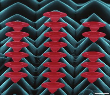
COVER IMAGES (top to bottom):
1st Place Winner | Timothy Pegg - Miami University of Ohio - Motley Spheres SLASH Prismatic Spheres
2nd Place Winner | Vivek Garg - IITB MOnash Research Academy - Microscopic Gardening Tiny Blossoms of Silicon
2nd Place Winner | Gary Bauchan - USDA Agricultural Research Service - Coffee Drinking Flat Mite

    Letter from the                                                                                                       2019
    Presidents
    On behalf of the Microscopy Society of America, the Microanalysis Society, and                               Questions?
    the International Field Emission Society, we invite you to join us August 4-8, 2019,
    for Microscopy & Microanalysis 2019 at the Oregon Convention Center in Portland,                             TECHNICAL MEETING
                                                                                                                 CONTENT:
    Oregon. This will be the fourth M&M meeting held in the City of Roses. It’s an
                                                                                                                 2019 Program Chair
    excellent venue with wonderful restaurants, lots of activities for the family, and a
                                                                                                                 Alice Dohnalkova,
    comfortable climate with August highs near 80°F (27°C).                                                      Pacific Northwest National
                                                                                                                 Laboratory
    The Program Committee, led by Alice Dohnalkova, Huolin Xin, Assel Aitkaliyeva,
                                                                                                                 MM2019ProgramChair@
    and Baptiste Gault, has developed an exciting group of symposia, spanning                                    microscopy.org
    advances in instrumentation and techniques development, as well as applications
    in the analytical, biological, and physical sciences. We encourage you to browse                             REGISTRATION:
    the Call for Papers for the complete symposium list, and to submit one or more                               Registrar
    scientific papers for platform or poster presentation. Tables will continue to be                            MMRegistration@
    provided at the poster boards for fully dynamic multimedia poster presentations.                             conferencemanagers.com

    The main meeting will be preceded by the ever-popular Sunday Short Courses, two                              EXHIBITS & EXHIBITORS:
    Pre-Meeting Congresses, and will officially start with the Opening Welcome Reception                         Exhibits Manager
    on Sunday evening. The Sunday reception is a great place for all attendees to                                doreen@corcexpo.com
    meet new colleagues and reconnect with old friends. Students and early-career
    professionals are especially encouraged to participate in the MSA Student Council-                           SPONSORS & SPONSORSHIPS:
    led Pre-Meeting Congress that highlights outstanding work by student and post-                               Sponsorship Manager
    doctoral fellow attendees. On Monday morning, the Plenary Session kicks off the                              mary@corcexpo.com
    scientific program with two exciting plenary lectures, and the presentations of the
    M&M meeting awards and awards from the sponsoring societies. We are excited to                               GENERAL:
    announce that two of the 2017 Nobel Prize winners in Chemistry, Professors Joachim                           Meeting Manager
    Frank and Richard Henderson, will be our 2019 Plenary Speakers. Please join us                               meetingmanager@microscopy.org
    in welcoming back these long-time MSA members and frequent M&M attendees as
    they discuss their groundbreaking work in cryo-electron microscopy.                                          Are You a
    In addition to the strong scientific program, what sets the M&M meeting apart is the                         Member?
    Exhibit Hall, the world’s largest annual microscopy exhibition, which showcases
    the latest in microscopy instrumentation and accessories. Don’t miss the highly                              Join Today and Save on M&M
    popular vendor tutorials, held Monday through Wednesday after hours in the                                   2019 Registration Fees
    Exhibit Hall. Other educational opportunities throughout the week include focused
    biological and physical science tutorials, educational outreach programs, and our                            Visit http://
    Technologists’ Forum special and roundtable sessions.                                                        microscopy.org
                                                                                                                 to join the Microscopy Society
    In short, M&M 2019 will be an outstanding opportunity to stay abreast of the latest                          of America online, or call
    technologies, hear about new developments in applications across all areas of                                1-800-538-3672 for more
    microscopy and microanalysis, and most importantly network with colleagues.                                  information about the benefits
                                                                                                                 of MSA membership.
    We hope to see you in Portland!
                                                                                                                 Visit http://
    Paul Kotula, Sandia                Rhonda Stroud, U.S. Naval                David Larson, AMETEK, Inc.
    National Laboratories              Research Laboratory                      President, International Field
                                                                                                                 microanalysissociety.org
    President, Microscopy              President, Microanalysis Society         Emission Society                 to find out the benefits
    Society of America                                                                                           of MAS membership.

                                                                                                                 Visit http://www.
                                                                                                                 fieldemission.org/
                                                                                                                 index.php to join the
                                                                                                                 International Field Emission
                                                                                                                 Society.
    Ted Kinsman - Rochester Institute of Technology - Extremophile Community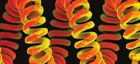
Biological Sciences Symposia
                                                                           Geoff Williams - Brown University - Mantodea Nymph

B01 Multi-Modal, Large-Scale, and 3D                  B04 Cutting Edge Microscopy in the                   • Root cause analysis of issues related to the
    Correlative Microscopy                                Pacific Northwest                                  manufacturing of drugs, medical products,
    James Fitzpatrick, Washington University School     Douglas Keene, Shriners Hospital for Children        and devices
                                                                                                           • Device challenges (failure mode analysis,
    of Medicine                                         Claudia Lopez, Oregon Health & Science               biocompatibility, sterility, etc.)
    Jacob Hoogenboom, Delft University of               University and Pacific Northwest Center
                                                                                                           • Regulatory and data integrity compliance
    Technology, Netherlands                             for Cryo-EM
                                                                                                             of instrumentation and methods in the
    Ben Giepmans, University Medical Center           • Cryo-electron microscopy: best                       pharmaceutical industry including challenges
    Groningen, Netherlands                              approaches for data collection and                   and best practices for 21 CFR part 11
• Multi-Modal Microscopy                                analysis                                           • Roundtable discussion with pharma peers and
• Correlative Microscopy (CLEM)                       • Pushing the limits of super-resolution               major equipment suppliers as well as invited
• Large-Scale Volume EM                                 fluorescence microscopy                              governing regulatory bodies.
• Data Recognition and Modeling                       • Quantitative analysis in microscopy
• Novel Probes and Sample                             • 3D microscopy
                                                      • Novel approaches for correlative light             B07 3D Structures: from Macromolecular
  Preparation Workflows
• New Hardware and Instrumentation                      and electron microscopy                                Assemblies to Whole Cells (3DEM FIG)
                                                      • Data mining, machine learning                         Melanie Ohi, University of Michigan Life
B02 Element Analysis of                                 approaches to analyze large electron                  Sciences Institute
                                                        and optical microscopy datasets
    Biological Materials                                                                                      Elitza Tocheva, University of British
                                                                                                              Columbia, Canada
    Peta Clode, University of Western Australia
    Stefan Vogt, Argonne National Laboratory
                                                      B05 Light and Fluorescence                              Teresa Ruiz, University of Vermont
                                                          Microscopy for Imaging Cell
    Nicole Hondow, University of Leeds,                                                                    • Structure and function of macromolecular
                                                          Surface and Structure
    United Kingdom                                                                                           complexes in vitro and in vivo
                                                        Justin Taraska, NIH - Laboratory of                • Single particle cryo-electron microscopy
•   Element & Isotopic analysis                         Molecular & Cellular Imaging
•   Cell & tissue mapping                                                                                  • Cryo-electron tomography
•   Electron, ion, & X-ray sources                      David Zenisek, Yale University
                                                        School of Medicine
•   Bio-nanotechnology                                                                                     B08 Cryo-EM - from Physics to Cell
•   Biominerals                                         David Perrais, CNRS UMR, Institut
                                                        interdisciplinaire de Neurosciences,
                                                                                                               Biology: Honoring the Remarkable
                                                        France                                                 Legacy of Ken Downing
B03 Utilizing Microscopy for Research                   Xiaolin Nan, Oregon Health                            Melanie Ohi, University of Michigan Life
    and Diagnosis of Diseases in                        Sciences University                                   Sciences Institute
    Humans, Plants and Animals                          Randall Smith, Portland State                         Eva Nogales, University of California-Berkeley,
                                                        University                                            Lawrence Berkeley National Laboratory
    Ru-ching Hsia, University of
    Maryland-Baltimore                                • Confocal, light sheet, and expansion               • Cryo-electron microscopy
    Marcela Redigolo, West Virginia University          microscopy research topics                         • Microtubule structure and regulation
                                                      • Immunocytochemistry in current cell                • Electron crystallography
    Han Chen, Penn State College of Medicine            biology research
• Microscopic characterization of cellular and        • Total Internal Reflection Microscopy
                                                                                                           B09 From Images to Insights:
  molecular structure in normal and diseased            (TIRF): Research derived from insights and
  humans, animals and plants                            works by Christien J. Merrifield                       Working with Large Data in
• Applications of microscopic imaging                 • Polarization optics                                    Cell Biological Imaging
  for basic and clinical research with                • Fluorescence reporter proteins,                       Kedar Narayan, National Cancer Institute
  emphases in technical development and                 fluorescent protein development and
                                                                                                              Camenzind Robinson, St. Jude Children’s
  implementation                                        characteristics                                       Research Hospital
• Investigation of organisms and their                • Related correlative microscopies
  related pathogens in clinical and research                                                                  Jonathan Lefman, NVIDIA Corporation
  laboratories                                                                                             • Processing, storing, and distributing
• Techniques that improve rapid detection
                                                      B06 Pharmaceuticals FIG – Imaging,
                                                          Analysis, and Regulation of                        large image data in cell biology
  of pathogens and accurate diagnosis,                                                                     • Correlating images and metadata from
  for example, quantitative microscopy,                   Medical Products, Devices and
                                                                                                             LM, EM, and other modalities (XRM,
  nanomaterials in diagnosis, imaging                     Data Integrity
                                                                                                             chemical imaging, etc.)
  cytometry, high throughput microscopy,                Gianpiero Torraca, Amgen, Inc.                     • Extracting, segmenting, and visualizing
  automation of microscopy, etc.
                                                        Daniel Skomski, Merck Research                       features of interest efficiently
                                                        Laboratories                                       • Adapting and applying open-source
                                                                                                             tools and frameworks
                                                      • Novel microscopic or spectroscopic                 • Scalable solutions for small and mid-
                                                        methods applied to biomedical/                       sized labs and facilities
                                                        biotechnological areas
                                                      • Pharmacology challenges (polymorphs,
                                                        contaminants, particles, etc.)

                                                                                                                                        Microscopy & Microanalysis 2019
                                                                                                                                               August 4-8 | Portland, OR   3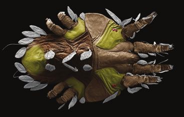
Physical Sciences Symposia
Erica Stevens - University of Pittsburgh - When the Particles Align
P01 In situ TEM Characterization P03 Revealing the Fundamental P05 Theory and Applications of
of Dynamic Processes Structure of Soft and Hard Electron Tomography in the
During Materials Synthesis Matter by Minimizing Materials Sciences
and Processing Beam-Sample Interactions Peter Ercius, Lawrence Berkeley
Dongsheng Li, Pacific Northwest Joerg Jinschek, The Ohio State University National Laboratory
National Laboratory Robert Hovden, University of Michigan
David Flannigan, University of Minnesota
Haimei Zheng, Lawrence Berkeley Sandra Van Aert, University of
Dalaver H. Anjum, King Abdullah
National Laboratory and University of Antwerp, Belgium
University of Science & Technology
California-Berkeley
(KAUST), Saudi Arabia
• Structure-property relationships in 3D
Liang Jin, Direct Electron
Stig Helveg, Haldor Topsoe A/S, Denmark across the nano- to atomic-scale
Yu Han, King Abdullah University of • Advanced reconstruction algorithms
• Development of new EM modes such
Science and Technology, Saudi Arabia and theory (discrete tomography,
as low-dose / low-dose-rate electron
atomic resolution, compressed sensing,
• Nucleation and crystal growth from microscopy, low-voltage electron
ptychography, etc.)
solutions, melts, and vapors microscopy, ultrafast TEM, or quantum
• Tomographic techniques that push the
• Technical advances, applications and electron microscopy, and their impact on
limits of spatial resolution, time, or in-situ
practical experiences associated with beam-sample interactions
environments
electrochemical processes including • Studying the effects of sample support,
• Multi-modal (multi-detector, spectroscopic)
batteries, water splitting, fuel cell, and temperature (e.g., cryogenic), and
3D reconstruction and visualization
photoelectrochemistry environment (gas, liquid) on beam-
• Developments in specialized holders sample interactions
and electron microscopes, data analysis • Minimizing electron dose and/or electron P06 In situ TEM of Nanoscale Materials
and mining, and practical challenges for dose-rates to prevent radiolysis and and Electronic Devices for Phase
microscopy displacement damage Transformation Studies
• Chemical and electrochemical reactions • Optimization of the detection of every
• Polymeric and organic/inorganic self- scattering event by techniques such as Leopoldo Molina-Luna, Technische Universität
assembly and nanoparticle mediated phase-plate imaging, direct electron Darmstadt, Germany
growth and oriented attachment detection, high-speed image acquisition, Lin Zhou, Ames Laboratory
• Solid-gas interaction and new techniques for image
processing Judy J. Cha, Yale University
• In situ TEM development: imaging,
P02 Microscopy and Microanalysis of acquisition, software
Nuclear and Irradiated Materials P04 Spectroscopy and Imaging of • Development of In situ TEM holders
Chad Parish, Oak Ridge National Nanostructured Low-Z Materials and chips for thermal, electrical, and
Laboratory in the Electron Microscope mechanical excitations
Khalid Hattar, Sandia National Laboratories Dan Hodoroaba, Federal Institute • Phase transformation of nanomaterials
Pater Hosemann, University of for Materials Research and Testing by in situ heating and cooling TEM
California–Berkeley (BAM), Germany experiments
• Phase transformation by mechanical
Assel Aitkaliyeva, University of Florida Andrew Stewart, University of
testing of in situ TEM experiments
Limerick, Ireland
• Fission, fusion, accelerator, and space • Electric field / current induced phase
materials: metals, ceramics, polymers, Meiken Falke, Bruker Nano GmbH, transformation of functional materials
semiconductors, fuels, etc. Germany
• Damage phenomena: dislocation loops, • Organic (e.g. polymers) and inorganic P07 Electron Crystallography of Nano-
segregation and precipitation, stacking (e.g. oxides) compunds, biomaterials;
fault tetrahedral, etc. structures in Nanotechnology,
also light elements to be quantified in
• Microscopy via SEM, TEM, aberration- Materials and Bio-Sciences
heavy matrices (e.g. carbon in steel)
correction, in situ microscopy, etc. and light matter with nano-inclusions Sergei Rouvimov, University of Notre Dame
• Microanalysis via microprobe, atom (e.g. oxide films containing metallic Roberto Reis, Lawrence Berkeley
probe, mass spectroscopy, etc. nanoparticles) National Laboratory
• Modelling and theory approaches that • High-resoluton spectroscopy
aid in interpretation of microscopy data techniques for SEM and STEM, Alex Eggeman, University of Manchester,
of these phenomena e.g. EDS, EELS United Kingdom
• Applications of data science to large or • Imaging techniques using e.g. • Scanning electron diffraction techniques for
sparse microscopy datasets annular detectors to quantify light micro-structure analysis including 4D-STEM,
elements in STEM EBSD,SPED, and others
• Correlative approaches combining • Electron diffraction methods for soft and
different spectroscopies and imaging biological materials
to study low-Z materials (e.g. SEM • New approaches in analysis and simulation
and Raman, SEM and Auger electron in electron crystallography to improve
spectroscopy, STEM ABF imaging and the speed and reliability of structure
spectroscopy) characterization
• Simulations and theoretical approaches • Solving and refining atomic arrangements
to quantify light element compositions from electron diffraction data
in electron imaging, EDS, EELS, etc. • Novel strategies in materials research
4 2019
www.microscopy.org/MandM/2019a
Eric Formo - University of Georgia - To Boldly Go
P08 Microscopy and Spectroscopy P11 Advances in Characterization of P13 Advanced Characterization
of Nanoscale Materials for Geological and Extraterrestrial of Components Fabricated by
Energy Applications Samples Additive Manufacturing
Chongmin Wang, Pacific Northwest Bradley De Gregorio, U.S. Naval Research Isabella van Rooyen, Idaho National
National Laboratory Laboratory Laboratory
Bobby Hooghan, Weatherford Laboratories Subhashish Meher, Idaho National
Matthew T. McDowell, Georgia Laboratory
Lori Hathon, University of Houston
Institute of Technology Federico Sciammarella, Northern
Yuanyuan Zhu, University of Connecticut
• Novel uses of various imaging and Illinois University
analytical techniques to characterize Cesar Terrazas, The University of
• Novel imaging and spectroscopy geological samples Texas-El Paso
techniques for structural and chemical • Investigations of natural materials
• TEM and STEM studies (imaging,
evolution of nanoscale materials requiring advanced microscopy and
EDS, electron diffraction, EELS) to
• Mass and charge transport in materials with microanalysis
understand phase transformations,
dimensionalities from 0 to 3D • Innovative solutions to long-standing
microstructural evolution in
• Multi-scale to atomic-resolution imaging technical challenges for sample
components produced by various
and spectroscopy of materials related to preparation and characterization
energy harvesting and storage additive manufacturing (AM) processes
• New insights into the formation, history,
• Low-dose and low dose-rate imaging • 3D microstructure analysis methods
and use of geological and extraterrestrial
and spectroscopy for beam-sensitive on the micro-, nano-, and atomic scale
samples enabled by microscopy
nanostructures to understand the integrity of AM
• Micro- and nano-scale studies of minerals,
• Advances in ultrafast imaging and fabricated products
both isolated and ensemble, on the
spectroscopy data collection and • Microstructural response of AM
generation, storage, and preservation of
interpretation components to post-processing
organic matter
conditions
• Applications of imaging technologies in
• Current challenges in analytical
P09 The Success of TMBA: TEM and petroleum exploration and production,
tools for microscopy and
STEM Developments in Techniques, including linking imaging and image
microanalysis of AM products
Applications and Education analysis with laboratory physical
• Microstructure and defect analysis by
property measurements (e.g. porosity,
Masashi Watanabe, Lehigh University permeability, wettability, strength and
both characterization and modeling for
insights into solidification and melt pool
Joseph Michael, Sandia National Laboratories acoustic properties), and upscaling
dynamics in AM processes
Paul Kotula, Sandia National Laboratories properties from the pore to the core scale
• In situ experiments on AM products
• Advances in TEM and STEM
• Improvements in the analysis of TEM P12 New Frontiers in Atom Probe
and STEM data Tomography Applications
• Educational approaches to teaching of Baishakhi Mazumder, University at Buffalo
STEM and TEM
• The role of tools and data in education Arun Devaraj, Pacific Northwest National
and research Laboratory
• APT analysis of minerals, biominerals, soft
matter, biological tissues
P10 Applications of Integrated • APT characterization of semiconductor
Electron Probe Microscopy materials and devices
and Microanalysis Techniques • Investigation of processes limiting the
in Characterizing Natural and lifetime of engineering materials in service;
Synthetic Materials e.g. corrosion, mechanical fracture,
Donggao Zhao, University of Missouri- radiation damage, etc.
Geoff Williams - Brown
Kansas City University - Tungsten Filament
Minghua Ren, University of Nevada-
Las Vegas
Owen Neill, University of Michigan
• Imaging from SE, BSE, X-ray, CL, charge
contrast, transmitted electron, diffracted
or scattered electron, etc.
• Qualitative and quantitative
determination of chemical compositions
of natural and synthetic materials
• Repeatability, reproducibility and
compatibility of quantitative microanalysis
standards
• Crystal structure determination
using electron diffraction or electron
backscatted diffraction (EBSD)
Microscopy & Microanalysis 2019
August 4-8 | Portland, OR 5Analytical Sciences Symposia
Tony Fearns - The Francis Crick Institute - You Take my Breath Away
A01 Advances in Phase A04 Recent Developments in A07 Vendor Symposium
Retrieval Microscopy Atom Probe Tomography Elizabeth Dickey, North Carolina State
Kai He, Clemson University Ty Prosa, Cameca Instruments Inc. University
Charudatta Phatak, Argonne National Baptiste Gault, Max-Planck-Institut für Deborah Kelly, Virginia Carilion
Laboratory Eisenforschung, Germany Research Institute
Toshiaki Tanigaki, Hitachi Ltd. David J. Larson, Cameca Instruments Inc. • New methods and techniques; new
Martha McCartney, Arizona State University developments and technologies
• New developments in field
• Four-dimensional scanning • Breakthroughs and new
evaporation theories and mechanisms
transmission electron microscopy instrumentation
• Advances in APT instrumentation and
(4D-STEM) • Improvements to existing
technique development (including FIM)
• In-line and off-axis electron holography instrumentation
• Reconstruction improvements and
• Electron and X-ray ptychography future directions
• Magnetic imaging (Lorentz TEM, DPC, • Standards development for atom A08 Current Trends and Challenges
EMCD, phase plate, etc.) probe tomography in Electron Energy-Loss
• New theory, instrumentation, and Spectroscopy
computational algorithms Patricia Abellan, SuperSTEM Laboratory,
A05 Leveraging 3D Imaging
• In-situ phase retrieval methods United Kingdom
and Analysis Methods
for New Opportunities in Matthieu Bugnet, University of Lyon –
A02 Data Acquisition Schemes, Material Science CNRS, France
Machine Learning Algorithms, Ashwin Shahani, University of Michigan Peter Crozier, Arizona State University
and Open Source Software
Roland Brunner, Materials Center Leoben Xiaoqing Pan, University of California-Irvine
Development for Electron
Forschung GmbH, Germany
Microscopy • Recent advances in acquisition,
Wil Harris, Carl Zeiss Microscopy processing and modelling of low
Francisco de la Peña, University of
Lille, France Erdmann Spiecker, Universität Erlangen- energy-loss EELS
Philippe T. Pinard, Oxford Instruments Nürnberg, Germany • Latest developments in core-loss
NanoAnalysis, United Kingdom EELS acquisition
• 3D, including repetitive time-lapse
Eric Prestat, University of Manchester and
• Fundamental electron matter
‘4D’, microscopy methods for
SuperSTEM, United Kingdom interaction (molecular/solid level)
materials science
at the (sub)nanometer scale
• Open-source and/or community- • Challenges with respect to big
• Non-destructive characterization
driven software development data handling
of chemical bonding of functional
• Machine learning • Challenges with respect to image
groups and adsorbates at
• New algorithms and processing processing/analysis
surfaces and interfaces
workflow • Linking imaging data with
• Fundamentals and applications of
• Novel data acquisition schemes computational methods and modeling
vibrational EELS
A03 Low-Energy X-ray Spectroscopy: A06 Low Voltage, Low Energy Electron
A09 Microscopy and
Novel Applications Using Soft Microscopy Imaging and Analysis
Microanalysis for Real-World
X-ray Emission Spectroscopy David C. Bell, Harvard University Problem Solving
(SXES), Cathodoluminescence (CL)
Natasha Erdman, JEOL USA Inc. Janet H. Woodward, Buckman
and Synchrotron Techniques
Hector Calderon, Instituto Politécnico Ke-Bin Low, BASF Corporation
Anette von der Handt, University of
Nacional, Mexico
Minnesota Xiaofeng Zhang, Nanosys Inc.
Emma Bullock, Carnegie Institution • Low voltage application of TEM, SEM
• Real-world problem solving using all
for Science and STEM
• Analyical possibilities with low voltage forms of microscopy and microanalysis
Juliane Gross, Rutgers University • Practical applications of correlative
and low energy electron microscopy
Zach Gainsforth, University of methods employing microscopy and
California-Berkeley • New designs of electron microcopes
for low voltage or energy operation related techniques
• Low energy spectroscopy • Materials preparation consideration • Quantitative approaches for
• Chemical state analysis for low voltage/ low energy operation increased confidence in results
• Trace chemistry from non-ideal samples
• SXES, CL, XAS, EELS, XPS • Creative methodologies for
• Geological and extraterrestrial preparation and analysis of real
materials world samples
• Biological materials • Equipment testing, calibration and
quality assurance
6 2019
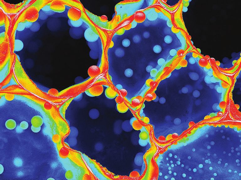
www.microscopy.org/MandM/2019Sunanda Sharma - MIT - Deep Blood Vision
A10 Advances in Focused Ion A11 Current and Emerging A12 Advances in Cryo-EM
Beam Instrumentation, Microscopy for Quantum Technology
Applications and Information Sciences Mike Marko, Wadsworth Center
Techniques Miaofang Chi, Oak Ridge
Anchi Cheng, NY Structural
Suzy Vitale, Carnegie Institution of National Laboratory
Biology Center
Washington Sonia Conesa-Boj, Delft University of
Radostin Danev, Tokyo University, Japan
Joshua Sugar, Sandia National Laboratories Technology, Netherlands
Bruce Arey, Pacific Northwest Lena F. Kourkoutis, Cornell University
• Sample preparation,
National Laboratory including cryo-FIB
• New instrumentation to enable the • EM instrumentation (cameras,
Alan Bahm, Thermo Fisher Scientific
investigation of quantum materials phase plates, automation)
• Beyond Ga: FIB applications using in S/TEMs • Image processing for single-
Xe, He, Ne, and development of new • Current and emerging imaging particle and tomographic
ion sources including Cs and spectroscopy techniques reconstruction
• Advances in cryo-FIB and working for understanding quantum • Applications using cutting-
with beam-sensitive materials phenomena in materials edge technology
• Advanced circuit edit and in situ • New insights into the behavior of
device characterization electrons, ions, lattice and spin, and/
• Novel geometries, milling or their correlations in materials
strategies and non-standard lift • Probing spin states and
outs for TEM/STEM charge transfer in energy and
• Alternative gas chemistries, etching, quantum materials
and complex structure fabrications • Qualitative and quantitative analysis
• Enhancing analytical SEM with FIB, of charge distributions in materials
including 3D EDS/EBSD or SIMS, • Development of new, ultra-stable in
and other correlative analytics situ stages for biasing, cooling, and
including WDS, CL, Raman mechanical contact, etc.
spectroscopy, EBIC, TKD
Joseph Mowery - USDA Agricultural Research Center - Happy Little Protozoa
Technologists’ Forum Sessions
X30 Utilization of the National X31 Roundtable: Technical Careers X32 Imaging Resin Embedded
NIH funded Cryo-EM Centers: in Microscopy – For the Love of Samples for Serial
Transformative High Resolution Microscopy Block Face Imaging or
Cryo-Electron Microscopy CHAIRS: Phoebe J. Doss, University of Texas Array Tomography
CHAIRS: Claudia Lopez, Oregon Health & Southwestern Medical Center CHAIRS: Janice G. Pennington, University
Science University Janice G. Pennington, University of of Wisconsin-Madison
Janice G. Pennington, University of Wisconsin-Madison Phoebe J. Doss, University of Texas
Wisconsin-Madison Southwestern Medical Center
• Technologists from diverse
• Sample preparation “Do’s & Don’ts” backgrounds in microscopy will • Array tomography, a technique
• Best approaches for data collection speak about their careers. for imaging serial sections for 3D
• Direct Electron detectors: uses • How did they find out about reconstruction, will be compared
and preferences microscopy as a career? Why did with SBFSEM and FIB SEM
• Data processing and handling they choose that instead of all the • Tips for resin embedding of
• Best practices in a national other options available? samples for SEM imaging
laboratory • How has their career developed • Tips for preparing serial sections
• “Personalities” of different centers through the years and what advice for array tomography
do they have for technologists new • Techniques for correlative light
to the field? and electron microscopy
• Learn how to become a Certified
Electron Microscopy Technologists
(CEMT) and what it can do for you
to promote your career.
• Become a part of the conversation
and share your story!
Microscopy & Microanalysis 2019
August 4-8 | Portland, OR 7Tutorials
Spencer Reisbeck - University of Minnesota - TaS2 Nano Lava Pits
PHYSICAL SCIENCES TUTORIALS
X40 Following the Electrons: X41 Entrepreneurship in the X42 Efficient Phase Contrast Imaging
Simulation for High-Resolution Microscopy Community via Electron Ptychography
STEM and CBEDs PRESENTER: PRESENTER:
PRESENTER: Daniel Masiel, Integrated Timothy J. Pennycook,
Mark P. Oxley, Oak Ridge Dynamic Electron Solutions Max Planck Institute for Solid
National Laboratory Dan founded Integrated State Research, Germany
Mark Oxley is a research Dynamic Electron Solutions Timothy Pennycook
scientist in the Materials (IDES) fresh out of grad is a Scientist at
Science and Technology school. IDES allows researchers the Max Planck Institute for
Division at Oak Ridge to illuminate nanoscale Solid State Research. His
National Laboratory. His expertise is the dynamics with its line of time-resolved research focuses on developing methods
simulation and quantification of scanning imaging products spanning femtosecond to to extract the maximum information out of
transmission electron microscopy images millisecond time scales. samples, including using dose efficient 4D
and spectroscopy. He is also working on STEM methods such as ptychography to
• Instrumentation development and see beam sensitive materials more clearly
the accurate simulation of 4D STEM data
sets to be used as training sets for deep commercialization before they are destroyed. He programmed
learning algorithms. • Practical steps to take when the first implementation of single side band
starting your own business ptychography which has now evolved into
• Introduction to basic STEM simulation • Business start-up best practices ptychoSTEM, a free and open source package
techniques and the requirement for
• Financing a scientific for performing ptychography.
convergence
instrumentation company • Introduction to ptychography
• Simulation of electron energy loss
• Hardware considerations; fast cameras
spectroscopy for core and low loss
• Introduction to the free and open source
excitations
ptychoSTEM package
• The importance of including the
• Processing the data and performing post
contribution of electrons that have
collection aberration correction and
undergone thermal diffuse scattering
optical sectioning
• Convergent beam diffraction patterns:
requirements for quantitative simulation
BIOLOGICAL SCIENCES TUTORIALS
X43 Expanding the Computational X44 Electron Optics for CryoEM: X45 Tips and Tricks for High-Pressure
Toolbox for CryoEM Facts and Myths Freezing / Freeze Substitution
PRESENTER: PRESENTER: PRESENTER:
Alberto Bartesaghi, Duke Wim Hagen, The European Martin Schauflinger,
University Molecular Biology Laboratory University of Missouri
– Heidelberg, Germany
Alberto Bartesaghi, Martin Schauflinger, PhD is
PhD is currently an Wim Hagen is currently the currently a Senior Research
Associate Professor of senior engineer in electron Specialist at University of
Computer Science, Electrical Engineering microscopy, after working for FEI for Missouri’s Electron Microscopy Core. Martin
and Biochemistry. He pushed the resolution many years in multiple roles – building has been refining advanced biological
of cryoEM protein structure determination transmission electron microscopes on the specimen preparative techniques, with his
during his tenure as a post-doctoral fellow factory floor, writing software to control major focus on obtaining optimal contrast
and staff scientist at the National Cancer them, and finally as a senior applications of high pressure frozen cellular membranes
Institute in the Sriram Subramaniam lab. cryoEM specialist. upon freeze substitution.
• Robust strategies for particle picking • Electron optics • Sample preparation for high pressure
and sorting • Microscope alignment freezing
• Per-particle frame alignment for high- • Optimizing data collection settings • Sample loading into a high
resolution cryoEM • Sample quality pressure freezer
• Data-driven approaches for optimal • Modifying freeze substitution
exposure weighting solutions
• Unsupervised image sorting using • Quick freeze substitution
Machine Learning algorithms
• Towards fully automated cryoEM
workflows
8 2019
www.microscopy.org/MandM/2019Meeting Awards
Eric Formo
How to Apply For an M&M Meeting Award: University of Georgia - Yellow Brick Road
1. As part of the on-line paper submission process, an applicant
must flag his or her paper for award consideration. Only one
paper may be designated per applicant. ONSITE AWARDS Modified A
2. The applicant must appear as first author and presenter of the The M&M meeting’s co-sponsoring societies confer
paper submitted for award. competitively judged awards at the meeting.
3. The applicant must provide the name, title, institution, and
e-mail address of his or her supervisor, who will be contacted MSA Student Poster Awards
to provide a supporting letter and confirmation of applicability We believe poster presentations
for the indicated award category (e.g. student, post-doc, or are an excellent format for
technical staff). all participants to engage in
intensive discussion with other researchers in the field.
To especially encourage students to take advantage
GENERAL CONSIDERATIONS:
of this opportunity and submit papers for poster
Award applicants will automatically be considered for
presentation, MSA provides cash awards to the most
memorial scholarships, conferred by MSA based on the
outstanding student posters (first author) each day (up
generous support of society sponsors.
to one in each of three categories).
Applicants who have previously received an M&M Meeting
Award will not be considered for a second award in the Diatome Poster Awards
same category. All posters illustrating the use of diamond knife
ultramicrocrotomy are eligible. Prizes include cash
STUDENTS: and Swiss watches.
All full-time students enrolled at accredited academic
institutions are eligible. High school, undergraduate, and MAS Best Paper Awards
graduate students are encouraged to apply. Applicants are MAS annually confers awards
not required to be members of the sponsoring society. for papers presented at the M&M
meeting deemed to be best in four categories. Modified A
POSTDOCTORAL RESEARCHERS: Each comes with a cash award generously
All full-time postdoctoral researchers are eligible. Applicants provided by MAS Sustaining Members.
are not required to be members of the sponsoring society.
DISCONTINUED:
MSA Micrograph Competition
PROFESSIONAL TECHNICAL STAFF MEMBERS:
The MSA Micrograph Competition
Full-time technologists are eligible. In addition, the applicant
at the M&M annual meeting has
must be a member of the sponsoring society, current in his or
been replaced by a year-round
her dues for the year of the meeting.
micrograph contest sponsored by Microscopy Today.
AMOUNT OF AWARD:
M&M Meeting Awards and memorial awards consist of full
meeting registration and up to $1,000 for travel-related
expenses. Original receipts must be provided to receive A NEW MICROGRAPH COMPETITION!
travel reimbursement. Microscopy Today Micrograph Awards
All award winners also receive an invitation to the Presidents’ Scientifically significant micrographs:
Reception, held on the Tuesday evening of the meeting.
• Published category (images published in 2018)
NOTIFICATION OF AWARD: • Open category (unpublished images)
All award applicants will be notified of their award • Video category (movies and 3-D reconstructions)
status approximately eight weeks following the Call
Submission site will be available in January through the
for Papers deadline.
M&M and MSA websites.
Unsuccessful applicants will be permitted to withdraw their
papers, should their ability to attend the meeting be contingent Deadline for submission is February 21, 2019
on the award, within one week following notification.
Prizes awarded at M&M 2019 in Portland, Oregon!
REQUIREMENTS OF AWARD:
All award winners must present their paper in person at the
M&M meeting in order to receive their award.
Awardees are expected to attend and participate in the entire
meeting, which runs from Sunday evening’s opening reception
through late Thursday afternoon.
Awardees are required to attend the Monday morning plenary
session, at which their award will be conferred. Microscopy & Microanalysis 2019
9
August 4-8 | Portland, ORSunday Short Courses
Geoff Williams - Brown University - Tungsten Filament
X-10 High-Resolution Structure Determination by X-13 Modern Electron Crystallography for
Cryo-EM: What Could Possibly Go Wrong? Materials Sciences and Biology
LEAD INSTRUCTORS: LEAD INSTRUCTORS:
Anchi Cheng, New York Structural Biology Center Sergei Rouvimov, University of Notre Dame
Steve Ludtke, Baylor College of Medicine Peter Moeck, Portland State University
• Specimen preparation and plunge-freezing choices • Recent developments in electron crystallography for
and considerations nanomaterials including soft and biological materials
• Data collection – Camera and TEM parameters, TEM • Basics of scanning electron diffraction methods for
automation microstructure analysis including bio-crystals
• Initial image processing from raw data • Electron crystallography applications for structural biology,
• Single-particle reconstruction, choices and validation including protein crystals
• Structure modeling and results presentation • Cryo-electron crystallography, including single-particle cryo-EM
• New experimental and computer-simulation techniques to
X-11 Super-Resolution Microscopy: Potential, improve the speed and reliability of structure characterization
Mechanics, Implementation, and Practicalities
LEAD INSTRUCTORS: X-14 In Situ and Operando Approaches to TEM
Bryan Millis, Vanderbilt University LEAD INSTRUCTORS:
Simon Watkins, University of Pittsburgh Robert Sinclair, Stanford University
Peter Crozier (tentative), Arizona State University
• What is “super-resolution microscopy” and do you need it?
• What are the various approaches available and how does This course will introduce the fundamental concepts for in situ
each work? electron microscopy, and will include:
• What are the strengths and weaknesses of each method? • Hot stages
• Practicalities of running super-resolution imaging in a • Gas cells
multi-user facility • Liquid cells
• How practical is live-cell super-resolution microscopy? • Biasing holders
• Common pitfalls of super-resolution microscopy • Magnetic field
• Light illumination
X-12 Selecting and Optimizing Image Information in
the SEM and VPSEM X-15 Data Analysis in Materials Science
LEAD INSTRUCTOR: INSTRUCTORS:
Brendan Griffin, University of Queensland, Australia Duncan Johnstone, University of Cambridge, United Kingdom
• Understanding the imaging options for materials and Katherine E. MacArthur, Forschungszentrum Jülich, Germany
biological application-specific SEM use Magnus Nord, University of Antwerp, Belgium
• Determining specific conditions for the most-relevant Francisco de la Peña, University of Lille, France
sample imaging Eric Prestat, University of Manchester, United Kingdom
• Modern electron detection systems and stage/column Joshua Taillon, National Institute of Standards and Technology
variables for SEM/VPSEM
• Introduction to HyperSpy and related Python libraries for multi-
• Tools for measurement and resolution determination
dimensional image and spectra processing and analysis
• Machine learning
• Big data analysis strategies
• Curve fitting of multi-dimensional datasets
Microscopy • EELS and EDS analysis
• Atomic resolution image analysis
Outreach Sessions
X91 Microscopy Explorations for Families and Kids of All Ages
Check the M&M 2019 website “Outreach” section under Scientific Program for updated information about this session.
X92 Microscopy Outreach – ProjectMICRO
The Project MICRO workshop is located in the MSA Megabooth all week after the Exhibit Hall opens. Visit the Outreach booth every
day to see how to set up different stations in a classroom, and share your experiences with how you have fun with microscopy
outreach. See different microscope systems for use in a classroom, in action; peruse the books suitable for elementary school age
children; and put your name into a draw for the daily door prize.
10 2019
www.microscopy.org/MandM/20192019 Pre-Meeting
Congresses Kyun Seong Dae - Korea Advanced Institute of
Science and Technology - NANOLEGO
X60 Third Annual Pre-Meeting Congress for Students, X61 NexTEM: Next-Generation Transmission
Post-Docs, and Early-Career Professionals in Electron Microscopy
Microscopy and Microanalysis
Sunday, August 4, 2019 l 8:30 AM – 5:00 PM
Saturday, August 3, 2019 l 8:30 AM – 5:00 PM Separate registration and fee required.
Separate registration required.
INCLUDED IN REGISTRATION FEE:
INCLUDED IN REGISTRATION FEE: Breakfast, AM Break, Lunch, PM Break
Friday evening social event; breakfast, AM Break, Lunch,
ORGANIZERS:
PM Break, Saturday evening banquet
Steven R. Spurgeon, Pacific Northwest National
Organized by the Microscopy Society of America Laboratory
Student Council (StC) Mitra L. Taheri, Drexel University
PROGRAM CHAIR: Demie Kepaptsoglou, SuperSTEM, United Kingdom
Ethan Lawrence, Arizona State University
Topics covered within this PMC include:
This pre-meeting congress is organized by and for students,
postdocs, and early-career professionals, and provides: • Integration of advanced instrumentation, in situ
environments, and data analytics tools for more
• A forum for early-career professionals to deliver comprehensive characterization of real-world materials
presentations to peers ahead of the meeting; • Emerging instrumentation and approaches to examine
• Opportunities to share research and data in an nanoscale systems with high spatial resolution and
engaging, non-intimidating, and interactive setting; chemical sensitivity
• Expanded professional networking, and career • Novel in situ and operando methods to study the
development mentoring from recent graduates; dynamics of complex materials systems, including
• The opportunity to win awards, determined by peer voting. alloys, thin films, nanoparticles, and liquids
• Machine learning and analytics approaches for
Plenary Speakers
high-throughput data collection, processing, and
feature classification
The M&M 2019 Executive Program Committee is pleased to present two of the three 2017 Laureates of the
Nobel Prize in Chemistry “for developing cryo-electron microscopy for the high-resolution structure determination
of biomolecules in solution” and shared with Jacques Dubochet.
Joachim Frank, Ph.D. Richard Henderson, Ph.D.
Professor of Biochemistry, Molecular Biophysics, and Medical Research Council Laboratory of Molecular
Biological Sciences, Columbia University Biology (MRC LMB) – Cambridge, United Kingdom
Dr. Joachim Frank’s major contribution to the field has Single Particle CryoEM: Potential for
been in developing mathematical and computational Further Improvement
methods for processing and analyzing cryo-EM images
of multiple randomly-oriented molecules within a sample Dr. Richard Henderson developed TEM into a
and compiling them into a representative 3D structure. tool for the direct determination of the structure
Dr. Frank used his algorithms to generate the first 3D images of of proteins, and applied it most notably to two-dimensional (2D)
the ribosome – a large structure made of several proteins and crystals of the purple light-harvesting protein, bacteriorhodopsin.
RNA strands, which is responsible for translating RNA into proteins Images and electron diffraction patterns of many 2D crystals of
inside cells in all organisms. With this distinctive technique, when bacteriorhodopsin from multiple angles were acquired using low-
combined with Dubochet’s method of ice-embedding, information dose electron exposures, and combined to generate a 3D image
on conformational changes of macromolecules in their native states of the protein. He continued to refine this technique over many
can be obtained, which enables a deeper understanding of the way years until he produced images at similar resolutions as those
‘molecular machines’ function in cells. Structures of many molecules from X-ray diffraction. Later, Dr. Henderson turned his attention
that resist crystallization and hence cannot be studied by X-ray to the development and improvement of methods of high-
crystallography can now be elucidated. Initially, the resolution that resolution electron cryo-microscopy and single particle structure
could be obtained was limited by the poor performance of recording determination. With colleagues, he advanced these techniques
media. This technical problem was solved 7 years ago with the for exploring high resolution ultrastructure of membrane proteins,
introduction of cameras capable of detecting single electrons. The protein complexes and other non-crystalline biomolecules
development of cryo-electron microscopy has revolutionized the in solution. During this journey, Dr. Henderson made critical
imaging of biomolecules and propelled biochemistry into a new era. contributions to many of the single particle electron microscopy
By now, about 1500 structures of proteins and RNA-protein complexes approaches, including pioneering the development of direct
have been solved and entered in a public database, making this electron detectors.
knowledge a fast growing and increasingly important contribution to Dr. Richard Henderson was presented with MSA’s Distinguished
molecular medicine and the development of drug therapies. Biological Scientist award in 2005, and was named as an MSA
Dr. Frank’s achievements were recognized with MSA’s Distinguished Fellow in 2009.
Biological Scientist award in 2003, and he was named an MSA
Fellow in 2009. Microscopy & Microanalysis 2019
August 4-8 | Portland, OR 11Thank you to our Sustaining Members
3Scan International Centre for Diffraction Data
Advanced MicroBeam, Inc IXRF Systems, Inc.
Advanced Microscopy Techniques JEOL USA, Inc
Angstrom Scientific Inc. Lehigh Microscopy School
Applied Physics Technologies, Inc. Leica Microsystems, Inc.
Australian Centre for Microscopy Mager Scientific, Inc.
and Microanalysis
Micro Star Technologies, Inc.
Birla Carbon Company
Micron, Inc.
Bruker Nano Analytics
NanoSpective
Carl Zeiss Microscopy, LLC
Nion Co.
Carnegie Mellon University
Oxford Instruments
Denton Vacuum LLC
PIE Scientific LLC
Diatome U.S.
PNDetector
Direct Electron, LP
Probe Software, Inc.
Duniway Stockroom Corp.
Protochips, Inc.
E.A. Fischione Instruments, Inc.
Quantum Design Inc.
EDAX Inc
Raith America, Inc.
Electron Microscopy Sciences
RaySpec Ltd
EMSIS GmbH
Scientific Instrumentation Services, Inc
EXpressLO LLC
SEMTech Solutions, Inc
Gatan, Inc.
SPI Supplies/ Structure Probe, Inc.
Georgia Tech, Materials Science
Ted Pella Inc.
and Engineering
Tescan USA Inc.
Grant Scientific Corp.
Thermo Fisher Scientific
High-Field Consultants, Inc.
Tousimis Research
Hitachi High Technologies America
Corporation
HREM Research Inc
TSS Microscopy LLC
Hummingbird Precision Machine Co.
XEI Scientific, Inc.
ibss Group, Inc.
Integrated Dynamics Engineering Inc.
2019
www.microscopy.org/MandM/2019You can also read