CALL FOR PAPERS PAPER SUBMISSION DEADLINE: FEBRUARY 18, 2021 - www.microscopy.orgMandM/2021 - Microscopy Society of America
←
→
Page content transcription
If your browser does not render page correctly, please read the page content below

CALL FOR PAPERS
PAPER SUBMISSION DEADLINE: FEBRUARY 18, 2021

                                 www.microscopy.org/MandM/2021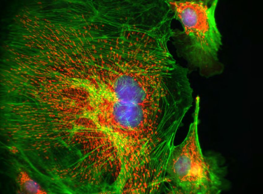
Questions?
TECHNICAL MEETING CONTENT:
                                                        Letter from the Presidents
2021 Program Chair
Elizabeth Wright                                        On behalf of the Microscopy Society of America and the Microanalysis
University of Wisconsin-Madison                         Society, we invite you to join us, in-person, August 1-5, 2021, for Microscopy
MM2021ProgramChair@microscopy.org                       & Microanalysis 2021 in Pittsburgh, PA. With a distinguished industrial history,
                                                        Pittsburgh has transformed itself into a great summer destination with diverse,
REGISTRATION:                                           interesting and walkable neighborhoods. Combining comfortable temperatures
Registrar                                               in the 80s, with lots of outdoor and riverside activities, as well as a healthy pub
MMRegistration@conferencemanagers.com                   and foodie culture, we know you will enjoy a great blend of science and social
                                                        activities! At this time, MSA and MAS are planning to move forward with the
EXHIBITS & EXHIBITORS:                                  in-person meeting. However, in this age of COVID-19, there is uncertainty on
Exhibits Manager                                        the public health situation next year. If the conditions do not improve and we
doreen@corcexpo.com                                     are unable to meet in person, we plan to bring a vibrant and outstanding virtual
                                                        meeting that builds off our experience from M&M 2020. We will create an even
SPONSORS & SPONSORSHIPS:                                better virtual experience featuring platform and poster presentations, a robust
Sponsorship Manager                                     trade show experience, and opportunities to connect with old friends and make
mary@corcexpo.com
                                                        new friends.

GENERAL:
                                                        The Program Committee, led by Elizabeth Wright, Eric Stach, Vincent
Meeting Manager
                                                        Smentkowski, and Andrew Herzing, has developed an exciting group of
meetingmanager@microscopy.org
                                                        symposia, spanning advances in instrumentation and techniques development,
                                                        as well as applications in the analytical, biological, and physical sciences.
                                                        We encourage you to browse this Call for Papers for complete symposium
Are You a Member?                                       descriptions and to submit one or more scientific papers for platform or poster
                                                        presentations. Tables will continue to be provided at the poster boards for fully
Join Today and Save on M&M 2021                         dynamic multimedia poster presentations.
Registration Fees!
                                                        The main meeting will be preceded by the ever-popular Sunday Short Courses
                                                        and three Pre-Meeting Congresses. Students and early-career professionals
                                                        are especially encouraged to participate in the MSA Student Council’s 5th
                                                        Annual Pre-Meeting Congress that highlights outstanding work by student and
Visit http://microscopy.org to join the                 postdoctoral fellow attendees. Join us Sunday evening to officially kick off the
Microscopy Society of America online, or for            meeting at the Opening Welcome Reception and reconnect with colleagues
more information about the benefits of MSA              and meet new friends. On Monday morning, the Plenary Session kicks off the
membership.                                             scientific program with an exciting set of lectures in Physical and Biological
                                                        sciences and the presentations of the M&M meeting awards and awards from
                                                        the sponsoring societies.

                                                        In addition to the strong scientific program, what sets the M&M meeting apart
Visit http://the-mas.org to find out the                is the Exhibit Hall, the world’s largest annual microscopy exhibition, which
benefits of MAS membership.                             showcases the latest instrumentation and accessories. Don’t miss the highly
                                                        popular vendor tutorials, held Monday through Wednesday after hours in the
                                                        Exhibit Hall. Other educational opportunities throughout the week include
IMAGES:                                                 focused biological and physical science tutorials, educational outreach
                                                        programs, and our Technologists’ Forum special and roundtable sessions.
Cover, top; footer of page 3:
Rat endothelial cells by Damon Strom,
WITec GmbH, Ulm, Germany                                As always, M&M 2021 will be the premier meeting to attend to stay abreast
Cover, bottom left:                                     of the latest technologies, hear about new developments in applications
Aloe vera leaf copy by Jose Martinez-Lopez,             across all areas of microscopy and microanalysis, and most importantly
Química Tech Microscopy and Microanalysis,
Juarez, Mexico                                          network with colleagues.

Cover, bottom right:                                    Peter A. Crozier, Arizona State University
Native vanadium dendrites by Sarah Gain, Centre
for Microscopy, Characterisation and Analysis,          President, Microscopy Society of America
University of Western Australia, Perth, Australia
                                                        Heather Lowers, United States Geological Survey
Footer on page 2:                                       President, Microanalysis Society
Detail from Ciliate expels its organelles byJulia Van
Etten, Rutgers University, New Brunswick, NJ

2       www.microscopy.org/MandM/2021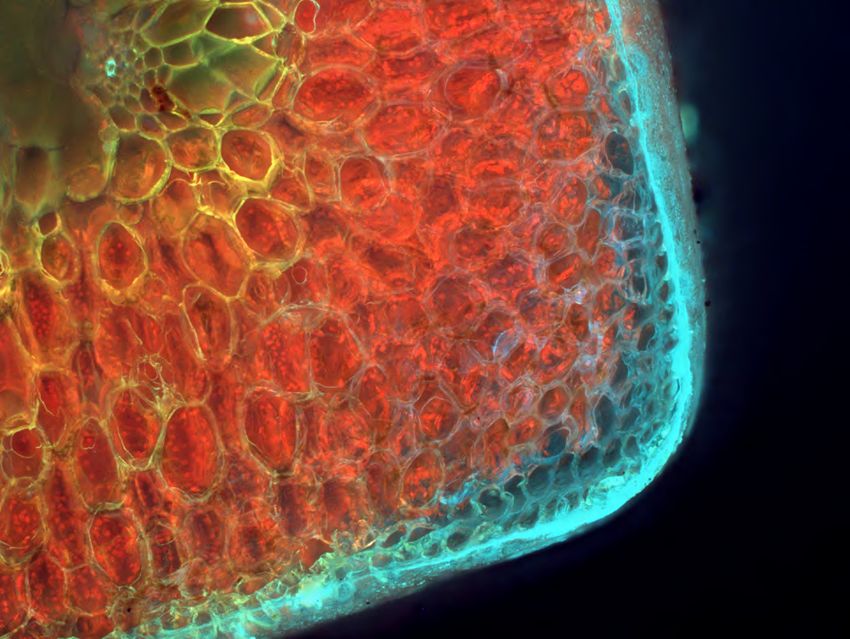
PLENARY SPEAKERS
For more information on each of the M&M 2021 Plenary Speakers, visit the
website at www.microscopy.org/2021/plenaryspeakers

Physical Science Plenary Speakers                                                       Biological Science Plenary Speakers

Maximilian Haider      Harald Rose       Ondrej Krivanek     Knut Urban                         Kizzmekia S. Corbett                 Jason McLellan
  CEOS Gmbh           Ulm Universität       Nion Co      Forschungszentrum                   National Institutes of Health,    University of Texas at Austin,
                                                               Jülich                         Vaccine Research Center          College of Natural Sciences

                           KAVLI AWARDEES                                                              COVID-19 VACCINE DEVELOPERS

                                                     ANALYTICAL SCIENCES SYMPOSIA
A01    Diffraction Imaging Across                      •    Enhancing analytical SEM with FIB,           A04    New Frontiers in In-Situ Electron
       Disciplines                                          including 3D EDS/EBSD and other                     Microscopy in Liquids and Gases
                                                            correlative analytics including WDS, CL,            (L&G EM FIG Sponsored)
ORGANIZERS:
Andrew Minor, Professor of Materials Science
                                                            Raman spectroscopy, EBIC, TKD, u-CT,
                                                                                                         ORGANIZERS:
and Engineering, Facility Director of the National          SIMS, and atom probe
                                                       •    Advances in cryo-FIB and working with        Huolin Xin, University of California, Irvine
Center for Electron Microscopy, Molecular
Foundry, Lawrence Berkeley National Laboratory              beam-sensitive materials                     Wei-Chang D. Yang, NIST
                                                       •    Innovative micro and nano-structure          Stephen House, University of Pittsburgh
Jose Rodriguez, Department of Chemistry
& Biochemistry, University of California, 		                prototyping                                    •   Solid/gas and solid/liquid interaction,
Los Angeles                                                                                                    electrochemistry and biological and soft matters
 •    4D-STEM techniques                              A03    Microscopy and                                    in liquids
 •    Micro/Nano diffraction for biology and                                                               •   Multidimensional and multidimenional in situ
                                                             Microanalysis for Real                            electron microscopy
      soft materials
 •    Analysis methods for diffraction patterns              World Problem Solving                         •   In situ electron microscopy driven by artificial
      and image reconstruction                        ORGANIZERS:                                              intelligence
 •    In situ diffraction imaging                     Ke-Bin Low, BASF Corporation                         •   Big data analysis using deep learning and neural
 •    Cryo diffraction imaging                        Xiaofeng Zhang, Nanosys Inc.                             networks
 •    Cryo electron microscopy                        Jeremy Beebe, Dow
                                                      Abigail Lindstrom, NIST                            A05    Advances in Analytical STEM-in-SEM
A02    Advances in Focused Ion Beam                    •    Real world problem solving using all         ORGANIZERS:
       Instrumentation, Applications                        forms of microscopy and microanalysis        Jason Holm, National Institute of Standards
                                                                                                         and Technology
       and Techniques in and Materials                 •    Practical applications of correlative
                                                            methods employing microscopy and             Dagmar Gerthsen, Karlsruhe Institute of Technology
       and Life Sciences
                                                            related techniques                           Hendrix Demers, Hydro-Québec, Center of Excellence
ORGANIZERS:                                                                                              in Transportation Electrification and Energy Storage,
                                                       •    Quantitative approaches for increased
Suzy Vitale, Carnegie Institution for Science                                                            Dmers
                                                            confidence in results from non-ideal
Annalena Wolff, Queensland University of
                                                            samples                                        •   Transmission imaging in the SEM (BF, DF, mixed
Technology
                                                       •    Creative methodologies for preparation             imaging modes, Z-contrast, diffraction contrast,
Joshua Sugar, Sandia National Laboratories                                                                     phase contrast, defect analysis)
                                                            and analysis of real world samples
 •    Latest developments in novel ion sources         •    Equipment testing, calibration and             •   Transmission electron diffraction and
      and FIB instrumentation                               quality assurance                                  channeling in the SEM (t-EBSD/TKD)
 •    Advances in helium ion microscopy                                                                    •   Transmission spectroscopy in the SEM
      including scripting, SIMS, nanofabrication,                                                              (EDS, EELS)
      lithography and floodgun imaging                                                                     •   Combined FIB-SEM transmission methods for
 •    Novel geometries, milling strategies                                                                     sample prep and analysis
      and non-standard lift outs for TEM/                                                                  •   Instrumental developments and new detectors
      STEM and APT                                                                                         •   4D STEM-in-SEM applications (data acquisition,
                                                                                                               analysis, instrumentation, and control schemes)

                                                               Microscopy & Microanalysis 2021 Meeting | August 1-5 | Pittsburgh, PA                            3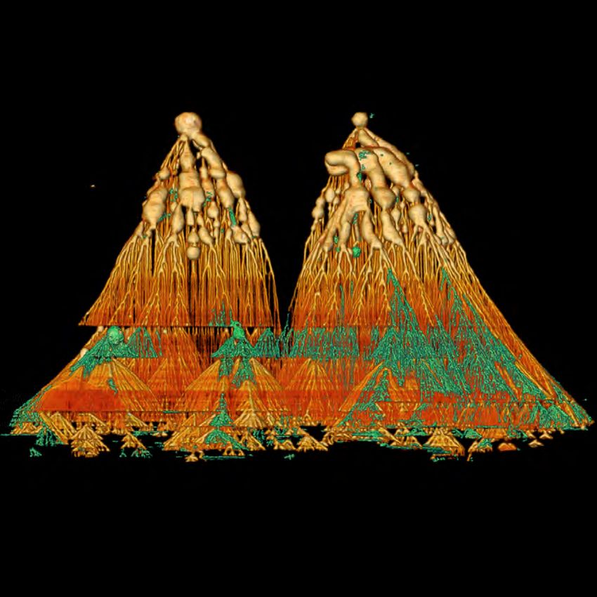
ANALYTICAL SCIENCES SYMPOSIA                                                                         continued

A06    Full System and Workflow                      A09    Moon Dust, Minerals and                              A11    Portable- and Laboratory-
       Automation for Enabling Big                          Microscopy                                                  based Approaches to Analysis
       Data and Machine Learning in                  ORGANIZERS:                                                        in Cultural Heritage
       Electron Microscopy                           Kate Burgess, U.S. Naval Research Laboratory                ORGANIZERS:
ORGANIZERS:                                          Michelle Thompson, Purdue University                        Thomas Lam, Smithsonian Institution, Museum
Andrew Barnum, Thermo Fisher Scientific              Jessica Barnes, University of Arizona                       Conservation Institute, Suitland, MD
Joerg Jinschek, Ohio State University                 •    Investigations of natural, experimental,              Barbara Berrie, National Gallery of Art,
Wouter Van den Broek, Humboldt University                  and analog materials to further                       Washington, DC
of Berlin                                                  understanding of terrestrial and planetary             •    Portable- or laboratory-based XRF, FTIR,
                                                           environments                                                NIR or Raman
 •    Big data in electron microscopy enabled
                                                      •    Advances in sample preparation and                     •    Correlative laboratory-based optical,
      through full system automation
                                                           analysis relevant to lunar, planetary, and                  electron microscopy, or atomic force
 •    Methodologies and software created
      by the community to combine machine                  terrestrial materials                                       microscopy
                                                      •    Use of microscopy to explore the origins               •    Other relevant or emerging approaches
      learning with microscope operation
 •                                                         and evolution of Earth and planetary                        of portable or laboratory-based
      Data augmentation strategies to increase
                                                           materials                                                   approaches will be considered
      data diversity and close the simulation-
                                                      •    Lunar samples, meteorites and
      experiment gap
                                                           micrometeorites, terrestrial samples,
 •    Workflows and data tracking from sample
                                                           interplanetary dust particles, minerals,
      preparation through to imaging and
                                                           planetary building blocks, returned
                                                                                                                 A12    Microscopy and Microanalysis
      analysis                                                                                                          of Biomineralized and
                                                           samples
                                                      •    TEM, SIMS, FIB, SEM, Microprobe, STXM,                       Biomimetic Materials and
A07    Vendor Symposium                                    atom probe, uXCT                                             Structures
ORGANIZERS:                                                                                                      ORGANIZERS:
Jay Potts, University of South Carolina              A10    Unresolved Challenges                                Kenneth Livi, Johns Hopkins University
Lena Kourkoutis, Cornell University                         in Quantitative X-Ray                                Sue Okerstrom, Lichen Labs LLC
 •    New methods and techniques; new                       Microanalysis                                         •    Advances in microscopy of combined
      developments and technologies                                                                                    hard and soft materials
                                                     ORGANIZERS:
 •    Breakthroughs and new instrumentation                                                                       •    Natural, biomimetic, experimental, and
 •    Improvements to existing instrumentation       Aaron Torpy, CSIRO Mineral Resources,                             engineered materials
                                                     Clayton, Australia
 •    New analysis programs and data                                                                              •    Correlative microscopy and microanalysis
      processing packages                            Nick Wilson, CSIRO Mineral Resources,                             techniques
                                                     Clayton, Australia
                                                                                                                  •    Approaches, applications and discoveries
                                                     Hendrix Demers, Hydro-Québec’s Center of                     •    Biominerals as archives of Earth
A08    Data Management, Version                      Excellence in Transportation Electrification and
                                                                                                                       environmental history
                                                     Energy Storage, Varennes, Canada
       Control, and Multiformat
                                                     Aurélien Moy, Department of Geoscience,
       Analysis in Electron Microscopy               University of Wisconsin-Madison, Madison,
ORGANIZERS:                                          WI, USA
Josh Sugar, Sandia National Laboratories              •    Quantification using low-energy x-rays
Suzy Vitale, Carnegie Institution for Science              (e.g. Li K, Fe L, REE M lines)
Joe McKeown, Lawrence Livermore National              •    Valence / chemical state determination
Laboratory                                                 by x-ray spectroscopy
 •    Advances in backed-up permanent                 •    Improvements to quantification
      archival systems for storage of large data           algorithms, procedures, and standard
      sets and methods for transfer to these               reference materials
      systems                                         •    Quantification of micro/nano-scale
 •    Methods for file version control that allow          particles and structures
      multiple users to share and analyze the         •    Quantification of beam sensitive materials
      same data set, with the ability to return to    •    Improvements to detector/instrument
      the original when needed                             technology
 •    Methods for analyzing data between
                                                                                                                  Native vanadium dendrites by Sarah Gain, Centre
      multiple proprietary formats across                  Diatom triangle by Larry Gouliard, Jr.,                for Microscopy, Characterisation and Analysis,
      different microscopy/software platforms,             Independent Microscopist, Pekin, IL                    University of Western Australia, Perth, Australia
      including file format converters
 •    Information architecture for local and
      server-based data analysis software and
      data storage
 •    Novel methods for analyzing and
      visualizing large correlated datasets (e.g.,
      3D serial section, tomography, and 3D plus
      analytical EDS, EBSD, SIMS, CL, etc.)
 •    Methods for determining the scientifically
      interesting data when overwhelmed with
      information (e.g., finding the needle in the
      haystack)

 4      www.microscopy.org/MandM/2021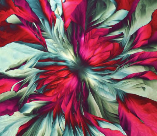
BIOLOGICAL SCIENCES SYMPOSIA
B01    3D Structures: From                         •    Historical perspectives and current            •    Instrument hardware development
       Macromolecular Assemblies to                     developments in the field of high-             •    Cryogenic sample handling / transfer
                                                        resolution cryo-EM                             •    Artificial Intelligence based Image
       Whole Cells (3DEM FIG)                      •    Hybrid structural approaches to study               Analysis, Big Data Management
ORGANIZERS:                                             macromolecular complexes
Melanie Ohi, University of Michigan Life           •    Assembly and architecture of plant,
Sciences Institute
                                                                                                      B07    Challenges and Advances in
                                                        animal, and bacterial viruses
Teresa Ruiz, University of Vermont                 •    In-situ structural investigations of viral           Electron Microscopy Research
Cheri Hampton, UES, Inc. · AFRL/RXAS                    infection and assembly                               and Diagnosis of Diseases in
Wright-Patterson Air Force Base                    •    Structural biology of proteins involved in           Humans, Plants and Animals
William Rice, NYU Langone Health                        metabolism, immunity and pathogenesis                (FIG associated)
 •    Structure and function of macromolecular                                                        ORGANIZERS:
      complexes in vitro and in vivo              B05    Imaging, Microscopy,                         Claudia Lopez, Oregon Health & Science
 •    Single particle cryo-electron microscopy                                                        University
 •    Cryo-electron tomography
                                                         and Micro/Nano-Analysis
                                                                                                      Marcela Redigolo, West Virginia University
                                                         of Pharmaceutical,                           Ru-Ching Hsia, University of Maryland
                                                         Biopharmaceutical, and                       Han Chen, Penn State
B02    Visualizing Cells with Cryo-ET
                                                         Medical Health Products–                      •    Enhancement of sample preparation
ORGANIZERS:
Grant Jensen, Caltech
                                                         Research, Development,                             workflow for EM analysis of complex
                                                         Analysis, Regulation, and                          tissue for diagnosis or research
Yi-Wei Chang, University of Pennsylvania                                                               •    Application of advanced or innovative
Danielle Grotjahn, Scripps Research Institute            Commercialization 		                               EM techniques to study host-pathogen
                                                         (FIG associated)
Matt Swulius, Penn State College of Medicine                                                                interactions, virulence factors and
                                                  ORGANIZERS:                                               ultrastructural changes
 •    Cryo-ET allows large macromolecular
                                                  Daniel Skomski, Merck & Co. Inc.                     •    Ultrastructural characterization of
      assemblies to be visualized within cells
      in a near-native state                      Annie Muske-Dukes, Thermo Fisher Scientific               diseases including the use of organoids,
 •    The range of targets that can be             •    Novel microscopic or spectroscopic                  xenografts and animal models
      analyzed by cryo-ET is rapidly expanding          methods applied to pharmaceutical,             •    Novel approaches and applications for
      to both larger and smaller                        biopharmaceutical, medical, and health              correlative or hybrid microscopy
 •    Sub-tomogram averaging methods can                fields (API, excipient, drug, device, etc.)
      result in high-resolution structures         •    Root cause analysis of issues related
 •    Many technical advances in                        to the manufacturing of drugs, medical
                                                                                                      B08    Cryo-EM in Drug Discovery
      instrumentation, methods, and software            products, and devices in both R&D and         ORGANIZERS:
      are dramatically expanding both the               good manufacturing practice (GMP)             Leah Frye, Schrodinger
      power and applicability of the method             settings                                      Giovanna Scapin, NanoImaging Services
                                                   •    Utilization of techniques and methods to      Anke Mulder, Thermo Fisher Scientific
                                                        overcome unique product performance            •    Cryo-EM in structure-based drug
B03    From Images to Insights:                         and pharmacology challenges                         discovery
       Working with Large Multi-                        (polymorphs, contaminants, particles,          •    Cryo-EM in optimization of potency and
       Modal Data in Cell Biological                    etc.)                                               selectivity
                                                   •    Regulatory and data integrity compliance       •    Use of cryo-EM to address classical
       Imaging                                          of instrumentation and methods in the               ADMET issues such as hERG inhibition
ORGANIZERS:                                             pharmaceutical industry                        •    Rapid and efficient determination of cryo-
Kedar Narayan, NIH/NCI & FNL                       •    Investigations and evaluations of testing           EM structures for drug discovery
Cam Robinson, St. Jude Children’s                       and drug and vaccine development
Research Hospital                                       throughout a product lifecycle
Jonathan Lefman, nvidia                                 (identification, development, testing,        B09    To Fix or Not to Fix? A Question
 •    Correlating images and appropriate                supply chain, regulatory, etc.)                      for Biological Samples
      metadata from LM, EM, other modalities       •    Device and throughput challenges (failure     ORGANIZERS:
      (XRM, chemical imaging...)                        mode analysis, biocompatibility, sterility,
                                                                                                      Alice Dohnalkova, Pacific Northwest National
 •    Efficiently segmenting and visualizing            etc.)                                         Laboratory
      features of interest                                                                            Gail Celio, University of Minnesota
 •    Adapting and applying open-source tools
      and frameworks for image processing
                                                  B06    Multi-Modal Multi-		                          •    Legitimate advantages and deficiencies,
      and analysis                                       Dimensional Microscopy                             and how to control them
 •    Implementing scaleable solutions for        ORGANIZERS:                                          •    Recognizing artifacts in chemically and
      small and mid-sized labs and facilities                                                               cryo- processed material
                                                  James Fitzpatrick, Washington University
                                                  School of Medicine
                                                                                                       •    Preparing problematic samples, e.g.,
                                                                                                            volume limitations, high material
                                                  Xiao-Ying Yu, Pacific Northwest National
B04    Michael Rossmann                           Laboratory
                                                                                                            density, etc.
       Memorial Symposium                                                                              •    Immunolabelling in cryo- and
                                                  Si Chen, Argonne National Laboratory
                                                                                                            chemical fixation
ORGANIZERS:                                       Ben Giepmans, University Medical Center              •    Handling damage from the electron beam
S. Saif Hasan, Department of Biochemistry         Groningen, Groningen, NL
                                                                                                       •    Development and applications of post-
and Molecular Biology, University of Maryland      •    Multi-Modal Microscopy, Volume EM,                  fixation preparation
School of Medicine, Baltimore MD
                                                        CLEM
Terje Dokland, Department of Microbiology,         •    X-Ray Imaging & Analysis
University of Alabama at Birmingham,               •    Analytical Spectroscopy / SIMS
Birmingham AL

                                                          Microscopy & Microanalysis 2021 Meeting | August 1-5 | Pittsburgh, PA                        5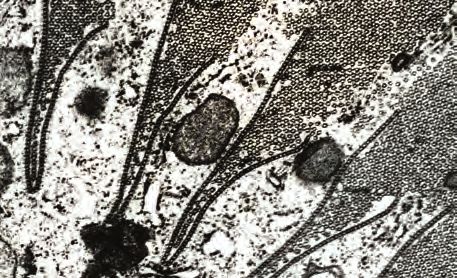
BIOLOGICAL SCIENCES SYMPOSIA                                                                    continued

B10    Cryo-EM at Local, Regional, and                •    Hardware and software upgrades to            ORGANIZERS:
       National Cryo-EM Centers                            optimize data collection                     Michelle Digman, University of California Irvine
                                                      •    On the fly data processing. When to          Matthew Lew, Washington University in
ORGANIZERS:
                                                           stop data collection?                        St. Louis
Claudia Lopez, Oregon Health & Science                •    Best practices in a cryoEM facility          Kevin Welsher, Duke University
University and Pacific North West CryoEM              •    How to best store, analyze, and
Center (PNCC)                                                                                           Andreas Gahlmann, University of Virginia
                                                           share your data
Elizabeth Wright, University of Wiscsonsin-           •    Data processing: what are we missing?            •   Fluorescence lifetime imaging microscopy
Madison and Midwest Center for CryoET                                                                       •   Super-resolution microscopy
(MCCET)                                                                                                     •   Single molecule spectroscopy
Clint Potter, New York Structural Biology
                                                     B11    Frontiers in Fluorescence                       •   Computational image analysis
Center and National Center for CryoEM                       Lifetime and Super-Resolution
Access and Training (NCCAT)                                 Imaging of Biological Structures
 •    Sample preparation: screening optimization            and Dynamics

                                                     PHYSICAL SCIENCES SYMPOSIA
P01    Advanced Imaging and                           •    Measurement of light elements in earth       P05      Evaluation of Materials for
       Spectroscopy for Nanoscale                          and material sciences.                                Nuclear Applications
       Materials Characterization                                                                       ORGANIZERS:
ORGANIZERS:                                          P03    Exploring Beam-Sample                       Mukesh Bachhav, Idaho National Laboratory
Lin Zhou, Ames Laboratory                                   Interactions for Uncovering                 Assel Aitkaliyeva, University of Florida
David Cullen, Oak Ridge National Laboratory                 the Atomic or Dynamic                       Jing Wang, Pacific Northwest National
Ping Lu, Sandia National Laboratories                                                                   Laboratory
                                                            Nature of Matter
 •    Advances in imaging and spectroscopy                                                                  •   Current and advanced nuclear fuels and
                                                     ORGANIZERS:
      methods for structural and chemical                                                                       materials
                                                     Joe Patterson, University of California, Irvine
      characterization                                                                                      •   Irradiation-induced defect analysis:
 •    Correlative approaches combining               Stig Helveg, Technical University of Denmark               dislocations, loops, RIS, RIP, SFT, etc.
      spectroscopic and imaging methods (e.g.        Jennifer Cookman, University of Limerick               •   Microscopy and microanalysis advances
      ABF/HAADF, 4D-STEM, EDS/EELS) to                •    Development of theories for electron-                in nuclear research
      provide new insight into material structure          sample interactions                              •   Data science and data mining of
 •    Real-time and multi-dimensional structural      •    Precise control over electron probes in              microscopy-based datasets
      and chemical imaging                                 time and space                                   •   Standards and interpretation of data for
 •    Defect structural studies in materials          •    Precise, accurate and reliable                       high Z materials
      such as nanoparticles, thin-films, and               measurement and reporting of electron
      multiplayers                                         flux and dose
 •    Optimization of imaging conditions (low         •    High spatial / temporal resolution imaging
                                                                                                        P06      Defects in Materials: How We
      voltage, low dose, cryo etc.) for structure-         of beam-sensitive materials                           See and Understand Them
      property relationship studies                   •    Using the electron beam to modulate          ORGANIZERS:
                                                           structures and processes                     Jinwoo Hwang, The Ohio State University
                                                      •    Understanding structure and dynamics         Tyler Grassman, The Ohio State University
P02    Many Detectors Make                                 from noisy data                              Honggyu Kim, University of Florida
       Lights Work: Advances in
                                                                                                            •   Imaging, diffraction, and spectroscopic
       Microanalysis of Light Elements
                                                     P04    Emerging Low-Dimensional                            methods that directly investigate
       in Synthetic and Natural                                                                                 defects of any nature in both crystalline
                                                            Nanomaterials and Their
       Materials                                                                                                and amorphous (disordered) materials
                                                            Heterostructures                                •   Determining atomic and electronic
ORGANIZERS:
                                                     ORGANIZERS:                                                structure of point defects, extended
Anette von der Handt, University of Minnesota        Moon Kim, University of Texas at Dallas
Jed Mosenfelder, University of Minnesota
                                                                                                                defects, defect complexes
                                                     Zonghoon Lee, Ulsan National Institute of              •   Dynamics of defects – nucleation,
Owen Neill, University of Michigan                   Science and Technology (UNIST) / Institute for
                                                                                                                interaction, migration, diffusion,
Jamie Weaver, National Institute of Standards        Basic Science (IBS)
                                                                                                                annihilation, formation of complexes
and Technology                                        •    Various allotropes of carbon such as             •   Defects in next-generation
 •    Advances in technologies for the detection           carbyne, nanotube, graphene, and                     semiconductors and metals, low-
      of light elements.                                   diamond                                              dimensional materials, energy-
 •    Improvements to existing methods for light      •    Heterostructure with other emerging                  generating materials, materials for
      element analysis.                                    nanomaterials such as TMDs and MXenes                quantum information
 •    Soft X-ray spectrometry, neutron                •    Growth morphology, defects, surfaces             •   Novel and quantitative digital data
      activation, EDS, WDS, SIMS, FTIR.                    and interfaces                                       processing methods for visualizing
 •    Analysis of light elements for energy           •    Applications in nano-electronics, energy,            defects with high sensitivity, accuracy,
      technology applications.                             and other emerging devices                           and precision

6      www.microscopy.org/MandM/2021 for up-to-date meeting information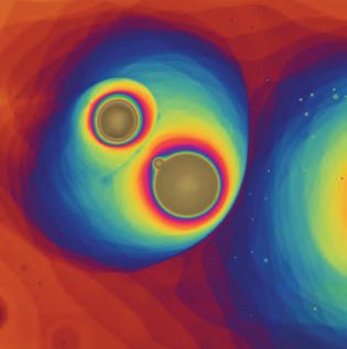
PHYSICAL SCIENCES SYMPOSIA                                                              continued

P07     Quantum Materials Probed                      P10    Investigating Phase                              P12    Microscopy & Spectroscopy
        by High Spatial and Energy                           Transitions in Functional                               of Energy Conversion and
        Resolution in Scanning/                              Materials and Devices by In                             Storage Materials
        Transmission Electron                                Situ/Operando TEM                                ORGANIZERS:
                                                                                                              Lianfeng Zou, Pacific Northwest
        Microscopy                                    ORGANIZERS:
                                                                                                              National Laboratory
ORGANIZERS:                                           Michele Conroy, University of Limerick
                                                                                                              Katherine Jungjohann, Sandia
Nasim Alem, Penn State University                     Trevor Almeida, University of Glasgow                   National Laboratories
Mary Scott, University of California Berkeley         Leopoldo Molina-Luna, TU Darmstadt                      Pengfei Yan, Beijing University of
 •    High resolution S/TEM imaging of                Judy Cha, Yale                                          Technology, China
      nanostructures and quantum materials             •    Phase transitions in functional materials         Michael Zachman, Oak Ridge National
 •    Electronic and chemical structure of                  and devices                                       Laboratory
      nanostructures and quantum materials             •    In-situ TEM capabilities (heating, biasing,        •    Characterization of materials related to
 •    Understanding of the phonon and plasmon               cooling, magnetic fields)                               energy generation, capture, conversion, or
      resonances and their coupling behavior,          •    Combination with advanced TEM                           storage across multiple length scales.
      particularly at defects, interfaces, and              techniques (Phase related, spectroscopy,           •    In situ/operando techniques for studying the
      surfaces in nanostructures                            4D STEM)                                                dynamic structural and chemical evolution
 •    Studies of topological, skyrmionic,              •    Controlled electron-beam-induced                        of energy-related materials under synthesis,
      superconducting and other quantum                     transitions                                             working, or processing conditions.
      materials                                                                                                •    Developments and applications of
                                                                                                                    emerging techniques such as 4D-STEM and
                                                      P11    Fast and Ultrafast Dynamics                            monochromated EELS for characterization of
P08     Advanced Characterization
                                                             Using Electron Microscopy                              energy-related materials
        of Components Fabricated by                                                                            •    Low-dose, low-voltage, and cryogenic
                                                      ORGANIZERS:
        Additive Manufacturing                        Ilke Arslan, Argonne National Laboratory,                     techniques that provide new insights into
ORGANIZERS:                                           Center for Nanoscale Materials                                the structure, chemistry, structural-property
Isabella van Rooyen, Idaho National laboratory        David Flannigan, University of Minnesota, Dept.               relationships of energy-related materials
Subhashish Meher, Idaho National Laboratory           of Chemical Engineering and Materials Science            •    New computational tools for imaging,
Federico Sciammarella, MXD USA                        Pietro Musumeci, University of California, Los                diffraction, spectroscopy, or tomography
                                                      Angeles, Physics Dept.                                        datasets collection and analysis
 •    TEM and STEM studies (imaging, EDS,
      electron diffraction, EELS) and atom probe       •    Techniques and instrumentation for
      tomography (APT) to understand phase                  high frame-rate S/TEM and SEM                     P13    Advanced Application of Atom
      transformations, and microstructural             •    Method and instrument development for                    Probe Tomography: Specimen
      evolution in components produced by                   improved combined temporal, spatial,
      various AM processes                                  and energy resolutions
                                                                                                                     preparation, Instrumentation,
 •    3D microstructure analysis methods on            •    Methods for reaching sub-femtosecond                     and Data analysis
      the micro-, nano-, and atomic scale to                stroboscopic and sub-nanosecond                   ORGANIZERS:
      understand the integrity of AM fabricated             single-shot resolutions                           Daniel Perea, Pacific Northwestern National Labs,
      products                                         •    Advances and new developments in high-            Richland, Washington, USA
 •    Microstructural response of AM components             precision generation of discrete electron         James Douglas, Dept of Materials, University of
      to post-processing conditions                         packets in S/TEM and SEM                          Oxford, Oxford, UK
 •    Microstructure and defect analysis by both       •    Discoveries, new insights, and                    Daniel Haley, Dept of Materials, University of
      characterization and modeling for insights            paradigm-changing observations                    Oxford, Oxford, UK
      into solidification and melt pool dynamics in         resulting from UEM in physical,                    •    Advances in data processing for improved
      AM processes                                          chemical, and materials systems                         accuracy and signal detection in APT,
 •    Current challenges in analytical tools
                                                                                                                    including noise processing, chemical
      for microscopy and microanalysis of
                                                                                                                    quantification and error processing
      AM products
                                                                                                               •    New methods for improving reproducibility
 •    In-situ characterization experiments on
                                                                                                                    and reliability of APT data reconstruction
      AM products
                                                                                                                    and spatial analysis, including advanced
                                                                                   Gold nanocubes by
                                                                                                                    data modeling, improved cluster analysis
                                                                                   Vasile-Dan Hondoroaba,
P09 -   Nanoscale X-Ray and Electron                                               Federal Institute for            and machine learning approaches.
        Microscopy Techniques and                                                  Materials Research and      •    Atom probe analysis and sample
                                                                                   Testing, Berlin, Germany         preparation of materials exposed to
        Applications in Material Science                                                                            extreme environments such as temperature,
ORGANIZERS:                                                                                                         irradiation, corrosion and pressure.
Xianghui Xiao, Brookhaven National Laboratory                                                                  •    Multi-length scale and complementary
Hanfei Yan, Brookhaven National Laboratory                                                                          techniques for optimized APT analysis of
Huolin Xin, University of California, Irvine                                                                        complex material systems.
 •    Correlative high-resolution electron and                                                                 •    Advanced specimen preparation,
      x-ray microscopy applications in material                                     Aloe vera leaf                  instrumentation, and experimental protocols
      science                                                                       by Jose Martinez-Lopez,         that enable atom probe analysis of
 •    Correlative microscopy and bulk technique                                     Química Tech Microscopy         environmentally sensitive materials for direct
      applications in material science                                              and Microanalysis,              observation of hydrogen, hydrated phases,
 •    Advanced data analysis and modeling for                                       Juarez, Mexico                  biological and other soft materials, and
      multimodal and multiscale experiments                                                                         liquid/solid in
 •    Challenges in correlative microscopy
      applications

                                                             Microscopy & Microanalysis 2021 Meeting | August 1-5 | Pittsburgh, PA                              7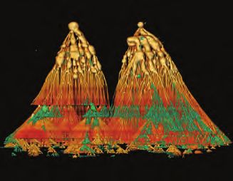
AWARDS
    How to Apply For an M&M Meeting Award:                                       Onsite Awards
    1.    As part of the on-line paper submission process, an applicant          The M&M meeting’s co-sponsoring societies confer
          must flag their paper for award consideration. Only one paper          competitively judged awards at the meeting.
          may be designated per applicant.
                                                                                 MSA Student Poster Awards
    2. The applicant must appear as first author and presenter of the            We believe poster presentations are
       paper submitted for award.                                                an excellent format for all participants
                                                                                 to engage in intensive discussion
    3. The applicant must provide the name, title, institution, and              with other researchers in the field. To especially encourage
       e-mail address of their supervisor, who will be contacted to              students to take advantage of this opportunity and submit
       provide a supporting letter and confirmation of applicability             papers for poster presentation, MSA provides cash awards to
       for the indicated award category (e.g. student, post-doc, or              the most outstanding student posters (first author) each day
       technical staff).                                                         (up to one in each of three categories).

    GENERAL CONSIDERATIONS:                                                      Ultramicrotomy Award by Diatome
    Award applicants will automatically be considered for memorial               All posters illustrating the use of diamond knife
    scholarships, conferred by MSA based on the generous support of              ultramicrocrotomy are eligible. Prizes include a trip
    society sponsors.                                                            to Switzerland with a visit to the Diatome factory, and
                                                                                 Swiss watches.
    Applicants who have previously received an M&M Meeting Award
    will not be considered for a second award in the same category.
                                                                                 MAS Best Paper Awards
                                                                                 MAS annually confers awards for papers
    STUDENTS:
                                                                                 presented at the M&M meeting deemed to
    All full-time students enrolled at accredited academic institutions
                                                                                 be best in four categories. Each comes with a cash award
    are eligible. High school, undergraduate, and graduate students are
                                                                                 generously provided by MAS Sustaining Members.
    encouraged to apply. Applicants are not required to be members of
    the sponsoring society.
                                                                                 Microscopy Today
    POSTDOCTORAL RESEARCHERS:                                                    Micrograph Awards
    All full-time postdoctoral researchers are eligible. Applicants are not      This competition rewards the innovative blending of art and
    required to be members of the sponsoring society.                            science. Winning micrographs will be selected on the basis
                                                                                 of artistic merit and general audience appeal. See M&M 2021
    PROFESSIONAL TECHNICAL STAFF MEMBERS:                                        website for complete information.
    Full-time technologists are eligible. In addition, the applicant must
    be a member of the sponsoring society, current in their dues for the         All images used in this brochure are 2020 Microscopy Today
    year of the meeting.                                                         Micrograph Award Winners.

    AMOUNT OF AWARD:
    M&M Meeting Awards and memorial awards consist of full meeting
    registration and up to $1,000 for travel-related expenses. Original
    receipts must be provided to receive travel reimbursement.                Thin Film Crystals by Karl Gaff, K Gaff Microscopy, Dublin, Ireland
    All award winners also receive an invitation to the Presidents’
    Reception, held on the Tuesday evening of the meeting.

    NOTIFICATION OF AWARD:
    All award applicants will be notified of their award status
    approximately eight weeks following the Call for Papers deadline.
    Unsuccessful applicants will be permitted to withdraw their papers,
    should their ability to attend the meeting be contingent on the
    award, within one week following notification.

    REQUIREMENTS OF AWARD:
    All award winners must present their paper in person at the M&M
    meeting in order to receive their award.

    Awardees are expected to attend and participate in the entire
    meeting, which runs from Sunday evening’s opening reception
    through late Thursday afternoon.

    Awardees are required to attend the Monday morning plenary
    session, at which their award will be conferred.

8        www.microscopy.org/MandM/2021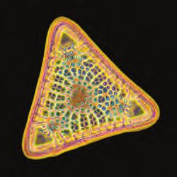
MICROSCOPY OUTREACH SESSIONS
  X90    Microscopy in the Classroom                                                X91    Microscopy Explorations for Families and
  ORGANIZERS:                                                                              Kids of All Ages (formerly “Family Affair”)
  Jane Howe, University of Toronto, Canada                                          ORGANIZERS:
  Donovan N. Leonard, Oak Ridge National Laboratory                                 Elaine Humphrey, University of Victoria, Canada
  Josh Silverstein, Pacific Northwest National Laboratory                           Pat Connelly, National Institutes of Health
  Rengasayee (Sai) Veeraraghavan, The Ohio State University                         Please check back on the M&M 2021 website
  At the frontiers of science, the microscope is an interdisciplinary tool          (“Scientific Program” – “Outreach”) for updated
  which allows students to glimpse into the unknown and link structure              information about this session.
  and function. The Education Outreach Committee of MSA seeks to
  connect individuals and institutions and ensure that the pathways to
  microscopy careers and education are exciting, engaging and clear.
                                                                                    X92    Project MICRO
   • Best practices for incorporating microscopy into K-12 and post-
                                                                                    ORGANIZERS:
       secondary classrooms and curricula
   • Local and national initiatives emphasizing STEM education                      Elaine Humphrey, University of Victoria, Canada
       and outreach                                                                 Janet Schwarz, University of Vermont
   • Methods to expose students to microscopy in a fun,                             Pat Connelly, National Institutes of Health
       engaging and impactful manner
                                                                                    The Outreach booth is part of the MSA Megabooth and is
                                                                                    available every day the exhibit hall is open. Learn how to set up
                                                                                    different stations in a classroom and share your fun microscopy
                                                                                    outreach classroom experiences! See different microscope
                                                                                    systems in action for use in a classroom; peruse a selection of
                                                                                    books suitable for elementary school-age children; and put your
                                                                                    name into the draw for a daily door prize.

                                                  Soap Film
                                                  by Gerd Günther,
                                                  independent microscopist,
                                                  Düsseldorf, Germany

                                                   TECHNOLOGISTS’ FORUM
X30     Technologists’ Forum                       X31    Technologists’ Forum Roundtable:                       X32    Technologists’ Forum Workshop
        Roundtable: Histology                             Technical Careers in Microscopy –                             Technique Tips: Special Stains
        Helpline                                          PhD Not Required                                              and Serial Sectioning
ORGANIZERS:                                        ORGANIZERS:                                                   ORGANIZERS:
Page Baluch, Arizona State University              Page Baluch, Arizona State University                         Page Baluch, Arizona State University
Ru-Ching Hsia, University of Maryland              Richard Martins                                               Ru-Ching Hsia, University of Maryland
 •    Where to find answers to technical            •    There are many careers in advanced                       •    Training in the use of special stains is
      questions                                          analytical and scientific fields that do not                  usually provided on the job but not as
 •    Available networks to find laboratory              require a PhD                                                 part of a formal training program so many
      or clinical experts                           •    Learn about careers from professionals that                   technicians feel unprepared
 •    Where to get advice when shopping                  entered the microscopy community with                    •    Learn new techniques using serial
      for laboratory equipment                           skills and expertise garnered from previous                   sectioning and array tomography
 •    How to find labs that provide                      employment, effective networking and                     •    Learn about stains that are common in
      specialized histological/lab services              military service                                              quantification-based research
                                                    •    Commercial technical careers in microscopy               •    This session will be useful to histologists
                                                         include instrument technicians, service                       at all levels by providing protocols and
                                                         engineers, applications, sales, marketing and                 technical advice regarding techniques in
                                                         account managers                                              special stains and serial sectioning/array
                                                    •    Gather advice that can help in pursuing a new                 tomography
                                                         or different career path

                                                                 Microscopy & Microanalysis 2021 Meeting | August 1-5 | Pittsburgh, PA                       9
SHORT COURSES
X-10 High-Resolution Structure • Electron detector technology suitable for • Biasing holders
4D-STEM experiments • Magnetic field
Determination by Cryo-EM • List of possible 4D-STEM experimental • Light illumination
LEAD INSTRUCTORS: configurations and references
Tim Grant, Morgridge Institute / University of • Analysis software for characterizing large
Wisconsin-Madison numbers of STEM diffraction pattern X-15 Data Analysis in
Mike Cianfrocco, University of Michigan images and visualization of the results Materials Science
• Cryo-EM specimen preparation • Software and tutorial for simulating LEAD INSTRUCTORS:
• Introduction to TEM 4D-STEM datasets Eric Prestat, University of Manchester and
• Single-particle data collection SuperSTEM Laboratory, United Kingdom
• Single-particle image processing
Joshua Taillon, National Institute of Standards
• Validation of results X-13 Serial EM for EM Data and Technology
Acquisition • Introduction to HyperSpy and related
LEAD INSTRUCTORS:
X-11 Explaining the New Python libraries for multi-dimensional
Cindi Schwartz, Rocky Mountain Laboratories/ image and spectra processing and
World Order of Biological NIAID/National Institutes of Health analysis
Fluorescence Microscopy Guenter Resch, Nexperion, Austria • Curve fitting of multi-dimensional datasets
LEAD INSTRUCTORS: • Installation, calibration, and operating • Machine learning
Bob Price, University of South Carolina School concept of SerialEM • Big data analysis strategies
of Medicine • Image acquisition techniques such as for • EELS and EDS analysis
Jay Jerome, Vanderbilt University tilt-series, single-particle, and micro-ED • Optional: application to the analysis
• Basics of fluorescence • Ancillary hardware such as direct-electron of atomic resolution images, scanning
• Basics of confocal microscopy detectors, energy filters, and phase plates electron diffraction and 4D STEM datasets
• New fluorescence imaging modes • Scripting to extend SerialEM
• Selection of appropriate imaging modes For this hands-on and interactive short
course, attendees will need to install
X-14 In situ and Operando software on their own laptop in advance
X-12 Guidelines for Performing Approaches to TEM and bring it with them to the short course
4D-STEM Characterization LEAD INSTRUCTORS: (instructions will be provided).
from the Atomic to Micrometer Robert Sinclair, Stanford University
Scales: Experimental Peter Crozier, Arizona State University
Considerations, Data Analysis This course will introduce the fundamental
and Simulation concepts for in situ electron microscopy,
including:
LEAD INSTRUCTORS:
• Hot stages
David Muller, Cornell University • Gas cells
Colin Ophus, Lawrence Berkeley National • Liquid cells
Laboratory
PHYSICAL SCIENCES TUTORIALS
X41 Entrepreneurship in the Microscopy Community X43 X-Ray Imaging & Computed Tomography
Several entrepreneurs from the microscopy community will be in attendance for PRESENTERS: Tara Selly, Assistant Professor of Research, Univ.
a round table Q&A with tutorial attendees on topics including, but not limited to: of Missouri; Jim Schiffbauer, Associate Professor,
Geological Sciences, Univ. of Missouri
• Instrumentation development and commercialization
• Practical steps to take when starting your own microscopy based business • Introduction to X-Ray microscopy and computed
• Panel discussion on business start-up best practices tomography
• Role of local affiliated microscopy societies in bringing microscopists and • How spot size, detector pixel size and gemoterical
businesses together magnification affect resolution
• Reconstruction strategies for isosurface determination
• Example applications in the geological sciences
X42 Monochromated Aberration Corrected STEM: Why?
PRESENTER: Dr. Jordan A. Hachtel, Oak Ridge National Laboratory
• Introduction to monochromated EELS
• Alignment and tuning in a monochromated Nion UltraSTEM
• Different types of monochromated EELS experiments: Aloof EELS, off-axis
EELS, momentum-resolved EELS Zosterograptus microtubules
by Andrea Brothers, A.B. Brothers
• Post-acquistion processing and analysis of ultralowloss EELS data.
Microscopy, Manassas, VA
Jordan Hachtel is a staff scientist at Oak Ridge National Laboratory. His research
focuses on applications of ultralow-loss monochromated EELS to optical,
biological, and quantum materials. Beyond this wide class of applications, he has
also focused on using novel techniques, unique to ultralow-loss EELS, to access
new aspects of the nanoscale infrared response in such systems.
10 www.microscopy.org/MandM/2021PRE-MEETING CONGRESSES
ORGANIZERS:
X60 Annual Pre-Meeting Congress for Students,
Ru-ching Hsia, University of Maryland-Baltimore
Post-Docs, and Early-Career Professionals in
Claudia Lopez, Oregon Health and Science University
Microscopy and Microanalysis
Marcela Redigolo, West Virginia University
Organized by the Microscopy Society of America Student Council (StC) Han Chen, Penn State Medical College
Saturday, July 31, 2021 • 8:30 AM - 5:00 PM Joe Mowery, Agricultural Research Service, US Department of Agriculture
Separate registration required – see registration form (Spring 2021) Mike Reichelt, Genentech
INCLUDED IN REGISTRATION FEE: This PMC will include four 90-minute platform sessions with 4-6 speakers each
Friday evening social event; breakfast, AM Break, Lunch, PM Break, session, and a poster session.
Saturday evening banquet • Automation and streamlining of workflow for diverse EM specimens
• Immuno-electron microscopy and correlative LM-EM workflow, advances
PROGRAM CHAIR: Piyush Haluai, Arizona State University and challenges
BIOLOGICAL SCIENCES CO-CHAIR: Joseph Kim, University of • Challenges and best practices in the preparation of plant, insect,
Wisconsin-Madison aquatic/marine, and pharmaceutical specimens for EM
PHYSICAL SCIENCES CO-CHAIR: Berit Goodge, Cornell University • Roundtable discussion: instrument-assisted and automated Bio EM
SOCIAL CHAIR: Dara Laczniak, Purdue University sample processing, challenges and applications
This pre-meeting congress is organized by and for students,
postdocs, and early-career professionals, and provides: X62 Recent Developments in Advanced Imaging
• A forum for early-career professionals to deliver presentations to and Spectroscopy
peers ahead of the meeting Organized by the MSA Aberration-Corrected Electron Microscopy
• Opportunities to share research and data in an engaging, non- Focused Interest Group
intimidating, and interactive setting
• Expanded professional networking, and career development Sunday August 1, 2021 v 8:30 AM – 5:00 PM
mentoring from recent graduates Separate registration required – see registration form (Spring 2021)
• The opportunity to win awards, determined by peer voting INCLUDED IN REGISTRATION FEE:
Breakfast, AM Break, Lunch, PM Break
X61 Contemporary Electron Microscopy Advances ORGANIZERS:
in Biomedical Research Juan Carlos Idrobo, PhD, CNMS, Oak Ridge National Laboratory
Robert Klie, PhD, University of Illinois
Organized by the MSA Diagnostic and Biomedical Microscopy Shize Yang, PhD, Arizona State University
Focused Interest Group
This PMC will focus on developments in instrumentation, techniques, and
Sunday August 1, 2021 • 8:30 AM - 5:00 PM analysis for functional aberration-corrected electron microscopy, including:
Separate registration required – see registration form (Spring 2021) • 4D-STEM
INCLUDED IN REGISTRATION FEE: • Aberration correction
Breakfast, AM Break, Lunch, PM Break • Monochromated EELS
• Field-free imaging
• Low temperature (cryo) characterization
BIOLOGICAL SCIENCES TUTORIALS
X44 Cryo-EM Structure Determination of Small X45 Traversing Spatial Scales with Correlative
Proteins Microscopy
PRESENTER: Dr. Deb Kelly, Professor of Biomedical Engineering, PRESENTER: James A.J. Fitzpatrick, Ph.D.
Pennsylvania State University Scientific Director, Center for Cellular Imaging
• Isolating proteins from COVID-19 patients Professor of Neuroscience and Cell Biology &
• Affinity-capture using alternative substrates Physiology, Washington University School
• Low-dose Image Acquisition in Milli-seconds (LIAM) of Medicine
• Reconstructing small proteins (~50 kDa range) • Introduction to Multi-Modal imaging and concept of
Correlative Microscopy
• Discussion of current CLEM, CXREM and Cryo-CLEM methods
• Practical use of different algorithms for correlating multi-modal data
• Examples of Correlative Microscopy in Studies of Disease
Pathogenesis
James Fitzpatrick is a Professor at Washington University School
of Medicine in St. Louis and the inaugural Scientific Director of
the Center for Cellular Imaging. His research focuses on the
development and application of multi-modal correlated imaging
approaches and AI-based image analysis methods to study the
pathogenesis of cancer and neurodegenerative disease.
Microscopy & Microanalysis 2021 Meeting | August 1-5 | Pittsburgh, PA 112021
Thank You to Our Sustaining Members
(As of Oct. 2, 2020)
Advanced Microscopy Techniques Corp International Centre for Diffraction Data
Angstrom Scientific, Inc. IXRF Systems
Applied Physics Technologies JEOL USA, Inc.
Birla Carbon Lehigh Microscopy School
Bruker Nano Analytics Leica Microsystems
Carl Zeiss Microscopy, LLC Micron, Inc.
CEOS GmbH Microscopy Innovations LLC
Dectris Ltd. NanoSpective
Diatome US Nion Co.
Direct Electron LP Oxford Instruments
Duniway Stockroom Corp. PIE Scientific LLC
EDAX, Inc. PNDetector GmbH
Electron Microscopy Sciences Probe Software, Inc.
EXpressLO LLC Quantum Design Inc.
Fischione Instruments RaySpec Ltd
Focus E-Beam Technology (Beijing) Co., Ltd RMC Boeckeler
Gatan SEMTech Solutions, Inc.
Hitachi High Technologies America, Inc. Ted Pella Inc.
High-Field Consultants TESCAN USA
HREM Research Inc. Thermo Fisher Scientific
Hummingbird Scientific Tousimis
ibss Group, Inc. TSS Microscopy LLC
Integrated Dynamics Engineering, Inc. XEI Scientific, Inc.
www.microscopy.org/MandM/2021You can also read