Enhanced cell survival and therapeutic benefits of IL-10-expressing multipotent mesenchymal stromal cells for muscular dystrophy
←
→
Page content transcription
If your browser does not render page correctly, please read the page content below
Nitahara-Kasahara et al. Stem Cell Research & Therapy            (2021) 12:105
https://doi.org/10.1186/s13287-021-02168-1

 RESEARCH                                                                                                                                         Open Access

Enhanced cell survival and therapeutic
benefits of IL-10-expressing multipotent
mesenchymal stromal cells for muscular
dystrophy
Yuko Nitahara-Kasahara1,2,3*, Mutsuki Kuraoka3,4, Yuki Oda1, Hiromi Hayashita-Kinoh1,2,5, Shin’ichi Takeda3 and
Takashi Okada1,2,5*

  Abstract
  Background: Multipotent mesenchymal stromal cells (MSCs) are potentially therapeutic for muscle disease because
  they can accumulate at the sites of injury and act as immunosuppressants. MSCs are attractive candidates for cell-
  based strategies that target diseases with chronic inflammation, such as Duchenne muscular disease (DMD). We
  focused on the anti-inflammatory properties of IL-10 and hypothesized that IL-10 could increase the typically low
  survival of MSCs by exerting a paracrine effect after transplantation.
  Methods: We developed a continuous IL-10 expression system of MSCs using an adeno-associated virus (AAV)
  vector. To investigate the potential benefits of IL-10 expressing AAV vector-transduced MSCs (IL-10-MSCs), we
  examined the cell survival rates in the skeletal muscles after intramuscular injection into mice and dogs. Systemic
  treatment with IL-10-MSCs derived from dental pulp (DPSCs) was comprehensively analyzed using the canine X-
  linked muscular dystrophy model in Japan (CXMDJ), which has a severe phenotype similar to that of DMD patients.
  Results: In vivo bioluminescence imaging analysis revealed higher retention of IL-10-MSCs injected into the
  hindlimb muscle of mice. In the muscles of dogs, myofiber-like tissue was formed after the stable engraftment of
  IL-10-MSCs. Repeated systemic administration of IL-10-DPSCs into the CXMDJ model resulted in long-term
  engraftment of cells and slightly increased the serum levels of IL-10. IL-10-hDPSCs showed significantly reduced
  expression of pro-inflammatory MCP-1 and upregulation of stromal-derived factor-1 (SDF-1). MRI and
  histopathology of the hDPSC-treated CXMDJ indicated the regulation of inflammation in the muscles, but not
  myogenic differentiation from treated cells. hDPSC-treated CXMDJ showed improved running capability and
  recovery in tetanic force with concomitant increase in physical activity. Serum creatine kinase levels, which
  increased immediately after exercise, were suppressed in IL-10-hDPSC-treated CXMDJ.
  Conclusions: In case of local injection, IL-10-MSCs could maintain the long-term engraftment status and facilitate
  associated tissue repair. In case of repeated systemic administration, IL-10-MSCs facilitated the long-term retention
  (Continued on next page)

* Correspondence: y-kasahara@nms.ac.jp; t-okada@ims.u-tokyo.ac.jp
1
 Department of Biochemistry and Molecular Biology, Nippon Medical School,
Bunkyo City, Tokyo, Japan
Full list of author information is available at the end of the article

                                      © The Author(s). 2021 Open Access This article is licensed under a Creative Commons Attribution 4.0 International License,
                                      which permits use, sharing, adaptation, distribution and reproduction in any medium or format, as long as you give
                                      appropriate credit to the original author(s) and the source, provide a link to the Creative Commons licence, and indicate if
                                      changes were made. The images or other third party material in this article are included in the article's Creative Commons
                                      licence, unless indicated otherwise in a credit line to the material. If material is not included in the article's Creative Commons
                                      licence and your intended use is not permitted by statutory regulation or exceeds the permitted use, you will need to obtain
                                      permission directly from the copyright holder. To view a copy of this licence, visit http://creativecommons.org/licenses/by/4.0/.
                                      The Creative Commons Public Domain Dedication waiver (http://creativecommons.org/publicdomain/zero/1.0/) applies to the
                                      data made available in this article, unless otherwise stated in a credit line to the data.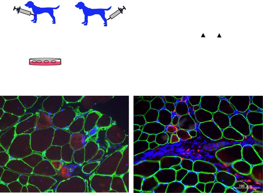
Nitahara-Kasahara et al. Stem Cell Research & Therapy   (2021) 12:105                                                Page 2 of 15

 (Continued from previous page)
 of the cells in the skeletal muscle and also protected muscles from physical damage-induced injury, which
 improved muscle dysfunction in DMD. We can conclude that the local and systemic administration of IL-10-
 producing MSCs offers potential benefits for DMD therapy through the beneficial paracrine effects of IL-10
 involving SDF-1.
 Keywords: Mesenchymal stromal cells, IL-10, DMD, Dental pulp stromal cells

Background                                                              effect after transplantation. IL-10-expressing MSCs
Multipotent mesenchymal stromal cells (MSCs) derived                    were previously developed using retroviral [15, 16]
from the bone marrow are conventionally termed                          and lentiviral vectors [17, 18], and transcription
“adherent non-hematopoietic cells.” The cells express                   activator-like effector nuclease (TALEN)-mediated
several cell-surface antigenic markers, including CD44,                 gene editing [19]. They were observed to offer thera-
CD73, CD90, and CD105 [1]. MSCs can self-renew and                      peutic benefits in collagen-induced inflammatory
differentiate into several different cell types. These                  arthritis [20], and also prevented lung ischemia-
include mesodermal cells, such as osteoblasts, chondro-                 reperfusion injury [15], endotoxin-induced acute
cytes, adipocytes, and myocytes [2–4] as well as non-                   injury [21], GVHD [16], traumatic brain injury [17],
mesodermal cells, such as hepatocytes [5], neural cells                 and left ventricular remodeling after myocardial
[6], and epithelial cells [7].                                          infarction [19].
   The multi-lineage potential of MSCs has been exploited                 We developed a continuous IL-10 expression system
for prospective use in therapies for various diseases. The              of MSCs using an adeno-associated virus (AAV) vector.
cells can be easily expanded in culture and are non-                    AAV vectors are safe and do not integrate into the host
tumorigenic. Furthermore, the use of MSCs as third-party                cell genome, and the risk of insertional mutagenesis is
materials in cell therapy reflects that MSCs are immune-                low. To investigate the potential benefits of using MSCs
privileged, unlike other stem cells or induced pluripotent              for treating muscle disease, we examined whether trans-
stem cells (iPS), as they do not express human leukocyte                duction with the IL-10-expressing AAV vector enhanced
antigen (HLA) class II, CD40, CD80, or CD86 molecules                   the survival rates of MSCs in skeletal muscles, and the
[8], and express only low levels of HLA class I. These cells            potential advantages offered by MSC transplantation.
are not lysed by natural killer cells or cytotoxic T lympho-            Systemic treatment using MSCs derived from dental
cytes [9]. MSCs can influence immune effector cell devel-               pulp (dental pulp stem cells, DPSCs) was comprehen-
opment, maturation, and function as well as reactive T cell             sively analyzed using the canine X-linked muscular dys-
responses through the production of bioactive cytokines                 trophy model in Japan (CXMDJ), which has a severe
and proteins [10, 11]. The mechanism underlying the im-                 phenotype similar to DMD in humans [22, 23]. DPSCs
munosuppressive effects of MSCs is unclear. Nonetheless,                are similar to bone marrow MSCs, which show high ex-
their immunosuppressive properties have been exploited in               pression of the surface markers CD29, CD73, CD90, and
clinical applications. MSCs are commercially authorized for             CD105, which are common stem cell markers in MSCs,
the treatment of acute graft-versus-host disease (GVHD).                but not CD34 or CD45. We have previously reported
MSCs are attractive candidates for cell-based strategies that           that DPSCs also express IL-10 and vascular endothelial
target diseases with chronic inflammation, such as                      growth factor (VEGF) at high levels and exhibit im-
Duchenne muscular dystrophy (DMD) [11].                                 munosuppressive activities [24, 25]. We evaluated
   Interleukin-10 (IL-10) is an anti-inflammatory                       whether DPSCs expressing IL-10 play an important role
cytokine with anti-apoptotic properties [12] that                       as a cell source for DMD therapy.
modulates the inflammatory immune response. IL-10
reduces M1 macrophage activation and inhibits the                       Methods
production of pro-inflammatory cytokines such as                        Animals
interferon-gamma (IFN-γ), tumor necrosis factor-                        NOD/SCID mice were purchased from Nihon CLEA
alpha (TNF-α), IL-1β, and IL-6 in inflamed tissues                      (Tokyo, Japan) and were housed at the National Center
[13, 14]. IL-10 also reduces the expression of CD54,                    of Neurology and Psychiatry (Tokyo, Japan). All experi-
CD80, CD86, and major histocompatibility complex                        ments using mice were performed in accordance with
class II molecules, resulting in incomplete T cell sig-                 the guidelines approved by the Nippon Medical School
naling and induction of antigen-specific antibodies.                    and National Center of Neurology and Psychiatry
We hypothesized that IL-10 could increase the typic-                    (NCNP) Animal Ethics Committees. Beagle dogs and
ally low survival of MSCs by exerting a paracrine                       CXMDJ colony dogs were maintained at NCNP
Nitahara-Kasahara et al. Stem Cell Research & Therapy   (2021) 12:105                                                    Page 3 of 15

according to the NCNP standard protocol for animal                      In vivo imaging analysis
care. Experiments were performed in accordance with                     After the injection of luciferase-expressing rat MSCs
the guidelines approved by the Ethics Committee for the                 on day 0 of the experiment, in vivo luminescence im-
Treatment of Laboratory Animals at NCNP.                                ages were acquired periodically to assess the engraft-
                                                                        ment efficiency and cell survival in the transplanted
Cell preparation                                                        mice. Prior to imaging, the mice were anesthetized by
MSCs derived from rat bone marrow were isolated and                     inhalation of 2.0% isoflurane and oxygen and injected
expanded as previously described [26]. For the experi-                  intraperitoneally with 150 mg luciferin (Summit Phar-
ments on dogs, healthy donor Beagle dogs were anesthe-                  maceuticals International Corp., Tokyo, Japan.) per
tized using thiopental and isoflurane, and 1.0 mL of                    kilogram body weight. In vivo images were acquired
bone marrow fluid was collected. The CD271+ MSCs                        using the IVIS charge-coupled-device camera system
were enriched and cultivated using the MSC Research                     (Xenogen Corp., Alameda, CA, USA) at multiple time
Tool Box-CD271 (LNGFR) containing CD271 (LNGFR)-                        points (0, 3, 7, 18, 27, 31, 34, 42, 49, 54, and 67 days
PE and anti-PE microbeads for cell separation (Miltenyi                 after transplantation). The region of interest (ROI) lu-
Biotec GmbH, Bergisch Gladbach, Germany), as previ-                     minescence signals from individual MSC-injected sites
ously reported [27]. Human DPSCs were provided by                       were measured using the Living Image® 3.2 software
JCR Pharmaceuticals (Hyogo, Japan). The cells were cul-                 package (Xenogen Corp.).
tured in Dulbecco’s modified Eagle’s medium (DMEM;
Thermo Fisher Scientific, Waltham, MA, USA) supple-                     Transplantation into dogs
mented with 10% fetal bovine serum (FBS; Thermo                         AAV1/IL-10-transduced Luc-CD271+ MSCs derived
Fisher Scientific) and 1% antibiotic-antimycotic solution               from a healthy dog (2.4–2.7 × 10⁷ cells/2 mL) were
(FUJIFILM Wako Pure Chemical Industries, Osaka,                         injected into the muscles of healthy Beagle dogs. Five
Japan) at 37 °C in a 5% CO2 atmosphere.                                 days before the treatment, muscle degeneration were
                                                                        induced in the tibialis anterior (TA) muscles by
Cell culture and gene transduction                                      injecting 10 nmol/kg cardiotoxin (C9759, Sigma-
To generate luciferase-expressing MSCs, MSCs isolated                   Aldrich) under anesthesia. For analgesia treatment,
from Sprague-Dawley rat bone marrow [26] were trans-                    0.02 mg/kg of buprenorphine hydrochloride (Lepetan,
duced with vesicular stomatitis virus-glycoprotein (VSV-                Otsuka Pharmaceutical, Tokyo, Japan) was injected
G)-pseudotyped retroviral vector encoding firefly lucifer-              intramuscularly before the effect of general anesthesia
ase [28]. Canine CD271+ MSCs were transduced with a                     wore off. On days 0 and 50 of the experiment, the
luciferase-expressing retroviral vector, followed by trans-             MSCs were injected into pretreated muscles without
duction with enhanced green fluorescent protein (eGFP)                  using immunosuppressants. The injected muscles
or MyoD-expressing adenoviral vector (Ad C2-eGFP or                     were then biopsied at 4 weeks after the treatment, or
Ad C2-MyoD), as reported previously [27]. To assess the                 the animals were sacrificed at 8 weeks after trans-
long-term effects of IL-10 expression, MSCs or DPSCs                    plantation. The dogs underwent periodic veterinary
were transduced with AAV1/eGFP or control AAV1/IL-                      examinations during the experiments.
10 vectors developed according to methods described                       hDPSCs or AAV1/IL-10-transduced hDPSCs (4.0 × 10⁶
previously [29, 30]. All cells were maintained in DMEM                  cells/mL/kg body weight at a rate of 1 mL/min) were ad-
supplemented with 10% FBS, 100 U/mL penicillin, and                     ministered via intravenous injection into CXMDJ that
100 μg/mL streptomycin (Sigma-Aldrich, St. Louis, MO,                   were pretreated with polaramine (chlorpheniramine
USA). For transplantation, the cells were washed with                   maleate, 0.15 mg/kg) using nine injections at 2-week in-
PBS to completely remove the culture medium contain-                    tervals (Table 1). After each injection, the activity, heart
ing vectors.                                                            rate, respiratory rate, and signs of abnormalities were
                                                                        carefully monitored. Weight measurement and blood
Transplantation of MSCs into mice                                       tests were performed weekly to examine the side effects
Luciferase-expressing rat MSCs (Luc-MSCs, 5.0–10.0 ×                    of repeated cell treatment.
106 cells) were injected intramuscularly into the right or                For biopsy and necropsy, the individual muscles
left hindlimb muscle of NOD/SCID mice pretreated with                   were sampled for tendon-to-tendon dissection, divided
cardiotoxin (10 μM, Merck KGaA, Darmstadt, Germany)                     into several fragments, and immediately frozen in li-
1 day before the treatment. AAV1/IL-10- or eGFP-                        quid nitrogen-cooled isopentane for histological ana-
vector-transduced Luc-MSCs (1.0 × 10⁷ cells, Fig. 1)                    lysis. Whole muscle tissue homogenates were
were intramuscularly injected into the right (eGFP-                     prepared using a POLYTRON homogenizer (150–180
MSCs) and left (IL-10-MSCs) hindlimb muscles of                         min− 1) and Multi-Beads Shocker (Yasui Kikai Corp.
NOD/SCID mice.                                                          Osaka, Japan).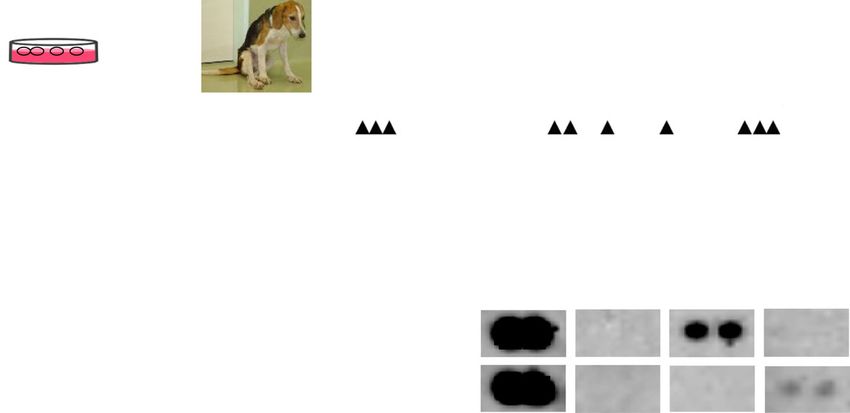
Nitahara-Kasahara et al. Stem Cell Research & Therapy (2021) 12:105 Page 4 of 15
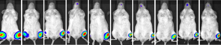
Fig. 1 Extended engraftment of IL-10-MSCs. Luciferase-expressing rat MSCs were transduced with control AAV1/enhanced green fluorescence
protein (GFP) or AAV1/IL-10 vector. a MSCs expressing GFP or IL-10 were injected into the right or left hindlimb muscle (IL-10 (−) or IL-10 (+)) of
NOD/SCID mice. In vivo bioluminescence imaging of mice treated with MSCs expressing GFP or IL-10 revealed the appearance of luciferase
signals between 3 and 67 days after intramuscular injection into the right or left hindlimb muscle: GFP-MSCs, IL-10 (−), or IL-10-MSCs, IL-10 (+). b
Monitoring of the quantitative luciferase counts at the GFP- or IL-10-MSC-injected site by imaging analysis conducted between 3 and 67 days
after treatment (n = 6–3). c Quantitative measurement of IL-10 levels in the MSC culture medium using ELISA 7 days after transduction with
AAV1/GFP or AAV1/IL-10 (n = 3). Data are presented as mean ± SD, and statistical differences between GFP-MSCs and IL-10-MSCs are indicated,
***
P < 0.001, ****P < 0.0001, t-test are indicated
Blood test were determined using a DRI-CHEM3506 automated
The dogs underwent periodic veterinary examinations at analyzer (Fuji Film, Tokyo, Japan).
1–2-week intervals until sampling. Hematological and
serum biochemical testing for creatine kinase (CK) was Histopathological and immunohistochemical analyses
performed using a model F-820 semi-automated Samples from MSC-treated TA muscles were collected
hematology analyzer (Sysmex, Hyogo, Japan). The levels and immediately frozen in liquid nitrogen-cooled iso-
of serum alanine aminotransferase (ALT), aspartate ami- pentane. Five mice from each group were used for ana-
notransferase (AST), and blood urea nitrogen (BUN) lysis at each time point. Transverse cryosections 8 μm in
Table 1 Summary of transplantation experiments
Dog ID Sex Agea BWb Cell Cell numbers Interval Injection numbers Route
4502MN M 51 11.3 IL-10-CD271+MSCs, MyoD-CD271+MSCs 2.5 × 10⁷ cells – 1 i.m.
5601MN M 40 11.2 IL-10-CD271+MSCs, MyoD-CD271+MSCs 4.0–10.0 × 10⁶ cells – 2 i.m.
14103MN M 3 5.0 – – – – –
14102MA M 3 3.3 – – – – –
14105MA M 3 3.4 hDPSCs 4.0 × 10⁶ cells/kg 2 weeks 9 i,v.
6
14108MA M 3 3.5 IL-10-hDPSCs 4.0 × 10 cells/kg 2 weeks 9 i,v.
M male, i.m intramuscular injection, i.v. intravenous injection
a
Age at injection (months)
b
BW body weight at first injection (kg)Nitahara-Kasahara et al. Stem Cell Research & Therapy (2021) 12:105 Page 5 of 15
thickness prepared from the skeletal muscles were manufacturer’s instructions. Luciferase levels were mea-
stained with H&E using standard procedures. For immu- sured using a Varioskan LUX Multimode Microplate
nohistochemical analyses, thick cryosections were fixed Reader (Thermo Fisher Scientific). Protein concentra-
in acetone for 5 min at − 20 °C. The tissue sections were tions were measured using a Pierce® BCA Protein Assay
then blocked using 0.5% bovine serum albumin (BSA) in Kit (Thermo Scientific Pierce, Rockford, IL, USA). Three
PBS. The following antibodies were used for antigen de- independent experiments were performed in duplicate.
tection at 1:40–1:50 dilutions: rabbit anti-firefly lucifer-
ase (ab21176; Abcam Plc., Cambridge, UK) and mouse Biodistribution of MSCs
anti-dystrophin (NCL-DYS3, Leica, Wetzlar, Germany). The tissue samples were disrupted in a Multi-Beads
These antibodies were diluted using 0.5% BSA in PBS Shocker (Yasui Kikai Co., Ltd., Osaka, Japan). DNA was
and used to treat the cells or tissue sections overnight at extracted from the tissue suspensions using a DNeasy
4 °C. The tissue sections were washed with PBS and then Blood and Tissue kit (QIAGEN, Valencia, CA) and quan-
probed with Alexa 568-conjugated anti-rabbit IgG anti- tified using a NanoDrop spectrophotometer (Thermo
bodies (Thermo Fisher Scientific) and Alexa 488- Fisher Scientific). Real-time qPCR was performed using
conjugated anti-mouse IgG antibodies (Thermo Fisher 125 ng of DNA in a total volume (20 μL) containing DNA
Scientific) at 1:250–1:100 dilution for 1 h at 4 °C. The Master SYBR Green I kit (Roche Diagnostics, Basel,
coverslip slides were washed with PBS and mounted in Switzerland) and primers for Alu or murine glyceralde-
Vectashield (Vector Laboratories Inc., Burlingame, CA, hyde 3-phosphate dehydrogenase (Gapdh). The primer
USA) with 4,6-diamidino-2-phenylindole (DAPI). Im- sequences used were as follows: human Alu, 5′-GTCAGG
munofluorescence analysis was performed using an IX71 AGATCGAGACCATCCC-3′ (forward) and 5′-TCCTGC
fluorescence microscope (Olympus, Tokyo, Japan). CTCAGCCTCCCAAG-3′ (reverse); murine Gapdh, 5′-
To confirm the presence of transplanted cells at the GATGACATCAAGAAGGTGGTGA-3′ (forward) and
injection sites, the MSCs were labeled with luciferase or 5′-TGCTGTAGCCGTATTCATTGTC-3′ (reverse). The
GFP. The tissue sections were incubated in a solution of PCR conditions were as follows: 95 °C for 2 min, followed
3% H2O2 to block endogenous peroxidase. The nonspe- by 40 cycles at 95 °C for 15 s, 68 °C for 30 s, and 72 °C for
cific binding sites were blocked using a 2% BSA solution. 30 s. The standard was generated by adding 10-fold serial
The tissue sections were probed with primary antibodies dilutions of human DPSCs to determine the number of
for 1 h and then treated using the 3,3′-diaminobenzidine human DPSCs in 125 ng of DNA that was used in the
(DAB) substrate kit (Vector Laboratories Inc.) contain- real-time PCR for each organ sample. We extrapolated
ing horseradish peroxidase (HRP) as an enzyme indica- the quantity of DNA isolated from each organ to deter-
tor. The slices were then stained with DAB chromogen mine the number of human DPSCs per organ.
to determine the form of the brown-antigen reaction
product. The tissue sections were visualized using an Proteome cytokine/cytokine array
IX71 microscope (Olympus). The FBS-free DPSC culture medium was collected after
2 days of incubation for array analysis. The relative
ELISA expression of cytokines and chemokines in the culture
The IL-10 expression levels were measured in the FBS- medium was quantified using the Proteome Profiler™
free MSC culture medium after 2 days of incubation, in Array (Mouse Cytokine Array, Panel A; R&D Systems
the TA muscle lysate, and in the serum obtained from Inc.), as previously described [28]. To achieve maximum
animals using the Quantikine ELISA mouse or canine assay sensitivity, the blots were incubated overnight with
IL-10 Immunoassay (Thermo Fisher Scientific) and ca- plasma. Enhanced chemiluminescence incubation was
nine IL-6 (R&D Systems Inc.) and the collagen type III performed for 5 min using the Super Signal West Femto
Immunoassay (Cloud-clone Corp. TX, USA), according Chemiluminescence Kit (Thermo Scientific Pierce), and
to the manufacturers’ recommendations. The final values the samples were imaged and analyzed using the Image
were normalized to the protein concentrations, which Quant LAS 4000 coupled with Image Quant TL software
were measured using the Pierce® BCA Protein Assay Kit (GE Healthcare Japan, Tokyo, Japan) and Image J soft-
(Thermo Fisher Scientific). ware (NIH, Bethesda, MD).
Luciferase reporter assays Locomotor activity analyses
Luciferase reporter assays were performed to evaluate The physical activity levels of CXMDJ and littermate
the retention of Luc-MSCs in the TA muscle. Firefly lu- normal dogs used as controls were monitored during the
ciferase activity was tested in whole tissue homogenates experimental period using an infrared sensor system
using the Bright-Glo™ Luciferase Assay System (Promega (Supermex, Muromachi Kikai Co., Ltd., Tokyo, Japan),
Corporation, Madison, WI) according to the as previously described [31]. These systems monitor andNitahara-Kasahara et al. Stem Cell Research & Therapy (2021) 12:105 Page 6 of 15
enumerate all spontaneous movements. The average of B). When luciferase (Luc)-MSCs were injected along
all counts of spontaneous locomotor activity in animals with AAV1/IL-10 or AAV1/LacZ control vectors, which
determined over 5 days and nights (12 h light/dark were also not transduced into the cells, a significant dif-
cycles) was calculated. Furthermore, we measured the ference was observed in cell survival in the Luc-MSC-
15-m running time of normal and CXMDJ littermates treated mice (Figure S1C, D). IL-10 plasmid-transfected
during the experimental period. The running speed was Luc-MSCs expressed higher levels of IL-10 and were
averaged four times. more effective at enhancing post-transplantation reten-
tion (Figure S2). We also confirmed a maximum 5.03-
Magnetic resonance imaging (MRI) fold stronger luciferase signal intensity in muscles trans-
CXMDJ anesthetized by injection (20 mg/kg) were intu- planted with IL-10-expressing MSCs than in control
bated using an endotracheal tube, and general MSC-treated muscles at day 7 (See also Figure S2C), al-
anesthetization was maintained using an inhalational though most of these cells disappeared within 12 days.
mixture of 2 to 3% isofluorane and oxygen. The heart We developed a continuous IL-10 expression system of
rate and oxygen saturation levels were monitored con- MSCs using an AAV vector to confirm the expected high
tinuously. Images of the T2-weighted and fat-saturated and prolonged cell survival rates following transplantation.
T2-weighted series were captured using a method The IL-10-Luc-MSCs displayed a higher survival rate im-
described in a previous study [32]. We examined the mediately after administration (luciferase signal at 3 days
crus muscles of the lower limbs using a superconducting after transplantation, 4.41 ± 1.78 × 10⁵ counts), with the
3.0-Tesla MRI device (MAGNETOM Trio; Siemens maximum value of 6.5-fold observed at 24 days (3.46 ±
Medical Solutions, Erlanger, Germany) with an 18-cm 1.12 × 10⁴ counts) compared to the signal corresponding
diameter/18-cm length human extremity coil. The im- to GFP-Luc-MSCs (2.98 ± 1.04 × 10⁵ counts, P < 0.0001;
ages were analyzed quantitatively using the Syngo 5.35 ± 2.93 × 10³ counts, < 0.005, respectively), as observed
MR2004A software (Siemens Medical Solutions), as pre- in vivo in imaging (Fig. 1a, b) and immunohistological
viously reported [32, 33]. Briefly, the ROIs were selected analyses (Figure S3). Significantly higher levels of IL-10
to avoid flow artifacts and large vessels, and the signal expression from AAV1/IL-10-transduced Luc-MSCs (IL-
intensities were measured for these ROIs. The SNRs for 10-Luc-MSCs) were confirmed in vitro (Fig. 1c) and in
each ROI were calculated using the following equation: treated muscles (See also Figure S4). The findings sug-
SNR = signal intensity/SDair, where SDair is the stand- gested that the higher retention in the early stage exerted
ard deviation (SD) of the background noise. The average a significant effect on long-term engraftment. Notably, IL-
SNR (Ave SNR) was calculated using the equation de- 10-Luc-MSCs were retained for more than 67 days after
scribed in a previous report [33]. The right and left TA transplantation (Fig. 1a, b).
muscles, EDL, gastrocnemius medial head, GL, flexor
digitorum superficialis, flexor digitorum longus, and Successful long-term engraftment of IL-10-MSCs in
flexor hallucis longus muscle were examined. injured muscle tissue
We also investigated the effects of IL-10 overexpression
Statistical analyses in CD271+MSCs derived from dog bone marrow using
Data are presented as the mean ± SD. Differences Beagle dogs as a larger animal model (Fig. 2a, Table 1).
between two groups were assessed using unpaired two- The high efficiency of adenovirus and AAV1 transduc-
tailed t-tests. Multiple comparisons between three or tion in CD271+MSCs was confirmed based on immuno-
more groups were performed using ANOVA (n = 3–6). fluorescence analysis or GFP signals (See also Figure S5).
Statistical significance was defined as *P < 0.05, **P < 0.01, CD271+MSCs transduced with AAV1/IL-10 (IL-10-Luc-
***
P < 0.001, and ****P < 0.0001 and was calculated using CD271+MSCs) exhibited IL-10 overexpression in the
Microsoft Excel (Microsoft, Redmond, WA, USA) and culture medium (MyoD-MSCs, 3.1 pg/mL; IL-10-MSCs,
GraphPad Prism 8 (GraphPad, La Jolla, CA, USA). 93.6 pg/mL). Four weeks after intramuscular injection,
the accumulation of IL-10-Luc-CD271+MSCs was ob-
Results served in the immunochemical analysis of cardiotoxin-
Enhanced engraftment of IL-10-expressing MSCs injured tibialis anterior (TA) muscle tissues. Immuno-
We investigated the effects of IL-10 after MSC injection fluorescence analysis revealed the accumulation of
into the skeletal muscle. In vivo bioluminescence im- luciferase-positive IL-10-Luc-CD271+MSCs around the
aging analysis showed that IL-10-expressing MSCs inflammatory region in MSC-treated TA muscle 4 weeks
tended to exhibit higher retention in the hindlimb after the secondary injection, comparable to the observa-
muscle of NOD/SCID mice than normal MSCs 4 days tion for MyoD-MSCs (Fig. 2b). Luciferase-positive
after injection; however, upon quantitative analysis, the muscle-like tissue was detected in the IL-10-Luc-
difference was not found to be significant (Figures S1A, CD271+MSC-treated muscle, similar to that observed inNitahara-Kasahara et al. Stem Cell Research & Therapy (2021) 12:105 Page 7 of 15 Fig. 2 Successful long-term engraftment of canine IL-10-MSCs in the skeletal muscles of dogs. a Transplantation schedule. Canine CD271+MSCs expressing luciferase (Luc-CD271+MSCs) were transduced with AAV1/IL-10 in the cardiotoxin-pretreated tibialis anterior (TA) muscle of the recipient dog. b Immunofluorescence analysis of the TA muscle derived from a MyoD-Luc-CD271+MSC- (left panel) and IL-10-Luc-CD271+MSC- treated dog (5601MN, right panels) 8 weeks after injection using antibodies specific for luciferase (red), dystrophin (green), and nuclear stain 4′,6′- diamidino-2-phenylindole (DAPI, blue). Arrow, luciferase-positive myofibers. Bar = 200 μm. c Luciferase assays to determine the cell number in the IL-10-Luc-CD271+MSC- (IL-10-MSCs) or MyoD-Luc-CD271+MSC (MyoD-MSC)-treated TA muscle lysate. The mean was the average value of three measurements from each group. d Quantitative measurement of IL-10 levels in the IL-10-MSC- or MyoD-MSC-treated TA muscle lysate (mg protein) using ELISA. Data are presented as mean ± SD, and statistical differences between values for MyoD-MSC- and IL-10-MSC-treated TA muscle are indicated, *P < 0.05, t-test the muscle after treatment with Luc-CD271+ MSCs muscles increased, while those in MyoD-Luc- transduced with MyoD (MyoD-Luc-CD271+MSC), CD271+MSC-treated muscles did not (Fig. 2d). These which is a key factor for myogenic determination, as de- data suggest that IL-10-expressing CD271+MSCs could scribed in a previous report [27]. These findings suggest survive long-term and engraft after intramuscular injec- that the Luc-CD271+MSCs transduced with AAV1/IL- tion during muscle regeneration. 10 formed myofibers. Luciferase activity, which corre- lated with the number of MSCs, tended to be higher in Safety and efficacy of systemic transplantation of IL-10- IL-10-Luc-CD271+MSC-treated whole TA muscles than DPSCs in the DMD model in MyoD-Luc-CD271+MSC-treated muscle (Fig. 2c). The Next, we evaluated the efficacy of IL-10-expressing IL-10 levels in IL-10-Luc-CD271+MSC-treated TA MSCs by performing systemic transplantation using
Nitahara-Kasahara et al. Stem Cell Research & Therapy      (2021) 12:105                                                               Page 8 of 15

hDPSCs (Fig. 3a). We confirmed that the extracellular                       CXMDJ model with the DMD phenotype (Table 1). No
secretion of IL-10 from AAV1/IL-10-transduced-human                         obvious abnormalities related to liver damage, kidney
DPSCs (IL-10-hDPSCs) significantly inhibited the ex-                        damage, or anemia were induced in response to the sys-
pression of pro-inflammatory monocyte chemotactic                           temic administration of hDPSCs in the CXMDJ model.
protein-1 (MCP-1), and conversely upregulated stromal-                      Transient increases in ALT, AST, and BUN levels were
derived factor-1 (SDF-1) in IL-10-hDPSCs compared                           observed occasionally in the CXMDJ model, independent
with the levels observed in hDPSCs (Fig. 3b, c). IL-10-                     of treatment with hDPSCs (See also Figure S6). During
hDPSCs or hDPSCs were intravenously injected nine                           the experiment, the IL-10-hDPSC-treated CXMDJ
times at biweekly intervals in the acute phase in the                       showed better growth compared to the untreated

 Fig. 3 Safe systemic treatment with IL-10-expressing hDPSCs in the CXMDJ model. a Transplantation schedule. b Quantitative measurement of IL-
 10 expression in 2-day culture medium and in the hDPSC lysate (100 mg protein) using ELISA. Data are presented as mean ± SD, and statistical
 differences between hDPSCs vs. IL-10-hDPSCs are indicated (****P < 0.001, n = 3). c Cytokine and chemokine expression in 2-day culture media of
 hDPSCs and IL-10-hDPSCs analyzed using the Proteome ProfilerTM Array. Changes in the expression levels of monocyte chemotactic protein-1
 (MCP-1), and stromal-derived factor-1 (SDF-1/CXCL12), compared to the positive control (PC) signals or negative control (NC). Signal intensity in
 the regions of interest (ROIs) quantified using array images (upper panels) and representative data (graph) are presented. ND, not detected. d
 Growth curve of untreated CXMDJ (control DMD; 14102MA), hDPSC-, and IL-10-hDPSC-treated CXMDJ (hDPSC-DMD, 14105MA; IL-10-hDPSC-DMD,
 14108MA) dogs. Data are presented as mean ± SD, and statistical differences between control DMD vs. IL-10-hDPSC-DMD (****P < 0.001), hDPSC-
 DMD vs. IL-10-hDPSC-DMD (####P < 0.001) are indicated; one-way ANOVA. e Serum levels of IL-10 at 6, 24, and 48 h, and 7 days after
 transplantation (n = 3), quantified using ELISA. Data are presented as mean ± SD, and statistical differences between control DMD vs. IL-10-hDPSC-
 DMD (****P < 0.0001), hDPSC-DMD vs. IL-10-hDPSC-DMD (###P < 0.001, ####P < 0.0001) are indicated; ns, not significant, two-way ANOVA
Nitahara-Kasahara et al. Stem Cell Research & Therapy (2021) 12:105 Page 9 of 15
littermates, CXMDJ (P < 0.0001), and hDPSC-treated diameter in the dystrophic muscles, spread muscle inter-
CXMDJ (< 0.0001), in terms of body weight (Fig. 3d). stitium, and cell infiltration interspersed in the muscle
Although the serum levels of IL-10 increased transiently interstitium (Fig. 4e). In contrast, the histopathological
6 h after IL-10-hDPSC injection (104.0 pg/mL vs. control observations of the hDPSC- and IL-10-hDPSC-treated
DMD, vs. hDPSC-DMD, < 0.0001), the levels decreased CXMDJ muscles revealed significantly limited infiltration
rapidly within 24 h of transplantation (32.6 pg/mL vs. of nuclei, which indicated a milder phenotype compared
control DMD, < 0.0067; vs. hDPSC-DMD, < 0.0278), and to untreated CXMDJ. These data suggest that the re-
did not differ significantly from the control CXMDJ 7 peated systemic administration of IL-10-hDPSCs induces
days after injection (21.6 pg/mL) (Fig. 3e). The levels of morphological improvement, including inflammation
IL-6 in blood increased transiently in CXMDJ (max- regulation, in CXMDJ. In addition, we performed quanti-
imum 742.9 pg/mL). In contrast, the levels in IL-10- tative analysis of collagen type III expression at 8 muscle
hDPSC-treated CXMDJ were within the normal range tissue sites to investigate fibrosis. However, we could not
(0–9.85 pg/mL) during the experiments. High levels of confirm a significant difference among the groups (con-
IFN-γ were also observed in CXMDJ (maximum 103.9 trol DMD, 2.27 ± 2.18 pg/μg protein; hDPSC- and IL-10-
pg/mL) and hDPSC-treated CXMDJ (114.8 pg/mL), hDPSC-DMD, 3.91 ± 1.66, and 6.17 ± 2.50 pg/μg protein,
whereas the levels tended to be marginally lower in the respectively, P = 0.317).
IL-10-hDPSC-treated CXMDJ (68.4 pg/mL). The trend
of cell retention in blood was similar for hDPSCs Long-term maintenance of muscle function in IL-10-
(17.9 ± 19.5 pg/100 ng genomic DNA) and IL-10- hDPSC-treated DMD dog
hDPSCs 24 h after transplantation (79.4 ± 7.89 pg/100 ng The CXMDJ developed progressive general weakness
genomic DNA), as revealed by human-specific Alu-PCR. owing to the reduced strength of the skeletal muscles.
Long-term engraftment in tissues was investigated. The tetanic force in the hind limbs in CXMDJ (2.55 ±
hDPSCs were not detected in the skeletal muscle, lung, 0.42 N m/s) was 41.2 ± 5.1% of that in normal dogs (P <
or liver tissues of the hDPSC-treated CXMDJ. The IL- 0.0001) (Figure S8). Conversely, significantly higher
10-hDPSCs only survived and maintained the engraft- torque values were observed in the IL-10-hDPSC-treated
ment status 4 months after treatment in the TA muscle CXMDJ (4.17 ± 1.28 N m/s, 66.5 ± 12.2% of the value in
(56.1 pg/100 ng genomic DNA), whereas the cells were the normal dog, < 0.0001) than in CXMDJ (< 0.0008),
not detected in other organs. Conversely, dystrophin similar to that in hDPSC-treated CXMDJ (3.68 ± 0.57
expression was undetectable in the muscle tissues of N m/s). These results suggest that the progressive loss of
hDPSC-treated CXMDJ (Figure S7A). limb muscle strength is ameliorated upon treatment
with hDPSCs and IL-10-hDPSCs.
Morphological improvement in IL-10-hDPSC-treated dog Additionally, the physical activity of CXMDJ in the
with DMD home cage also reduced drastically with age compared
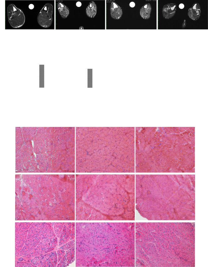
The high-intensity T2-signals observed in MRI, which to that in normal dogs (Fig. 5a) [31]. Improved activity
were detected in the necrotic/edematous and inflamma- was confirmed in both groups, the hDPSC-treated
tory lesions in the dystrophic muscle, were significantly (8340.4 ± 1090.3 counts; vs. control DMD, P = 0.0006)
reduced in the cross-sectional muscles of the IL-10- and IL-10-hDPSC-treated CXMDJ (8531.6 ± 1146.5
hDPSC-treated dog (82.5 ± 16.9 average signal-to-noise counts; vs. control DMD, < 0.0001), which was observed
ratios, SNRs) compared to the signals in hDPSC-treated upon the comparison with 1-year-old CXMDJ litter-
CXMDJ (97.4 ± 13.3, P = 0.008) after transplantation mates advanced symptoms (3954.4 ± 792.0 counts)
(Fig. 4a, b). The weight of each muscle obtained from (Fig. 5a). The IL-10-hDPSC-treated CXMDJ exhibited
IL-10-hDPSC-treated CXMDJ (3.73 ± 1.43 g) increased persistent and predominantly higher activity (6 months;
compared to that in the untreated (2.16 ± 1.04 g, vs. IL- 13,008.8 ± 1367.1 counts) than CXMDJ (9926.0 ± 1436.8
10-hDPSC- CXMDJ, < 0.005) and hDPSC-treated counts, < 0.005) as well as hDPSC-treated CXMDJ (12,
CXMDJ (2.33 ± 1.04 g, < 0.005) (Fig. 4c). We also con- 605.8 ± 1756.3 counts). Furthermore, hDPSC- or IL-10-
firmed that the area of the muscle fiber in the extensor hDPSC-treated CXMDJ maintained a 15-m running
digitorum longus (EDL) muscle from IL-10-hDPSC- speed and were active at 3 to 12 months of age (Fig. 5b,
treated CXMDJ (935.7 ± 601.9 μm2) increased compared see also Figure S9, and Table S1; normal vs. control
to that in the untreated (537.6 ± 439.2 μm2 vs. IL-10- DMD, P < 0.0001–0.0062; control DMD vs. hDPSC-
hDPSC-CXMDJ, < 0.0001) and hDPSC-treated CXMDJ DMD, < 0.005; control DMD vs. IL-10-hDPSC-DMD, <
(629.3 ± 352.8 μm2, < 0.0001) (Fig. 4d). The gastrocne- 0.005). There was no significant difference in the run-
mius lateral (GL), EDL, and flexor digitorum superficialis ning speed between hDPSC- and IL-10-hDPSC-treated
(FDS) muscles from CXMDJ displayed smaller (regener- CXMDJ. However, the increased serum CK levels after
ating fibers) and larger (hypertrophic fibers) fiber running exercise (50,595 ± 67,255 unit/L) were restoredNitahara-Kasahara et al. Stem Cell Research & Therapy (2021) 12:105 Page 10 of 15
Fig. 4 Improvement in the hDPSC-treated CXMDJ model observed by histological examination. a Cross-sectional magnetic resonance images
(MRI) in the lower leg muscles of a 7-month-old normal dog, untreated CXMDJ (control DMD), hDPSC-treated (hDPSC-DMD), and IL-10-hDPSC-
treated CXMDJ (IL-10-hDPSC-DMD). Muscle necrosis and inflammation based on the sequence of T2-weighted imaging of the lower legs for each
dog were comparable (R, right side; L, left side, left/right asymmetry). b The averaged signal-to-noise ratios (Ave SNR) were calculated in the
regions of interest (ROIs) from all muscles (n = 14) derived from each hindlimb of 2-month-old (before transplantation) and 7-month-old dogs
(after transplantation). Data are presented as mean ± SD, and statistical differences between normal vs. control DMD (****P < 0.0001), hDPSC-DMD
vs. IL-10-hDPSC-DMD (##P < 0.01) are indicated; ns, not significant, one-way ANOVA. c Weight of skeletal muscle (n = 16) from 1-year-old normal
dog, untreated CXMDJ (control DMD), hDPSC-treated (hDPSC-DMD), and IL-10-hDPSC-treated CXMDJ (IL-10-hDPSC-DMD) shown as dot plots.
Median values are presented as black bars. d Muscle fiber areas (μm2) measured from the extensor digitorum longus (EDL) muscle of 1-year-old
control DMD, hDPSC-DMD, and IL-10-hDPSC-DMD dogs by hematoxylin and eosin (H&E) staining. Each fiber area is indicated by a dot, and the
average fiber area is described as a red bar for each muscle. In total, 305 fibers are represented in the dot plot. Median values are indicated by
the red bars. Statistical differences between normal vs. control DMD (****P < 0.0001), control DMD, or hDPSC-DMD vs. IL-10-hDPSC-DMD (#P < 0.05,
####
P < 0.0001) are indicated; ns, not significant, one-way ANOVA. e H&E staining of the gastrocnemius lateral head (GL), EDL, and flexor digitorum
superficialis (FDS) muscle from 1-year-old control DMD, hDPSC-DMD, and IL-10-hDPSC-DMD dogs. Scale bar, 100 μmNitahara-Kasahara et al. Stem Cell Research & Therapy (2021) 12:105 Page 11 of 15 Fig. 5 Improved locomotor activity in CXMDJ treated with IL-10-expressing MSCs. a Day-time behavioral activity of normal (left graph), untreated CXMDJ (control DMD), hDPSC-treated CXMDJ (hDPSC-DMD), and IL-10-hDPSC-treated CXMDJ (IL-10-hDPSC-DMD) in home cage at 3, 6, and 12 months presented as mean count activity (average of value for 5 days). Statistical differences between control DMD vs. hDPSC-DMD or IL-10- hDPSC-DMD dog groups (#P < 0.05, ##P < 0.01, ###P < 0.001, ####P < 0.0001) are indicated; ns, not significant, two-way ANOVA. b 15-m running speed of normal, control DMD, hDPSC-DMD, and IL-10-hDPSC-DMD dogs at 12 months. The mean value was the average value of four measurements from each group. Statistical differences between normal vs. DMD (*P < 0.05, **P < 0.01, and ****P < 0.0001), control DMD vs. hDPSC- DMD, or IL-10-hDPSC-DMD (#P < 0.05, ##P < 0.01, ###P < 0.001) are indicated; ns, not significant, one-way ANOVA. Serum creatine kinase (CK) levels c and serum lactic acid levels d in each group before, 0, and 20 min after running exercise, which were four times greater than those after 15-m running, as measured using ELISA. Statistical differences between normal vs. DMD (*P < 0.05, **P < 0.01), control DMD vs. IL-10-hDPSC-DMD (#P < 0.05) are indicated; two-way ANOVA. n = 4 for each group. Data are presented as the mean ± SD immediately until 20 min in IL-10-hDPSC-treated L; 20 min, 95,300 ± 16,835 unit/L) as well as in the CXMDJ (Fig. 5c, 16,490 ± 4850 unit/L; vs. hDPSC- untreated CXMDJ (0 min, 76,650 ± 46,995 unit/L; 20 treated DMD, P = 0.0134). Conversely, a persistent and min, 81,425 ± 47,458 unit/L, P = 0.9277) after exercise. significant increase in the serum CK levels was observed CXMDJ also showed higher concentrations of lactic acid in hDPSC-treated CXMDJ (0 min, 96,075 ± 24,311 unit/ before and after exercise compared to normal dogs.
Nitahara-Kasahara et al. Stem Cell Research & Therapy   (2021) 12:105                                                  Page 12 of 15

However, no significant change in the levels of lactic                  enhance pro-angiogenic differentiation in injured tissue
acid was observed in hDPSC-treated CXMDJ (Fig. 5d).                     as a result of prolonged survival.
These findings suggest that IL-10-hDPSCs exert a pro-                     It was reasoned that IL-10-expressing hDPSCs exerted
tective effect against dystrophic damage caused by                      enhanced anti-inflammatory and protective effects on
exercise.                                                               damaged tissue owing to the downregulation of MCP-1
  Overall, we observed that IL-10-expressing hDPSCs                     and upregulation of SDF-1 in IL-10-hDPSCs (Fig. 3).
were able to ameliorate the dystrophic phenotype upon                   SDF-1 is a crucial factor that supports tissue regener-
systemic repeated administration in dogs with DMD.                      ation, and the roles of SDF-1 in MSC paracrine-
                                                                        mediated tissue repair have been reported [38]. Further-
                                                                        more, it was also reported that the expression of mul-
Discussion                                                              tiple pro-angiogenic factors, such as SDF-1, FGF-2, IGF-
To improve the functionality of MSCs as a cell source,                  1, and VEGF-A, is upregulated in IL-10-MSC-treated
we focused on the overexpression of IL-10 based on its                  cardiac muscle [19], suggesting changes in the surround-
anti-apoptotic and anti-inflammatory properties. In this                ing microenvironment.
study, bone marrow MSCs derived from rat and CD271+                       In the dog model, the repeated systemic transplant-
MSCs isolated from healthy dogs were injected intra-                    ation of hDPSCs and IL-10-expressing hDPSCs was safe
muscularly into NOD/SCID mice or healthy Beagle dogs                    and effective as a strategy for DMD therapy, as indicated
to study cell survival and engraftment in injured muscles               by the blood tests, growth, spontaneous activity, and
(Figs. 1 and 2). hDPSCs exhibit cellular properties that                running function (Figs. 3 and 4). Long-term engraftment
are highly similar to those of bone marrow MSCs.                        of hDPSCs was only confirmed in the dystrophic mus-
Human-origin DPSCs or IL-10-transduced hDPSCs were                      cles in IL-10-DPSC-treated DMD, which suggests that
also administered via intravenous injection into CXMDJ                  hDPSC engraftment was enhanced in response to IL-10
to investigate their systemic therapeutic effects (Figs. 3,             paracrine effects. For example, SDF-1 and growth factors
4, and 5). Our results indicate an improvement in sur-                  might enhance DPSC retention by altering the micro-
vival rate, engraftment, and protective effects of different            environment. SDF-1 and its receptor CXCR4 and
types of MSC in muscle tissues. This property is consid-                CXCR7 stimulate the production of paracrine mediators,
ered to be highly common to MSCs.                                       including VEGF, β-FGF-1, and HGF, which exert anti-
  The higher survival rate of IL-10-MSCs in the early                   apoptotic, pro-angiogenic, and anti-inflammatory effects
stage immediately after transplantation, as observed in                 [39]. In addition, it is revealed that muscle regeneration
the in vivo imaging analysis, is thought to facilitate long-            is associated with muscular re-expression of CXCR4 in
term cell retention (Fig. 1), suggesting that stable IL-10              dystrophic muscle [40]. Based on this, we consider that
expression enabled the long-term survival and engraft-                  IL-10-DPSC-specific engraftment and therapeutic effects
ment of MSCs after transplantation. Our findings sug-                   may be associated with DMD treatment.
gest that during skeletal muscle regeneration, prolonged                  These facts provide evidence of the accumulation of
engraftment of IL-10-expressing CD271+MSCs eased the                    hDPSCs at the site of inflammation after systemic ad-
formation of new myofiber-like tissue and preservation                  ministration, similar to that of MSCs. The functional re-
of a functional contractile apparatus, following exposure               covery in the dystrophic skeletal muscles was attributed
to the muscle stem cell niche/microenvironment (Fig. 2).                to the alleviation of the morphological pathologies, as in-
Rarely, MSCs differentiate into myogenic lineage cells in               dicated by the MRI findings and the histopathological
the absence of triggers such as MyoD (See also Figure                   appearance of samples from the hDPSC- and IL-10-
S5D, E) [34], 5-azacytidine [35], and Notch I intracellular             hDPSC-treated CXMDJ; however, fibrosis was not pre-
domain [4]. Although we could not confirm the myo-                      vented (Fig. 4). Indeed, both hDPSC- and IL-10-hDPSC-
genic differentiation of IL-10-MSCs in this study, as                   treated CXMDJ showed improved limb strength, as evi-
shown in Figure S7B, IL-10 is also considered to play a                 denced by the tetanic force, revealing an improvement
role in the long-term engraftment and survival of MSCs                  in spontaneous activity and running speed, while no sig-
in muscle tissue, resulting from their association with                 nificant difference was observed in the treated CXMDJ
surrounding myogenic stem/progenitor cell populations,                  while maintaining apparent function. We observed that
such as satellite cells, skeletal muscle-MSCs, fibro-                   the IL-10-hDPSC-treated CXMDJ was more stable than
adipogenic progenitors (FAB), and myo-endothelial pro-                  the hDPSC-treated CXMDJ because the increase in
genitors [36] for muscle repair. Furthermore, treatment                 serum CK levels after exercise was rapidly stabilized in
of myocardial infarction with CD271+ bone marrow                        the IL-10-hDPSC-treated CXMDJ (Fig. 5c). These facts
MSCs inhibited the expression of inflammatory cyto-                     suggest that treatment with IL-10-hDPSCs provides pro-
kines and significantly upregulated pro-angiogenic VEGF                 tection from physical damage-induced muscle injury in
[37]. CD271+ MSCs expressing IL-10 are also thought to                  CXMDJ, as opposed to the effect observed in dogs
Nitahara-Kasahara et al. Stem Cell Research & Therapy (2021) 12:105 Page 13 of 15
injected with untreated or non-transgenic cells, which is inflammatory properties of IL-10 in DMD treatment. In
further evidenced by the effects of the modified charac- case of local injection, the IL-10-MSCs could maintain
teristics of IL-10-hDPSCs involved in SDF-1 and VEGF. the long-term engraftment status and facilitate associ-
However, it was not clear whether the protective effects ated tissue repair. In case of repeated systemic adminis-
of IL-10-hDPSCs on the dystrophic muscle were caused tration, the IL-10-MSCs also protected the muscles from
by IL-10 directly or indirectly. The molecular mecha- physical damage-induced injury, which improved the
nisms underlying the action of IL-10-hDPSCs are ex- signs of muscle dysfunction in DMD. We can conclude
pected to be elucidated in future studies. that the local and systemic administration of IL-10-
This is the first report of increased cell survival, en- MSCs may exert beneficial IL-10 paracrine effects, which
graftment, and possible tissue formation induced by IL- have potential value in DMD therapeutics.
10-secreting MSCs in muscle tissues. In our previous re-
port, myogenic lineage-MSCs were successfully Supplementary Information
engrafted in muscle tissues [27]. However, a more effi- The online version contains supplementary material available at https://doi.
cient transplantation strategy is required for functional org/10.1186/s13287-021-02168-1.
improvement of muscle dystrophy. This study evaluated
the possibility of improving survival, engraftment, and Additional file 1: Figure S1. In vivo bioluminescence imaging for the
detection of injected MSCs. Figure S2. Enhanced engraftment of IL-10-
immune modulation of MSCs by AAV vector-mediated MSCs. Figure S3. MSC survival in skeletal muscle tissue. Figure S4. IL-10
stable expression of IL-10. expression in IL-10-MSC-treated muscle tissue. Figure S5.
We have previously provided evidence that severe phe- Characterization of gene-transduced MSCs. Figure S6. Serum chemistry
data from the hDPSC-treated CXMDJ model. Figure S7. Reverse transcrip-
notypes in IL-10 knockout mdx mice, such as increased tion PCR for evaluating human specific dystrophin expression. Figure S8.
M1 macrophage infiltration, high inflammatory factor Estimated isometric tetanic force in hDPSC-treated CXMDJ. Figure S9.
levels, and progressive cardiorespiratory dysfunction, 15-m running speed of hDPSC-treated CXMDJ. Table S1. Running speed
(15-m) of dogs aged 12 to 44 months
show a predisposition toward inflammation [41]. Gluco-
corticoids are widely used in patients to interrupt and
improve muscle strength during early stages, which may Abbreviations
AAV: Adeno-associated virus; ALT: Alanine aminotransferase; BSA: Bovine
also act directly on muscle fibers by stabilizing sarco- serum albumin; BUN: Blood urea nitrogen; CD: Cluster of differentiation;
lemma [42, 43]; however, this method is frequently asso- CK: Creatine kinase; CXMDJ: Canine X-linked muscular dystrophy model in
ciated with severe side effects. In our strategy, MSCs Japan; DAB: 3,3′-Diaminobenzidine; DAPI: 4′,6′-Diamidino-2-phenylindole;
DMD: Duchenne muscular dystrophy; DMEM: Dulbecco’s modified Eagle’s
appeared to exhibit inflammatory regulation effects and medium; DPSCs: Dental pulp stem cells; eGFP: Enhanced green fluorescent
a protective effect in the dystrophic muscle through the protein; EDL: Extensor digitorum longus; FAB: Fibro-adipogenic progenitors;
suppression of M1 macrophage infiltration by secreting FBS: Fetal bovine serum; FDS: Flexor digitorum superficialis; GAPD
H: Glyceraldehyde 3-phosphate dehydrogenase; GL: Gastrocnemius lateral;
IL-10. In addition, our stable IL-10 expression system GVHD: Graft-versus-host disease; HLA: Human leukocyte antigen;
was found to be safe, with a low risk of genome insertion HRP: Horseradish peroxidase; IFN-γ: Interferon-gamma; IL-10: Interleukin-10;
owing to the use of an AAV vector. Random integration, iPS: Induced pluripotent stem cells; MCP-1: Monocyte chemotactic protein-1;
MSCs: Multipotent mesenchymal stromal cells; MRI: Magnetic resonance
off-target effects, and poor specificity are associated with imaging; PBS: Phosphate-buffered saline; PCR: Polymerase chain reaction;
the use of other viruses and genome engineering tech- SD: Standard deviation; SDF-1: Stromal-derived factor-1; SNRs: Signal-to-noise
niques. IL-10-expressing MSCs are expected to have po- ratios; TA: Tibialis anterior; TALEN: Transcription activator-like effector nucle-
ase; TNF-α: Tumor necrosis factor-alpha; VEGF: Vascular endothelial growth
tential applicability in muscle regeneration and factor; VSV-G: Vesicular stomatitis virus-glycoprotein
treatment of muscle diseases. We have previously re-
ported that IL-10 overexpression promotes neuroprotec- Acknowledgements
tion in an experimental acute ischemic stroke model The authors express their gratitude to Akihiko Umezawa, Akinori Nakamura,
Tetsuya Nagata, Masanori Kobayashi, Yuko Shimizu-Motohashi, Jun Tanihata,
[44]. There is clinical interest in the applicability of IL- Michihiro Imamura, Yoshitsugu Aoki, Yoshitaka Miyagawa, and Kazuhiro Ya-
10-MSCs in ex vivo cell therapy owing to their anti- mamoto for their technical advice, support, and helpful discussions; Maya
inflammatory properties and ability to release cytokines Kawamura, Tomoko Fukatsu, Sonoko Shimazu, Tomoko Chiyo, Chiaki Masuda,
Kazue Kinoshita, Ryoko Nakagawa, and Rie Ogawa for technical assistance;
into the surrounding environment, which mediate their Hideki Kita, Shinichi Ichikawa, Yumiko Yahata-Kobayashi, Takayoshi Hikage, Aya
paracrine effects and modify the developmental fate of Kuriyama, Akane Hanaoka-Hayashi, Namiko Ogawa, and other staff members
neighboring cells. We expect that the multiple character- of JAC Co. for caring for the dogs (General Animal Research Facility, NCNP).
We are thankful to JCR Pharmaceuticals Co., Ltd. for their generous contribu-
istics and regenerative effects of MSCs alone as well as tion of DPSCs and MSCs. We also thank Posadas Herrera Guillermo for check-
in combination with CD271+ IL-10 MSCs will result in ing the quality of English in the manuscript.
an improved therapeutic impact in DMD.
Authors’ contributions
Conclusions Y. N-K. and T. O. conceived and planned the experiments. Y. N-K. and M.K.
performed the experiments, derived the models, and analyzed the data. H.
Our methods were developed to enhance MSC survival H-K. Y.O. and M.K. contributed to sample preparation and assisted with ex-
and improve their therapeutic effects using the anti- periments involving animal models. Y. N-K. wrote the manuscript inNitahara-Kasahara et al. Stem Cell Research & Therapy (2021) 12:105 Page 14 of 15
consultation, and T.O. and S.T. helped supervise the project. T.O. supervised 9. Rasmusson I, Ringden O, Sundberg B, Le Blanc K. Mesenchymal stem cells
the project. The author(s) read and approved the final manuscript. inhibit the formation of cytotoxic T lymphocytes, but not activated
cytotoxic T lymphocytes or natural killer cells. Transplantation. 2003;76:
Funding 1208–13.
This work was supported by a research grant from JCR Pharmaceuticals Co., 10. Zappia E, Casazza S, Pedemonte E, Benvenuto F, Bonanni I, Gerdoni E, Giunti
Ltd., Health Sciences Research Grants for Research on Human Genome and D, Ceravolo A, Cazzanti F, Frassoni F, Mancardi G, Uccelli A. Mesenchymal
Gene Therapy from the Ministry of Health, Labor and Welfare of Japan, and a stem cells ameliorate experimental autoimmune encephalomyelitis
Grant-in-Aid for Scientific Research from the Ministry of Education, Culture, inducing T-cell anergy. Blood. 2005;106:1755–61.
Sports, Science and Technology (grant number: 22390284, 22–40134). 11. Ichim TE, Alexandrescu DT, Solano F, Lara F, Campion Rde N, Paris E, Woods
EJ, Murphy MP, Dasanu CA, Patel AN, Marleau AM, Leal A, Riordan NH.
Mesenchymal stem cells as anti-inflammatories: implications for treatment
Availability of data and materials
of Duchenne muscular dystrophy. Cell Immunol. 2010;260:75–82.
The datasets used and/or analyzed during the current study are available
12. Aggarwal S, Pittenger MF. Human mesenchymal stem cells modulate
from the corresponding author on reasonable request.
allogeneic immune cell responses. Blood. 2005;105:1815–22.
13. MacDonald KP, Pettit AR, Quinn C, Thomas GJ, Thomas R. Resistance of
Ethics approval and consent to participate rheumatoid synovial dendritic cells to the immunosuppressive effects of IL-
Animal experiments using MSCs were conducted in accordance with the 10. J Immunol. 1999;163:5599–607.
protocol approved by the Institutional Animal Care and Use Committee of 14. Groux H, Bigler M, de Vries JE, Roncarolo MG. Inhibitory and stimulatory
Nippon Medical School (27-199) and National Center of Neurology and effects of IL-10 on human CD8+ T cells. J Immunol. 1998;160:3188–93.
Psychiatry (NCNP) Animal Ethics Committees (2012011, 2015004, and 19–30- 15. Manning E, Pham S, Li S, Vazquez-Padron RI, Mathew J, Ruiz P, Salgar SK.
06). Interleukin-10 delivery via mesenchymal stem cells: a novel gene therapy
approach to prevent lung ischemia-reperfusion injury. Hum Gene Ther.
Consent for publication 2010;21:713–27.
Not applicable. 16. Min CK, Kim BG, Park G, Cho B, Oh IH. IL-10-transduced bone marrow
mesenchymal stem cells can attenuate the severity of acute graft-versus-
Competing interests host disease after experimental allogeneic stem cell transplantation. Bone
Y. N-K., H. H-K., and T. O. were members of the Division of Cell and Gene Marrow Transplant. 2007;39:637–45.
Therapy, Nippon Medical School, which is an endowment department, sup- 17. Peruzzaro ST, Andrews MMM, Al-Gharaibeh A, Pupiec O, Resk M, Story D,
ported with an unrestricted grant from JCR Pharmaceuticals Co., Ltd. and Maiti P, Rossignol J, Dunbar GL. Transplantation of mesenchymal stem cells
Kaneka Corporation. hDPSCs were provided by JCR Pharmaceuticals Co., Ltd. genetically engineered to overexpress interleukin-10 promotes alternative
There is no COI related to other authors. inflammatory response in rat model of traumatic brain injury. J
Neuroinflammation. 2019;16:2.
Author details 18. Lu X, Ru Y, Chu C, Lv Y, Gao Y, Jia Z, Huang Y, Zhang Y, Zhao S. Lentivirus-
1
Department of Biochemistry and Molecular Biology, Nippon Medical School, mediated IL-10-expressing bone marrow mesenchymal stem cells promote
Bunkyo City, Tokyo, Japan. 2Division of Cell and Gene Therapy, Nippon corneal allograft survival via upregulating lncRNA 003946 in a rat model of
Medical School, Bunkyo City, Tokyo, Japan. 3Department of Molecular corneal allograft rejection. Theranostics. 2020;10:8446–67.
Therapy, National Institute of Neuroscience, National Center of Neurology 19. Meng D, Han S, Jeong IS, Kim SW. Interleukin 10-secreting MSCs via TALEN-
and Psychiatry, Kodaira, Tokyo, Japan. 4Laboratory of Experimental Animal mediated gene editing attenuates left ventricular remodeling after
Science, Nippon Veterinary and Life Science University, Musashino, Tokyo, myocardial infarction. Cell Physiol Biochem. 2019;52:728–41.
Japan. 5Division of Molecular and Medical Genetics, Center for Gene and Cell 20. Choi JJ, Yoo SA, Park SJ, Kang YJ, Kim WU, Oh IH, Cho CS. Mesenchymal
Therapy, Institute of Medical Science, University of Tokyo, Minato-ku, Tokyo stem cells overexpressing interleukin-10 attenuate collagen-induced arthritis
108-8639, Japan. in mice. Clin Exp Immunol. 2008;153:269–76.
21. Wang C, Lv D, Zhang X, Ni ZA, Sun X, Zhu C. Interleukin-10-overexpressing
Received: 8 September 2020 Accepted: 14 January 2021 mesenchymal stromal cells induce a series of regulatory effects in the
inflammatory system and promote the survival of endotoxin-induced acute
lung injury in mice model. DNA Cell Biol. 2018;37:53–61.
References 22. Shimatsu Y, Yoshimura M, Yuasa K, Urasawa N, Tomohiro M, Nakura M,
1. Friedenstein AJ, Petrakova KV, Kurolesova AI, Frolova GP. Heterotopic of Tanigawa M, Nakamura A, Takeda S. Major clinical and histopathological
bone marrow. Analysis of precursor cells for osteogenic and hematopoietic characteristics of canine X-linked muscular dystrophy in Japan, CXMDJ. Acta
tissues. Transplantation. 1968;6:230–47. Myol. 2005;24:145–54.
2. Bruder SP, Jaiswal N, Haynesworth SE. Growth kinetics, self-renewal, and the 23. Shimatsu Y, Katagiri K, Furuta T, Nakura M, Tanioka Y, Yuasa K, Tomohiro M,
osteogenic potential of purified human mesenchymal stem cells during Kornegay JN, Nonaka I, Takeda S. Canine X-linked muscular dystrophy in
extensive subcultivation and following cryopreservation. J Cell Biochem. Japan (CXMDJ). Exp Anim. 2003;52:93–7.
1997;64:278–94. 24. Jo YY, Lee HJ, Kook SY, Choung HW, Park JY, Chung JH, Choung YH, Kim ES,
3. Haynesworth SE, Goshima J, Goldberg VM, Caplan AI. Characterization of Yang HC, Choung PH. Isolation and characterization of postnatal stem cells
cells with osteogenic potential from human marrow. Bone. 1992;13:81–8. from human dental tissues. Tissue Eng. 2007;13:767–73.
4. Dezawa M, Ishikawa H, Itokazu Y, Yoshihara T, Hoshino M, Takeda S, Ide C, 25. Nito C, Sowa K, Nakajima M, Sakamoto Y, Suda S, Nishiyama Y, Nakamura-
Nabeshima Y. Bone marrow stromal cells generate muscle cells and repair Takahashi A, Nitahara-Kasahara Y, Ueda M, Okada T, Kimura K.
muscle degeneration. Science. 2005;309:314–7. Transplantation of human dental pulp stem cells ameliorates brain damage
5. Sun Y, Liu J, Qian F, Xu Q. Nitric oxide inhibits T cell adhesion and following acute cerebral ischemia. Biomed Pharmacother. 2018;108:1005–14.
migration by down-regulation of beta1-integrin expression in 26. Uchibori R, Okada T, Ito T, Urabe M, Mizukami H, Kume A, Ozawa K.
immunologically liver-injured mice. Int Immunopharmacol. 2006;6:616–26. Retroviral vector-producing mesenchymal stem cells for targeted suicide
6. Kopen GC, Prockop DJ, Phinney DG. Marrow stromal cells migrate cancer gene therapy. J Gene Med. 2009;11:373–81.
throughout forebrain and cerebellum, and they differentiate into astrocytes 27. Nitahara-Kasahara Y, Hayashita-Kinoh H, Ohshima-Hosoyama S, Okada H,
after injection into neonatal mouse brains. Proc Natl Acad Sci U S A. 1999; Wada-Maeda M, Nakamura A, Okada T, Takeda S. Long-term engraftment of
96:10711–6. multipotent mesenchymal stromal cells that differentiate to form myogenic
7. Ma Y, Xu Y, Xiao Z, Yang W, Zhang C, Song E, Du Y, Li L. Reconstruction of cells in dogs with Duchenne muscular dystrophy. Mol Ther. 2012;20:168–77.
chemically burned rat corneal surface by bone marrow-derived human 28. Nitahara-Kasahara Y, Hayashita-Kinoh H, Chiyo T, Nishiyama A, Okada H,
mesenchymal stem cells. Stem Cells. 2006;24:315–21. Takeda S, Okada T. Dystrophic mdx mice develop severe cardiac and
8. Nauta AJ, Fibbe WE. Immunomodulatory properties of mesenchymal respiratory dysfunction following genetic ablation of the anti-inflammatory
stromal cells. Blood. 2007;110:3499–506. cytokine IL-10. Hum Mol Genet. 2014;23:3990–4000.You can also read