Interaction between RECQL4 and OGG1 promotes repair of oxidative base lesion 8-oxoG and is regulated by SIRT1 deacetylase
←
→
Page content transcription
If your browser does not render page correctly, please read the page content below
6530–6546 Nucleic Acids Research, 2020, Vol. 48, No. 12                                                                 Published online 20 May 2020
doi: 10.1093/nar/gkaa392

Interaction between RECQL4 and OGG1 promotes
repair of oxidative base lesion 8-oxoG and is
regulated by SIRT1 deacetylase
Shunlei Duan1 , Xuerui Han1,† , Mansour Akbari1,† , Deborah L. Croteau2 ,
Lene Juel Rasmussen1 and Vilhelm A. Bohr 1,2,*
1
 Center for Healthy Aging, Department of Cellular and Molecular Medicine, University of Copenhagen,
DK-2200 Copenhagen, Denmark and 2 Laboratory of Molecular Gerontology, National Institute on Aging, 251 Bayview

                                                                                                                                                                  Downloaded from https://academic.oup.com/nar/article/48/12/6530/5841135 by guest on 29 September 2020
Blvd, Baltimore, MD, 21224, USA

Received November 19, 2019; Revised April 14, 2020; Editorial Decision May 03, 2020; Accepted May 04, 2020

ABSTRACT                                                                          functions in multiple cellular processes, including DNA
                                                                                  replication (7–10), non-homologous end joining (NHEJ)
OGG1 initiated base excision repair (BER) is the ma-                              (11,12) and homologous recombination (HR) (12,13) as
jor pathway for repair of oxidative DNA base damage                               well as telomere and mitochondrial DNA maintenance (14–
8-oxoguanine (8-oxoG). Here, we report that RECQL4                                19). However, much less is known about the function of
DNA helicase, deficient in the cancer-prone and pre-                              RECQL4 in base excision repair (BER).
mature aging Rothmund-Thomson syndrome, physi-                                       Accumulation of oxidative DNA damage has been im-
cally and functionally interacts with OGG1. RECQL4                                plicated in cancer and aging (20). Reactive oxygen species
promotes catalytic activity of OGG1 and RECQL4 de-                                (ROS) generated during normal cellular metabolism and
ficiency results in defective 8-oxoG repair and in-                               from exogenous sources such as ionizing radiation (IR),
creased genomic 8-oxoG. Furthermore, we show that                                 can generate various types of DNA base lesions, includ-
acute oxidative stress leads to increased RECQL4                                  ing 7,8-dihydro-8-oxoguanine (8-oxoG) which is thought to
                                                                                  be the most common oxidative DNA base damage (21). 8-
acetylation and its interaction with OGG1. The NAD+ -
                                                                                  oxoG is potentially mutagenic. During DNA replication, 8-
dependent protein SIRT1 deacetylates RECQL4 in                                    oxoG may mispair with adenine (A) causing G:C to T:A
vitro and in cells thereby controlling the interaction                            transversion mutations (22,23). In mammals, the base exci-
between OGG1 and RECQL4 after DNA repair and                                      sion repair (BER) pathway repairs 8-oxoG and a key pro-
maintaining RECQL4 in a low acetylated state. Col-                                tein for repair of 8-oxoG is DNA glycosylase 1 (OGG1)
lectively, we find that RECQL4 is involved in 8-oxoG                              (24). OGG1 variants including S326C, which has a lower
repair through interaction with OGG1, and that SIRT1                              base excision activity, are associated with increased risk of
indirectly modulates BER of 8-oxoG by controlling                                 developing cancer (25–27). OGG1-deficient mice accumu-
RECQL4–OGG1 interaction.                                                          late high levels of 8-oxoG lesions and increased mutations
                                                                                  rates (28,29). This data highlights the importance of 8-oxoG
INTRODUCTION                                                                      repair and OGG1 in maintaining genomic integrity and pre-
                                                                                  venting tumorigenesis.
RECQL4, one of the five RecQ helicases in mammalian                                  Sirtuins are evolutionarily conserved protein deacety-
cells, is associated with premature aging and cancer prone                        lases. Seven mammalian sirtuins (1–7) have been identi-
syndromes (1–4). Mutations in human RECQL4 con-                                   fied (30,31). SIRT1 is the mammalian orthologue of yeast
tribute to three autosomal recessive disorders: Rothmund–                         Sir2 (silent information regulator 2), which has emerged as
Thomson Syndrome (RTS), RAPADILINO syndrome                                       an important regulator of aging (32,33). SIRT1 participates
and Baller–Gerold Syndrome (2). Skeletal abnormali-                               in various cellular functions including gene silencing, stress
ties, growth retardation, and in case of RTS and RA-                              resistance, apoptosis, senescence, metabolism, and tumori-
PADILINO, predisposition to cancer, are the common clin-                          genesis (30,31). Targets of SIRT1 include histones and many
ical features of RECQL4 deficiency (5). Increased cellular                        DNA repair proteins (34–39). Evidence suggests that SIRT1
senescence because of the accumulation of DNA damage                              plays an important role in DNA repair. It deacetylates Ku70
was also observed in a mouse model of RTS deficient in                            and promotes non-homologous end-joining (NHEJ) fol-
RECQL4 (6). Numerous studies have shown that RECQL4

* To   whom correspondence should be addressed. Tel: +1 410 558 8162; Fax: +1 410 558 8157; Email: vbohr@nih.gov
†
    These authors contributed equally to this work.

C The Author(s) 2020. Published by Oxford University Press on behalf of Nucleic Acids Research.
This is an Open Access article distributed under the terms of the Creative Commons Attribution Non-Commercial License
(http://creativecommons.org/licenses/by-nc/4.0/), which permits non-commercial re-use, distribution, and reproduction in any medium, provided the original work
is properly cited. For commercial re-use, please contact journals.permissions@oup.com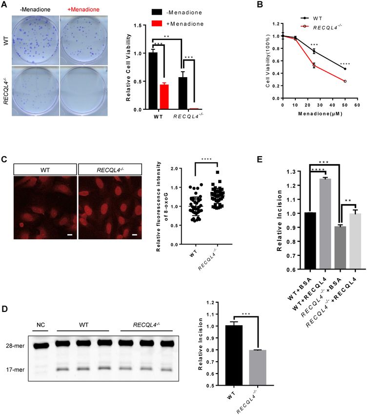
Nucleic Acids Research, 2020, Vol. 48, No. 12 6531
lowing exposure to ionizing radiation (IR) (35). SIRT1 reg- lysines mutated to glutamine: CAGGCTCCTCCGCC
ulates the enzymatic activity and subcellular localization AGCAGGCATGGCAGCAGCAGTGGCGGCAGCAG
of Werner helicase (WRN) after DNA damage through GGGGAGTGTTTTGG; and the primer for RECQL4
deacetylation of WRN (36). SIRT1 promotes homologous (KR) mutant with all five lysines mutated to arginine:
recombination (HR) repair of double-strand breaks (DSBs) CAGGCTCCTCCGCAGGCAGGCATGGAGGCAGA
through activation of NBS1 by deacetylation (37). In re- GGTGGCGGAGGAGAGGGGAGTGTTTTGG. All
sponse to oxidative stress and IR, SIRT1 redistributes from mutants were verified by sequencing.
repetitive DNA foci to DNA breaks to promote DNA re-
pair (40). Besides its role in DSB repair and oxidative stress Cell culture and generation of stable cell lines
response, SIRT1 has been shown to participate in the repair
of UV-light generated DNA damage through deacetylation U2OS and HEK293T cells were maintained in Dulbecco’s
of xeroderma pigmentosum group A (XPA) (38). More re- modified Eagle’s medium (DMEM supplemented with 10%
cently, SIRT1 has been shown to modulate BER activity fetal bovine serum (FBS) and 1% penicillin-streptomycin at
Downloaded from https://academic.oup.com/nar/article/48/12/6530/5841135 by guest on 29 September 2020
through deacetylation of OGG1 and apurinic/apyrimidinic 37◦ C in a humidified incubator with 5% CO2 . To generate
endonuclease-1 (APE1) (39,41). RECQL4 knock out cells, a double nicking strategy using
A recent study revealed that 8-oxoG lesions accumulate CRISPR/Cas9n with paired guide RNAs was applied (45).
genome-wide at DNA replication origins within transcribed The two paired single guide RNA (sgRNA) sequences were
long genes (42). Intriguingly, 8-oxoG and ␥ H2AX, a sensi- as follows: (i) sgRNA sense: 5 -AAGCAGAAACACTA
tive marker for DNA double-strand breaks (43), co-localize CGTGCG-3 ; (ii) sgRNA anti-sense: 5 -GTACGTAATT
at these DNA replication origins within the transcribed GCCCCTGTCA-3 (sc-403224-NIC, Santa Cruz). U2OS
long genes (42). Given the role of RECQL4 in replication cells were transiently transfected with 1 g of RECQL4
and DNA double-strand breaks repair, we hypothesize that double-nickase plasmids at 80% cell density in a six-well cul-
RECQL4 is involved in 8-oxoG repair, particularly in such ture plate using PolyJet™ transfection reagent (SL100688,
regions. Further, a previous study found that RECQL4 is SignaGen Laboratories). Transfected cells were selected us-
an acetylated protein (44), indicating that the function of ing 2 g/ml puromycin for one week. Subsequently, single
RECQL4 may be regulated by acetylation/deacetylation. cells were seeded on 150 mm dishes at 80 cells per dish to ob-
Because of the involvement of SIRT1 and RECQL4 in tain single clones and verified for knockout by genotyping
DNA repair and since RECQL4 is an acetylated protein, and western blot analysis. Stable shRNA-mediated knock-
we asked whether SIRT1 could regulate RECQL4 function down cell lines were generated by lentivirus transduction.
in DNA repair by deacetylation. All cell lines were tested for mycoplasma by MycoAlert My-
Here, we show that RECQL4 is required for efficient BER coplasma Detection Kit (Lonza).
of 8-oxoG. RECQL4 stimulates 8-oxoG repair by physical
and functional interactions with OGG1. Further, we show Lentivirus production and infection
that SIRT1 interacts with RECQL4, deacetylates it, and Lentivirus production was performed as previously de-
maintains it in a hypoacetylated state. The acetylation of scribed with some modifications (46). Briefly, for lentiviral
RECQL4 and its binding ability to OGG1 is enhanced af- packaging, HEK293T cells were cotransfected with pack-
ter oxidative stress, which is counteracted by SIRT1. More- aging plasmids psPAX2 and pMD2.G, and lentiviral plas-
over, the acetylation status of RECQL4 is tightly regulated mids targeting indicated genes. The virus-containing super-
by SIRT1 in response to oxidative stress. Taken together, natant was collected 48 h after transfection and used for cell
our study reveals a hitherto uncharacterized function of infection. Twenty four hours after infection, medium was
RECQL4 as a regulator of BER that is further regulated changed to 5 g/ml puromycin-containing medium, and
by SIRT1. cells were incubated for 1 week to select for infected cells.
Lentivirus based construct encoding short hairpin RNAs
MATERIALS AND METHODS targeting human SIRT1 (TRCN0000218734) was obtained
Reagents from Sigma-Aldrich.
Synthetic oligonucleotides were from TAG Copen- Western blot and immunoprecipitation
hagen. Menadione sodium bisulfite (74036), nicotinamide
(N0636), Trichostatin A (T8552) and piperidine (411027) For western blot analyses, cells were lysed in RIPA buffer
were from Sigma-Aldrich. Recombinant RECQL4 was (89900, Thermo Fisher Scientific) supplemented with pro-
used as previously described unless otherwise indicated tease inhibitor (4693159001, Sigma-Aldrich). Protein con-
(13). centration in the cell lysates was determined using the
protein assay dye reagent (#5000006, Bio-Rad). Proteins
were separated in NuPAGE 4–12% Bis–Tris gradient gels
Site-directed point mutation and plasmid construction
(Thermo Fisher Scientific), transferred to 0.2 m pore-sized
Substitution of lysines (376, 380, 382, 385 and 386) of PVDF membranes (GE10600021, Sigma-Aldrich) and the
RECQL4 with glutamine or arginine was carried out by blots were detected with the indicated antibodies. Im-
PCR using pCMV-Tag4A-3xFLAG-RECQL4 as tem- munoprecipitations were performed as previously described
plate using QuikChange Lightning Multi Site-Directed (12,13). Briefly, cells or cells transfected with the indicated
Mutagenesis Kit (Agilent Technologies, Inc., #210515). expression constructs were lysed in IP lysis buffer contain-
The primer for RECQL4 (KQ) mutant with all five ing 40 mM Tris–HCl pH 7.4, 2 mM MgCl2 , 150 mM NaCl,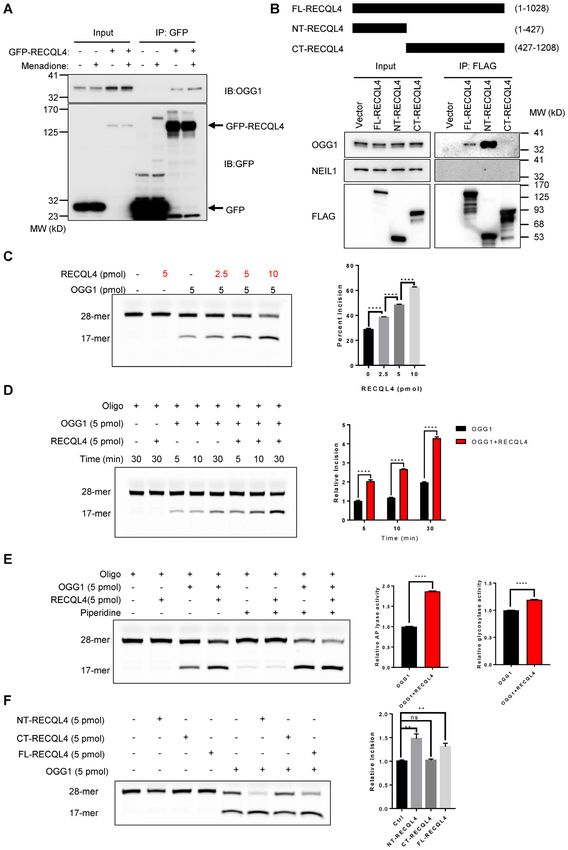
6532 Nucleic Acids Research, 2020, Vol. 48, No. 12
0.2% NP-40, 0.4% Triton X-100, supplemented with pro- segment, the primer sequences were: sense, 5 -TCT AAG
tease inhibitor (4693159001, Sigma-Aldrich) and 20 U/ml CCT CCT TAT TCG AGC CGA-3 , antisense, 5 -TTT
benzonase (E1014, Sigma-Aldrich). Lysates were incubated CAT CAT GCG GAG ATG TTG GAT GG-3 . PCR con-
with Protein A/G agarose beads (88802, Thermo Fisher ditions were: (i) 94◦ C, 1 min; (ii) 65◦ C, 12 min––repeat 1 and
Scientific) or GFP-TRAP beads (gtma-20, ChromoTek) or 2 for 19 cycles; (iii) 65◦ C, 10 min. For the 117 bp segment,
FLAG-magnetic beads (Sigma-Aldrich) overnight at 4◦ C. primer sequences were: sense, 5 -CCC CAC AAA CCC
The beads were washed with washing buffer (20 mM Tris– CAT TAC TAA ACC CA-3 ; antisense, 5 -TTT CAT CAT
HCl, pH7.4, 150 mM NaCl, 0.2% Triton X-100) for six GCG GAG ATG TTG GAT GG-3 . PCR conditions were:
times. The washed Protein A/G agarose beads or GFP- (a) 94◦ C, 1 min; (b) 58◦ C, 1 min; (c) 65◦ C, 1 min––repeat 1–3
TRAP beads were resuspended in 2 × SDS sample buffer, for 19 cycles; (d) 65◦ C, 2 min. PCR products were separated
boiled at 95◦ C, and then subjected to Western blotting. in 1% agarose gel, and visualized with ethidium bromide
The washed FLAG-magnetic beads were eluted with the staining. Quantitation was performed by Fiji software.
above washing buffer containing 300 g/ml 3xFLAG pep-
Downloaded from https://academic.oup.com/nar/article/48/12/6530/5841135 by guest on 29 September 2020
tide (F4799, Sigma-Aldrich). The following antibodies and
Measurement of mtDNA copy number
dilutions were used: anti-RECQL4 (2814; 1:1,000), anti-
SIRT1 (8469, 1:1000) antibodies were from Cell Signaling Total DNA was extracted from cell pellets using the QI-
Technology; anti-OGG1 (ab124741, 1:3000), anti-acetyl- Aamp DNA Mini kit (51304, QIAGEN). mtDNA copy
OGG1 (acK338 + acK341, ab93670, 1:500), anti-NEIL1 number analysis was performed by qPCR with the Platinum
(ab21337, 1:1000), anti-GFP (ab290, 1:5000), anti-Histone SYBR Green qPCR SuperMix-UDG reagents(11733038,
H3 (ab1791, 1:5000), anti-APE1 (ab194,1:1000) antibodies Thermo Fisher Scientific). mtDNA levels were assessed
were from Abcam; anti-Lamin B1 (sc-374015, 1:3000) was using primers against the mitochondrial 117bp segment.
from Santa Cruz Biotechnology; anti-Actin (A5441, 1:10 Primer sequences were: sense, 5 -CCC CAC AAA CCC
000); anti-FLAG (F1804, 1:10 000) antibodies were from CAT TAC TAA ACC CA-3 ; antisense, 5 -TTT CAT CAT
Sigma-Aldrich. For secondary antibodies, mouse anti-goat GCG GAG ATG TTG GAT GG-3 ; Nuclear actin served
IgG-HRP (sc-2354, Santa Cruz), rabbit anti-mouse IgG- as a loading control.
HRP (A9044, Sigma-Aldrich), goat anti-rabbit IgG-HRP
(A6667, Sigma-Aldrich) were used.
8-oxoG incision assay
Oligonucleotide containing an 8-oxoG lesion at a spe-
Immunofluorescence analysis
cific position was annealed to its complementary oligonu-
For staining of 8-oxoG in DNA, a previously published pro- cleotide. The oligonucleotide sequences are: 5 -GAA CGA
tocol was used with slight modifications (47,48). In brief, 2 CTG T(8-oxoG)A CTT GAC TGC TAC TGA T-3 -
× 104 cells were seeded on 12 mm diameter coverslip in 24- TAMRA, and 5 - ATC AGT AGC AGT CAA GTC ACA
well plates. After 24 h, cells were washed with ice-cold PBS, GTC GTT C-3 . This assay was carried out as previously de-
fixed in 4% paraformaldehyde (PFA) in PBS for 10 min. scribed with some modifications (52–54). For 8-oxoG inci-
Cells were washed with PBS three times for 5 min each with sion assay, the DNA substrate (5 pmol) was incubated with
agitation. Cells were then permeabilized with 0.4% Triton- the indicated amounts of recombinant human RECQL4 or
X-100 in PBS, followed by three times washes for 5 min human OGG1 (ab98249, Abcam) in 20 l reaction buffer
each. Cells were then treated with 2.5 mg/ml RNase A in (70 mM HEPES–KOH, 7.4, 5 mM EDTA, 1 mM DTT, 75
PBS for 1 h at 37◦ C, followed by three PBS washes. Nuclear mM NaCl, 10% glycerol, 100 g/ml BSA) for 30 min at
DNA was denatured by treating with freshly prepared 2 N 37◦ C. Reactions were terminated by adding 20 l of for-
HCl in distilled water for 10 min at room temperature, fol- mamide loading buffer (95% formamide, 0.025% w/v bro-
lowed by three PBS washes. Cells were then incubated with mophenol blue, 0.025% w/v xylene cyanol, 5 mM EDTA
mouse anti-8-oxo-dG antibody (ab48508, Abcam). Alexa pH 8.0). The samples were heated at 95◦ C for 3 min, and
Fluor 555 anti-mouse secondary antibodies were used. All put on ice. For glycosylase and AP lyase activity measure-
images were acquired with Carl Zeiss confocal microscope ment, the reactions were terminated by adding 20 l of for-
LSM 780 at the Core Facility for Integrated Microscopy at mamide loading buffer and 1 l piperidine (99%). The sam-
University of Copenhagen. Fluorescence intensity was mea- ples were then heated at 95◦ C for 5 min in order to cleave
sured using Fiji software. all AP sites, prior to loading on gels. For 8-oxoG incision
assay with whole cell extracts, 30 g protein extracts were
incubated with 5 pmol TAMRA-labeled DNA substrates
Measurement of mitochondrial DNA damage by long-range
in reaction buffer containing 20 mM HEPES–KOH pH 7.4,
PCR
5 mM EDTA, 5 mM DTT, 150 mM NaCl, 10% glycerol, 100
This assay was performed as previously described with mi- g/ml BSA. After incubation for 4 h at 37◦ C, the reac-
nor modifications (49–51). Total DNA was isolated from tions were terminated by adding 1 l 5 mg/ml Proteinase K
cell pellets using the QIAamp DNA Mini kit (51304, QI- and 1 l 10% SDS and incubated at 55◦ C for 30 min. DNA
AGEN). DNA concentration was measured by an Epoch was precipitated by adding 5 g glycogen, 4 l 11 M am-
Microplate Spectrophotometer (BioTek). Amplification of monium acetate, 60 l ethanol, and overnight incubation
an 8.9 kb segment and a 117 bp segment of mitochondrial at –80◦ C. Samples were pelleted, dried and suspended in
DNA was performed using the LongAmp Taq DNA Poly- 20 l of formamide loading buffer. All samples were elec-
merase (M0323L, New England Biolabs). For the 8.9 kb trophoresed on 15% polyacrylamide/8 M urea gel in 1×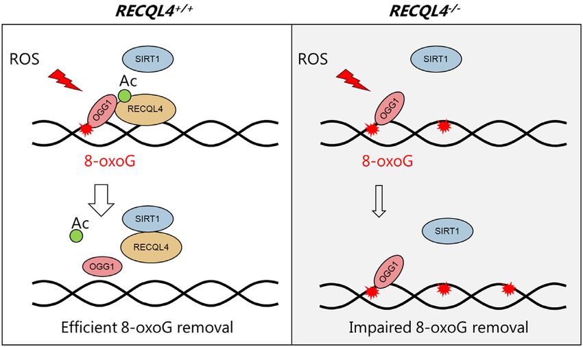
Nucleic Acids Research, 2020, Vol. 48, No. 12 6533
TBE buffer. Fluorescence was detected by ChemiDoc Imag- The reaction mixtures were resolved by SDS-PAGE and the
ing Systems (Bio-Rad). Quantifications were performed by acetylation status of RECQL4 was analyzed by western blot
Fiji software. with a monoclonal anti-acetylated lysine antibody (9441S,
CST).
In vitro acetylation assay
Recombinant human RECQL4 or human OGG1 (ab98249, Clonogenic survival assay
Abcam) (1 g) was incubated with different amounts of re- Cells were plated into six-well plates at 200 cells per well.
combinant human CBP (0.1 g, 0.2 g, 0.5 g) (catalytic After overnight incubation, cells were treated with 50 M
domain) (BML-SE452-0100, Enzo Life Sciences) in 20 l menadione for 1 h. Then the medium were removed, and
acetylation buffer containing 50 mM Tris, pH 8.0, 50mM cells were washed once with DMEM medium and changed
NaCl, 0.1 mM EDTA, 100 g/ml BSA, 10% glycerol, 1 to normal culture medium for another 12 days. The colonies
mM DTT,1 mM PMSF, and 10 mM sodium butyrate in the were washed with PBS for two times, fixed and stained
Downloaded from https://academic.oup.com/nar/article/48/12/6530/5841135 by guest on 29 September 2020
presence of 2 mM acetyl CoA (A2056-10G, Sigma). Reac- with staining buffer containing 0.5% crystal violet and 20%
tions were performed at 30◦ C for 1 h, stopped by adding ethanol for 1 h. The colonies were then gently washed in
2 × Laemmli buffer (S3401, Sigma-Aldrich), and resolved water and air-dried. Images were acquired with a CanoScan
by SDS-PAGE. The gels were analyzed by Coomassie blue LiDE 220 scanner and quantification of colonies were per-
staining and western blot with the indicated antibodies. For formed with Fiji software.
detection of the acetylation of RECQL4, an anti-acetylated
lysine antibody (9441S, CST) was used. For detection of
acetylation of OGG1, an anti-acetylated OGG1 antibody Cell viability assay
(ab93670, Abcam) was used. Cells (8 × 10³ per well) were seeded into 96-well plates. 24
h later, cells were treated as described in the figure legends.
Acetylation of RECQL4 in cells Cell viability was measured with CellTiter 96 AQueous One
Solution Cell Proliferation Assay MTS kit according to the
Cells were co-transfected with the expression constructs
manufacturer’s instructions (G3580, Promega).
for FLAG-tagged RECQL4 and CBP or p300. After 36
h, cells were lysed in lysis buffer (50 mM HEPES–KOH
7.5, 500 mM NaCl, 0.5% NP-40, 10% glycerol, 5 M TSA, EdU incorporation assay
10 mM nicotinamide, 10 mM sodium butyrate, 1× pro- EdU (5-ethynyl-2 -deoxyuridine) incorporation assays were
tease inhibitor cocktail) and sonicated on ice. FLAG-tagged performed using the Click-iT™ EdU Alexa Fluor™ 488
RECQL4 were pulled down with M2 FLAG-magnetic Flow Cytometry Assay Kit (Thermo Fisher Scientific,
beads at 4◦ C. After the beads were sequentially washed #C10425) according to the manufacturer’s instructions.
with wash buffer A (50 mM HEPES–KOH pH7.5, 1 M Briefly, the cells were incubated with 10 M EdU for 1 h,
NaCl, 0.5% NP-40, 10% glycerol, 10 mM nicotinamide), fixed with 4% PFA in DPBS for 15 min at room temperature,
wash buffer B (50 mM HEPES–KOH pH7.5, 500 mM permeabilized with 0.4% Triton X-100/1% BSA in PBS for
NaCl, 0.5% NP-40, 10% glycerol), and wash buffer C (50 15 min. After permeabilization, Click-iT reaction was per-
mM HEPES–KOH pH 7.5, 150 mM NaCl, 0.5% NP- formed. Cells were analyzed using a CytoFLEX flow cy-
40, 10% glycerol), the 3xFLAG-tagged RECQL4 were tometer (Beckman Coulter, Inc.).
eluted by incubating the beads in wash buffer C containing
300 g/ml 3xFLAG peptide (F4799, Sigma-Aldrich). The
eluted proteins were analyzed by SDS-PAGE, and acetyla- Statistical analyses
tion of RECQL4 was probed by western blot with an anti- Data were analysed using a two-tailed, unpaired Stu-
acetylated lysine antibody (9441S, CST). dent’s t test and are presented as mean ± SD or SEM as
indicated in the figure legends. P < 0.05 was considered as
In vitro deacetylation assay statistically significant.
This assay was carried out in two steps. First, recombinant
human RECQL4 or human OGG1 (ab98249, Abcam) (1 RESULTS
g) were acetylated by 0.1 g recombinant human CBP
Characterization of RECQL4 knockout cells
(BML-SE452-0100, Enzo Life Sciences) in 10 l of acety-
lation buffer containing 50 mM Tris, pH 8.0, 50 mM NaCl, Previous studies have greatly increased our knowledge of
0.1 mM EDTA, 10% glycerol, 1 mM DTT,1 mM PMSF and the molecular function of RECQL4 using RTS patient sam-
2 mM acetyl CoA. After incubation at 30◦ C for 1 h, the re- ples. However, large variations observed in RTS patient cells
action mixtures were incubated at 30◦ C for an additional 1 have hampered understanding of the role of RECQL4 in
h with the indicated amount of recombinant SIRT1 (1 U = BER (55–57). To rule out the potential influence of inter-
1 pmol/min at 37◦ C, 250M FLUOR DE LYS® substrate cellular genetic and epigenetic variations which may result
(Prod. No. BML-KI104), 500 M NAD+) (BML-SE239- in subtle differences including responses to stress and re-
0100, Enzo Life Sciences) in 20 l of deacetylation buffer pair, we generated isogenic RECQL4−/− and control cells
containing 50 mM Tris–HCl, pH 9.0, 50 mM NaCl, 4 mM using the double nicking CRISPR/Cas9n system. This ap-
MgCl2 , 1 mM DTT, 1 mM PMSF, 10% glycerol in the pres- proach is thought to significantly reduce off-target effects
ence of 50 M NAD+ co-factor (20-221, Sigma-Aldrich). and facilitate gene knockout with high on-target cleavage6534 Nucleic Acids Research, 2020, Vol. 48, No. 12
efficiency (45). The N-terminal 400 amino acids (aa) region initiated by the removal of 8-oxoG by OGG1 DNA glycosy-
of RECQL4 possesses homology to the yeast DNA repli- lase (24). Thus, increased sensitivity to oxidative stress and
cation initiation factor Sld2 (7,58), and also contains its increased endogenous levels of 8-oxoG may be due to re-
nuclear localization signals (59) and a mitochondrial tar- duced 8-oxoG repair capacity in these cells. Therefore, we
geting sequence (17), indicating that the N-terminal region assessed the 8-oxoG incision ability in RECQL4−/− cells us-
of RECQL4 mediates its important functions. Therefore, ing a TAMRA-labeled DNA substrate containing 8-oxoG
to completely disrupt the function of RECQL4, we used (Supplementary Figure S2C). Since RECQL4 and OGG1
paired sgRNAs targeting exon 5 of the RECQL4 gene lo- are both nuclear and mitochondrial proteins, we used whole
cus (Figure 1A), which encodes most of the amino acids cell extracts to better represent the function of these two
in N-terminal region. Biallelic disruption of RECQL4 was proteins in both compartments. 8-oxoG incision activity
confirmed by genomic PCR and Western blot analysis (Fig- was significantly lower in extracts from RECQL4−/− cells
ure 1B and C). A number of studies have demonstrated compared with extracts from wild-type control cells (Fig-
that RECQL4 plays an important role in DNA replica- ure 2D). Addition of purified recombinant RECQL4 pro-
Downloaded from https://academic.oup.com/nar/article/48/12/6530/5841135 by guest on 29 September 2020
tion (7,60–62). Consistent with previous findings, we found tein restored 8-oxoG incision in RECQL4−/− cell extracts
that depletion of RECQL4 leads to diminished prolifera- to the level of control cells, and significantly increased 8-
tive capacity, as assessed by colony formation assay (Fig- oxoG incision activity in extracts from control cells (Fig-
ure 1D). The number of colonies produced by RECQL4−/− ure 2E), suggesting that RECQL4 is required for efficient
cells was less and the colonies were smaller than those by 8-oxoG repair and may be directly involved in 8-oxoG re-
wild-type cells, indicating that depletion of RECQL4 re- pair. Thus, we conclude that RECQL4 participates in BER
sults in decreased cellular survival and proliferation. De- of 8-oxoG and as such RECQL4 deficiency leads to defec-
creased cell proliferation in RECQL4−/− cells was further tive repair of 8-oxoG.
confirmed by an EdU (5-ethynyl-2 -deoxyuridine) incorpo-
ration assay showing that the percentage of EdU-labeled
RECQL4 selectively interacts with OGG1 and promotes 8-
cells in RECQL4−/− cells was significantly less than that
oxoG repair
in wild type cells (Supplementary Figure S1A). Overex-
pression of RECQL4 in RECQL4−/− cells rescued these We asked whether the stimulation of 8-oxoG repair by
phenotypes, suggesting that depletion of RECQL4 was RECQL4 in cell extracts involves interaction between
responsible for decreased cellular survival and prolifera- RECQL4 and OGG1, the major DNA glycosylase for
tion observed in RECQL4−/- cells (Figure 1E and Supple- 8-oxoG repair. To test this, we performed the following
mentary Figure S1B). Previously, RECQL4 was shown to experiments. Using immunoprecipitation (IP) with GFP-
be present in mitochondria where it might contribute to RECQL4 as bait, we found that RECQL4 directly inter-
mtDNA maintenance (14,15,17,63). Therefore, we assessed acts with OGG1 (Figure 3A), and menadione-induced ox-
the effects of RECQL4 knockout on mitochondrial func- idative stress increased this interaction (Figure 3A), sug-
tion. RECQL4 depletion resulted in a significant reduc- gesting that RECQL4 may further enhance 8-oxoG re-
tion in mtDNA copy number (Figure 1F), and diminished pair following oxidative stress. Similarly, IP of endogenous
mtDNA repair capacity as RECQL4−/− cells accumulated RECQL4 showed direct interaction between RECQL4 and
more mtDNA damage than wild-type cells (Figure 1G). OGG1 in wild-type cells, but not in RECQL4−/- cells (Sup-
We generated two RECQL4-deficient cell lines. Analysis plementary Figure S3A). We then mapped the interacting
of the second RECQL4-deficient cell line (RECQL4−/− regions between these two proteins. RECQL4 contains a
#22) showed similar results (Supplementary Figure S1C– Sld2-like domain at the N-terminus, a highly conserved
E). Thus, we generated an isogenic RECQL4−/− cell line, ATPase and helicase domain in the middle, and a poorly
which exhibits several cellular phenotypes previously ob- characterized C-terminus (4,66). IP was performed with
served in RTS patient cells and in RECQL4-deficient cells 3× FLAG-tagged full-length RECQL4 (FL-RECQL4), 3×
(7,14–16,64), suggesting that it is suitable for RECQL4 FLAG-tagged truncated RECQL4 mutants (1−427aa, NT-
study. RECQL4; 427−1208aa, CT-RECQL4) (Figure 3B, up-
per panel) (12). Only the full-length RECQL4 and NT-
RECQL4 interacted with OGG1, but not the CT-RECQL4
RECQL4 is required for BER of 8-oxoG
(Figure 3B, lower panel). The absence of NEIL1, another
Several studies have shown that RTS patient cells were major DNA glycosylase for repair of 8-oxoG, in the IP com-
hypersensitive to oxidative stress (55,56), suggesting that plex indicates the selective RECQL4/OGG1 interaction for
RECQL4 deficiency may impair repair of oxidative DNA repair of 8-oxoG (Figure 3B, lower panel).
lesions. We observed a markedly increased sensitivity of To test whether RECQL4 functionally interacts with
RECQL4−/− cells to menadione, a ROS-generating com- OGG1, we performed the 8-oxoG incision assay in the
pound (65), as measured by colony formation assay and presence of RECQL4 and OGG1. RECQL4 significantly
MTS assay (Figure 2A, B and Supplementary Figure S2A). stimulated the 8-oxoG incision activity of OGG1 in a
Moreover, RECQL4−/− cells showed increased sensitivity concentration-dependent manner (Figure 3C and Supple-
to H2 O2 treatment (Supplementary Figure S2B). Consis- mentary Figure S3B), suggesting a functional interaction.
tent with some observations in RTS patient cells, we found Moreover, as shown in Figure 3D, RECQL4 stimulates
a significantly higher level of 8-oxoG in RECQL4−/− cells the 8-oxoG incision activity of OGG1 in a time-dependent
(Figure 2C) (47,48). 8-oxoG:C in the genomic DNA is pri- manner. OGG1 is a bifunctional DNA glycosylase, pos-
marily repaired by the base excision repair (BER) pathway sessing a strong DNA glycosylase activity and a relativeNucleic Acids Research, 2020, Vol. 48, No. 12 6535
Downloaded from https://academic.oup.com/nar/article/48/12/6530/5841135 by guest on 29 September 2020
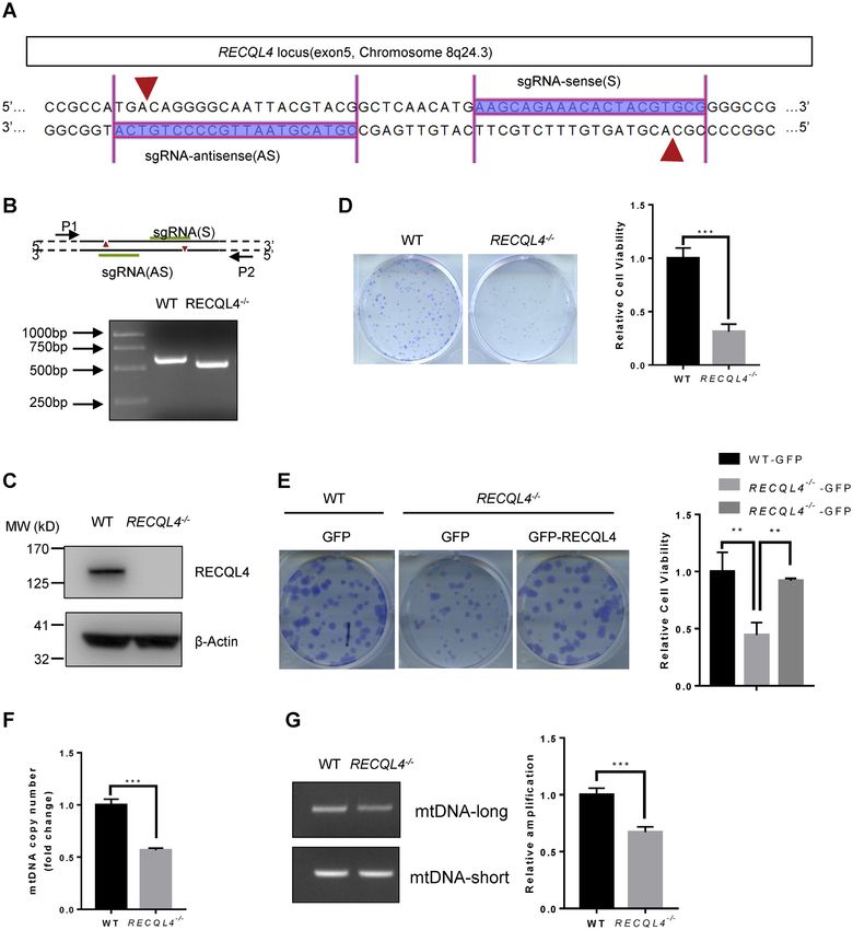
Figure 1. Characterization of RECQL4 knockout cells. (A) Schematic showing sgRNA sequences targeting the human RECQL4 locus using Cas9 D10A
nickases (Cas9n). Red arrows indicate the sites of nicks in DNA. (B) PCR analyses of WT and RECQL4−/− cells using primer pair (P1+P2) to amplify the
genomic region ranging from exon 5 to intron 6 of RECQL4. The result shows the RECQL4−/− clone with a homozygous deletion of the RECQL4 locus.
(C) Knockout of RECQL4 in U2OS cells was confirmed by immunoblotting. (D) Colony formation assay showing RECQL4−/− cells have clonogenic
survival defects. Images show representative colonies stained with crystal violet. Data are shown as mean ± SEM, n = 3. *** P < 0.001 (t-test). (E) Colony
formation assay showing that re-introduction of WT-RECQL4 in RECQL4−/− cells rescues the clonogenic survival defects of RECQL4−/- cells. Three
independent experiments were performed, each with four replicates. Representative images (left) and quantifications (right) are shown. ** P < 0.01, using
unpaired two-tailed Student’s t test. (F) mtDNA copy number in WT and RECQL4−/− cells. Data are shown as mean ± SEM, n = 3. *** P < 0.001 (t-test).
(G) mtDNA damage detection by a PCR-based assay assessing relative quantitative amplification of a small (117 bp) to a large (10 kb) segment of mtDNA.
A representative gel (left) and quantifications (right) are shown as mean ± SEM, n = 3. *** P < 0.001.6536 Nucleic Acids Research, 2020, Vol. 48, No. 12
Downloaded from https://academic.oup.com/nar/article/48/12/6530/5841135 by guest on 29 September 2020
Figure 2. RECQL4 is required for BER of 8-oxoG. (A) Colony formation ability of WT and RECQL4−/- cells treated with or without 50 M menadione.
Images show representative colonies stained with crystal violet. Data are shown as mean ± SEM, n = 3. ** P < 0.01 (t-test); *** P < 0.001 (t-test). (B)
MTS assay showing viability of WT and RECQL4 knockout cells treated with or without menadione. Data are shown as mean ± SEM, n = 3. ** P < 0.01
(t-test); *** P < 0.001 (t-test). (C) 8-oxoG base lesion detected in WT and RECQL4−/− cells by immunofluorescence microscopy. Scale bar, 10 m. The
apochromat 63×/1.4 DIC (oil) objective was used. The 8-oxoG level was measured by quantifying the fluorescence intensity with Fiji software. At least 50
cells were counted for each experiment. Data are shown as the mean ± SD of three independent experiments. *** P < 0.001 (t-test). (D) Base excision repair
(BER) assay measuring 8-oxoG incision activity using a TAMRA-labeled 8-oxoG containing oligonucleotide duplexes in whole cell extracts of WT and
RECQL4− /− cells. Representative gel (left) and quantification (right) of 8-oxoG incision levels are shown. The negative control (NC) was added with the
same cell extraction buffer. (E) Quantification of BER analysis of 8-oxoG repair in WT and RECQL4−/- whole cell extracts. WT and RECQL4−/− extracts
were supplemented with either recombinant wild-type human RECQL4 protein or BSA. Recombinant human RECQL4 and BSA were used at 50 ng/l.
Data are shown as mean ± SEM from three independent experiments. ** P < 0.01, *** P < 0.001, **** P < 0.0001, using unpaired two-tailed Student’s t test.Nucleic Acids Research, 2020, Vol. 48, No. 12 6537
Downloaded from https://academic.oup.com/nar/article/48/12/6530/5841135 by guest on 29 September 2020
Figure 3. RECQL4 selectively interacts with OGG1 and promotes 8-oxoG repair. (A) Menadione treatment enhances RECQL4/OGG1 interaction. Im-
munoprecipitated material from cells expressing GFP or GFP-tagged WT-RECQL4 with GFP-TRAP beads analyzed by immunoblotting (IB) with anti-
GFP and anti-OGG1 antibodies. (B) N-terminal domain of RECQL4 interacts with OGG1 in cells. Schematic diagram of 3× FLAG-tagged RECQL4 and
the truncated fragments (upper panel). FL-RECQL4, NT-RECQL4, and CT-RECQL4 were all tagged with both 3× FLAG and SV40 nuclear-location
sequence (NLS). (C) Representative gel (left) and quantification (right) of the stimulation of catalytic activity of OGG1 by RECQL4 in a dose-dependent
manner. Data are presented as mean ± SEM from three independent experiments. **** P < 0.0001, using unpaired two-tailed Student’s t test. (D) Repre-
sentative gel (left) and quantification (right) of the stimulation of catalytic activity of OGG1 by RECQL4 in a time-course dependent manner. Data are
presented as mean ± SD from two independent experiments. **** P < 0.0001, using unpaired two-tailed Student’s t test. (E) Representative gel (left) of the
stimulation of catalytic activity of OGG1 by RECQL4 treated with or without piperidine. Quantification (right) of the glycosylase and AP lyase activities
of OGG1 stimulated by RECQL4. Data are presented as mean ± SD from two independent experiments. **** P < 0.0001, using unpaired two-tailed Stu-
dent’s t test. (F) BER assay showing that the N-terminal domain of RECQL4 functionally interacts with OGG1. 3 × FLAG-tagged RECQL4 proteins
were purified from normal U2OS cell. A representative image (left) and quantifications (right) are shown. ** P < 0.01; ns, not significant, using unpaired
two-tailed Student’s t test. Data are shown as mean ± SEM, n = 4.6538 Nucleic Acids Research, 2020, Vol. 48, No. 12
weak AP lyase activity (67,68). Next, we tested which ac- idative DNA damage can trigger RECQL4 acetylation. In
tivity of OGG1 was most affected by RECQL4. To sep- addition, we found that under our conditions OGG1 can
arate these two activities, after termination, the reactions be acetylated by CBP as well and acetylation significantly
were followed by treatment with or without hot piperi- increases 8-oxoG incision activity of OGG1 in vitro (Sup-
dine. RECQL4 significantly stimulated both the glycosy- plementary Figure S4B and Supplementary Figure S4C),
lase and AP lyase activities of OGG1 (Figure 3E). How- which are consistent with previous observations (39,72).
ever, the stimulation of the AP lyase activity of OGG1 by
RECQL4 was more prominent than that of its glycosy- SIRT1 interacts with and deacetylates RECQL4
lase activity (Figure 3E). These results suggest RECQL4
Protein acetylation is a highly dynamic post-translational
may coordinate the glycosylase activity and AP lyase ac-
modification that can be reversed by Sirtuin deacetylases.
tivity of OGG1 in BER of 8-oxoG. Moreover, only full-
Among the Sirtuins, the nuclear SIRT1 is involved in var-
length RECQL4 and NT-RECQL4 stimulated 8-oxoG inci-
ious cellular processes including DNA repair (73–75). Be-
sion activity of OGG1 (Supplementary Figure S3C and Fig-
Downloaded from https://academic.oup.com/nar/article/48/12/6530/5841135 by guest on 29 September 2020
cause RECQL4 is an acetylated protein and the SIRT1 in-
ure 3F), further supporting that RECQL4 interacted with
hibitor, nicotinamide (NAM), affects the acetylation status
OGG1 through its N-terminus. Thus, our results strongly
of RECQL4 (44), we asked whether RECQL4 is a poten-
support that RECQL4 physically and functionally interacts
tial target of SIRT1. To test this, we first checked our re-
with OGG1, and that RECQL4 plays a role in 8-oxoG re-
cently published two separate mass spectrometry data sets
pair through interacting with OGG1.
of RECQL4 interactome (12,13). SIRT1 was identified in
both studies as a potential deacetylase for RECQL4. To
RECQL4 is an acetylated protein and its acetylation is stim- further test this, we immunoprecipitated GFP-RECQL4
ulated by oxidative stress from U2OS cells and detected a direct interaction between
RECQL4 and SIRT1 (Figure 5A). We then mapped the in-
Increasing number of studies show that the functions and
teraction region between RECQL4 and SIRT1 to the N-
activities of DNA repair proteins are regulated by post-
terminus of RECQL4, which was within the previously
translational acetylation (69,70). p300, and the closely re-
reported acetylation region of RECQL4 by p300 (Fig-
lated protein CBP, are highly conserved acetyltransferases
ure 5B) (44). To determine whether SIRT1 deacetylates
involved in multiple cellular processes including DNA re-
RECQL4, we performed a two-step deacetylation assay in
pair (71). Previously, RECQL4 was shown to become
vitro. First, we acetylated RECQL4 by incubating recom-
acetylated in the N-terminus by p300 (44). We transiently
binant RECQL4 with recombinant CBP acetylase in the
co-transfected cells with 3 × FLAG-WT-RECQL4 to-
presence of acetyl-CoA. Second, we incubated the acety-
gether with either p300 or CBP. The level of acetylated
lated form of RECQL4 with recombinant human SIRT1.
RECQL4 was significantly increased in cells co-transfected
RECQL4 was deacetylated by SIRT1 in a dose-dependent
with RECQL4 together with p300 or CBP (Figure 4A).
manner in the presence of NAD+ co-substrate. In addition,
These results showed that RECQL4 is an acetylated pro-
the deacetylation was inhibited in the presence of NAM
tein, which is consistent with a previous report (44) and
(Figure 5C). Thus, we conclude that RECQL4 is a novel tar-
suggested that RECQL4 is a potential new target of CBP
get of SIRT1. To further investigate the five lysine residues
acetyltransferase. To further confirm that CBP acetylates
as possible targets of SIRT1 deacetylation, we performed
RECQL4, we performed an in vitro acetylation assay using
the in vitro deacetylation assay using the RECQL4 mutants.
purified recombinant CBP (catalytic domain) together with
Mutations of the five lysines residues did not completely
recombinant RECQL4. RECQL4 is acetylated by CBP in
abolish the deacetylation of RECQL4 by SIRT1 (Figure
vitro in the presence of acetyl-coenzyme A (Figure 4B), sug-
5D), indicating that deacetylation of lysines other than the
gesting that RECQL4 is a possible substrate for CBP. A pre-
five lysine residues by SIRT1 is possible.
vious study showed that five lysines (376, 380, 382, 385 and
It has been reported that SIRT1 negatively regulates the
386) of the total 32 lysines can be acetylated within the N
catalytic activity of OGG1 in vivo (39). However, it remains
terminus of RECQL4 (44). Therefore, to determine whether
unclear whether SIRT1 deacetylates OGG1 because in a
CBP may acetylate these residues on RECQL4, we gener-
separate study OGG1 activity was not affected by NAM
ated two RECQL4 mutants, RECQL4 (KQ) with all five
(72). To clarify this, we used an in vitro deacetylation assay
lysine residues mutated to glutamine and RECQL4 (KR)
using recombinant human OGG1 and recombinant human
with all five lysine residues mutated to arginine using site-
SIRT1. SIRT1 directly deacetylated OGG1 in the presence
directed mutagenesis to mimic acetylated lysine and non-
of NAD+ co-substrate, and the deacetylation was inhib-
acetylated lysine, respectively. Compared to the wild type,
ited in the presence of NAM (Supplementary Figure S5A).
RECQL4 (WT), both the RECQL4 (KQ) mutant and the
Thus, this data provides evidence consistent with OGG1 be-
RECQL4 (KR) mutant exhibited significantly reduced ca-
ing a substrate for SIRT1 (39), suggesting that SIRT1 is a
pacity to become acetylated by CBP in vitro (Figure 4C
potential regulator of BER.
and Supplementary Figure S4A). This data indicates that at
least one of the five lysine residues is the target of acetylation
SIRT1 controls the interaction between OGG1 and RECQL4
by CBP. Further, it also suggests that CBP may acetylate
following oxidative stress and maintains RECQL4 in a hy-
other lysine residues on RECQL4 in addition to these five
poacetylated state
lysine residues. Interestingly, the RECQL4 acetylation level
was significantly increased following menadione treatment Our results showed that SIRT1 deacetylates RECQL4
(Figure 4D), suggesting that oxidative stress and likely ox- in vitro, and we next asked whether SIRT1 deacety-Nucleic Acids Research, 2020, Vol. 48, No. 12 6539
Downloaded from https://academic.oup.com/nar/article/48/12/6530/5841135 by guest on 29 September 2020
Figure 4. RECQL4 is an acetylated protein and its acetylation is stimulated by oxidative stress. (A) RECQL4 acetylation in transiently transfected U2OS
cells with 3xFLAG-tagged RECQL4, p300 or CBP, treated with 2 M TSA (trichostatin A) and 5 mM NAM (nicotinamide). Immunoprecipitated
RECQL4 proteins were then detected by Western blot with anti-acetylated lysine antibody. A representative gel is shown on the left. The graph on the
right shows quantification of relative acetylated RECQL4 levels normalized to total RECQL4. Data are presented as mean ± SD from two independent
experiments. * P < 0.05, using unpaired two-tailed Student’s t test. (B) Acetylation of RECQL4 by CBP in an in vitro acetylation assay. Recombinant
RECQL4 (1 g), Acetyl-CoA (2 mM), and different amounts of recombinant CBP (0.1, 0.2, 0.5 g) were incubated at 30◦ C for 1 h. Acetylated RECQL4
proteins were detected with anti-acetylated lysine antibody (top). Total amounts of RECQL4 were assessed with Coomassie blue staining (bottom). The
graph on the right shows quantification of relative acetylated RECQL4 levels normalized to total RECQL4. Data are shown as mean ± SD from three
independent experiments. *** P < 0.001, **** P < 0.0001, using unpaired two-tailed Student’s t test. (C) Acetylation of wild type RECQL4 (WT), RECQL4
(KQ) and RECQL4 (KR) mutants by CBP in an in vitro acetylation assay. 3 × FLAG-tagged RECQL4 proteins were purified from normal U2OS cell.
FLAG-tagged RECQL4 proteins (1 g), Acetyl-CoA (2 mM), and recombinant CBP (0.2 g) were incubated at 30◦ C for 1 h. Acetylated FLAG-tagged
RECQL4 proteins were detected with anti-acetylated lysine antibody. A representative gel is shown on the left. The graph on the right shows quantification
of relative acetylated FLAG-tagged RECQL4 protein levels normalized to total. Data are shown as mean ± SD from two independent experiments. ** P <
0.01, *** P < 0.001, using unpaired two-tailed Student’s t test. (D) Western blot showing increased endogenous acetylated RECQL4 levels in cells treated
with 50 M menadione for 1 h. Endogenous acetylated proteins were immunoprecipitated with anti-acetyl-lysine antibody-conjugated beads. The presence
of RECQL4 in the immunoprecipitated complex and acetylation levels in the extract were measured by Western blot with anti-RECQL4 antibody. Input
for immunoprecipitation was used as a loading control for total RECQL4 (bottom). A representative gel is shown on the left. The graph on the right shows
quantification of relative acetylated RECQL4 levels normalized to total RECQL4. Data are shown as mean ± SD from two independent experiments. * P
< 0.05, using unpaired two-tailed Student’s t test.6540 Nucleic Acids Research, 2020, Vol. 48, No. 12
Downloaded from https://academic.oup.com/nar/article/48/12/6530/5841135 by guest on 29 September 2020
Figure 5. SIRT1 interacts with and deacetylates RECQL4. (A) Immunoprecipitation (IP) in cells expressing GFP or GFP-tagged WT-RECQL4 with GFP-
TRAP beads analyzed by immunoblotting (IB) with anti-GFP and anti-SIRT1 antibodies. Representative blot is shown from two independent experiments.
(B) Immunoprecipitation (IP) performed in cells expressing vector alone or FLAG-tagged FL-RECQL4, NT-RECQL4, and CT-RECQL4 with FLAG-
M2 magnetic beads and analyzed by immunoblotting (IB) with the indicated antibodies shows that the N-terminal domain of RECQL4 interacts with
SIRT1 in cells. Representative blot is shown from two independent experiments. (C) Deacetylation of RECQL4 by SIRT1 in an in vitro deacetylation assay.
Recombinant RECQL4 (1 g) was first acetylated by CBP (0.1 g) for the first 1 h, then acetylated RECQL4 was incubated with NAD+ (50 M), and
different amounts of recombinant SIRT1 (0.5, 1, 2 U) at 30◦ C for an additional 1 h. Acetylated RECQL4 was probed with anti-acetylated lysine antibody.
Total amounts of RECQL4 were assessed with an anti-RECQL4 antibody. NAM served as a SIRT1 inhibitors. A representative gel is shown on the left.
The graph on the right shows quantification of relative acetylated RECQL4 levels normalized to total RECQL4. Data are shown as mean ± SD from
three independent experiments. ** P < 0.01, *** P < 0.001, using unpaired two-tailed Student’s t test. (D) Deacetylation of wild type RECQL4 (WT) and
RECQL4 (KQ) mutant by SIRT1 in an in vitro deacetylation assay. 3 × FLAG-tagged RECQL4 proteins were purified from normal U2OS cells. FLAG-
tagged RECQL4 proteins (1 g), were first acetylated by CBP (0.1 g) for the first 1 h, then acetylated proteins were incubated with NAD+ (50 M), and
recombinant SIRT1 (2 U) at 30◦ C for an additional 1 h. Acetylated FLAG-tagged RECQL4 proteins were probed with anti-acetylated lysine antibody.
Total amounts of FLAG-tagged RECQL4 proteins were assessed with an anti-FLAG antibody. A representative gel is shown on the left. The graph on
the right shows quantification of relative acetylated RECQL4 protein levels normalized to total. Data are shown as mean ± SD from three independent
experiments. * P < 0.05, ** P < 0.01, using unpaired two-tailed Student’s t test.Nucleic Acids Research, 2020, Vol. 48, No. 12 6541
Downloaded from https://academic.oup.com/nar/article/48/12/6530/5841135 by guest on 29 September 2020
Figure 6. SIRT1 controls the interaction between OGG1 and RECQL4 following oxidative stress and maintains RECQL4 in a hypoacetylated state. (A)
Western blot analysis of acetylation of RECQL4 in shCtrl or shSIRT1 cells. Knockdown of SIRT1 significantly increased RECQL4 acetylation. shCtrl and6542 Nucleic Acids Research, 2020, Vol. 48, No. 12
lates RECQL4 in cells. We assessed RECQL4 acetylation hanced in the presence of CBP and acetyl CoA, and was
in SIRT1 knockdown cells by immunoprecipitating 3 × reduced in the presence of SIRT1 and NAD+ co-substrate
FLAG-WT-RECQL4 and then immunoblotting with anti- (Supplementary Figure S6B).
bodies against acetylated lysine. Acetylation of RECQL4
was markedly increased in SIRT1 knockdown cells (Figure DISCUSSION
6A and Supplementary Figure S6A), indicating that SIRT1
The novel findings in this study are that (i) RECQL4 in-
deacetylates RECQL4 in cells.
teracts with OGG1 and stimulates the glycosylase activ-
As shown in Figure 4D, we observed that the level of
ity and AP lyase activity of OGG1, most prominently the
acetylated RECQL4 was significantly increased in cells
AP lyase activity, (ii) SIRT1 interacts with and deacetylates
treated with menadione, suggesting that RECQL4 acety-
RECQL4, and (iii) SIRT1 modulates the interaction be-
lation is regulated by oxidative stress. To understand how
tween RECQL4 and OGG1 in response to oxidative stress.
oxidative stress stimulates RECQL4 acetylation, we asked
In this study, we provide new evidence that RECQL4 reg-
whether oxidative stress affects the interaction between
Downloaded from https://academic.oup.com/nar/article/48/12/6530/5841135 by guest on 29 September 2020
ulates BER of the major oxidative DNA base lesion 8-
RECQL4 and SIRT1. To test this, we examined the time
oxoG. Cellular phenotypes in RECQL4−/- cells suggest de-
course of interaction between RECQL4 and SIRT1 after
fective BER including hypersensitivity to oxidative stress,
menadione treatment. The interaction between RECQL4
increased 8-oxoG levels in the genome, and decreasing 8-
and SIRT1 was significantly disrupted immediately after
oxoG incision capacity. We demonstrated that RECQL4
menadione treatment (Figure 6B), suggesting that the in-
directly interacts with OGG1 DNA glycosylase in cells,
creased acetylation of RECQL4 after menadione treat-
and that RECQL4 stimulated OGG1 dependent 8-oxoG
ment is likely due to a reduction in the interaction between
repair in vitro. Thus, our results are consistent with previ-
RECQL4 and the deacetylase SIRT1. Notably, we observed
ous studies suggesting possible roles of RECQL4 in BER
that the interaction between RECQL4 and SIRT1 was grad-
(55,56). However, we show here for the first time that
ually restored after menadione treatment and reached the
RECQL4 stimulates repair of 8-oxoG through physical
maximum after 6h recovery (Figure 6C), suggesting that the
and functional interaction with OGG1, and that the possi-
acetylation of RECQL4 following oxidative stress is tightly
ble mechanism for the regulation of this interaction is via
controlled by SIRT1. As shown in Figure 3A, the inter-
acetylation/deacetylation of RECQL4 in response to ox-
action between RECQL4 and OGG1 was enhanced after
idative stress. Our findings may have important implications
menadione treatment, which in turn can be counteracted
for the role RECQL4 in genome stability maintenance and
by SIRT1. Indeed, the interaction between RECQL4 and
telomere maintenance against aging and cancer.
OGG1 was significantly enhanced in SIRT1 knockdown
Many DNA repair proteins have been identified as SIRT1
cells (Figure 6D). These results suggest that SIRT1 controls
substrates, including WRN (36), Ku70 (35), XPA (38),
the interaction between RECQL4 and OGG1 following
NBS1 (37), RPA (76,77). In addition, studies have shown
acute oxidative stress, and potentially maintains RECQL4
that SIRT1 regulates resistance to oxidative stress through
in a hypoacetylated state. Interestingly, the RECQL4 (KQ)
different mechanisms (78–80). Indeed, a number of studies
and RECQL4 (KR) mutants almost completely disrupted
show that the acetylation/deacetylation status of APE1 is
the interaction between RECQL4 and OGG1 while increas-
regulated by SIRT1, thereby modulating its DNA repair ca-
ing the interaction between RECQL4 and SIRT1 (Figure
pacity (41,81–83). Here, we report that SIRT1 interacts with
6E). We did not detect any interaction between RECQL4
RECQL4 and deacetylates RECQL4. Our data shows that
and APE1 (Figure 6E). Furthermore, the stimulatory ef-
oxidative stress readily results in increased acetylation of
fect of RECQL4 on the catalytic activity of OGG1 was en-
RECQL4. We found that the interaction between RECQL4
←−−−−−−−−−−−−−−−−−−−−−−−−−−−−−−−−−−−−−−−−−−−−−−−−−−−−−−−−−−−−−−−−−−−−−−−−−−−−−−−−−−−−−−−−−−−
shSIRT1 cells were transfected with FLAG-RECQL4. 24 hours after transfection, whole-cell lysates were subjected to immunoprecipitation with FLAG-
M2 magnetic beads followed by Western blot analysis of the immunoprecipitated material with anti-acetylated lysine and anti-RECQL4 antibodies. A
representative gel is shown on the left. The graph on the right shows quantification of relative acetylated RECQL4 levels normalized to total RECQL4.
Data are shown as mean ± SD from two independent experiments. *** P < 0.001, using unpaired two-tailed Student’s t test. (B) Immunoprecipitation (IP)
was performed in shCtrl and shSIRT1 cells expressing FLAG-tagged RECQL4 with FLAG-M2 magnetic beads and analyzed by western blot with the
indicated antibodies. shCtrl and shSIRT1 cells expressing FLAG-tagged RECQL4 were treated with 50M menadione for 1 h, then the media were replaced
with fresh media and the cells were collected at indicated time points. Interaction between RECQL4 and SIRT1 decreased immediately after menadione
treatment (0 h recovery) both in shCtrl and shSIRT1 cells. This interaction increased at 6 h recovery, compared with 0 h recovery. Lamin B1 was used as a
loading control. A representative gel is shown on the left. The graph on the right shows the relative ratio of immunoprecipitated SIRT1 to FLAG-RECQL4
from shCtrl cells. Data are shown as mean ± SD from two independent experiments. * P < 0.05, ** P < 0.01, using unpaired two-tailed Student’s t test.
(C) Time-course dependent interaction between RECQL4 and SIRT1 following 1 h 50 M menadione treatment. Cells were transfected with FLAG-
RECQL4 and cell lysates were collected at 0, 1, 2, 4, 6, and 12 h after menadione treatment. Western blot analysis of the IP complexes was carried out by
anti-FLAG and anti-SIRT1 antibodies. A representative gel is shown on the left. The graph on the right shows the relative ratio of immunoprecipitated
SIRT1 to FLAG-RECQL4 from shCtrl cells. Data are shown as mean ± SD from three independent experiments. * P < 0.05, *** P < 0.001, using unpaired
two-tailed Student’s t test. (D) Immunoprecipitation (IP) was performed in shCtrl and shSIRT1 cells expressing FLAG-tagged RECQL4 with FLAG-M2
magnetic beads and analyzed by Western blot using the indicated antibodies. SIRT1 knockdown significantly affects the interaction between RECQL4 and
OGG1. A representative gel is shown on the left. The graph on the right shows the relative ratio of immunoprecipitated OGG1 to FLAG-RECQL4 from
shCtrl and shSIRT1 cells. Data are shown as mean ± SD from three independent experiments. *** P < 0.001, using unpaired two-tailed Student’s t test.
(E) Immunoprecipitation (IP) was performed in cells expressing FLAG-tagged RECQL4 (WT), RECQL4 (KQ) mutant, and RECQL4 (KR) mutant with
FLAG-M2 magnetic beads treated with or without menadione, and analyzed by Western blot using the indicated antibodies. Five lysine mutants impaired
the interaction between RECQL4 and OGG1. A representative gel is shown on the left. The graph on the right shows relative ratio of immunoprecipitated
OGG1 to FLAG-RECQL4. Data are shown as mean ± SD from two independent experiments. * P < 0.05, using unpaired two-tailed Student’s t test.Nucleic Acids Research, 2020, Vol. 48, No. 12 6543
Downloaded from https://academic.oup.com/nar/article/48/12/6530/5841135 by guest on 29 September 2020
Figure 7. Model for the involvement of RECQL4 in OGG1-mediated removal of 8-oxoG and its regulation by SIRT1 deacetylase. In response to oxidative
stress, RECQL4 becomes hyperacetylated, which enhances its interaction with OGG1 to promote 8-oxoG repair. After repair, SIRT1 outcompetes OGG1
from interaction with RECQL4 to return it to a hypoacetylated state. However, 8-oxoG repair is impaired in RECQL4- deficient cells, because of the loss
of stimulatory effect of RECQL4 on OGG1, which leads to increased genomic 8-oxoG lesions.
and SIRT1 was disrupted at an early stage of oxidative stress A previous study showed that RECQL4 is acetylated
(Figure 6B), and that the interaction between RECQL4 and by the p300 acetyltransferase on five lysine residues (376,
SIRT1 tends to be restored at a late stage of stress response, 380, 382, 385 and 386) (44). In this study, we showed
possibly to return RECQL4 to a hypoacetylated state when that the CBP acetyltransferase was also able to acetylate
interaction between RECQL4 and OGG1 after acute ox- RECQL4 in cells and in vitro (Figure 4A and B). Muta-
idative stress is no longer urgently needed. This mechanism tions of five lysine residues (376, 380, 382, 385 and 386)
of regulation of RECQL4 by SIRT1 seems to be differ- out of a total of 18 lysine residues identified within the
ent from the regulation of APE1, where the association be- N-terminus domain of RECQL4 significantly reduced the
tween APE1 and SIRT1 is increased after oxidative stress capacity of RECQL4 to become acetylated by CBP in
and SIRT1 promotes the binding of APE1 to X-ray repair vitro. However, the RECQL4(KQ) and RECQL4 (KR) mu-
cross-complementing protein 1 (XRCC1) after stress (41). tants can still be acetylated by CBP, suggesting that there
Furthermore, consistent with previous reports that OGG1 are other lysine residues on RECQL4 can be acetylated
may also be a potential target of SIRT1 (39,84), we observed by CBP in addition to at least one of these five lysine
that SIRT1 could deacetylate OGG1 in vitro. Additional ex- residues. Further, we showed that these five lysine residues
periments are needed to determine whether SIRT1 directly may be involved in the regulation of the interaction be-
regulates 8-oxoG repair under normal conditions. Finally, tween RECQL4 and OGG1, because RECQL4 (KQ) and
the observation that SIRT1 interacts with RECQL4 in the RECQL4 (KR) mutants significantly disrupted the interac-
absence of oxidative stress, suggests that SIRT1 may also tion between RECQL4 and OGG1. However, determining
regulate other functions of RECQL4 in the nucleus, such as which lysine residue/residues are responsible for the inter-
DNA replication. action disruption still remains to be done. Further, it re-
RECQL4 possesses a Sld2-like domain at the N- mains to be determined which lysine/lysines in RECQL4
terminus, a highly conserved ATPase and helicase domain are deacetylated by SIRT1.
in the middle of the protein, and a poorly characterized C- Increasing evidence suggests that OGG1-initiated BER
terminus (2). Our results demonstrate that RECQL4 inter- of 8-oxoG in the gene promoter region plays a role in
acts with OGG1 through its N-terminus domain (1−427 aa) regulating gene expression, especially gene promoters con-
without its ATPase and helicase domain (Figure 3B), and taining the potential G-quadruplex (G4) forming sequence
the same N-terminus domain (1−427 aa) of RECQL4 alone (PQS) (88–91). The N-terminus of RECQL4 has a remark-
is able to stimulate the enzymatic activity of OGG1 (Figure ably high affinity for G4 DNA, 60-fold higher than for other
3F). In addition, RECQL4 stimulates the enzymatic activity DNA structures (92). In this study, we find that the N-
of OGG1 in the absence of ATP (Figure 3C−E). These re- terminus of RECQL4 strongly interacts with OGG1. This
sults suggest that the ATPase and helicase of RECQL4 may raises the possibility that OGG1 together with RECQL4
not be required for the stimulation of OGG1 activity. As we are involved in the regulation of gene expression in those
know, RECQL4 possesses a relatively weak helicase activ- promoters that contains the potential G-quadruplex (G4)
ity and a strong strand annealing activity (85–87). Thus, it forming sequence (PQS). Indeed, a list of RNA Poly-
is reasonable to assume that the RECQL4 strand anneal- merase II subunits such as POLR2A, POLR2B, POLR2C,
ing activity may be involved in the stimulation of OGG1 POLR2E, POLR2H, POLR3D, POLR2A and POLR2G,
activities, where double-strand DNA is the substrate for were identified as RECQL4 interacting proteins (12,13),
OGG1. which suggests a possible role of RECQL4 in transcription.You can also read