POSTERIOR LOCALIZATION OF APVAS1 POSITIONS THE PREFORMED GERM PLASM IN THE SEXUAL OVIPAROUS PEA APHID ACYRTHOSIPHON PISUM - LIN ET AL.
←
→
Page content transcription
If your browser does not render page correctly, please read the page content below
Posterior localization of ApVas1 positions the
preformed germ plasm in the sexual oviparous
pea aphid Acyrthosiphon pisum
Lin et al.
                                                  Lin et al. EvoDevo 2014, 5:18
                                 http://www.evodevojournal.com/content/5/1/18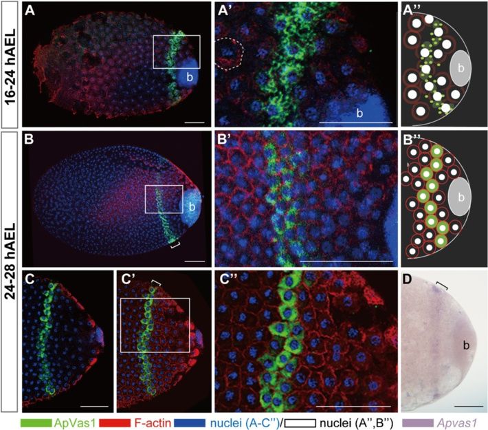
Lin et al. EvoDevo 2014, 5:18
http://www.evodevojournal.com/content/5/1/18

 RESEARCH                                                                                                                                        Open Access

Posterior localization of ApVas1 positions the
preformed germ plasm in the sexual oviparous
pea aphid Acyrthosiphon pisum
Gee-way Lin1,3, Charles E Cook2, Toru Miura3* and Chun-che Chang1,4,5*

  Abstract
  Background: Germline specification in some animals is driven by the maternally inherited germ plasm during early
  embryogenesis (inheritance mode), whereas in others it is induced by signals from neighboring cells in mid or late
  development (induction mode). In the Metazoa, the induction mode appears as a more prevalent and ancestral
  condition; the inheritance mode is therefore derived. However, regarding germline specification in organisms with
  asexual and sexual reproduction it has not been clear whether both strategies are used, one for each reproductive
  phase, or if just one strategy is used for both phases. Previously we have demonstrated that specification of germ
  cells in the asexual viviparous pea aphid depends on a preformed germ plasm. In this study, we extended this work
  to investigate how germ cells were specified in the sexual oviparous embryos, aiming to understand whether or
  not developmental plasticity of germline specification exists in the pea aphid.
  Results: We employed Apvas1, a Drosophila vasa ortholog in the pea aphid, as a germline marker to examine
  whether germ plasm is preformed during oviparous development, as has already been seen in the viviparous
  embryos. During oogenesis, Apvas1 mRNA and ApVas1 protein were both evenly distributed. After fertilization,
  uniform expression of Apvas1 remained in the egg but posterior localization of ApVas1 occurred from the fifth
  nuclear cycle onward. Posterior co-localization of Apvas1/ApVas1 was first identified in the syncytial blastoderm
  undergoing cellularization, and later we could detect specific expression of Apvas1/ApVas1 in the morphologically
  identifiable germ cells of mature embryos. This suggests that Apvas1/ApVas1-positive cells are primordial germ cells
  and posterior localization of ApVas1 prior to cellularization positions the preformed germ plasm.
  Conclusions: We conclude that both asexual and sexual pea aphids rely on the preformed germ plasm to specify
  germ cells and that developmental plasticity of germline specification, unlike axis patterning, occurs in neither of
  the two aphid reproductive phases. Consequently, the maternal inheritance mode implicated by a preformed germ
  plasm in the oviparous pea aphid becomes a non-canonical case in the Hemimetabola, where so far the zygotic
  induction mode prevails in most other studied insects.
  Keywords: Aphid, Asymmetric localization, Developmental plasticity, Germ cells, Germline specification, Vasa

* Correspondence: miu@ees.hokudai.ac.jp; chunche@ntu.edu.tw
3
 Laboratory of Ecological Genetics, Graduate School of Environmental Science,
Hokkaido University, N10 W5, Kita-ku, Sapporo, Hokkaido 060-0810, Japan
1
 Laboratory for Genetics and Development, Department of Entomology/
Institute of Biotechnology, College of Bioresources and Agriculture, National
Taiwan University, No. 27, Lane 113, Roosevelt Road, Sec. 4, Taipei 106, Taiwan
Full list of author information is available at the end of the article

                                          © 2014 Lin et al.; licensee BioMed Central Ltd. This is an Open Access article distributed under the terms of the Creative
                                          Commons Attribution License (http://creativecommons.org/licenses/by/2.0), which permits unrestricted use, distribution, and
                                          reproduction in any medium, provided the original work is properly credited. The Creative Commons Public Domain
                                          Dedication waiver (http://creativecommons.org/publicdomain/zero/1.0/) applies to the data made available in this article,
                                          unless otherwise stated.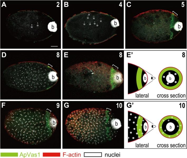
Lin et al. EvoDevo 2014, 5:18                                                                                  Page 2 of 16
http://www.evodevojournal.com/content/5/1/18

Background                                                    of the asexual aphid [14]. After cellularization, signals of
Studying model organisms can expand our knowledge in          Vas and Nos detected using cross-reacting antibodies
developmental biology and enrich our understanding of         were specifically recruited into the primordial germ cells
developmental diversity. One issue, developmental plas-       (PGCs), suggesting that germline specification in the
ticity, has long been a difficult subject to access in        asexual aphid relies on a germ plasm preformed within
animal models displaying little phenotypic plasticity.        the egg prior to cellularization [14]. However, we have
However, the pea aphid Acyrthosiphon pisum, a hemi-           not found any published descriptions of the assembly of
metabolous hemipteran insect with abundant adaptive           maternal germ plasm in oocytes or early embryos in the
capacity in response to specific environmental cues, has      sexual phase. To date, early segregation of germ cells in
proven an excellent model for study, and the recently         oviparous embryos has been reported in just two species:
published genome has made research on this species            the black willow aphid Melanoxanthus spp. [15] and the
more accessible [1,2]. With a sequenced genome, scien-        spring grain aphid Toxoptera graminum [16]. In the
tists can start exploring - at molecular and systematic       black willow aphid, newly segregated ‘primitive germ
levels - how and why a genome can direct the alteration       cells’ were identified in the posterior region of the inva-
of phenotypic and reproductive traits by stimuli from         ginating blastoderm. In the spring grain aphid, germline
outside. For example, environmental stresses such as          segregation was observed slightly later during gastrula-
high population density or predation trigger unwinged         tion and segregated germ cells were located aside the
aphid females to produce winged offspring for dispersal       posterior abdomen of the elongating germ band (embryo
[3,4], and aphid females also sense changing photope-         proper). In both aphid species, the presence of germ
riods to alter reproductive modes [5-7]. However, how         (polar) granules - an ultrastructural feature of the pre-
the environmental signals are received by the genome and      formed germ plasm - was not described. Taken together,
where the sequences required for polyphenic switches are      this suggests that germline specification in oviparous
located in the genome remain largely unknown.                 aphids is not germ-plasm dependent and, if this is the
   Embryogenesis in aphid life cycles can be categorized      case, aphids may adopt two programs to specify germ
as two types: asexual viviparous (bringing forth living       cells in asexual and sexual phases.
young without fertilization) and sexual oviparous (pro-          In some existing animal models such as Drosophila
ducing fertilized eggs). In the remaining paragraphs of       melanogaster (fly), Caenorhabditis elegans (nematode),
this article we abbreviate ‘asexual viviparous’ and ‘sexual   Danio rerio (zebrafish), and Xenopus laevis (frog), it is
oviparous’ to ‘viviparous’ and ‘oviparous’, respectively.     clear that specification of germ cells is driven by the ma-
Recent studies suggest that there are two developmental       ternal germ plasm assembled within the developing oo-
programs underlying early development of the vivipar-         cytes (reviewed in [17,18]). Removal of maternal germ
ous and oviparous embryos in the pea aphid. For               plasm leads to the loss of PGCs or sterility in adults
example, the expressions of axis patterning genes             [19-21]. In Mus musculus (mouse), however, germline
(hunchback, orthodenticle, caudal, nanos) and the ter-        specification occurs much later during gastrulation and
minal gene torso-like display distinctly different patterns   there is no maternally inherited germ plasm. Instead,
during early development in viviparous compared to            specification of germ cells is triggered via induction of
oviparous embryos [8-11]. Comparison of transcrip-            signals (the ‘induction mode’): Bmp4, Bmp8b, and
tomes of synchronized embryos in asexual and sexual           Bmp2, members of the bone morphogenetic protein
phases shows that more than 30 genes are differentially       family, are secreted from the extra-embryonic ectoderm
transcribed, further supporting the existence of divergent    to induce certain cells in the neighboring proximal epi-
programs of development underlying these two repro-           blast - the primary ectoderm - to form the precursors of
ductive cycles [12]. It appears that the change of devel-     PGCs [22-24]. After PGC precursors migrate to the pos-
opmental programs occurs during the transition period         terior primitive streak, germline fate is further specified
from asexual to sexual phases - our previous work indi-       through the function of Blimp1, a key transcription fac-
cates that the germline-specific identity of Api-piwi6 and    tor known to repress the somatic program in the PGCs
Api-ago3a, both of which encode components of the             [25]. Although cytoplasmic inheritance of maternal germ
Piwi-interacting RNA (piRNA) pathway, is lost from            plasm, the ‘inheritance mode’, is prevalent in most
viviparity to oviparity [13]. Accordingly, we would like to   current animal models, the induction mode actually ap-
know whether embryonic development is globally chan-          pears more common, and is likely ancestral, when other
ged in distinct reproductive phases or whether some de-       published data about germline development in emerging
velopmental mechanisms remain conserved.                      and non-model animals are collated (reviewed in [26]).
   Previously we identified posterior localization of Vasa       Among insect species, the inheritance mode for germ-
(Vas) and Nanos (Nos), two of the most conserved              line specification is mostly confined to Holometabola
germline markers in animals, in the syncytial blastoderm      such as flies, mosquitoes, and wasps [27,28], all of which
Lin et al. EvoDevo 2014, 5:18                                                                                 Page 3 of 16
http://www.evodevojournal.com/content/5/1/18

undergo complete metamorphosis. The lack of evidence         label germline cell fate in viviparous embryos [42]. How-
to support the existence of maternal germ plasm in           ever, asymmetric localization of Apvas1 mRNA, unlike
some holometabolous species like Bombyx mori (silk-          that of the Vas signals, was not identified in the vivipar-
worm) [29,30], Apis mellifera (honeybee) [31,32], and        ous pea aphid. In order not to miss detecting Vas pro-
Tribolium castaneum (beetle) [33,34], however, indicates     tein restricted to the potential germ plasm, we produced
that the inheritance mode is not a universal mechanism       an antibody against ApVas1 for immunostaining. Through
in Holometabola. By contrast, evidence from classical        comparing germline specification in the viviparous and
histological studies argues that insects adopting the in-    oviparous embryos, we aimed to identify similarities and
duction mode are the majority in Hemimetabola, which         differences between the development of germ cells in both
in contrast to Holometabola undergo incomplete meta-         of the reproductive cycles. Most importantly, this would
morphosis and are less derived. Ubiquitous expression        reveal whether aphids adopt one or two versions of mech-
of conserved germline genes such as vas, piwi, and nos       anisms for specifying germ cells in their life.
in oocytes and early embryos of Hemimetabola further
supports the above conclusion [35-37]. A recent report
                                                             Methods
concerning the abolishment of PGC formation via
                                                             Pea aphid culture
knockdown of the mesodermal marker twist in the
                                                             The pea aphid strain ApL, formerly known as Sap05Ms2,
cricket Gryllus bimaculatus - a model for the basally
                                                             was collected in Hokkaido, Japan and reared on broad
branching insects - suggests the presence of signal in-
                                                             beans (Vicia faba) in growth chambers under a long-day
duction for germline specification in hemimetabolous in-
                                                             photoperiod (16 h light/8 h dark) at 20°C [43]. Induction
sects [35], though the actual signaling molecules are not
                                                             of the sexual phase was carried out under a short-day
yet identified.
                                                             photoperiod (8 h light/16 h dark) at 15°C. Details of in-
   Current data show that germline specification in
                                                             duction were described in Ishikawa et al. [44,45]. Fertil-
Hemiptera is diverse. As described above, we have iden-
                                                             ized eggs for embryonic assays were maintained under
tified a presumptive germ plasm assembled in the
                                                             short-day photoperiod at 15°C for 42 days. Embryonic sta-
posterior syncytial blastoderm of the viviparous pea
                                                             ging for oviparous and viviparous embryogeneses followed
aphid - a hallmark of the inheritance mode [14]. In the
                                                             Miura et al. [46].
milkweed bug Oncopeltus fasciatus (Hemiptera: Lygaei-
dae), however, asymmetric localization of germline genes
is not detected in oocytes and early embryos, and the        Production and purification of antibody against ApVas1
segregation of PGCs is not identified until the late         In order to avoid the conserved motifs of the DEAD (Glu-
blastoderm stage. Moreover, knockdown of the con-            Asp-Ala-Glu)-box protein family, highly divergent se-
served germline markers vas and tudor does not affect        quences in the N-terminus of ApVas1 were selected and
the formation of PGCs. All of this evidence implies the      synthesized to induce the ApVas1 antibody (Additional
absence of a maternal germ plasm and the existence of        file 1: Figure S1A). This ApVas1 antigen contains 451
an inductive mechanism in Oncopeltus [36]. Similar con-      amino acids (aa) in the N-terminal region (aa 4-454). A
clusions have also been described in germline develop-       translation template of Apvas1 was cloned using a forward
ment in other hemipteran species such as the assassin        primer (5'-GATCAGATCTGGGTGGTTGGGATGATGA
bug Rhodnius prolixus (Hemiptera: Reduviidae) [38] and       ATCTGG-3', encoding GGWDDESG) and a reverse pri-
several species of scale insects [39,40]. Accordingly, the   mer (5'-AATTGAATTCCTATTCTCTCTCTGGTTGTT
germ plasm-dependent mechanism found in the vivipar-         CACGATCAC-3', encoding GDREQPERE). Products of
ous pea aphid so far appears to be a non-canonical case      the polymerase chain reaction (PCR) were then subcloned
in the Hemiptera in contrast to most other hemipterans.      into the pET32a(+) vector (Novagen, San Diego, CA,
Recent studies show that the oviparous embryos adopt         USA) to produce a His-tagged fusion protein in Escheri-
conserved mechanisms to pattern early embryogenesis          chia coli C41(DE3) cells. Affinity purified His-tagged
while the viviparous embryos use a distinct and less con-    ApVas1 protein in homogenized polyacrylamide gel slices
served mechanism. Given the gross morphological simi-        was used to immunize rabbits. For antibody purification,
larities in oviparous aphid development with other           antisera were passed through a column packed with the
hemipterans [38-41], it would be interesting to know if      cyanogen bromide (CNBr)-activated Sepharose 4B (GE
germline specification is also similar.                      Healthcare, Uppsala, Sweden) coupled with a polypeptide
   In order to explore how germ cells are specified during   containing 186 aa in the N-terminus of ApVas1 (aa 4-189)
oviparous development in the pea aphid, we employed          (Additional file 1: Figure S1B). After non-specific bindings
whole-mount in situ hybridization of Apvas1 (previously      were washed off, the ApVas1 antibody was eluted with
known as Apvas [42], a Drosophila vas ortholog in the        0.1 M Glycine at pH 2.5 and immediately neutralized to
pea aphid), which we successfully used previously to         pH 7.0 using 1 M Tris pH 8.0. The neutralized ApVas1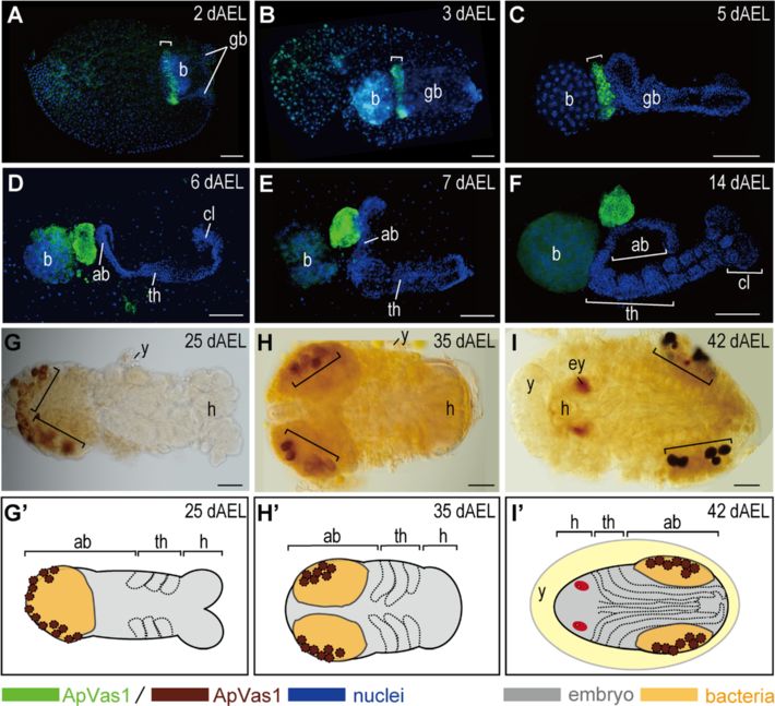
Lin et al. EvoDevo 2014, 5:18                                                                                   Page 4 of 16
http://www.evodevojournal.com/content/5/1/18

antibody was dialyzed against 1× phosphate buffered sa-        (2) secondary antibodies: anti-rabbit IgG conjugated with
line (PBS) and then stored at -80°C in 50% glycerol.           Cy5 (Invitrogen, Paisley, UK), Alexa Fluor 488 goat anti-
                                                               mouse (Invitrogen), biotinylated goat anti-rabbit IgG
Fixation and dissection of oviparous eggs                      (Vector labs, Burlingame, CA, USA) (all diluted 1:500).
Aphid eggs were dechorionated in a 1:1 solution of             Nuclear staining of embryos older than 2 dAEL used 4',6-
methanol and heptane for 1 min with vigorous shaking.          diamidino-2-phenylindole (DAPI) (2 μg/mL) (Sigma) at
The heptane-methanol solution was removed without              37°C for 2 h, but those collected within 48 hours after egg
disturbing the dechorionated eggs after they had sunk to       laying (hAEL) were stained using propidium iodide (PI)
the bottom of the 1.5 mL Eppendorf tube. Eggs were             (10 μg/mL) (Invitrogen) at 37°C for 2 h to avoid interfer-
then sequentially washed with mixture of methanol and          ence by autofluorescence from the yolk. F-actin staining
4% paraformaldehyde using ratios of 3:1, 1:1, and 1:3.         on oviparous ovarioles was performed with Rhodamine
After that, they were fixed in 4% paraformaldehyde for         Phalloidin (1:40) (Invitrogen); however, this was carried
20 min with mild shaking on a rotator. Fixed embryos           out using the anti-actin antibody (Sigma) in embryos pre-
collected before the end of 2 dAEL (days after egg lay-        fixed with solutions containing methanol.
ing) could be directly stained with riboprobe or anti-            Bright-field images were photographed using a BX51
body. From 3 dAEL onward, however, the serosal                 microscope (Olympus, Melville, NY, USA) connected
membrane that would turn black during staining had to          with a DP50 CCD camera (Olympus); fluorescence im-
be removed using a tungsten needle before staining. For        ages were taken by the laser-scanning microscope Zeiss
embryos collected by the end of 4 dAEL we did not sep-         LSM510 META (Carl Zeiss, Jena, Germany).
arate them from the yolk, but we remove yolk for those
collected from 5 dAEL to increase the accessibility of
                                                               Western blot
riboprobe or antibody to the embryos.
                                                               About 50 ng of each ApVas fusion protein was loaded for
                                                               protein electrophoresis (Figure 1A). Unfertilized eggs after
In situ hybridization, immunostaining, and imaging
                                                               oviposition were collected from females without mating;
Whole-mount in situ hybridization (WISH) on ovipar-
                                                               fertilized eggs were collected in parallel within 16 hAEL.
ous embryos was performed according to the protocol
                                                               Protein extracted from 10 unfertilized/fertilized eggs was
previously applied to the viviparous ovarioles [47]. Be-
                                                               dissolved in 100 μL of 2× sample buffer (57.7 mM Tris,
fore application of the riboprobe, further fixation of
                                                               10% Glycine, 2% SDS, 10% glycerol, 12.5 mM EDTA,
dechorionated eggs or dissected embryos using 4% para-
                                                               0.02% bromophenol blue 0.02%, 1% beta-mercaptoethanol)
formaldehyde for 4 h at 25°C was required. In order to
                                                               and 6 μL of extracts was loaded onto each lane (Figure 1A).
increase the probe specificity, we synthesized a Digoxi-
                                                               Primary antibody: anti-ApVas1 antibody (1:1,000); second-
genin (DIG)-labeled antisense riboprobe complementary
                                                               ary antibody: goat peroxidase-labeled antibody against
to the 149 nucleotides located in the 5’ untranslated re-
                                                               rabbit IgG (1:2,000) (Kirkegaard and Perry Laboratories
gion (UTR) of Apvas1, assuming that cross-hybridization
                                                               (KPL), Gaithersburg, MD, USA). Signals were developed
would not occur to Apvas2-4 mRNAs. In situ hybridi-
                                                               using the VisGlow™ Chemiluminescent Substrate, Horse-
zation showed that the expression pattern detected using
                                                               radish Peroxidase (HRP) system (Visual Protein, Taipei,
the 149-bp Apvas1 antisense riboprobe differed from the
                                                               Taiwan).
expressions of Apvas2-4 (Additional file 2: Figure S2). This
indicates that the 149-bp probe is Apvas1-specific and we
have used it for all detections of Apvas1 expression in this   Results
study. Sequences of the forward and reverse primers for        Maternal expression of Apvas genes in ovarioles of the
amplifying the 5’ UTR of Apvas1 are 5'- TTTGGCGG               sexual female
TGATGATGGAGAAG-3' and 5'-AATCGAGTCTAGGT                        The pea aphid has four vasa homologs (Apvas1-4), all of
GGCAACG-3', respectively. WISH was carried out at              which encode an ATP-dependent RNA helicase of the
66°C for 36 h.                                                 DEAD-box protein family [48]. For detecting the expres-
  Immunostaining followed the protocol of Chang et al.         sions of Apvas mRNA and protein, we synthesized anti-
[14], where viviparous ovarioles were stained using            sense riboprobes of Apvas1-4 and antibodies against
cross-reacting antibodies against Vas and Nos. For ovip-       ApVas1-4, respectively. Among the four Apvas genes,
arous embryos, an additional fixation using 4% parafor-        Apvas1 is the sole germline marker throughout vivipar-
maldehyde for 30 min at 25°C was required before               ous development while expressions of Apvas2-4 are not
application of the primary antibody. Antibodies and their      germline specific (Additional file 2: Figure S2) [2,42].
dilution ratios were: (1) primary antibodies: ApVas1           We therefore examined the expression of Apvas1 in the
antibody (1:20), ApVas2-4 antibodies (1:20), mouse anti-       sexual phase, assuming that it was also a germline
actin antibody (1:500) (Sigma, St Louis, MO, USA); and         marker in the oviparous embryos.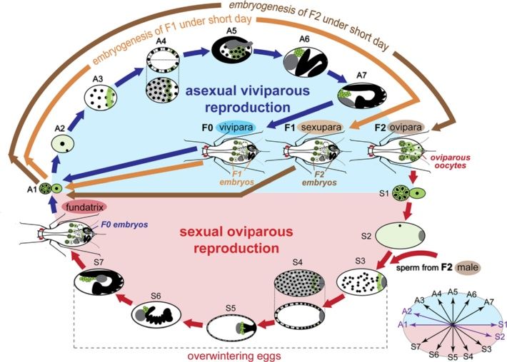
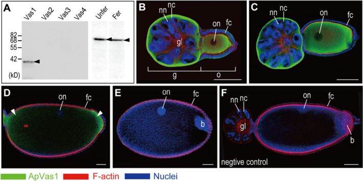
Lin et al. EvoDevo 2014, 5:18                                                                                                                        Page 5 of 16
http://www.evodevojournal.com/content/5/1/18

 Figure 1 Expression of ApVas1 in the oviparous ovarioles. Each ovariole is composed of a germarium plus 1 to 2 egg chambers accommodating
 the developing oocytes. Color keys that indicate staining signals of ApVas1, F-actin (Rhodamine Phalloidin), and nuclear DNA (DAPI) in the ovariole are
 highlighted under the figure. Anterior of egg chamber is to the left. (A) Detection of ApVas1 in vitro: western blot. On a membrane blotted with the
 ApVas1-4 fusion proteins, only ApVas 1 was detected by the ApVas1 antibody. On another membrane blotted with the total protein extracted from
 unfertilized eggs and fertilized eggs, ApVas1 antibody detected a major band with the expected molecular weight (62.8 kD, arrowhead) of ApVas1. This
 major band could not have been ApVas2 (75.7 kD) and ApVas3 (71.7 kD), but it was close to ApVas4 (61.65 kD). (B-E) Detection of ApVas1 in vivo:
 immunostaining. (B, C) Previtellogenic oocytes. Oocyte in (C), with longer egg length, was more mature than that shown in (B). Expression of ApVas1
 was evenly distributed in the cytoplasm of nurse cells (in germaria) and oocytes. Nuclei of nurse cells and oocytes were devoid of staining. (D) Vitellogenic
 oocytes. Preferential expression of ApVas1 (arrowheads) was identified in both anterior and posterior regions of the egg. (E) Mature oocytes subjected
 to oviposition. Signals of ApVas1 were almost not detected. Invading bacterial endosymbionts (bacteria) were observed in the egg posterior.
 (F) Negative control. Antibody against ApVas1, the primary antibody, was not applied to staining. Abbreviations: b, bacteria; fc, follicle cells; Fer, fertilized
 eggs; g, germaria; gl, germarial lumen; kD, kilodalton; nc, nurse cells; nn, nurse-cell nuclei; o, oocytes; on, oocyte nuclei; Unfer, unfertilized eggs. Scale
 bars: 100 μm.

  In situ hybridization results showed that none of the                            oocytes can be obtained from the adult aphids. Staining
four Apvas mRNAs, including Apvas1, was asymmetric-                                results showed that ApVas1 was uniformly expressed in
ally localized in the oocytes (Additional file 3: Figure S3).                      the cytoplasm of nurse cells and that of the previtello-
In order to examine whether ApVas1 was asymmetric-                                 genic oocytes (Figure 1B, C). In the vitellogenic oocytes,
ally localized or uniformly distributed, we stained the                            we detected transient localization of ApVas1 in both an-
ovarioles with the affinity-purified antibody against                              terior and posterior regions (Figure 1D). We conclude
ApVas1. Purification of this antibody was specifically                             that localized signals observed in the vitellogenic oocytes
carried out using an immobilized antigen composed of                               were ApVas1: (1) localization of ApVas1 was not de-
186 amino acids in the N-terminal region of ApVas1 to                              tected in oocytes that were not stained using the ApVas1
avoid antisera cross-reacting with downstream se-                                  antibody (negative control, Figure 1F); and (2) expres-
quences that contain the eight conserved motifs of the                             sion patterns of ApVas2-4 (Additional file 4: Figure S4)
DEAD-box protein family that is in all ApVas proteins                              were different from those of ApVas1 (Figure 1B-E). In
(Additional file 1: Figure S1B). Western blots showed                              mature oocytes subjected to oviposition, ApVas1 expres-
that the ApVas1 antibody could specifically detect                                 sion became undetectable (Figure 1E). We infer that the
ApVas1 and a major protein with expected size of the                               enlargement of egg chambers dilutes the intensity of the
full-length ApVas1 (62.8 kD) from the unfertilized and                             ApVas1 signal, because a major band corresponding to
fertilized eggs collected within 16 hAEL (Figure 1A).                              the size of ApVas1 could still be detected on the western
This suggests: (1) the affinity-purified antibody is specific                      blot (Figure 1A).
to ApVas1; and (2) the newly laid eggs inherit maternal
ApVas1 from the ovaries.                                                           Posterior localization of ApVas1/Apvas1 in the syncytial
  Immunostaining was performed on dissected ovarian                                and cellularized blastoderm
tubules (ovarioles) encompassing the developing oocytes.                           In the newly laid eggs, we did not identify localized sig-
In order to cover all developmental stages of oogenesis,                           nals of ApVas1 before the completion of the fourth cycle
we stained ovarioles dissected from both nymphs and                                of nuclear division (Figure 2A, B). At the time that we first
adults. The nymphal aphids contain a higher proportion                             observed localized ApVas1 (fifth nuclear cycle, Figure 2C),
of previtellogenic oocytes whilst many more vitellogenic                           energid nuclei had not yet reached the embryonic surface,
Lin et al. EvoDevo 2014, 5:18                                                                                                                  Page 6 of 16
http://www.evodevojournal.com/content/5/1/18

 Figure 2 Localization of ApVas1 in the posterior region of newly laid eggs. Staining was performed on fertilized eggs collected within 16
 hAEL. From 0 to 16 hAEL, embryos underwent 10 consecutive nuclear divisions. In each panel the nuclear cycle is highlighted in the upper right
 corner. DNA staining of bacteria (b) was detected in the posteriormost region of all egg chambers. (A, B) Second to fourth nuclear divisions.
 Asterisks mark the locations of cleaved nuclei in the shown focal plane. Localization of ApVas1 was not visualized. (C) Fifth nuclear division.
 Localization of ApVas1 (bracket) was first identified in the posterior region, anteriorly to the bacteria. (D, E) Eighth nuclear division. (D) and (E) are
 the same preparation but shown at two different focal planes. In (D), with the middle region of the egg in focus, most cleaved nuclei were
 observed but the ApVas1 stripe (bracket) was barely detected. In (E), with the egg cortex in focus, nuclear staining in the middle region was
 absent but the ApVas1 stripe (bracket) could be clearly identified. (E’) Schematic illustration indicating that cleaved nuclei have not reached the
 cortex region where ApVas1 is localized. (F, G) Ninth and tenth nuclear divisions. In contrast to eggs undergoing the eighth nuclear cycle,
 dividing nuclei could be observed in the cortex from the ninth nuclear cycle onward. In addition, the divisions began to progress in waves -
 nuclei located in the posterior third of the egg proceeded into anaphase but other nuclei remained at interphase or prophase. (G’) Schematic
 illustration indicating that some cleaved nuclei have reached the cortex region, being co-localized with the ApVas1 signals. Scale bars: 100 μm.

suggesting that ApVas1 is not zygotically synthesized and                         In order to monitor the status of cellularization, we
is more likely maternally provided. By the end of the                           co-stained embryos using an antibody against the fila-
eighth cycle, we did not identify any energid nuclei on the                     mentous actin (F-actin). In insects such as Drosophila,
cortex (Figure 2D, E). Migration of nuclei to the inner per-                    crickets, and grasshoppers, formation of the ‘actin cap’
iphery of the egg - a hallmark of the formation of the syn-                     that surrounds each superficial energid nucleus has been
cytial blastoderm - was first identified from the ninth                         regarded as a sign of cellularization and after cellulariza-
nuclear division onward (Figure 2F). Hence, we could find                       tion F-actin is located to the inner periphery of the cell
dividing nuclei within the ApVas1 stripe (Figure 2F, G). As                     membrane [49-51]. We adopted these conserved features
previously described by Miura et al. [46], we also identi-                      of F-actin polymerization to monitor cellularization in the
fied a mitotic wave along the anteroposterior axis from                         pea aphid. During 16 to 24 hAEL, morphology of the actin
the ninth to tenth nuclear cycles, which displayed asyn-                        caps was visualized and meanwhile we identified the ex-
chronous nuclear divisions in different regions of the egg.                     pression of ApVas1 associated with the cytoplasm sur-
However, localization of ApVas1 remained unaffected ad-                         rounding the energid nuclei (Figure 3A-A”). In the time
jacent to the endosymbiotic bacteria (bacteria) at the pos-                     window between 24 and 28 hAEL, we found that ApVas1
terior end of the egg, regardless of the status of nuclear                      was incorporated by the forming cell membrane whose
divisions within the ApVas1 stripe (Figure 2F, G, G’).                          inner periphery was enriched with F-actin (Figure 3B, B’,
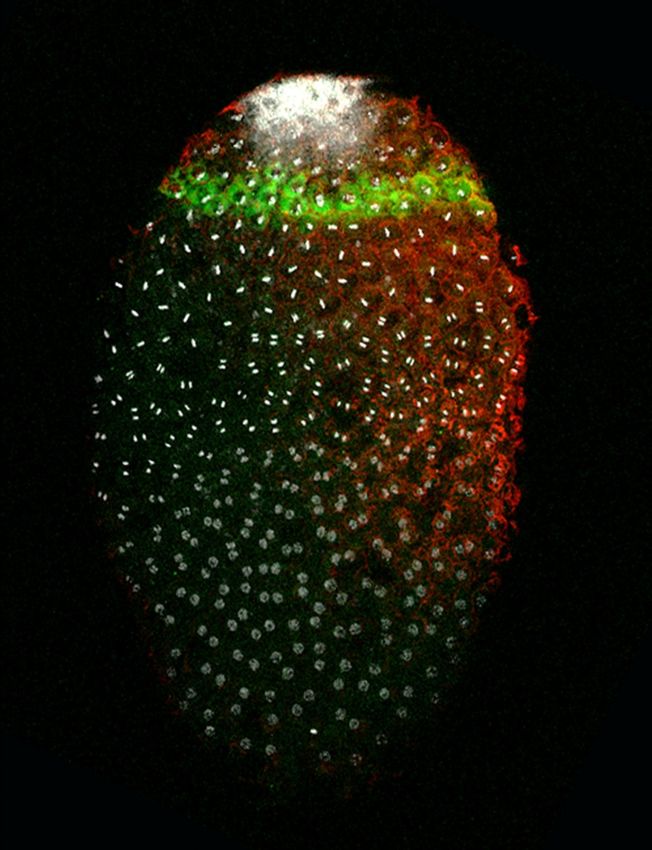
Lin et al. EvoDevo 2014, 5:18                                                                                                                     Page 7 of 16
http://www.evodevojournal.com/content/5/1/18

 Figure 3 Posterior localization of ApVas1 during cellularization of the syncytial blastoderm. Staining with ApVas1 antibody was performed
 on eggs collected within 16 to 28 hAEL. Embryo presented in (D) was hybridized with the Apvas1 antisense riboprobe. Status of cellularization
 was monitored by the expression of the polymerized F-actin using an anti-actin antibody. Anterior of egg chamber is to the left. Bacteria (b) are
 located in the posteriormost region of the eggs. (A-A”) Eggs collected during 16 to 24 hAEL. Actin caps (dashed circle) were visualized. In (A),
 localization of ApVas1 remained in the posterior cortex, forming a stripe anterior to the bacteria. (A’) is a magnification of the inset in (A): expression of
 ApVas1 was identified in the cytoplasm associated with the energid nuclei. (A”) Schematic illustration of ApVas1 expression shown in (A) and (A’).
 (B-D) Eggs collected during 24 to 28 hAEL. In (B), localization of ApVas1 remained in the egg posterior. (B’) is a magnification of the inset shown in
 (B): F-actin was located at the inner periphery of the forming cell membrane. (B”) Schematic illustration of ApVas1 expression shown in (B) and (B’).
 Embryos in (C) and (C’) belong to the same preparation but are shown at different focal planes. In (C), where the focal plane was internal, activity of
 F-actin was not detected; however, F-actin expression was detected in the cortex region shown in (C’). (C”) is a magnification of the inset shown in
 (C’): the intensity of ApVas1 and F-actin, in contrast to (B’), is increased. This suggests that the embryo in (C) is more mature than that in (B).
 (D) Posterior localization of Apvas1 mRNA. Transcripts of Apvas1 were restricted within a cortical stripe in the posterior region of the egg. Brackets in
 (C’) and (D) highlight locations of ApVas1/Apvas1 enrichment. Scale bars: 100 μm.

C’, C”). We note that cellularization may not have been                           optimal conditions to perform dual immunostaining and
completed because within the interior region of the                               in situ hybridization on pea aphid embryos, the similar
ApVas1-positive energids signals of F-actin staining are                          locations of the ApVas1 and Apvas1 stripes (Figure 3C’, D)
not detected (Figure 3C).                                                         and the synchronous migration of ApVas1/Apvas1-positive
   Apart from the ApVas1 protein, we also monitored the                           cells in later embryogenesis (Figure 4) suggest that ApVas1
expression of Apvas1 mRNA in parallel. In situ results                            and Apvas1 are co-localized.
showed that Apvas1 transcripts were ubiquitously dis-
tributed in germaria, oocytes (Additional file 3: Figure                          Migration of cells expressing ApVas1/Apvas1 during germ
S3A-C, F), and eggs collected within 24 hAEL (Additional                          band formation
file 5: Figure S5A). Posterior localization of Apvas1 was not                     Prior to invagination of the blastoderm, we observed the
identified until 28 hAEL (Figure 3D). Like posterior lo-                          increase of ApVas1-positive cells. Immunostaining re-
calization of ApVas1 in embryos collected within the same                         sults showed that the ApVas1 stripe expanded from two
developmental period (24 to 28 hAEL; Figure 3C, C’),                              to three cells in width, and the total cell numbers
Apvas1 mRNA was enriched to a stripe area within the                              increased from about 40 to 160 within the stripe
egg posterior (Figure 3D). Although we have not found                             (Figure 4A, A’, B, B’). Initiation of invagination could be
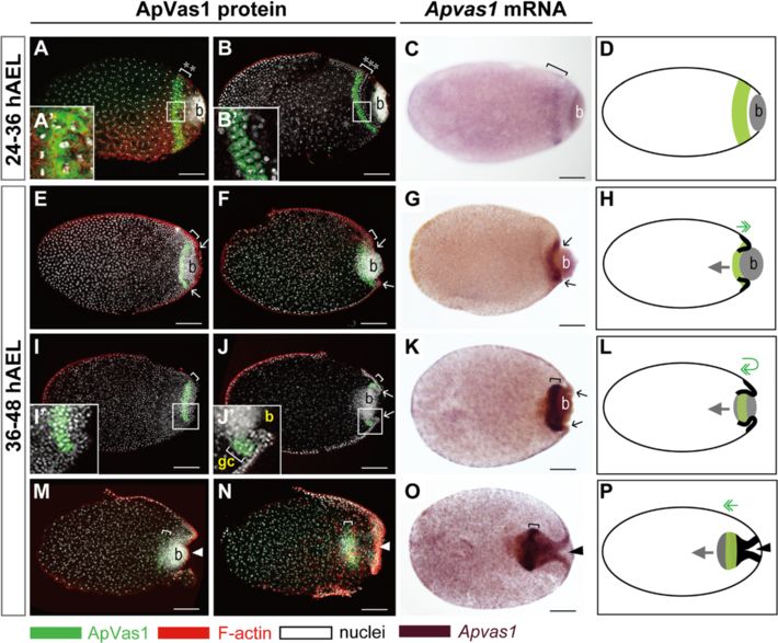
Lin et al. EvoDevo 2014, 5:18                                                                                                              Page 8 of 16
http://www.evodevojournal.com/content/5/1/18

 Figure 4 Expression of ApVas1/Apvas1 during germ band formation. (A, B, E, F, I, J, M, N) ApVas1 antibody staining; (C, G, K, O) Apvas1 in situ
 hybridization; ApVas1/Apvas1 stripes: brackets; (D, H, L, P) Schematic illustrations: migration of bacteria and ApVas1/Apvas1-positive cells are
 indicated with grey arrows and green double arrows, respectively; invaginating blastoderm is marked with bold lines. Anterior of egg chamber is
 to the left; in (M-P), anterior of the germ band is to the right. (A-D) Blastoderm embryos prior to invagination. (A’) and (B’) are magnifications of
 insets in (A) and (B), respectively. Increasing number of ApVas1-positive cells was evidenced by the expansion of stripe width (A: 2 rows; B: 3 rows;
 cell rows: asterisks). (C) Apvas1 mRNA was restricted to the same area as that of ApVas1 expression presented in (B). (E-H) Invagination of the
 posterior blastoderm. Invagination furrows marked with arrows in (F) and (G) appear more prominent than those in (E). Posterior boundary of the
 ApVas1/Apvas1 stripe moved to encounter the anterior border of the bacteria. (I-L) Co-migration of bacteria and the ApVas1/Apvas1 stripe into
 the yolk. Embryos in (I) and (J) belong to the same preparation but are shown at different focal planes: (I), cortex; (J), interior. (I’) and (J’) are
 magnifications of insets in (I) and (J), respectively. (K) Apvas1 expression. (L) ApVas1/Apvas1 stripe first reaches to the posteriormost end of the
 egg and then migrate back inside the yolk. After that, they follow the migration of bacteria. (M-P) Immersion of the ApVas1/Apvas1 stripe into
 the yolk during anatrepsis. (M) Continuous invagination of the posterior blastoderm pushed bacteria and ApVas1 stripes toward the central region
 of the egg chamber. (O, N) Cells expressing ApVas1/Apvas1 and bacteria were located at the posterior region of the germ band. Abbreviation: b,
 bacteria; gc, germ cells. Scale bars: 100 μm.

visualized by the appearance of invaginating furrows in                       are shown at different focal planes (4I, I’: cortex; 4 J, J’:
the posterior region of the eggs (Figure 4E, F). Accord-                      interior region). If PGCs really traveled through the bac-
ing to our observations, invagination of the blastoderm                       teria, we would not be able to observe an integrative
formed the blastopore, pulled bacteria into the interior                      stripe composed of ApVas1 positive cells without the
region of the egg, and meanwhile dragged the ApVas1                           interference of bacteria (Figure 4I) or the absence of
stripe on the cortex to migrate further posteriorly to-                       ApVas1 positive cells within the bacteria (Figure 4J).
ward the posterior pole of the egg (Figure 4E, F, H).                            Like ApVas1, corresponding patterns of migration were
When bacteria became fully immersed within the yolk,                          also identified in the migration of Apvas1-positive cells
the ApVas1 stripe located to the posterior cortex                             during blastoderm invagination (Figure 4C, G, K, O). The
followed the movement of bacteria, migrating backward                         chromogenic in situ results, moreover, allowed us to
to the inner region of the egg (Figure 4I-L). Later, bac-                     visualize the morphology of the invaginating blastoderm
teria and ApVas1-positive cells further migrated toward                       more clearly (Figure 4O), because under a bright-field
the egg center (Figure 4N-P). Here, we confirm that                           microscope the shape of the blastoderm was not masked
PGCs travel over, rather than through, the bacteria. Tak-                     by the adjacent bacterial mass that emitted strong nuclear
ing embryos presented in Figure 4I and J as an example,                       signals under a confocal microscope (Figure 4M, N). Iden-
these two embryos belong to the same preparation but                          tical patterns of blastoderm invagination and germ band
Lin et al. EvoDevo 2014, 5:18                                                                                                                Page 9 of 16
http://www.evodevojournal.com/content/5/1/18

formation have been described in embryogenesis of the                          a consequence, became located between the bacteria and
black willow aphid Melanoxanthus spp. [15] and the                             the elongating germ band (Figure 5B, C). We infer that
spring grain aphid T. graminum (Additional file 6: Figure                      further migration of bacteria and ApVas1-positive cells
S6C) [16], suggesting that movement of Vas/vas-positive                        toward the egg anterior was driven by the extension of
cells is likely conserved among the aphid species.                             the germ band.
                                                                                 During germ band extension, the morphology of the
Migration of cells expressing ApVas1/Apvas1 during germ                        cephalic lobe, thorax, and abdomen became distinct.
band extension, katatrepsis, and late embryogenesis                            Meanwhile, we found that the morphology of the
Although bacterial cells were migrating ahead of the                           ApVas1 stripe was also changed: the stripe was trans-
ApVas1/Apvas1-positive cells during germ band forma-                           formed into a globular structure, extra-embryonically lo-
tion (Figure 4E-P), these two populations of cells                             cated adjacent to the elongating abdomen (Figure 5D-F).
reached the posterior end of the germ band that was                            When embryos were fully segmented and the limb buds
about to elongate almost at the same time (Figure 5A).                         became visible ApVas1-positive cells aggregated to the
However, their co-localization broke up while the germ                         posterior region, forming a U shaped ring (Figure 5G,
band continued to extend. The ApVas1-positive cells, as                        G’). Prior to katatrepsis (embryo flip), we found that

 Figure 5 Expression of ApVas1 during germ band extension, katatrepsis, and late embryogenesis. Dissected embryos were collected from
 2 to 42 dAEL. Before katatrepsis (A-H): embryonic anterior, right; after katatrepsis (I): embryonic anterior, left. ApVas1 stripes: brackets. (A-C) Germ
 band extension. (A) Embryos at 2 dAEL of development. ApVas1 stripe and bacteria were co-localized to the posteriormost region of the germ
 band. (B, C) Embryos at 3 to 5 dAEL of development. Bacteria migrated through the ApVas1 stripe, locating posteriorly to the ApVas1 stripe.
 (D-F) Morphogenesis of the germ band. (D, E) Embryos at 6 to 7 dAEL of development. Structures of the cephalic lobe, thorax, and abdomen could be
 identified. Cephalic lobe in (E) was truncated; see Additional file 8: Figure S8B, D for intact morphology. (F) Embryos at 14 dAEL of development.
 Segments in the head and thorax were visible. During 6 to 14 dAEL, the ApVas1 stripe was reorganized into a globular shape. (G-I) Fully-segmented
 embryos before and after katatrepsis. Dorsal view of embryos attached with bacteria is presented, and follow the style of Miura et al. and Shingleton
 et al. [46,52]. (G) Embryos at 25 dAEL of development. ApVas1-positive cells associated with the bacteria were located to the posteriormost region of
 the embryo, forming a ‘U’ shaped cap. (H) Embryos at 35 dAEL of development. ApVas1-positive cells were bilaterally located in the abdomen.
 Another embryo at similar age in lateral view was shown in Additional file 7: Figure S7A. (I) Embryos at 42 dAEL of development (after katatrepsis).
 The distribution of ApVas1-positive cells remained similar to that seen in embryos of 35 dAEL. (G’-I’) Schematic illustrations shown in (G-I).
 Abbreviations: ab, abdomen; b, bacteria; cl, cephalic lobe; ey, eyes; gb, germ band; h, head; th, thorax; y, yolk. Scale bars: 100 μm.
Lin et al. EvoDevo 2014, 5:18 Page 10 of 16
http://www.evodevojournal.com/content/5/1/18
ApVas1-positive cells, together with the bacteria, were embryo, we conclude that the Apvas1 gene is a germline
separated into clusters within the dorsal side of the em- marker of pea aphids in both sexual and asexual phases.
bryo (Figure 5H, H’; Additional file 7: Figure S7A, B).
After katatrepsis, the ApVas1-positive cells were located bi- Identification and assembly of the preformed germ plasm
laterally in the dorsal region of the abdomen (Figure 5I, I’) in the sexual female
[46,52]. In parallel, we also examined the expression of The preformed germ plasm is usually assembled in a
Apvas1 in the elongating germ band (Additional file 8: subcellular area within the egg prior to cellularization.
Figure S8A, B), finding that it was restricted to the same Therefore, asymmetric localization of germline markers
places where ApVas1 was expressed (Additional file 8: that will be inherited by the PGCs has become a geo-
Figure S8C, D). This implies that in later stages of graphical signature of the preformed germ plasm
development Apvas1 mRNA and ApVas1 protein are co- (reviewed in [59]). In the oviparous embryo, we could
localized in the germ cells. detect the asymmetric localization of ApVas1 in the eggs
prior to cellularization (Figure 2C-G) and monitor the
Discussion incorporation of ApVas1 into presumptive PGCs during
Apvas1 gene as a germline marker in both sexual and cellularization (Figure 3B’). After katatrepsis, locations of
asexual females the ApVas1-positive PGCs (Figure 5I, I’) in the dorsal re-
The ultimate goal of this study is to understand how gion of the embryos correspond to the morphologically
germ cells are specified in the oviparous pea aphid. In identifiable germ cells in the viviparous aphids at equiva-
particular, we are interested in whether or not germline lent developmental stages [42,46]. Accordingly, we con-
specification relies on a preformed germ plasm. Here, clude that the subcellular cytoplasm restricting ApVas1
we employed vas as a germline marker to monitor the expression in the posterior cortex of the early syncytial
assembly of germ plasm and migration of germ cells. Al- blastoderm is the preformed germ plasm (Figure 2C).
though vas has been regarded as the most conserved In early embryos of Drosophila and the pea aphid, Vas
germ-cell specific gene in animals, in some species vas protein is localized to the germ plasm whereas vas
mRNA and Vas protein are not concurrently localized to mRNA is uniformly distributed (Figure 2C-G, Additional
the preformed germ plasm. For example, in Drosophila file 5: Figure S5A) [14,42,53,54]. This suggests that Vas
it is Vas protein, rather than vas mRNA, that is localized protein, rather than vas mRNA, is involved in the as-
to the germ (pole) plasm [53,54]. By contrast, in zebra- sembly of germ plasm in both species. However, the tim-
fish preformed germ plasm within the cleavage planes is ings of Vas localization are different: in Drosophila
enriched in vas mRNA, rather than the uniformly posterior localization of Vas to the germ plasm occurs
expressed Vas protein, in two cell-stage embryos [55-58]. during mid oogenesis, but in the oviparous embryos a
In order to maximize the opportunities for identifying subcellular cytoplasm containing ApVas1 expression -
the germ plasm in the oviparous pea aphid, we accord- the presumptive germ plasm - cannot be identified
ingly explored the expression of both Apvas1 (mRNA) through oogenesis (Figure 1B, C). Although in some
and ApVas1 (protein). vitellogenic oocytes posterior localization of ApVas1 is
Among previous studies of embryogenesis in sexual fe- identified (Figure 1D), we do not consider this the onset
males, germline development has been best character- of germ-plasm assembly because later ApVas1 localization
ized in T. graminum (spring grain aphid). According to cannot be continuously detected in the posterior region of
Webster and Phillips [16], segregation of the ‘primitive mature oocytes subjected to oviposition (Figure 1E) and
germ cells’ - an old term for the primordial germ cells newly laid eggs (Figure 2A, B). Besides, locations of
(PGCs) - in T. graminum was first detected extra- ApVas1 localization in the oocytes and embryos do not
embryonically adjacent to the posterior abdomen of the exactly correspond to each other - ApVas1 is localized to
elongated germ band. This location corresponds to where the posteriormost end of the oocyte (Figure 1D) while in
Apvas1/ApVas1 was expressed in embryos at equivalent the embryo the ‘ApVas1 stripe’ is already located anteriorly
developmental stage in the pea aphid (Figure 5D, E; to the bacteria (Figure 2C).
Additional file 8: Figure S8). Specific expression of Compared with Drosophila, the oviparous pea aphid
Apvas1/ApVas1, moreover, could be identified in germ segregates germ plasm much later: not until the fifth nu-
cells that were morphologically identifiable in the dorsal clear division after fertilization (Figure 2C). In other in-
region of embryos after katatrepsis (Apvas1: data not sects that adopt the inheritance mode, the assembly of
shown; ApVas1: Figure 5I, Additional file 7: Figure S7A). germ plasm also takes place during oogenesis [27,28,60].
The above evidence suggests that Apvas1/ApVas1-positive Thus the ‘post-fertilization’ assembly of germ plasm in the
cells are PGCs in the oviparous embryo. Taken together oviparous aphid is so far unusual. Nonetheless, a similar
with our previous work showing that Apvas1 [42] and situation can be found in the nematode C. elegans, where
ApVas1 [14] are also germline-specific in the viviparous fertilization triggers the posterior localization of maternalLin et al. EvoDevo 2014, 5:18 Page 11 of 16
http://www.evodevojournal.com/content/5/1/18
P granules (germ granules) [61]. At present we do not [8,10,11], our data show that the germ-plasm dependent
have experimental data to show whether the assembly of specification of germ cells is conserved in both modes
germ plasm in the oviparous aphid is fertilization of aphid development. The early segregation of germ
dependent or independent. However, the segregation of plasm, we infer, allows germ cells to locate in an
germ plasm in the viviparous aphid does not depend on extra-embryonic position so that the germline fate
sperm entry [14]. If aphids of both reproductive phases can be isolated from the somatic body plans. At a
share the same mechanism of germline specification then molecular level, differential expression of duplicated
fertilization may not play a role in the assembly of germ germline genes (for example, vas, nos, piwi, and ago3)
plasm in the oviparous embryo. On the other hand, unless during the switch of reproductive cycles and the es-
we can examine the distribution of all transcripts specific tablishment of sexual/asexual-specific germline tran-
to germ cells, we cannot exclude the possibility that the scriptomes may be required for germline reprogramming
germ plasm has already been assembled during oogenesis [13], but not for the specification of germ cells. Con-
via depositing germline determinants to localize ApVas1, sequently, germline specification - at the morpho-
or locally translate Apvas1, after fertilization. logical level - does not display developmental plasticity
in the pea aphid and perhaps in other aphid species
Comparison of germline specification and migration in as well.
oviparous and viviparous pea aphids Additionally, we find that segregation of germ plasm
Although distinct developmental programs have been and migration of germ cells in both oviparous and vivip-
identified in oviparous and viviparous embryogenesis arous embryos follows a very similar pattern (Figure 6)
Figure 6 Comparison of germline development during sexual and asexual reproductive phases. Induction of reproductive modes relies
on the length of photoperiods: long days induce the asexual phase; short days induce the sexual phase (for details see Methods). From asexual
to sexual reproduction (F0 to F2), embryos pregnant by the F0 females and F1 females must develop under short light period days. Expression
of ApVas1 in germaria, oocytes, presumptive germ plasm, and germ cells is designated in green. (S1-S7): germline development during oviparous
reproduction; (A1-A7): germline development during viviparous reproduction. S1/A1, germaria and oocytes; S2/A2, mature oocytes; S3/A3,
syncytial blastoderm; S4/A4, cellularized blastoderm (blastula); S5/A5, germ band formation; S6/A6, extending germ band; S7/A7, embryos
undergoing katatrepsis. Corresponding stages of development are highlighted in the bottom right corner. Presumptive germ plasm is specified in
the posterior syncytial blastoderm (S3/A3); cellularization of germ cells occurs in the posterior blastula (S4/A4); germ cells are migrating during
mid embryogenesis (S5-S7/A5-A7). Migrating germ cells are settled in the putative gonads of late embryos in both reproductive phases. In the
sexual phase, the oviparous embryo will give rise to the fundatrix (stem mother hatched from overwintering eggs). The asexual phase begins
from the birth of fundatrices bearing viviparous embryos. Data resources of germline development: oviparous development, Figures 1, 2, 3, 4,
and 5 and Additional file 3: Figure S3, Additional file 5: Figure S5, Additional file 7: Figure S7, Additional file 8: Figure S8; viviparous development:
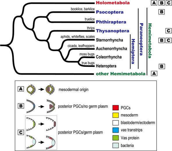
[14,42] and Additional file 2: Figure S2.Lin et al. EvoDevo 2014, 5:18 Page 12 of 16 http://www.evodevojournal.com/content/5/1/18 [14,42]: (1) the preformed germ plasm expressing Vas Evolution and development of germline specification in protein is not identified during oogenesis (S1/A1; S2/ aphids and other hemimetabolous insects A2); (2) segregation of the preformed germ plasm occurs The preformed germ plasm, a morphological feature of in the posterior region of syncytial blastoderm (S3/A3); the inheritance mode, has not been reported in any he- (3) PGCs form in the posterior blastoderm after cellular- mipterans apart from the aphids. For example, Pseudo- ization (S4/A4); (4) after germ band formation, PGCs coccus mcdanieli (mealybug), Lecanodiaspis pruinosa are located posteriorly to the germ band regardless of (false pit scale), and Icerya purchasi (cottony cushion its orientation in the egg chamber (S5/A5); (5) during scale) are scale insects that all belong to the same sub- germ band elongation, PGCs are aggregated and lo- order Sternorrhyncha as the aphids; however, in these cated extra-embryonically aside the elongating abdo- species germ plasm was undetectable using classical men (S6/A6); and (6) from katatrepsis onward, PGCs histological approaches and segregated germ cells were migrate toward the anteriormost region of egg cham- first identified adjacent to the posterior blastoderm bers and afterwards they migrate posteriorly (S7/A7). (Figure 7) [39]. In Oncopeltus fasciatus (milkweed bug), However, we find that migrating PGCs are already a true bug belonging to the suborder Heteroptera, how grouped into clusters prior to katatrepsis in the ovip- Vasa protein is distributed during early development has arous embryos (Figure 5G, H) but in the viviparous not been reported; nevertheless, mRNAs of 19 homo- embryos PGCs remain unseparated at the equivalent logues to Drosophila germline genes are not asymmetric- stage of development [42]. ally localized during early development, and formation of Figure 7 Phylogenetic distribution of mechanisms for germline specification within the Paraneoptera. In the phylogenetic tree we display four orders of the superorder Paraneoptera: Psocoptera (booklice, barklice), Phthiraptera (trulice), Thysanoptera (thrips), and Hemiptera (true bugs). For discussing germline specification within the Hemiptera, we present four suborders including Sternorrhyncha (aphids, whiteflies, scales), Auchenorrhyncha (cicadas, leafhoppers), Coleorrhyncha (moss bugs), and Heteroptera (true bugs). Squares A, B, and C designate mechanisms of germline specification: A, germ cells are derived from the mesoderm during mid/late embryogenesis; B, germ cells are segregated adjacent to the posterior blastoderm, which takes place after formation of the blastoderm, but a preformed germ plasm has not been identified; C, germline specification is driven by a preformed germ plasm located in the posterior pole (Drosophila) or slightly anterior to the posterior end (aphid) of the egg. With the exceptions of booklice (Liposcelis divergens; B) [62], thrips (Haplothrips verbasci; C) [63], and scale insects (Pseudococcus mcdanieli, Lecanodiaspis pruinosa, and Icerya purchase; B) [39], where study of germline segregation was carried out with traditional microscopic approaches, mechanisms of germline specification are deduced from expression data or functional analysis of the germline marker vas/Vas. Sources for molecular data: Hemiptera: aphids (Acyrthosiphon pisum; C) [14], true bugs (Oncopeltus fasciatus; B) [36,64]; other Hemimetabola: crickets (Gryllus bimaculatus; A) [35]; Holometabola: honeybees (Apis mellifera; A) [32], the flour beetle (Tribolium castaneum; B) [34], fruit flies (Drosophila melanogaster; C) [53]. Phylogenetic relationships of the Paraneoptera are based upon Grimaldi and Engel [65]; monophyletic clades within the Hemiptera are from Wheeler et al. [66] and Cryan and Urban [67]. Abbreviation: PGCs, primordial germ cells.
Lin et al. EvoDevo 2014, 5:18                                                                                                      Page 13 of 16
http://www.evodevojournal.com/content/5/1/18

germ cells is not affected by the knockdown of conserved        Additional files
germline markers [36].
   Taken together, the above cases suggest that absence          Additional file 1: Figure S1. Sequence alignment of the ApVas1-4
                                                                 proteins. (A) Alignment of the N-terminal sequences of ApVas1-4. The
of the preformed germ plasm is the norm in the                   aligned region includes divergent sequences as well as conserved amino
Hemiptera although how germ cells are specified in               acids in the helicase domain (green bar). Black boxes indicate amino
the other two hemipteran suborders Auchenorrhyncha and           acids identical to that of ApVas1; gray boxes indicate more than two
                                                                 amino acids that are identical between ApVas2-4 proteins. (B) Schematic
Coleorrhyncha remains unclear (Figure 7). Accordingly, the       comparison of ApVas1-4. Green boxes highlight the conserved helicase
common ancestor of hemipterans likely employed the               domain and blue bars mark the locations of the antigen sequences. The
induction mode to specify germ cells and the presence of         red bar beneath ApVas1 highlights the sequence region of the ligand
                                                                 that was used for affinity purifying the ApVas1 antibody.
germ plasm in aphids might be the result of independent
                                                                 Additional file 2: Figure S2. Expression of Apvas1-4 mRNA during
evolution within the Hemiptera. Nonetheless, we cannot           viviparous development. Germaria, oocytes, and embryos in the ovariole
exclude the possibility that the gain of a germ plasm in         were hybridized with the antisense riboprobes of Apvas1-4. Locations of
aphids occurred after the parthenogenetic phase was added        germ cells are indicated with hollow arrowheads. Anterior of egg
                                                                 chambers is to the left. (A-E) Apvas1. (A) In germaria and oocytes, Apvas1
to the sexual life cycle more than 200 million years ago         mRNA was expressed in the cytoplasm of nurse cells and oocytes. (B)
(reviewed in [68]), but how the inheritance mode was             During nuclear divisions, expression of Apvas1 remained in the cytoplasm
adopted in the sexual phase is still an unanswered question.     of the syncytial blastoderm. (C-E) Expression of Apvas1 was specifically
                                                                 restricted to the germ cells from blastoderm formation (stage 6) till late
   Though the inheritance mode is common neither in              embryogenesis. (F-J) Apvas2. (F, G) Expression of Apvas2 was undetectable
Hemiptera nor in other orders of the Hemimetabola, this          in germaria, oocytes, and syncytial blastoderm, but in the follicle cells
mechanism has also been reported in the mullein thrips           weak expression could be identified. (H, I) Except in the germ cells,
                                                                 transcripts of Apvas2 were evenly distributed in embryos before
Haplothrips verbasci, a member of the order Thysanoptera         katatrepsis. (J) In embryos after katatrepsis, expression of Apvas2 was
[63]. According to Heming, a putative germ (pole) plasm          ubiquitous including the germ-cell region. (K-O) Apvas3. (K, L) Expression
could be identified in the posterior end of the unfertilized     of Apvas3 was not detected in germaria, oocytes, and syncytia (including
                                                                 the follicle cells). (M, N) Like Apvas2 (panels H and I), uniform expression
egg and polar granules in the germ plasm can be inherited        of Apvas3 was detected except in the germ cells. (O) Expression patterns
by the newly segregated germ cells after fertilization in H.     of Apvas3 in late embryos are very similar to those of Apvas2 (panel J).
verbasci [63]. Taking together with the germ plasm identi-       (P-T) Apvas4. (P, Q) Expression of Apvas4 was detected in the germarial
                                                                 lumen, oocytes, and syncytial blastoderm. (R-T) Apvas4 was uniformly
fied in the pea aphid, we find that both cases - one in the      distributed in germ cells and somatic cells after blastoderm formation
Hemiptera (aphid) and one in the Thysanoptera (thrips) -         onward. Abbreviations: ab, abdomen; b, bacteria; cl, cephalic lobe; fc,
occur within the Paraneoptera, a monophyletic super-             follicle cells; g, germaria; h, head; nc, nurse cells; o, oocytes; on, oocyte
                                                                 nuclei; th, thorax. Scale bars: 20 μm in (A-C, F-H, K-M, P-R), 50 μm in (D, I,
order of insects and a sister group to the Holometabola          N, S) and 100 μm in (E, J, O, T).
(Figure 7) (reviewed in [65]). This implies that the inherit-    Additional file 3: Figure S3. Expression of Apvas1-4 mRNA in the
ance mode can be identified in derived orders of hemi-           oviparous ovarioles. (A-F) Apvas1. (A, B, C, F): antisense riboprobes; (D, E):
metabolous insects and the gain of a preformed germ              sense riboprobes as negative controls. In germaria and previtellogenic
                                                                 oocytes, Apvas1 mRNA was expressed in the cytoplasm of nurse cells and
plasm is achieved via independent evolution.                     oocytes (A). During vitellogenesis, expression of Apvas1 remained in the
                                                                 cytoplasm but the intensity of signals decreased as the egg chambers
                                                                 enlarged (B, C, F). (G-I) Apvas2. Expression of Apvas2 was detected in
Conclusions                                                      germaria (G), previtellogenic oocytes (G), and early vitellogenic oocytes
Identification of the preformed germ plasm in both ovip-         (H). In late vitellogenic oocytes (I), Apvas2 mRNA was preferentially
                                                                 expressed in the cortex of oocytes as well as the nuclei. (J-L) Apvas3.
arous and viviparous embryos of the pea aphid suggests           Expression of Apvas3 was detected in germaria (J), previtellogenic
that developmental plasticity of the germline-specification      oocytes (J), and early vitellogenic oocytes (K). However, it was almost
mode does not occur in the alternate reproductive cycles         undetected in late vitellogenic oocytes (L). (M-O) Apvas4. Expression of
                                                                 Apvas4 was detected in germaria (M), previtellogenic oocytes (M), early
in the aphids. This differs from the distinct developmental      vitellogenic oocytes (N), and late vitellogenic oocytes (O). Anterior of egg
programs known to regulate axis patterning in the asexual        chambers is to the left. Abbreviations: b, bacteria; -Ctrl, negative control;
and sexual phases. The preformed germ plasm in the pea           fc, follicle cells; g, germaria; gl, germarial lumen; nc, nurse cells; nn, nurse-
                                                                 cell nuclei; o, oocytes; on, oocyte nuclei. Scale bars: 100 μm.
aphid so far is the first maternal germ plasm identified
                                                                 Additional file 4: Figure S4. Expression of ApVas2-4 proteins in the
among hemimetabolous insects using molecular approa-             ovarioles dissected from the oviparous females. (A-C) ApVas2. (A) Signals
ches, and thus provides an opportunity for studying how a        of ApVas2 were not detected in the germaria and developing oocytes.
germ plasm is assembled in the Hemimetabola. It has              (B, C) During vitellogenesis, expression of ApVas2 was detected in the
                                                                 oocytes and follicle cells. Development of signals was performed using
been clear that a homolog of oskar (osk), a molecular            the chromogenic substrate 3,3’-Diaminobenzidine (DAB) Liquid Substrate
anchor known to localize Vas protein in the germ plasm           System (Sigma). (D-F) ApVas3. Expression of ApVas3 was detected in the
of Drosophila, does not exist in the pea aphid genome [1].       nuclei throughout oogenesis. (D) Signals of ApVas3 were barely detected
                                                                 in the germaria. (E) ApVas3 expression was restricted to the follicle cells
Finding molecular anchors for ApVas1 localization in the         during mid vitellogenesis. (F) In the late vitellogenic oocytes, expression
germ plasm will thus enable us to understand how an              of ApVas3 was almost undetectable. (G-I) ApVas4. (G) Expression of
‘osk-independent’ molecular networking for germ plasm            ApVas4 was identified in germaria and previtellogenic oocytes. (H)
                                                                 ApVas4 was preferentially expressed in the oocyte posterior but, unlike
assembly functions in less derived insects.
You can also read