Pharmacological activation of SERCA ameliorates dystrophic phenotypes in dystrophin-deficient mdx mice
←
→
Page content transcription
If your browser does not render page correctly, please read the page content below
Human Molecular Genetics, 2021, Vol. 30, No. 11             1006–1019

                                                                             doi: 10.1093/hmg/ddab100
                                                                             Advance Access Publication Date: 5 April 2021
                                                                             General Article

GENERAL ARTICLE

Pharmacological activation of SERCA

                                                                                                                                                         Downloaded from https://academic.oup.com/hmg/article/30/11/1006/6210386 by guest on 15 September 2021
ameliorates dystrophic phenotypes in
dystrophin-deficient mdx mice
Ken’ichiro Nogami1,2 , Yusuke Maruyama1,3 , Fusako Sakai-Takemura1 ,
Norio Motohashi1 , Ahmed Elhussieny1,4 , Michihiro Imamura1 ,
Satoshi Miyashita5 , Megumu Ogawa6 , Satoru Noguchi6,7 , Yuki Tamura8,9 ,
Jun-ichi Kira2 , Yoshitsugu Aoki1 , Shin’ichi Takeda10 and
Yuko Miyagoe-Suzuki1,*
1 Department  of Molecular Therapy, National Institute of Neuroscience, National Center of Neurology and
Psychiatry, Tokyo, Japan, 2 Department of Neurology, Neurological Institute, Graduate School of Medical
Sciences, Kyushu University, Fukuoka, Japan, 3 Department of Gene Regulation, Faculty of Pharmaceutical
Sciences, Tokyo University of Science, Noda, Chiba, Japan, 4 Department of Neurology, Faculty of Medicine,
Minia University, Minia, Egypt, 5 Department of Biochemistry and Cellular Biology, National Institute of
Neuroscience, National Center of Neurology and Psychiatry, Tokyo, Japan, 6 Department of Neuromuscular
Research, National Institute of Neuroscience, Translational Medical Center, National Center of Neurology and
Psychiatry, Tokyo, Japan, 7 Department of Clinical Development, Translational Medical Center, National Center
of Neurology and Psychiatry, Tokyo, Japan, 8 Graduate School of Health and Sport Science, Nippon Sport
Science University, Tokyo, Japan, 9 Research Institute for Sport Science, Nippon Sport Science University, Tokyo,
Japan and 10 National Center of Neurology and Psychiatry, Tokyo, Japan
*To whom correspondence should be addressed at: Department of Molecular Therapy, National Institute of Neuroscience, National Center of Neurology
and Psychiatry, 4-1-1, Ogawa-Higashi, Kodaira, Tokyo 187-8502 Japan. Tel: +81-42-346-1720, Fax: +81-42-346-1750, Email: miyagoe@ncnp.go.jp

Abstract
Duchenne muscular dystrophy (DMD) is an X-linked genetic disorder characterized by progressive muscular weakness
because of the loss of dystrophin. Extracellular Ca2+ f lows into the cytoplasm through membrane tears in
dystrophin-deficient myofibers, which leads to muscle contracture and necrosis. Sarco/endoplasmic reticulum Ca2+ -ATPase
(SERCA) takes up cytosolic Ca2+ into the sarcoplasmic reticulum, but its activity is decreased in dystrophic muscle. Here, we
show that an allosteric SERCA activator, CDN1163, ameliorates dystrophic phenotypes in dystrophin-deficient mdx mice.
The administration of CDN1163 prevented exercise-induced muscular damage and restored mitochondrial function. In
addition, treatment with CDN1163 for 7 weeks enhanced muscular strength and reduced muscular degeneration and

Received: February 17, 2021. Revised: March 30, 2021. Accepted: March 31, 2021
© The Author(s) 2021. Published by Oxford University Press. All rights reserved. For Permissions, please email: journals.permissions@oup.com
This is an Open Access article distributed under the terms of the Creative Commons Attribution Non-Commercial License (http://creativecommons.org/
licenses/by-nc/4.0/), which permits non-commercial re-use, distribution, and reproduction in any medium, provided the original work is properly cited.
For commercial re-use, please contact journals.permissions@oup.com

                                                                                                                                                1006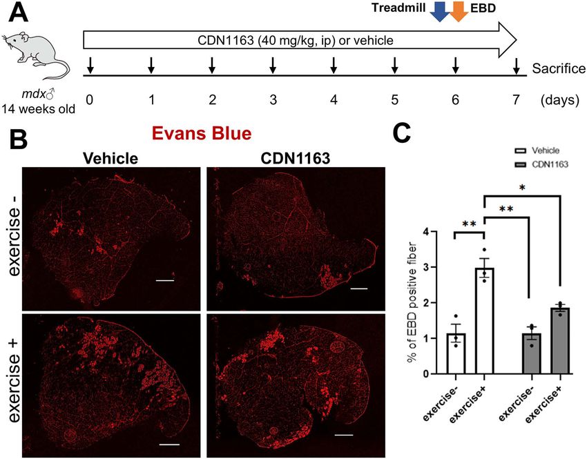
Human Molecular Genetics, 2021, Vol. 30, No. 11       1007

fibrosis in mdx mice. Our findings provide preclinical proof-of-concept evidence that pharmacological activation of SERCA
could be a promising therapeutic strategy for DMD. Moreover, CDN1163 improved muscular strength surprisingly in
wild-type mice, which may pave the new way for the treatment of muscular dysfunction.

Introduction                                                         skeletal muscles. Importantly, CDN1163 is a pan-activator for
                                                                     SERCA and is not isoform-specific (22). Recently, Lindsay et al.
Duchenne muscular dystrophy (DMD) is a severe, progressive,
                                                                     (27) reported that the incubation of isolated mdx muscle with
X-linked muscle-wasting disease caused by mutations in the
                                                                     CDN1163 for 30 min attenuated the loss of eccentric contraction-
DMD gene (1). Recent studies estimated the incidence of DMD
                                                                     induced force ex vivo, suggesting that CDN1163 may be applica-
to be 1:3500–1:10 000 newborn males (2,3). Clinical symptoms of
                                                                     ble for DMD treatment.
DMD appear at 2–3 years of age, and the loss of independent
                                                                         As a next step, we treated dystrophin-deficient mdx mice by
ambulation occurs by age 11–13 years. The mean age at death

                                                                                                                                          Downloaded from https://academic.oup.com/hmg/article/30/11/1006/6210386 by guest on 15 September 2021
                                                                     the administration of CDN1163. In this study, we demonstrated
due to respiratory and cardiac complications without ventilator
                                                                     the reduction of cytosolic Ca2+ level in mdx myotubes and whole
support is ∼19 years (4).
                                                                     tibialis anterior (TA) muscles isolated from mdx mice by phar-
    The DMD gene encodes the dystrophin protein, which local-
                                                                     macological activation of SERCA. We found that the adminis-
izes under the sarcolemma and forms a complex with gly-
                                                                     tration of CDN1163 for 1 week restored mitochondrial function
coproteins (the dystrophin-glycoprotein complex, DGC) at the
                                                                     and prevented exercise-induced muscular damage in mdx mice.
sarcolemma, linking the extracellular matrix and cytoskeleton
                                                                     We further revealed that treatment with CDN1163 for 7 weeks
(5,6). In the absence of DGC, the sarcolemma is disrupted during
                                                                     mitigated DMD-associated pathology and improved muscular
muscular contraction/relaxation and extracellular Ca2+ flows
                                                                     strength in mdx mice. Our findings provide preclinical proof-
into the cytoplasm through membrane tears (7). In addition,
                                                                     of-concept evidence that pharmacological activation of SERCA
Iwata et al. reported that a stretch-activated channel, TRPV2,
                                                                     ameliorates the dystrophic phenotypes of DMD model mice and
is translocated to the plasma membrane in dystrophic muscle
                                                                     could be a promising therapeutic strategy for DMD.
fibers and allows abnormal Ca2+ entry (8). More recent studies
further showed that stretch- and store-operated Ca2+ channels
are activated in dystrophic muscle and elevate cytoplasmic Ca2+      Results
levels (9,10).
                                                                     Administration of CDN1163 reduced cytosolic Ca2+
    Abnormally elevated cytosolic Ca2+ causes muscle contrac-
                                                                     levels in vitro and ex vivo
ture, mitochondrial dysfunction, activation of proteases, and
finally muscular degeneration and necrosis (11). In addition,        CDN1163 increases SERCA activity in vitro and in vivo in an
calcium regulation in the sarcoplasmic reticulum (SR) in the         allosteric manner (20,21,24). Importantly, a previous report
dystrophic muscle of mdx mice, an animal model of DMD, was           showed that treatment of isolated mdx muscle with CDN1163
shown to be chronically impaired (12,13). Ryanodine receptor         modestly attenuated loss of eccentric contraction-induced
1 (RyR1), a Ca2+ -release channel of the SR, was shown to be         force ex vivo (27). Therefore, we first determined whether
leaky due to S-nitrosylation by aberrantly activated nitric oxide    CDN1163 would reduce Ca2+ levels in the H2K-mdx cell line. The
synthase. FKBP12 (calstabin1) binds and stabilizes RyR1 but was      fluorescence intensity of Fluo-4 AM in H2K-mdx myotubes was
reported to be reduced from the RyR1 complex when RyR1 is S-         significantly reduced after 30 min of incubation with CDN1163
nitrosylated (12). Furthermore, the activity of Sarco/Endoplasmic    (Fig. 1A and B). In whole TA muscle dissected from mdx mice, the
Reticulum Ca2+ -ATPase (SERCA) was shown to be decreased             signal intensity was also significantly reduced after 30 min of
by upregulation of sarcolipin (SLN), an inhibitor of SERCA (13),     incubation with CDN1163 to the same level as that in wild-type
in dystrophic muscle. SERCA removes >70% of Ca2+ from the            C57BL/6J (BL6) mice (Fig. 1C and D). This result confirmed that
cytosol (14); therefore, the dysfunction of this Ca2+ pump of the    CDN1163 reduced cytosolic Ca2+ levels in dystrophin-deficient
SR further increases Ca2+ levels in the cytoplasm (11). These        mdx myofibers.
reports suggest that the restoration of Ca2+ regulation of the SR
to reduce the cytosolic Ca2+ level might be a good therapeutic
                                                                     Exercise-induced muscular damage was prevented
strategy for DMD.
                                                                     by 1-week administration of CDN1163
    Previous reports showed that SERCA overexpression by a
transgene (15–18) or enhancement through Hsp72 upregula-             Next, we daily injected CDN1163 (40 mg/kg) to mdx mice for 1
tion (19) rescued dystrophic phenotypes in mdx mice, sgcd−/−         week (Fig. 2A). We selected this dose based on a previous report
mice, and dystrophin- and utrophin-deficient dko mice; however,      that it effectively restored muscle mass and force in Sod−/−
whether pharmacological activation of SERCA is beneficial to         mice without harmful side effects (25). A higher dose of CDN1163
DMD phenotypes is still unknown. CDN1163 is an allosteric            was not applicable to mice, because CDN1163 is insoluble in an
SERCA activator that was identified in high-throughput screen-       aqueous solution. Before histological analysis, we forced vehicle-
ing assays and increases the activity at saturating (Ca2+ ) (Vmax)   and CDN1163-treated mdx mice to run for 60 min on a treadmill
(20,21). The therapeutic effects of CDN1163 have been shown in       to induce muscular damage and then intraperitoneally injected
various animal models of oxidative stress-related diseases, such     Evans blue dye (EBD) as previously described (28). Rupture of
as 6-OHDA-lesioned rats as a model of Parkinson’s disease (22),      the sarcolemma often causes uptake of this dye in dystrophin-
APP/PS1 mice as a model of Alzheimer’s disease (23), ob/ob mice      deficient myofibers (29). As expected, 1 h of running on the
as a model of diabetes (24), SOD1-deficient mice (25) and aging      treadmill caused a significant increase in EBD-positive fibers
mice (26).                                                           in the TA muscle of mdx mice (Fig. 2B and C). Importantly,
    SERCA1a is the major isoform in fast-twitch skeletal muscle,     the administration of CDN1163 significantly reduced EBD
and SERCA2a is expressed in cardiac, smooth, and slow-twitch         uptake into myofibers after treadmill exercise, indicating that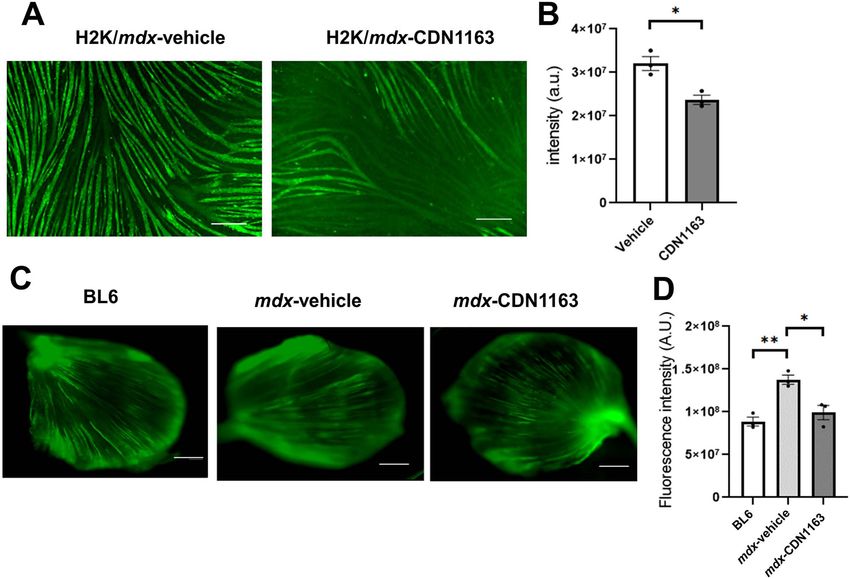
1008        Human Molecular Genetics, 2021, Vol. 30, No. 11

                                                                                                                                                                              Downloaded from https://academic.oup.com/hmg/article/30/11/1006/6210386 by guest on 15 September 2021
Figure 1. CDN1163 decreased cytosolic Ca2+ level in vitro and ex vivo. (A) Representative images showing cytosolic Ca2+ (Fluo-4 AM) of H2K-mdx myotubes established
from C57BL10/mdx mice after 30 min of treatment with vehicle or CDN1163. Scale bar, 200 μm. (B) Quantitative analysis of Fluo-4 AM signal intensity in (A). n = 3. (C)
Representative images showing cytosolic Ca2+ (Fluo-4 AM) in whole TA muscles dissected from wild-type C57BL/6J mice without treatment (BL6) or mdx mice after
30 min of treatment with vehicle (mdx-Vehicle) or CDN1163 (mdx-CDN1163). Muscles were incubated with Fluo-4 AM for 30 min. Scale bar, 1 mm. (D) Quantitative
analysis of Fluo-4 AM signal intensity (n = 3 mice per group) in (C). ∗ P < 0.01 by Student’s t-test (B) or ANOVA with the Tukey–Kramer test (D). Data are presented as the
means ± SEMs.

CDN1163 protected dystrophic muscle against exercise-induced                            mice treated with CDN1163 showed a significant increase in
damage.                                                                                 the maximal absorbance with a high-Ca2+ solution (Fig. 3B),
                                                                                        indicating that the administration of CDN1163 reversed the
                                                                                        mitochondrial swelling in mdx mice.
Mitochondrial respiratory function was restored                                            Next, we measured the glutamate malate-supported oxy-
by pharmacological SERCA activation                                                     gen consumption rate (OCR) and reactive oxygen species (ROS)
Sustained elevation of cytosolic Ca2+ levels was reported                               production from mitochondria. Previous reports showed that
to cause mitochondrial dysfunction by the formation of                                  cytosolic Ca2+ overload opens the mPTPs, leading to mitochon-
mitochondrial permeability transition pores (mPTPs) (30,31).                            drial depolarization, oxidative stress, inhibition of mitochondrial
Previous reports showed that mitochondria isolated from                                 ATP synthesis, and finally death of the myofibers in dystrophic
dystrophic mice were swollen because of mPTPs, leading to                               mice (11,30–34). Mitochondria isolated from the gastrocnemius
myofiber necrosis (15,30). To test whether CDN1163 prevents                             (GC) muscles of vehicle-treated mdx mice showed significantly
mPTP formation, we daily administrated CDN1163 (40 mg/kg) to                            lower OCR and higher production of ROS than those of BL6
mdx mice. After 1 week, freshly isolated mitochondria from each                         mice (Fig. 3C and D). The administration of CDN1163 signifi-
group were assessed. The baseline absorbance of mitochondria                            cantly increased OCR and reduced ROS production in mdx mice.
isolated from vehicle-treated mdx mice tended to be lower
than that of CDN1163-treated mdx mice or BL6 mice. This
suggests that mdx mitochondria are swollen at the baseline,
                                                                                        Administration of CDN1163 for 7 weeks decreased
though the difference was not statistically significant (Fig. 3A).                      muscular degeneration and fibrosis in mdx mice
The maximal absorbance change with a high-Ca2+ solution                                 To know the effects of CDN1163 on dystrophic phenotypes, we
(200 μm) for mitochondria isolated from the vehicle-treated mdx                         injected the compound into mdx mice for 7 weeks (Fig. 4A). Six-
mouse muscles was significantly smaller than that from the                              week-old male mdx mice were randomly divided into two exper-
BL6 mouse muscle. These results confirmed that mitochondria                             imental groups (n = 7/per group): the vehicle-injected group and
were swollen in mdx mouse muscles, as previously described                              the CDN1163-treated group. CDN1163 was injected intraperi-
(15,30). On the other hand, mitochondria isolated from mdx                              toneally at 40 mg/kg into mdx mice three times per week for 7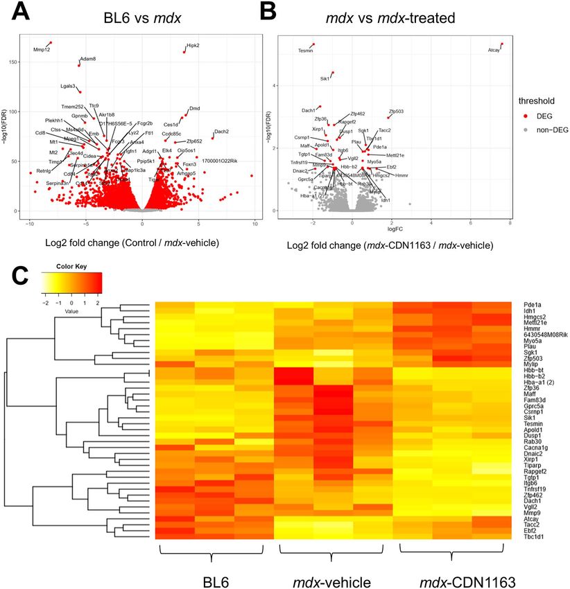
Human Molecular Genetics, 2021, Vol. 30, No. 11 1009
Downloaded from https://academic.oup.com/hmg/article/30/11/1006/6210386 by guest on 15 September 2021
Figure 2. One-week administration of CDN1163 decreased the uptake of EBD by damaged myofibers after treadmill exercise load. (A) Experimental design of EBD
injection and exercise load in 14-week-old male mdx mice administered vehicle or CDN1163. n = 3 mice/group. Just after treadmill exercise on day 6, EBD was injected
into the peritoneal cavity. (B) Representative images of EBD uptake (red fluorescence) in the TA muscles of mdx mice treated with vehicle or CDN1163 with or without
treadmill running (exercise + or −). Scale bar, 500 μm. (C) Percentages of EBD-positive fibers in the TA muscles in (B). Data are presented as the means ± SEMs. ∗ P < 0.05,
∗∗ P < 0.01 by ANOVA with the Tukey–Kramer test.
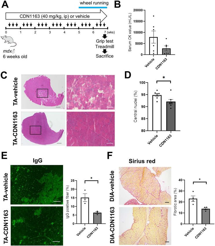
weeks. The administration of CDN1163 did not change the body significantly reduced collagen deposits in diaphragm (DIA) mus-
weight or organ weight (brain, liver, kidneys and spleen) of mdx cles, which is one of the most severely compromised muscles in
mice (Supplementary Material, Fig. S1A and B). mdx mice (Fig. 4F). Amelioration of the dystrophic histopathology
Mdx mouse muscle originally shows hypertrophy, which is of mdx mice was also obtained by 16 days of treatment in 14-
a compensatory effect because of muscular degeneration and week-old mdx mice (Supplementary Material, Fig. S2C), whereas
subsequent activation of satellite cells to regenerate myofibers that of 3-week-old mdx mice was unchanged (Supplementary
(18); however, neither muscular atrophy nor hypertrophy was Material, Fig. S3C).
observed in mdx muscle treated with CDN1163 compared with
the vehicle-injected group (Supplementary Material, Fig. S1C).
SERCA Ca2+ -ATPase activity was enhanced by CDN1163
The serum creatine kinase (CK) level, which is an indirect marker
without a change in protein expression of SERCA
of muscular damage, was reduced in treated mdx mice, although
this difference was not statistically significant (Fig. 4B). A sig- The expression level of SERCA1 (ATP2A1) in the GC muscle of
nificant reduction in CK level was observed in 14-week-old mdx mdx mice was the same as that of non-treated BL6 mice, and
mice treated daily with CDN1163 for 16 days (Supplementary not changed by the administration of CDN1163 (Fig. 5A). SERCA2
Material, Fig. S2A and B). On the other hand, a 16-day treatment (ATP2A2) expression was elevated in mdx mice compared with
of 3-week-old mdx mice tended to increase the CK level (Supple- non-treated BL6 mice, but not affected by CDN1163 treatment
mentary Material, Fig. S3A and B). Next, we examined whether (Fig. 5B). Importantly, the SERCA activity of mdx mice treated
CDN1163 administration ameliorated the histopathology in mdx with CDN1163 for 7 weeks was increased by 50% compared with
mouse muscle. Hematoxylin and eosin (H&E) staining of the TA that of vehicle-treated mdx mice (Fig. 5C).
muscle showed a reduction in the number of necrotic fibers We also examined the expression of SLN, which binds SERCA
(Fig. 4C). The presence of centrally located nuclei in the mouse and inhibits its function. The protein level of SLN in the DIA mus-
muscle indicates that the myofibers had been regenerated after cle of mdx mice was upregulated, as previously described (13),
damage. Importantly, the percentage of myofibers with cen- but unchanged in mdx mice treated with CDN1163 (Fig. 5D), sug-
trally nucleated fibers was significantly decreased by CDN1163 gesting that CDN1163 directly activates SERCA function without
treatment (Fig. 4D). The administration of CDN1163 also signifi- regulating the protein expression of SERCA nor SLN.
cantly reduced endogenous IgG accumulation in necrotic fibers A Ca2+ overload in dystrophic skeletal muscle is thought
(Fig. 4E). In addition, Sirius red staining showed that CDN1163 to activate Ca2+ -dependent proteases, such as calpain and1010 Human Molecular Genetics, 2021, Vol. 30, No. 11
increased grip strength in mdx mice (Fig. 6A). Treadmill running
time until exhaustion tended to be increased by the administra-
tion of CDN1163 (Fig. 6B). In the voluntary wheel running test,
there was no difference in total activity between the vehicle-
injected and CDN1163-treated groups (Fig. 6C). In addition,
CDN1163 administration increased both the specific twitch and
tetanic force of TA and GC muscles isolated from mdx mice
(Fig. 6D). Furthermore, the improvement of grip strength in 14-
week-old mdx mice and 3-week-old mdx mice was observed even
after 16 days of daily treatment with CDN1163 (Supplementary
Material, Figs S2D and S3D). Treadmill running time significantly
increased in 3-week-old mdx mice, though there was no
difference in 14-week-old mdx mice (Supplementary Material,
Figs S2E and S3E).
Downloaded from https://academic.oup.com/hmg/article/30/11/1006/6210386 by guest on 15 September 2021
We also performed the same set of experiments using BL6
mice to investigate the effects of pharmacological activation of
SERCA on muscular function in wild-type mice. Interestingly, the
administration of CDN1163 (40 mg/kg) for 16 days significantly
increased the grip strength and the specific muscle force even
in BL6 mice, although treadmill running time did not change
significantly (Fig. 6E–G). The performance of mdx mice treated
with CDN1163 was lower than that of vehicle-injected BL6 mice
(Fig. 6B, F, D and G).
Genes associated with inflammation were
downregulated by CDN1163 administration
To further clarify the mechanisms by which CDN1163 amelio-
rated the dystrophic phenotypes of mdx mice, we examined
the gene expression profiles using the TA muscles of 14-week-
Figure 3. Pharmacological activation of SERCA restored mitochondrial swelling, old mdx mice treated with vehicle or CDN1163 (10 mg/kg) daily
OCR and ROS production in mdx mice. (A) Absorbance at 540 nm by mitochondria for 16 days by RNA sequencing (RNA-seq). Many genes were
in response to an exogenous high-Ca2+ solution (200 μm CaCl2). After daily differentially expressed in mdx mouse muscle compared with
administration of CDN1163 to 14-week-old male mdx mice for 1 week, mito-
non-treated wild-type muscle (Fig. 7A). In contrast, only 40 genes
chondria were isolated from the TA and GC muscles. Absorbance was measured
before and 10 min after Ca2+ addition. A decrease in absorbance indicated
were significantly altered of which 15 were upregulated and
swelling of the mitochondria. (B) The maximal change in absorbance at 540 nm 25 were downregulated by CDN1163 treatment in mdx mice
by mitochondria in response to high external Ca2+ solution. (C and D) The OCR (FDR < 0.05 and log2-fold change > 0.5, Fig. 7B). We found that
(OCR) and ROS production in mitochondria isolated from BL6 and mdx mice several genes associated with inflammation (Matrix metallopro-
treated with vehicle or CDN1163. Data are presented as the means ± SEMs. teinase 9 (Mmp-9), Il-1β, Ptgs2, and CXC chemokine family) were
∗ P < 0.05, ∗∗ P < 0.01, ∗∗∗ P < 0.001 by ANOVA with the Tukey–Kramer test.
downregulated by CDN1163. There was a significant difference
in the expression of Mmp-9 (Fig. 7B and C). Mmp-9 is involved
caspase-3, and contribute to muscular degeneration in DMD in tissue remodeling, inflammation, and interstitial fibrosis in
muscles (11). Therefore, we assessed calpain activity in mdx many diseases (35,36). Importantly, Mmp-9 is upregulated in
muscles after CDN1163 administration for 7 weeks. Calpain mdx mice, and the inhibition of Mmp-9 improves myopathy and
activity in GC muscles tended to be lower in CDN1163-treated myofiber regeneration in mdx mice (37).
mdx muscle than in vehicle-treated mdx muscle, but the effects Increased cytosolic Ca2+ levels were reported to induce fast-
were not large enough to explain the therapeutic effects of to-slow fiber transformation (38–40). Dusp1 and Vgll2, which pro-
CDN1163 on the mdx dystrophic phenotype (Supplementary mote fast-to-slow fiber-type switching (41–43), were significantly
Material, Fig. S4A). Caspase-3 is also a key protease that leads downregulated; however, the expression of myosin heavy chain
to apoptosis. The protein level of caspase-3 was slightly but genes (Myh1, Myh2, Myh4, Myh7) was not significantly altered
significantly upregulated in mdx muscle; however, it was not (data not shown). Furthermore, the percentage of type I and
decreased by the administration of CDN1163 (Supplementary type IIa fibers in the GC/plantaris muscle of mdx mice treated
Material, Fig. S4B). Technically, however, it was difficult to with CDN1163 for 7 weeks were not different from those in
accurately measure calpain activity in homogenized tissue vehicle-treated mdx mice (Supplementary Material, Fig. S5).
samples, because artificial activation of calpains might occur.
Therefore, we cannot exclude the possibility that CDN1163
changed calpain activity.
Discussion
In this study, we revealed that pharmacological activation of
SERCA attenuated dystrophic phenotypes in mdx mice. We
Pharmacological activation of SERCA improved in vivo
demonstrated that the administration of CDN1163 indeed
exercise capacity and contraction force of isolated
decreased the cytosolic Ca2+ level in vitro and ex vivo. Notably,
muscle ex vivo 1-week administration of CDN1163 reduced EBD uptake by
We further assessed in vivo muscle function in mdx mice after damaged myofibers after exercise load. OCR and ROS production
7 weeks of CDN1163 administration. CDN1163 significantly in isolated mitochondria were also restored after the treatment.Human Molecular Genetics, 2021, Vol. 30, No. 11 1011
Downloaded from https://academic.oup.com/hmg/article/30/11/1006/6210386 by guest on 15 September 2021
Figure 4. Pharmacological activation of SERCA decreased muscular degeneration and fibrosis in mdx mice. (A) Experimental design for CDN1163 administration. Male
mdx mice (6-week-old, n = 7 per group) were randomly selected and treated with vehicle or CDN1163 (40 mg/kg BW) three times per week for 7 weeks. ip: intraperitoneal
injection. (B) Quantitation of serum CK levels (n = 6 per group). (C) Representative H&E staining of TA muscles from mdx mice treated with vehicle (Vehicle-TA) or CDN1163
(CDN1163-TA) for 7 weeks. Scale bars, 500 μm (left) and 100 μm (right). (D) Percentage of myofibers with central nuclei (n = 6 mice per group). (E) Representative images
of endogenous IgG detected with Alexa 488-labeled anti-mouse IgG antibody in TA muscles of vehicle-treated (Vehicle-TA) or CDN1163- treated mice (CDN1163-TA).
Scale bar, 100 μm. Quantitative analysis of IgG positive fibers in TA muscle (right panel, n = 3 mice per group). (F) Representative images of Sirius red staining of the
DIA muscles of vehicle-treated (Vehicle-DIA) or CDN1163-treated mice (CDN1163-DIA). Scale bar, 100 μm. Quantitative analysis of the fibrotic area in the DIA muscle
(n = 4 mice per group). We injected DMSO and Tween-80 at a final concentration of 10% v/v each into all experimental mice in vehicle group. Data are presented as the
means ± SEMs. ∗ P < 0.05 by Student’s t-test.
Our findings suggest that the therapeutic effects of CDN1163 on The expression of SLN, which binds SERCA and inhibits its
mdx pathophysiology can partly be explained by the restoration function, did not change in mdx mice treated with CDN1163,
of mitochondrial function, because previous reports showed that suggesting that CDN1163 directly activated SERCA function
the mitochondrial dysfunction due to cytosolic Ca2+ overload without regulating the protein expression of SLN. A previous
in dystrophic mice is related to dystrophic phenotypes (11,30– report showed that allosteric SERCA activators directly perturbed
34). In particular, recovered mitochondrial function might have the structure of SERCA and altered SERCA-phospholamban FRET
positive effects on EBD uptake after exercise load because as a result (20). In this study, we did not examine the effects
mitochondria are indispensable for cell membrane repair (44). of CDN1163 on the interaction between SERCA and SLN, but1012 Human Molecular Genetics, 2021, Vol. 30, No. 11
Downloaded from https://academic.oup.com/hmg/article/30/11/1006/6210386 by guest on 15 September 2021
Figure 5. SERCA Ca2+ -ATPase activity was enhanced by the administration of CDN1163. (A and B) Representative western blot analyses of SERCA1 (A) and SERCA2 (B)
in GC muscles of non-treated BL6 and mdx mice treated with vehicle or CDN1163 for 7 weeks. Quantitation of the signal intensities of SERCA1 (A) and SERCA2 (B) are
also shown. n = 3 mice per group. GAPDH was used as a loading control. (C) Ca2+-ATPase activity in enriched SR fractions isolated from the GC muscles of mdx mice
treated with vehicle or CDN1163 for 7 weeks. n = 4 mice per group. (D) Western blot analysis and quantification of the signal intensities of SLN in the DIA muscles of
non-treated BL6 and mdx mice treated with vehicle or CDN1163 for 7 weeks. n = 3 mice per group. GAPDH was used as a loading control. Data are presented as the
means ± SEMs. ∗ P < 0.05, ∗∗ P < 0.01, ∗∗∗ P < 0.001 by ANOVA with the Tukey–Kramer test (A, B and D) or Student’s t-test (C).
it is possible that CDN1163 changed the binding of SLN to DMSO, which itself has anti-inflammatory effects (46). Neverthe-
SERCA. less, the therapeutic effects of CDN1163 were observed in each
Treatment with CDN1163 for 7 weeks successfully decreased experiment even with the anti-inflammatory effects.
muscular degeneration. We also found that CDN1163 signifi- Previous studies showed that transgene-mediated SERCA
cantly reduced fibrosis in the DIA muscle, which represents overexpression or indirect activation through Hsp72 induction
one of the most severely compromised muscles in mdx mice. ameliorated dystrophic phenotypes in mdx and Sgcd−/− mice
These findings indicate that pharmacological activation of (15–19). Mázala et al. (18) showed that transgenic SERCA
SERCA may be a promising therapeutic strategy for DMD. In overexpression reduced the weights of the TA, soleus, plantaris,
terms of elevation of the serum CK level in 3-week-old mdx and extensor digitorum longus muscles in mdx mice. Muscular
mice treated with CDN1163, there is a possibility that increased hypertrophy is commonly observed in mdx mice, and the authors
muscular strength due to CDN1163 treatment rather leads considered that upregulated SERCA1 rescued pathological
to muscular damage in 3-week-old mdx mice because they hypertrophy in mdx mice. In the current study, however, the
are at the severe early necrosis-degeneration stage of mdx administration of CDN1163 did not reduce muscular weight,
mice (45). even with a 50% increase in SERCA Ca2+ -ATPase activity.
Grip strength and contraction force of isolated muscles were Similarly, the therapeutic effects of CDN1163 were milder than
increased by CDN1163 treatment. Interestingly, the administra- those in SERCA1-transgenic mice. Previous reports showed a 2–
tion of CDN1163 significantly improved the muscular function 3-fold increase in SR Ca2+ -ATPase activity and SR Ca2+ uptake
in wild-type mice also. This suggests that SERCA activation has in SERCA1-overexpressing mice (15,18). It is likely that the high
the potential for enhancing the muscular function, even in wild- levels of transgenic SERCA1 expression from the neonatal period
type muscle, by increasing the speed of the return to baseline had much stronger effects on mdx mice than pharmacological
of a Ca2+ transient. In this study, we dissolved CDN1163 in activation of SERCA.Human Molecular Genetics, 2021, Vol. 30, No. 11 1013
Downloaded from https://academic.oup.com/hmg/article/30/11/1006/6210386 by guest on 15 September 2021
Figure 6. Pharmacological activation of SERCA increased muscular strength in mdx mice and C57BL/6J mice. (A and B) Grip strength measurement (A) and time to
exhaustion in the treadmill test (B) in mdx mice treated with vehicle or CDN1163 for 7 weeks (n = 7 mice per group). (C) Voluntary wheel running test (n = 4 mice per
group). The number of wheel revolutions was recorded over a week, and the total wheel count per week is shown. (D) Specific twitch and tetanic force of TA and GC
muscles (n = 4 mice per group). (E and F) Grip strength measurement (E) and time to exhaustion in the treadmill test (F) in BL6 mice treated with vehicle or CDN1163 for
16 days (n = 3 mice per group). (G) Specific twitch and tetanic force of isolated TA and GC muscles of BL6 mice (n = 3 mice per group). We injected DMSO and Tween-80
at a final concentration of 10% v/v each into all experimental mice in vehicle group. Data are presented as the means ± SEMs. ∗ P < 0.05, ∗∗ P < 0.01, by Student’s t-test.
To further clarify the mechanisms by which CDN1163 APMK signaling and suppressed MEF2C and p38 MAPK signaling.
decreased muscular damage in mdx mice, we assessed the Based on the findings, the authors speculate that CDN1163
gene expression profiles by RNA-seq. Unexpectedly, a very small mitigated age-related muscular atrophy and weakness, at
percentage of the genes were differentially expressed between least in part by restoring energy production and muscular
vehicle- and CDN1163-treated mdx muscles; however, several regeneration activity. In dystrophic mdx mice, however, RNA-
genes associated with inflammation were downregulated seq analysis did not suggest that CDN1163 altered these
by CDN1163, indicating that CDN1163 treatment prevented signaling pathways. This could be because we treated young
muscular degeneration. Because CDN1163 activates SERCA2b mdx mice, which actively regenerate injured skeletal muscle.
which is expressed in macrophages, improvement of the Unexpectedly, the expression level of many genes in mdx
function and viability of macrophages may also contribute muscle treated with CDN1163 did not show intermediate
to the downregulation of the genes associated with inflam- values between vehicle-treated mdx mice and wild-type mice.
mation. The expression of genes related to fast-to-slow fiber- This might be because CDN1163 did not completely suppress
type conversion was also significantly downregulated by the muscular degeneration. Many myofibers were still in active
administration of CDN1163, but the conversion of fiber type degeneration/regeneration cycles, and regenerating myofibers
was not evident in our study. Recently, Qaisar et al (26). might show different gene expression patterns from mature
showed that CDN1163 treatment of 16-month-old male mice intact myofibers. Therefore, it may look like an unfavorable gene
for 10 months mitigated age-related muscular atrophy and expression pattern.
weakness. Interestingly, ingenuity upstream regulator analysis Another therapeutic strategy focuses on RyR1, a Ca2+ -release
suggested that CDN1163 activated PGC1-α, UCP1, HSF1, and channel on the SR. Compounds that stabilize the binding of1014 Human Molecular Genetics, 2021, Vol. 30, No. 11
Downloaded from https://academic.oup.com/hmg/article/30/11/1006/6210386 by guest on 15 September 2021
Figure 7. RNA-seq gene expression analysis of mdx mouse muscle after the administration of CDN1163 for 16 days. (A) Volcano plot showing differential expression of
all genes between BL6 (non-treated) and vehicle-treated mdx mice. (B) Volcano plot showing differential expression of all genes between mdx mice treated with vehicle
and CDN1163 (1.0 mg/ml in 10% DMSO/PBS, 10 mg/kg BW) for 16 days (14 weeks old, n = 3 per group). Top 50 DEGs with FDR < 0.05 and log2 fold change (log2FC) > 0.5
are indicated. (C) Heat map illustrates DEGs in non-treated BL6 mice, mdx mice treated with vehicle, and treated with CDN1163. The heat map is based on normalized
expression levels of significantly changed genes with the Benjamini–Hochberg adjusted P-value < 0.05 between vehicle and CDN1163-treated mdx mice. Z-scores for
the DEGs were transformed from the TMM value by using the gene filter package and visualized as a heat map by using the gplots package.
RyR1 to calstabin are called rycals, and a previous report showed dystrophic phenotypes, such as the double knock-out mdx
that rycals reduced RyR1 Ca2+ leakage and improved dystrophic mouse or the dystrophic rat, remain to be performed.
phenotypes in mdx mice (12). The combination of CDN1163 and
rycals might have synergistic therapeutic effects on dystrophic
phenotypes. Materials and Methods
CDN1163 would have effects on cardiac muscle which
Animals
express SERCA2a. Further investigations, including assessments
of the safety of CDN1163 and its therapeutic effects on the C57BL/6J (BL6) mice were purchased from Nihon Crea (Tokyo,
heart, ideally using other dystrophic murine models with severe Japan). mdx mice with a BL6 background were a generous giftHuman Molecular Genetics, 2021, Vol. 30, No. 11 1015
from Dr Sasaoka (Niigata University). The experimental mice of stimulation pulses (83 Hz) was recorded. Specific twitch and
were 6–14 weeks old. Only male mice were used in the study. The tetanic forces were calculated by normalizing to the physiolog-
mice were bred and kept in plastic cages in a 12:12-h light–dark ical cross-sectional area, which was determined by the ratio of
cycle at the specific pathogen-free animal facility at the National muscular weight to L0 and the density of mammalian skeletal
Institute of Neuroscience, NCNP. The mice were allowed free muscle.
access to food and drinking water. Body weight was measured
weekly during the experiment. Our experimental design and
procedures were based on the three R principle (replacement,
Grip strength measurements
reduction and refinement) and approved by the Experimental
Animal Care and Use Committee of the National Institute of The grip strength of the combined forelimbs and hind limbs
Neuroscience of the NCNP (approval ID: 2018041). The mice were was recorded using a grip strength meter (Muromachi Kikai Co.,
randomly assigned to experimental groups. Ltd; model MK-380M). The grip strength meter was positioned
horizontally, and the mouse was held by its tail and allowed
to securely grip the metal mesh. After the mouse obtained a
Downloaded from https://academic.oup.com/hmg/article/30/11/1006/6210386 by guest on 15 September 2021
CDN1163 solid grasp of the metal mesh, the mouse was pulled backward
CDN1163, an allosteric SERCA activator, was purchased from parallel to the device. The force that was applied to the mesh
Sigma-Aldrich (St. Louis, MO). CDN1163 was reported to be selec- at the time of release was recorded as the peak grip strength.
tive against > 160 off-targets (23). CDN1163 showed an accept- The measurement was repeated three times, and the average
able pharmacokinetic profile in mice (20,21), and membranes force was determined for each mouse. Grip strength values were
showed high permeability to CDN1163. Vehicle (10% DMSO, 10% normalized to the body weight (kg) of each mouse (mg/kg BW).
Tween-80 in PBS) or CDN1163 (1.0 mg/ml stock solution, final
dose of 40 mg/kg) was intraperitoneally injected into mdx mice.
Treadmill running test
2+
Cell culture and intracellular Ca measurements The mice were placed in individual lanes of an electrically
driven 10-lane treadmill (MK-680, Muromachi, Tokyo, Japan) and
Murine H2K-mdx myoblasts (47) were seeded in plates coated
acclimated to the treadmill at a speed of 5 m/min for 30 min
with matrigel (Corning, Corning, NY, USA) at 8 × 10⁴ cells/well in
twice a week before the experiment. The test began at a speed
growth medium. After 2 days, they were differentiated using 5%
of 5 m/min for 5 min and gradually increased by 1 m/min every
horse serum-containing differentiation medium. Intracellular
minute until exhaustion. Exhaustion was defined as the time
Ca2+ was monitored using Fluo-4 AM as previously described
when the mouse could no longer run despite repeated gentle
(48). Briefly, whole fresh TA muscles and H2K-mdx myotubes
nudges (52).
were incubated in buffer containing 4 μm Fluo-4 AM (Dojindo,
Kumamoto, Japan), 140 mm NaCl, 5 mm KCl, 2.5 mm CaCl2 , 1 mm
MgCl2 , 10 mm HEPES, and 10 mm glucose (pH 7.4) combined
with vehicle or CDN1163 (100 μm) for 30 min at room tempera- Voluntary exercise test
ture. The fluorescence intensity was evaluated using a BZ-X810
Voluntary exercise in an individual cage for 1 week was recorded
fluorescence microscope (Keyence, Osaka, Japan).
using a running wheel (SW-15, Melquest) as described previously
(53). The mice were kept in plastic cages with a running wheel
Serum CK level for 2 weeks. The first week was used to acclimate the mice to the
wheel. Voluntary exercise over the second week was quantified
Mice were anesthetized with isoflurane, and blood was taken via
as the average of the total number of wheel revolutions per day.
the inferior vena cava and kept at room temperature overnight.
The serum was then collected from the blood by centrifugation
at 3000 × g for 10 min. The serum level of CK, an indicator of
muscular damage, was assayed using the Fuji Drychem sys- Tissue preparation
tem (Fuji Film Medical Co. Ltd, Tokyo, Japan) according to the Mice were sacrificed by cervical dislocation, and the body was
manufacturer’s instructions. weighed. The TA, GC, EDL, SOL, DIA, and H muscles and organs
(brain, liver, kidneys and spleen) were collected using standard
dissection methods, and their weights were measured. Muscles
Measurement of muscle contraction force ex vivo
and organs were then quickly frozen in isopentane cooled by
The twitch and tetanic force of TA and GC muscles were mea- liquid nitrogen for histological analysis and RNA and protein
sured using an in vitro muscle test system (Aurora Scientific isolation. Frozen samples were stored at −80◦ C.
Inc., Aurora, ON, Canada) as previously described (49). Briefly,
the entire TA and GC muscles were dissected and kept in a
buffer containing 137 mm NaCl, 24 mm NaHCO3 , 5 mm KCl, 2 mm
CaCl2 , 1 mm MgSO4 , 11 mm glucose, 1 mm NaH2 PO4 (pH 7.4), and
Histology and IgG staining
0.025 mm d-tubocurarine at 25◦ C with continuous perfusion with The TA and DIA muscles were sectioned at 8 μm using a CryoStar
95% O2 –5% CO2 gas (50,51). After a muscle was placed in the NX70 cryostat (Thermo Fisher) and stained with H&E or Sirius
bath, a single 0.4-msec stimulation pulse was used to generate a red. IgG staining was performed to detect degenerating fibers as
twitch force, and the length of the muscle was carefully adjusted previously described (54). In brief, the cryosections were prein-
to determine the optimal muscular length (L0 ) to obtain the cubated with 5% BSA in PBS for 1 h, followed by incubation with
maximal twitch force. Tetanic force was obtained by stimulation Alexa 488-conjugated goat anti-mouse IgG antibody (A-11029,
of the muscle at L0 for a period of 300 msec (TA) or 600 msec (GC) Invitrogen, Carlsbad, CA). Fluorescence images were obtained
with a series of 3-msec pulses. The force developed during trains using a BZ-X810 fluorescence microscope (Keyence).1016 Human Molecular Genetics, 2021, Vol. 30, No. 11
Acute exercise load and EBD uptake Isolation of mitochondria and measurement of OCR
After the daily administration of vehicle or CDN1163 (40 mg/kg) After daily administration of the vehicle or CDN1163 (40 mg/kg)
for 1 week, muscular damage was induced by a modified tread- for 1 week, GC muscles were collected. The mitochondrial frac-
mill protocol as previously described (26). Briefly, the exercise tion was obtained by differential centrifugation as previously
load began at a speed of 5 m/min for 5 min and gradually described (55). Immediately after tissue collection, GC muscles
increased by 1 m/min every minute until the speed reached were placed in buffer [PBS, 10 mm EDTA (349–01885, Dojindo
10 m/min. The mice were kept on the treadmill for 30 min under Molecular Technologies, Kumamoto, Japan), pH 7.4]. Tissues were
repeated gentle nudges. After a 10-min interval, the session was well-minced using scissors, supplemented with 0.025% trypsin
repeated. All mice were able to carry out the test until the end of (209–16 941, Fujifilm Wako Pure Chemical Corporation), and then
the experiment. After treadmill exercise, the mice were injected incubated for 5 min on ice. To remove the trypsin, tissue suspen-
intraperitoneally with EBD (10 mg/ml, 0.1 ml/10 g BW). The next sions were centrifuged at 200 g for 5 min at 4◦ C, and then super-
day, the mice were sacrificed, and their GC muscles were cut into natants were discarded. The tissue pellets were resuspended
cryosections to observe EBD uptake (red fluorescence) by degen- in buffer [50 mm MOPS (194 837, BM Bio Japan, Tokyo, Japan),
Downloaded from https://academic.oup.com/hmg/article/30/11/1006/6210386 by guest on 15 September 2021
erating fibers. Images were taken with a BZ-X810 fluorescence 100 mm KCl (163–03545, Fujifilm Wako Pure Chemical Corp.),
microscope (Keyence, Osaka, Japan). 1 mm EGTA (342–01314, Dojindo Molecular Technologies), 5 mm
MgSO4 (83585–0401, Junsei Chemical, Tokyo, Japan) and 2.0 g/L
BSA (015–21 274, Fujifilm Wako Pure Chemical), pH 7.1] and were
Western blot analysis homogenized by 20 strokes using a Potter glass homogenizer.
The tissue homogenates were centrifuged at 500 g for 10 min
The protein was extracted with a sample buffer containing 2%
at 4◦ C, and the supernatants derived were collected. The super-
SDS, 15% glycerol, 125 mm Tris–HCl, 1 mm dithiothreitol, and
natants were centrifuged at 10000 g for 10 min at 4◦ C to obtain
cOmplete protease inhibitor cocktail (11 873 580 001; Roche, Indi-
mitochondrial pellets. The mitochondrial pellets were washed
anapolis, IN, USA). The protein lysate was then denatured at
in buffer (50 mm MOPS, 100 mm KCl, 1 mm EGTA and 5 mm
100◦ C for 5 min and centrifuged at 15 000 rpm for 5 min. After the
MgSO4) and resuspended in buffer (10 mm Tris, 30 mm KCl, 10 mm
supernatant was collected, the protein concentration was deter-
KH2PO4, 5 mm MgCl2, 1 mm EGTA and 2.5 g/L BSA, pH 7.2). The
mined using a protein assay (Bio-Rad Laboratories, Inc., Hercules,
protein concentration of each was determined using the BCA
CA, USA) with BSA as a standard. The protein lysate was sepa-
method and then adjusted to 2.0 mg/ml.
rated on an SDS-polyacrylamide gel and electrically transferred
The mitochondrial OCR was measured as previously
to polyvinylidene difluoride membranes (Millipore, Darmstadt,
described with minor modifications (55). Briefly, freshly isolated
Germany). The blots were then incubated with primary antibod-
mitochondria (20 μg) were incubated in a reaction buffer (105 mm
ies at 4◦ C overnight. The next day, the membranes were incu-
potassium-MES, 10 mm Tris, 30 mm KCl, 10 mm KH2PO4, 5 mm
bated with secondary antibodies. Signals were detected using
MgCl2, 1 mm EGTA, 2.5 g/L BSA, pH 7.2). Complex I-driven state III
ECL Prime Western Blotting Detection Reagent (GE Healthcare,
(ATP synthesis coupled) respirations were stimulated by adding
Buckinghamshire, UK; #RPN2232) and a ChemiDoc MP imaging
10 mm glutamate (070–00502, Fujifilm Wako Pure Chemical
system (Bio-Rad, Hercules, CA, USA). Data were analyzed using
Corp.), 2 mm malate (138–07512, Fujifilm Wako Pure Chemical
Image Lab 6.0 (Bio-Rad).
Corp.) and 2.5 mm ADP (306–50 501, Fujifilm Wako Pure Chemical
Corp.). Mitochondrial oxygen consumption was measured using
a Tecan Spark multimode plate reader (Spark 20 M, Tecan,
Antibodies Männedorf, Switzerland) with an oxygen-monitoring 96-well
Antibodies against SERCA1 (#12293) and SERCA2a (#4388) were microplate (OP96C, PreScan Precision Sensing, Regensburg,
purchased from Cell Signaling Technologies (Beverly, MA, USA). Germany) (excitation (Ex): 560 (20) nm; emission (Em): 670
Anti-caspase-3 antibody (#67341A) was obtained from BD (25) nm). The relative change in fluorescence per minute was
Pharmingen (San Diego, CA, USA). BA-F8 (type I MHC), SC- measured, and this was converted to the OCR according to
71 (type IIa MHC), and BF-F3 (type IIb MHC) were obtained the manufacturer’s instructions. To normalize the efficiency
from the Developmental Studies Hybridoma Bank (Iowa City, of mitochondrial isolation and purification, citrate synthase
IA, USA). An antibody against SLN was a kind gift from Dr (CS) activity in isolated mitochondria was measured as follows:
Muthu Periasamy (University of Central Florida). HRP-conjugated 1 μg of mitochondrial protein was mixed with reaction buffer
secondary antibodies (anti-rabbit IgG, horseradish peroxidase- [100 μm DTNB (346–08551, Dojindo Molecular Technologies),
linked F(ab )2 fragment; #NA9340 V) were purchased from GE 300 μm acetyl-CoA (00546–54; Nacalai Tesque, Kyoto, Japan),
Healthcare. HRP-conjugated rabbit anti-goat IgG (H + L) antibody 50 μm oxaloacetate (25804–81, Nacalai Tesque)] in a 96-well plate.
(#611620) was obtained from Invitrogen (Carlsbad, CA, USA). Absorbance changes at 412 nm/min were calculated. The OCR
was normalized to CS activity.
Mitochondrial swelling assay
After the daily administration of vehicle or CDN1163 (40 mg/kg)
Mitochondrial ROS emission kinetics
for 1 week, mitochondria were isolated from the GC muscles of Mitochondrial ROS emission was measured as previously
mdx mice. Light scattering from the isolated mitochondria was described with minor modifications (56). Briefly, mitochondria
measured using 200 μg of mitochondria suspended in 400 μL of were incubated in a black 96-well plate with mitochondrial
isolation buffer, and 200 μm CaCl2 was used to induce mitochon- respiration buffer and 5 μm Amplex Red (Thermo Fisher
drial swelling and shrinking as previously described (15). The Scientific, Waltham, MA, USA), 1 U/ml Horseradish peroxidase
change in absorbance at 540 nm for 10 min after the addition (169-10791, Fujifilm Wako Pure Chemical Corp.), and 5 U/ml
of CaCl2 was measured by a plate reader (BioTek, Winooski, superoxide dismutase (192-11281, Fujifilm Wako Pure Chemical
VT, USA). Corp.). The kinetics of ROS emission were assessed under stateHuman Molecular Genetics, 2021, Vol. 30, No. 11 1017
3 respiratory conditions through the addition of 2.5 mm ADP, Statistics
10 mm glutamate and 2 mm malate. The relative changes in
Data were analyzed and plotted using GraphPad Prism 8 soft-
fluorescence per minute were measured using a fluorescence
ware. All values are expressed as the means ± standard error of
plate reader (Spark 20 M, Tecan) (Ex: 560 [20] nm; Em: 620
mean (SEMs). Statistical differences were assessed by unpaired
[20] nm). The relative change in fluorescence per minute was
Student’s t-test or one-way analysis of variance (ANOVA) with
measured and normalized to OCR.
Tukey–Kramer post hoc analysis. Differences with probabilities1018 Human Molecular Genetics, 2021, Vol. 30, No. 11
the dystrophin era. Proc. Jpn. Acad. Ser B. Phys. Biol. Sci., 86, D.D. (2014) Discovery of enzyme modulators via high-
798–821. throughput time-resolved FRET in living cells. J. Biomol.
7. Matsumura, K. and Campbell, K.P. (1994) Dystrophin- Screen., 19, 215–222.
glycoprotein complex: its role in the molecular 22. Dahl, R. (2017) A new target for Parkinson’s disease: small
pathogenesis of muscular dystrophies. Muscle Nerve, 17, molecule SERCA activator CDN1163 ameliorates dyskinesia
2–15. in 6-OHDA-lesioned rats. Bioorg. Med. Chem., 25, 53–57.
8. Iwata, Y., Katanosaka, Y., Arai, Y., Komamura, K., Miyatake, 23. Krajnak, K. and Dahl, R. (2018) A new target for Alzheimer’s
K. and Shigekawa, M. (2003) A novel mechanism of myocyte disease: A small molecule SERCA activator is neuroprotec-
degeneration involving the Ca2+ -permeable growth factor- tive in vitro and improves memory and cognition in APP/PS1
regulated channel. J. Cell Biol., 161, 957–967. mice. Bioorg. Med. Chem., 28, 1591–1594.
9. Ruegg, U.T. (2013) Pharmacological prospects in the treat- 24. Kang, S., Dahl, R., Hsieh, W., Shin, A., Zsebo, K.M., Buettner, C.,
ment of Duchenne muscular dystrophy. Curr. Opin. Neurol., Hajjar, R.J. and Lebeche, D. (2016) Small Molecular Allosteric
26, 577–584. Activator of the Sarco/Endoplasmic Reticulum Ca2+ -ATPase
10. Zhao, X., Moloughney, J.G., Zhang, S., Komazaki, S. (SERCA) Attenuates diabetes and metabolic disorders. J. Biol.
Downloaded from https://academic.oup.com/hmg/article/30/11/1006/6210386 by guest on 15 September 2021
and Weisleder, N. (2012) Orai1 mediates exacerbated Chem., 291, 5185–5198.
Ca2+ entry in dystrophic skeletal muscle. PLoS One, 7, 25. Qaisar, R., Bhaskaran, S., Ranjit, R., Sataranatarajan, K.,
e49862. Premkumar, P., Huseman, K. and Van Remmen, H. (2019)
11. Allen, D.G., Whitehead, N.P. and Froehner, S.C. (2016) Restoration of SERCA ATPase prevents oxidative stress-
Absence of dystrophin disrupts skeletal muscle signaling: related muscle atrophy and weakness. Redox Biol., 20, 68–74.
roles of Ca2+ , reactive oxygen species, and nitric oxide in 26. Qaisar, R., Pharaoh, G., Bhaskaran, S., Xu, H., Ranjit, R., Bian, J.,
the development of muscular dystrophy. Physiol. Rev., 96, Ahn, B., Georgescu, C., Wren, J.D. and Van Remmen, H. (2020)
253–305. Restoration of sarcoplasmic reticulum Ca2+ ATPase (SERCA)
12. Bellinger, A.M., Reiken, S., Carlson, C., Mongillo, M., Liu, X., activity prevents age-related muscle atrophy and weakness
Rothman, L., Matecki, S., Lacampagne, A. and Marks, A.R. in mice. Int. J. Mol. Sci., 22, 37.
(2009) Hypernitrosylated ryanodine receptor calcium release 27. Lindsay, A., Baumann, C.W., Rebbeck, R.T., Yuen, S.L., South-
channels are leaky in dystrophic muscle. Nat. Med., 15, ern, W.M., Hodges, J.S., Cornea, R.L., Thomas, D.D., Ervasti, J.M.
5–330. and Lowe, D.A. (2020) Mechanical factors tune the sensitivity
13. Schneider, J.S., Shanmugam, M., Gonzalez, J.P., Lopez, H., Gor- of mdx muscle to eccentric strength loss and its protection
dan, R., Fraidenraich, D. and Babu, G.J. (2013) Increased sar- by antioxidant and calcium modulators. Skelet. Muscle, 10, 3.
colipin expression and decreased sarco(endo)plasmic retic- 28. Terrill, J.R., Radley-Crabb, H.G., Grounds, M.D. and Arthur,
ulum Ca2+ uptake in skeletal muscles of mouse models of P.G. (2012) N-Acetylcysteine treatment of dystrophic mdx
Duchenne muscular dystrophy. J. Muscle Res. Cell Motil., 34, mice results in protein thiol modifications and inhibition of
349–356. exercise induced myofibre necrosis. Neuromuscul. Disord., 22,
14. Periasamy, M. and Kalyanasundaram, A. (2007) SERCA pump 427–434.
isoforms: their role in calcium transport and disease. Muscle 29. Matsuda, R., Nishikawa, A. and Tanaka, H. (1995) Visualiza-
Nerve, 35, 430–442. tion of dystrophic muscle fibers in mdx mouse by vital stain-
15. Goonasekera, S.A., Lam, C.K., Millay, D.P., Sargent, M.A., Haj- ing with Evans blue: evidence of apoptosis in dystrophin-
jar, R.J., Kranias, E.G. and Molkentin, J.D. (2011) Mitigation deficient muscle. J. Biochem., 118, 959–964.
of muscular dystrophy in mice by SERCA overexpression in 30. Millay, D.P., Sargent, M.A., Osinska, H., Baines, C.P., Bar-
skeletal muscle. J. Clin. Invest., 121, 1044–1052. ton, E.R., Vuagniaux, G., Sweeney, H.L., Robbins, J. and
16. Morine, K.J., Sleeper, M.M., Barton, E.R. and Sweeney, H.L. Molkentin, J.D. (2008) Genetic and pharmacologic inhibition
(2010) Overexpression of SERCA1a in the mdx diaphragm of mitochondrial-dependent necrosis attenuates muscular
reduces susceptibility to contraction-induced damage. Hum. dystrophy. Nat. Med., 14, 442–447.
Gene Ther., 21, 1735–1739. 31. Reutenauer, J., Dorchies, O.M., Patthey-Vuadens, O., Vuagni-
17. Shin, J.H., Bostick, B., Yue, Y., Hajjar, R. and Duan, D. (2011) aux, G. and Ruegg, U.T. (2008) Investigation of Debio 025,
SERCA2a gene transfer improves electrocardiographic per- a cyclophilin inhibitor, in the dystrophic mdx mouse, a
formance in aged mdx mice. J. Transl. Med., 9, 132. model for Duchenne muscular dystrophy. Br. J. Pharmacol.,
18. Mázala, D.A., Pratt, S., Chen, D., Molkentin, J.D., Lovering, 155, 574–584.
R.M. and Chin, E.R. (2015) SERCA1 overexpression minimizes 32. Hajnóczky, G., Davies, E. and Madesh, M. (2003) Calcium
skeletal muscle damage in dystrophic mouse models. Am. J. signaling and apoptosis. Biochem. Biophys. Res. Commun., 304,
Physiol. Cell. Physiol., 308, C699–C709. 445–454.
19. Gehrig, S.M., van der Poel, C., Sayer, T.A., Schertzer, J.D., Hen- 33. Ascah, A., Khairallah, M., Daussin, F., Bourcier-Lucas, C.,
stridge, D.C., Church, J.E., Lamon, S., Russell, A.P., Davies, K.E., Godin, R., Allen, B.G., Petrof, B.J., Des Rosiers, C. and Burelle, Y.
Febbraio, M.A. et al. (2012) Hsp72 preserves muscle function (2011) Stress-induced opening of the permeability transition
and slows progression of severe muscular dystrophy. Nature, pore in the dystrophin-deficient heart is attenuated by acute
484, 394–398. treatment with sildenafil. Am. J. Physiol. Heart Circ. Physiol.,
20. Cornea, R.L., Gruber, S.J., Lockamy, E.L., Muretta, J.M., Jin, D., 300, H144–H153.
Chen, J., Dahl, R., Bartfai, T., Zsebo, K.M., Gillispie, G.D. et al. 34. Percival, J.M., Siegel, M.P., Knowels, G. and Marcinek, D.J.
(2013) High-throughput FRET assay yields allosteric SERCA (2013) Defects in mitochondrial localization and ATP syn-
activators. J. Biomol. Screen., 18, 97–107. thesis in the mdx mouse model of Duchenne muscular
21. Gruber, S.J., Cornea, R.L., Li, J., Peterson, K.C., Schaaf, T.M., dystrophy are not alleviated by PDE5 inhibition. Hum. Mol.
Gillispie, G.D., Dahl, R., Zsebo, K.M., Robia, S.L. and Thomas, Genet., 22, 153–167.Human Molecular Genetics, 2021, Vol. 30, No. 11 1019
35. Page-McCaw, A., Ewald, A.J. and Werb, Z. (2007) Matrix metal- derivation of tissue-specific and mutation-specific cell lines.
loproteinases and the regulation of tissue remodelling. Nat. Dev. Biol., 162, 486–498.
Rev. Mol. Cell. Biol., 8, 221–233. 47. Ito, N., Ruegg, U.T., Kudo, A., Miyagoe-Suzuki, Y. and Takeda,
36. Vu, T.H. and Werb, Z. (2000) Matrix metalloproteinases: effec- S. (2013) Activation of calcium signaling through Trpv1 by
tors of development and normal physiology. Genes Dev., 14, nNOS and peroxynitrite as a key trigger of skeletal muscle
2123–2133. hypertrophy. Nat. Med., 19, 101–106.
37. Li, H., Mittal, A., Makonchuk, D.Y., Bhatnagar, S. and Kumar, 48. Moorwood, C., Liu, M., Tian, Z. and Barton, E.R. (2013) Iso-
A. (2009) Matrix metalloproteinase-9 inhibition ameliorates metric and eccentric force generation assessment of skeletal
pathogenesis and improves skeletal muscle regeneration in muscles isolated from murine models of muscular dystro-
muscular dystrophy. Hum. Mol. Genet., 18, 2584–2598. phies. J. Vis. Exp., 71, e50036.
38. Sreter, F.A., Lopez, J.R., Alamo, L., Mabuchi, K. and Gergely, 49. Lynch, G.S., Hinkle, R.T., Chamberlain, J.S., Brooks, S.V. and
J. (1987) Changes in intracellular ionized Ca concentration Faulkner, J.A. (2001) Force and power output of fast and slow
associated with muscle fiber type transformation. Am. J. skeletal muscles from mdx mice 6-28 months old. J. Physiol.,
Phys., 253, C296–C300. 535, 591–600.
Downloaded from https://academic.oup.com/hmg/article/30/11/1006/6210386 by guest on 15 September 2021
39. Chin, E.R., Olson, E.N., Richardson, J.A., Yang, Q., Humphries, 50. Malicdan, M.C., Noguchi, S., Hayashi, Y.K. and Nishino, I.
C., Shelton, J.M., Wu, H., Zhu, W., Bassel-Duby, R. and (2008) Muscle weakness correlates with muscle atrophy and
Williams, R.S. (1998) A calcineurin-dependent transcrip- precedes the development of inclusion body or rimmed vac-
tional pathway controls skeletal muscle fiber type. Genes uoles in the mouse model of DMRV/hIBM. Physiol. Genomics,
Dev., 12, 2499–2509. 35, 106–115.
40. Allen, D.L. and Leinwand, L.A. (2002) Intracellular calcium 51. Brunelli, S., Sciorati, C., D’Antona, G., Innocenzi, A., Covarello,
and myosin isoform transitions. Calcineurin and calcium- D., Galvez, B.G., Perrotta, C., Monopoli, A., Sanvito, F., Bot-
calmodulin kinase pathways regulate preferential activation tinelli, R. et al. (2007) Nitric oxide release combined with
of the IIa myosin heavy chain promoter. J. Biol. Chem., 77, nonsteroidal antiinflammatory activity prevents muscular
45323–45330. dystrophy pathology and enhances stem cell therapy. Proc.
41. Shi, H., Scheffler, J.M., Pleitner, J.M., Zeng, C., Park, S., Hannon, Natl. Acad. Sci. U. S. A., 104, 264–269.
K.M., Grant, A.L. and Gerrard, D.E. (2008) Modulation of skele- 52. Yonekawa, T., Malicdan, M.C., Cho, A., Hayashi, Y.K., Nonaka,
tal muscle fiber type by mitogen-activated protein kinase I., Mine, T., Yamamoto, T., Nishino, I. and Noguchi, S. (2014)
signaling. FASEB J., 22, 2990–3000. Sialyllactose ameliorates myopathic phenotypes in symp-
42. Boyer, J.G., Prasad, V., Song, T., Lee, D., Fu, X., Grimes, K.M., Sar- tomatic GNE myopathy model mice. Brain, 137, 2670–2679.
gent, M.A., Sadayappan, S. and Molkentin, J.D. (2019) ERK1/2 53. Weller, B., Karpati, G. and Carpenter, S. (1990) Dystrophin-
signaling induces skeletal muscle slow fiber-type switching deficient mdx muscle fibers are preferentially vulnerable to
and reduces muscular dystrophy disease severity. JCI. Insight, necrosis induced by experimental lengthening contractions.
4, e127356. J. Neurol. Sci., 100, 9–13.
43. Honda, M., Hidaka, K., Fukada, S.I., Sugawa, R., Shirai, M., 54. Kitaoka, Y., Tamura, Y., Takahashi, K., Takeda, K., Takemasa,
Ikawa, M. and Morisaki, T. (2017) Vestigial-like 2 contributes T. and Hatta, H. (2019) Effects of Nrf2 deficiency on mito-
to normal muscle fiber type distribution in mice. Sci. Rep., 7, chondrial oxidative stress in aged skeletal muscle. Physiol.
7168. Rep., 7, e13998.
44. Vila, M.C., Rayavarapu, S., Hogarth, M.W., Van der Meulen, 55. Fisher-Wellman, K.H., Davidson, M.T., Narowski, T.M., Lin,
J.H., Horn, A., Defour, A., Takeda, S., Brown, K.J., Hathout, Y., C.T., Koves, T.R. and Muoio, D.M. (2018) Mitochondrial diag-
Nagaraju, K. et al. (2017) Mitochondria mediate cell mem- nostics: A multiplexed assay platform for comprehensive
brane repair and contribute to Duchenne muscular dystro- assessment of mitochondrial energy fluxes. Cell Rep., 24,
phy. Cell Death Differ., 24, 330–342. 3593–3606.e10.
45. Pastoret, C. and Sebille, A. (1995) mdx mice show progressive 56. Robinson, M.D., McCarthy, D.J. and Smyth, G.K. (2010) edgeR:
weakness and muscle deterioration with age. J. Neurol. Sci., a Bioconductor package for differential expression analysis
129, 97–105. of digital gene expression data. Bioinformatics, 26, 139–140.
46. Morgan, J.E., Beauchamp, J.R., Pagel, C.N., Peckham, M., Atal- 57. McCarthy, D.J., Chen, Y. and Smyth, G.K. (2012) Differential
iotis, P., Jat, P.S., Noble, M.D., Farmer, K. and Partridge, T.A. expression analysis of multifactor RNA-Seq experiments
(1994) Myogenic cell lines derived from transgenic mice with respect to biological variation. Nucleic Acids Res., 40,
carrying a thermolabile T antigen: a model system for the 4288–4297.You can also read