Increased anxiety in corticotropin-releasing factor type 2 receptor-null mice requires recent acute stress exposure and is associated with ...
←
→
Page content transcription
If your browser does not render page correctly, please read the page content below
Edinburgh Research Explorer
Increased anxiety in corticotropin-releasing factor type 2
receptor-null mice requires recent acute stress exposure and is
associated with dysregulated serotonergic activity in limbic brain
areas
Citation for published version:
Issler, O, Carter, RN, Paul, ED, Kelly, PA, Olverman, HJ, Neufeld-Cohen, A, Kuperman, Y, Lowry, CA,
Seckl, JR, Chen, A & Jamieson, PM 2014, 'Increased anxiety in corticotropin-releasing factor type 2
receptor-null mice requires recent acute stress exposure and is associated with dysregulated serotonergic
activity in limbic brain areas', Biology of Mood and Anxiety Disorders, vol. 4, no. 1, pp. 1-10.
https://doi.org/10.1186/2045-5380-4-1

Digital Object Identifier (DOI):
10.1186/2045-5380-4-1

Link:
Link to publication record in Edinburgh Research Explorer

Document Version:
Publisher's PDF, also known as Version of record

Published In:
Biology of Mood and Anxiety Disorders

Publisher Rights Statement:
© 2014 Issler et al.
This is an open access article distributed under the terms of the Creative Commons Attribution License
(http://creativecommons.org/licenses/by/2.0), which permits unrestricted use, distribution, and reproduction in
any medium, provided the original work is properly cited.

General rights
Copyright for the publications made accessible via the Edinburgh Research Explorer is retained by the author(s)
and / or other copyright owners and it is a condition of accessing these publications that users recognise and
abide by the legal requirements associated with these rights.

Take down policy
The University of Edinburgh has made every reasonable effort to ensure that Edinburgh Research Explorer
content complies with UK legislation. If you believe that the public display of this file breaches copyright please
contact openaccess@ed.ac.uk providing details, and we will remove access to the work immediately and
investigate your claim.

Download date: 26. Oct. 2021
Biology of Mood & Anxiety
Disorders

 This Provisional PDF corresponds to the article as it appeared upon acceptance. Fully formatted
                  PDF and full text (HTML) versions will be made available soon.

  Increased anxiety in corticotropin-releasing factor type 2 receptor-null mice
   requires recent acute stress exposure and is associated with dysregulated
                    serotonergic activity in limbic brain areas
                Biology of Mood & Anxiety Disorders 2014, 4:1                                   doi:10.1186/2045-5380-4-1

                                       Orna Issler (orna.issler@weizmann.ac.il)
                                    Roderick N Carter (rcarter1@staffmail.ed.ac.uk)
                                       Evan D Paul (Evan.Paul@Colorado.EDU)
                                         Paul AT Kelly (Paul.Kelly@ed.ac.uk)
                                      Henry J Olverman (H.Olverman@ed.ac.uk)
                                   Adi Neufeld-Cohen (adi.neufeld@weizmann.ac.il)
                                   Yael Kuperman (yael.kuperman@weizmann.ac.il)
                               Christopher A Lowry (Christopher.Lowry@Colorado.EDU)
                                         Jonathan R Seckl (J.seckl@ed.ac.uk)
                                        Alon Chen (alon.chen@weizmann.ac.il)
                                  Pauline M Jamieson (Pauline.Jamieson@ed.ac.uk)

                                              ISSN         2045-5380

                                   Article type            Research

                          Submission date                  26 July 2013

                          Acceptance date                  11 December 2013

                           Publication date                21 January 2014

                                   Article URL             http://www.biolmoodanxietydisord.com/content/4/1/1

This peer-reviewed article can be downloaded, printed and distributed freely for any purposes (see
                                     copyright notice below).

   Articles in Biology of Mood & Anxiety Disorders are listed in PubMed and archived at PubMed
                                              Central.

   For information about publishing your research in Biology of Mood & Anxiety Disorders or any
                                   BioMed Central journal, go to

                              http://www.biolmoodanxietydisord.com/authors/instructions/
                                                                   © 2014 Issler et al.
 This is an open access article distributed under the terms of the Creative Commons Attribution License (http://creativecommons.org/licenses/by/2.0), which
                     permits unrestricted use, distribution, and reproduction in any medium, provided the original work is properly cited.
Biology of Mood & Anxiety
Disorders

                            For information about other BioMed Central publications go to

                                                     http://www.biomedcentral.com/

                                                                   © 2014 Issler et al.
 This is an open access article distributed under the terms of the Creative Commons Attribution License (http://creativecommons.org/licenses/by/2.0), which
                     permits unrestricted use, distribution, and reproduction in any medium, provided the original work is properly cited.
Increased anxiety in corticotropin-releasing factor
type 2 receptor-null mice requires recent acute
stress exposure and is associated with dysregulated
serotonergic activity in limbic brain areas
      Orna Issler1
      Email: orna.issler@weizmann.ac.il

      Roderick N Carter2
      Email: rcarter1@staffmail.ed.ac.uk

      Evan D Paul3
      Email: Evan.Paul@Colorado.EDU

      Paul AT Kelly4
      Email: Paul.Kelly@ed.ac.uk

      Henry J Olverman4
      Email: H.Olverman@ed.ac.uk

      Adi Neufeld-Cohen1
      Email: adi.neufeld@weizmann.ac.il

      Yael Kuperman1
      Email: yael.kuperman@weizmann.ac.il

      Christopher A Lowry3
      Email: Christopher.Lowry@Colorado.EDU

      Jonathan R Seckl2
      Email: J.seckl@ed.ac.uk

      Alon Chen1
      Email: alon.chen@weizmann.ac.il

      Pauline M Jamieson2*
      *
        Corresponding author
      Email: Pauline.Jamieson@ed.ac.uk
      1
        Department of Neurobiology, Weizmann Institute of Science, Rehovot 76100,
      Israel
      2
       Centre for Cardiovascular Science, Queens Medical Research Institute, 47 Little
      France Crescent, Edinburgh EH16 4TJ, UK
      3
       Department of Integrative Physiology and Center for Neuroscience, University
      of Colorado Boulder, Boulder, CO 80309, USA
4
Centre for Cognitive and Neural Systems, University of Edinburgh, 1 George
Square, Edinburgh EH8 9JZ, UK
Abstract
Background
Corticotropin-releasing factor type 2 receptors (CRFR2) are suggested to facilitate successful
recovery from stress to maintain mental health. They are abundant in the midbrain raphe
nuclei, where they regulate serotonergic neuronal activity and have been demonstrated to
mediate behavioural consequences of stress. Here, we describe behavioural and serotonergic
responses consistent with maladaptive recovery from stressful challenge in CRFR2-null mice.
Results
CRFR2-null mice showed similar anxiety levels to control mice before and immediately after
acute restraint stress, and also after cessation of chronic stress. However, they showed
increased anxiety by 24 hours after restraint, whether or not they had been chronically
stressed.
Serotonin (5-HT) and 5-hydroxyindoleacetic acid (5-HIAA) contents were quantified and the
level of 5-HIAA in the caudal dorsal raphe nucleus (DRN) was increased under basal
conditions in CRFR2-null mice, indicating increased 5-HT turnover. Twenty-four hours
following restraint, 5-HIAA was decreased only in CRFR2-null mice, suggesting that they
had not fully recovered from the challenge. In efferent limbic structures, CRFR2-null mice
showed lower levels of basal 5-HT in the lateral septum and subiculum, and again showed a
differential response to restraint stress from controls.
Local cerebral glucose utilization (LCMRglu) revealed decreased neuronal activity in the
DRN of CRFR2-null mice under basal conditions. Following 5-HT receptor agonist
challenge, LCMRglu responses indicated that 5-HT1A receptor responses in the DRN were
attenuated in CRFR2-null mice. However, postsynaptic 5-HT receptor responses in forebrain
regions were intact.
Conclusions
These results suggest that CRFR2 are required for proper functionality of 5-HT1A receptors in
the raphe nuclei, and are key to successful recovery from stress. This disrupted serotonergic
function in CRFR2-null mice likely contributes to their stress-sensitive phenotype. The 5-HT
content in lateral septum and subiculum was notably altered. These areas are important for
anxiety, and are also implicated in reward and the pathophysiology of addiction. The role of
CRFR2 in stress-related psychopathologies deserves further consideration.
Keywords
5-HT1A receptor, anxiety, corticotropin-releasing factor type 2 receptor, raphe nuclei,
serotonin, stressBackground Serotonin (5-HT) is a key neurotransmitter in the control of mood. It is the major target of current antidepressant medications, and often also of treatments for anxiety disorders [1,2]. The principal sources of 5-HT neurons projecting to the forebrain are the midbrain dorsal (DRN) and median (MRN) raphe nuclei [3,4]. Corticotropin-releasing factor (CRF) is a key mediator of the stress response [5-7], and anxiety and affective disorders have been associated with CRF hyperactivity [8]. Corticotropin-releasing factor receptors are abundant in both DRN and MRN [9-11], where they are expressed in serotonergic and non-serotonergic neurons, including regulatory GABAergic neurons [12,13], suggesting the potential for complex interactions between CRF and serotonergic systems. Electrophysiological studies show that exogenous CRF administered to the raphe modulates serotonergic neuronal firing activity [14-16], and therefore CRF receptor-mediated effects on stress-related behaviours may be mediated via 5- HT in vivo [17-20]. Type 1 (CRFR1) and type 2 (CRFR2) CRF receptors [21-23] are preferentially activated by CRF or urocortin neuropeptides (Ucn1, Ucn2, Ucn3), respectively [24-28]. The raphe nuclei receive inputs from both CRF and Ucn1 expressing neurons [14,15,29-31], and a potentially important role for the CRF system in controlling 5-HT neurons here is emerging. CRFR2 is expressed at high levels in the raphe nuclei, while CRFR1 is expressed at lower levels in the raphe nuclei in rats and appears to be absent from this area in mice and human beings [9-11]. Exogenously administered CRFR2 agonists induce c-Fos expression in DRN 5-HT neurons, increase their firing rate, and increase 5-HT release in efferent stress-related nuclei [32-36]. In pharmacological studies, CRFR2 activation in the DRN potentiates immediate fear responses [35], fear conditioning and escape deficits 24 hours later in a model of learned helplessness [37,38], and decreases exploratory behaviours [19] in rodents. Recently, altered anxiety-like behaviour in Ucn-knockout or Ucn-overexpressing mice has been linked to disturbances in serotonergic activity in the neural circuitry controlling anxiety [39-41]. The Ucn1/Ucn2/Ucn3 triple knockout mouse phenotype suggests that CRFR2 and particularly Ucn3 are involved in successful recovery from stress [41]. This interaction with the 5-HT system may provide a major link between the two main arms of the central stress response; the CRF/Ucns peptidergic pathways and the sympathetic monoaminergic system. 5-HT1A receptors (5-HT1AR) are also particularly associated with modulating anxiety [42] and pharmacological stimulation of CRF receptors in the raphe nuclei has been demonstrated to regulate serotonergic neuronal firing here [43,44]. Thus, CRF-containing neuronal projections from the central amygdala (CeA) to the raphe nuclei [45] may modulate activity at postsynaptic 5-HT1AR by directly regulating activity of efferent 5-HT projections or may have wider-ranging effects on 5-HT function via altered raphe 5-HT1AR autoreceptor activity. Conversely, 5-HT1AR activity can influence CRF-induced changes in behaviour; 5-HT1AR- selective agonists can attenuate CRF-induced grooming [46]. We have previously shown that 5-HT1AR responsiveness plays a key role in stress-related behaviours associated with chronic activation of CRFR2 [39] and that interaction is further explored in the studies presented here.
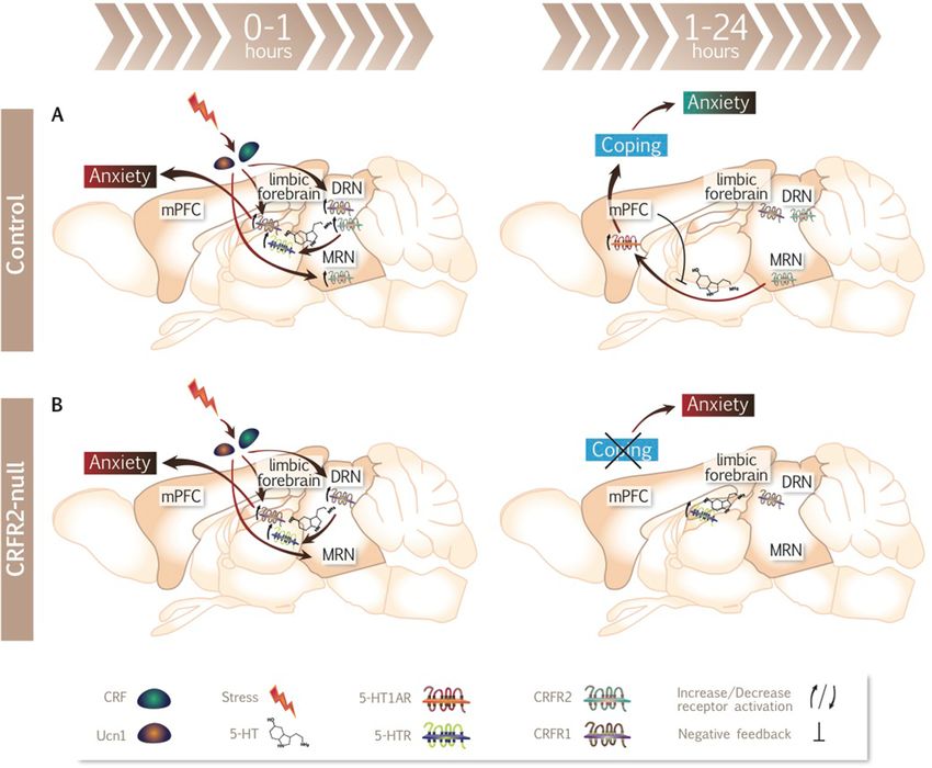
Activation of CRFR2 affects anxiety-like behaviour under stressed conditions [47-49] and
CRFR2-null mice have an anxiogenic phenotype [50,51]. This raises the question of what
role CRFR2 might play in the pathophysiology of anxiety-related and affective disorders in
human beings. To further investigate the mechanisms underlying this, we examined the
anxiety phenotype of CRFR2-null mice in detail, and characterized their serotonergic
responses to stress.

Methods
Animals

Mice were housed in temperature- and lighting-controlled rooms (lights on, 12 h) with free
access to laboratory chow and water. CRFR2-null mice, as previously described [50], and
control littermates (C57BL6 × 129) were the adult male offspring of parents heterozygous for
the knockout allele. For CRFR2 mRNA studies, adult male wild type C57BL6/J mice (Harlan
Laboratories) were used. Mice were group housed, except for the chronic variable mild stress
(CVMS) protocols, for which they were singly housed. Principles of laboratory animal care
(NIH No. 85-23, 1985) were followed. All procedures were approved by The Weizmann
Institute Animal Use and Care Committee or the United Kingdom Animals (Scientific
Procedures) Act, 1986.

Behavioural testing

Tests were carried out during the dark phase of the light cycle on adult male mice (2 to 4
months). Mice were habituated in the home cage in a dark room for 2 hours prior to each
behavioural test. Separate groups of mice were tested under: (a) basal conditions with no
stress applied prior to testing, n = 12 for control group, n = 14 for CRFR2-null group; (b)
immediately following 30 min of acute restraint stress (ARS), n = 13, both groups; (c) 24 to
48 hours following ARS, n = 5 for control group, n = 8 for CRFR2-null group (light/dark
transfer test performed at 24 hours post-stress, open-field at 48 hours post-stress); (d) 3 to 4
days following a 4-week CVMS protocol, (light/dark transfer test performed at 3 days post-
stress, open-field at 4 days post-stress), n = 10 for control group, n = 11 for CRFR2-null
group. The mice of group d were then retested 3 weeks later, when an ARS was applied and
testing was performed at 24 to 48 hours. Figure 1 shows the timeline of the experimental
protocols with stress procedures.

Figure 1 Schematic representation of experimental protocols and timelines. (A) Separate
cohorts of CRFR2-null and control mice were tested for anxiety-like behaviour in the
light/dark transfer and open-field tests: under basal conditions; immediately following ARS;
following CVMS and again 24 to 48 h after an ARS applied 3 weeks after the end of CVMS;
24 to 48 h following ARS. (B) CRFR2-null and control mice were exposed to no stress, ARS
or CVMS, and mRNA expression of stress-related genes and serotonin transporter (SERT)
binding were quantified 12 h after the end of stress. 5-HT/5HIAA content in brain nuclei
were quantified in unstressed mice and 24 h after ARS. (C) LCMRglu was measured in
CRFR2-null and control mice one hour after administration of saline or 5-HTR agonist. (D)
CRFR2 mRNA levels in brain were quantified over a 48-h time course following ARS or 7
days after CVMS in control mice. , ARS;                , CVMS; d, days; w, weeks.
Open-field (OF) test

The apparatus and experimental conditions were as previously described [50]. Mice were
placed in the centre of the apparatus to initiate a 10-min test session. Visits to, and distance
travelled and time spent in the inner zone of the arena were quantified using a video tracking
system (VideoMot2; TSE Systems, Bad Hamburg, Germany).

Light/dark transfer test (LDT)

Apparatus and experimental conditions were as previously described [50]. During a 5-min
test session, visits to, and distance travelled and time spent in the light compartment were
measured.

Stress procedures

Mice were subjected to 30 min ARS in a ventilated 50 ml plastic centrifuge tube. The CVMS
regime was modified from Ducottet et al. [52]. Mice were singly housed and a variety of mild
stressors were applied on an unpredictable schedule, 2 to 3 stressors per day for 4 weeks;
these included disruptions to the light-dark cycle, cage shift to one previously inhabited by
another male, cage tilt, damp bedding, low-intensity stroboscopic illumination, white noise,
restraint stress, short periods of food or water restriction, and housing with no bedding
followed by water in the cage. Controls were housed under stress-free conditions.

In the CVMS paradigm, mice were behaviourally tested 48 hours following termination of
the last stressor, which was standardized and was 24 hours of constant light for all mice (n =
10 or 11). For in-situ hybridization and 5-HT transporter (SERT) binding studies, mice (n = 6
for control basal group, n = 8 for CRFR2-null basal group, n = 7 for all stress groups) were
killed 12 hours after ARS, or after the last variable stressor, by decapitation within 15 s of
disturbing the home cage. The brains were removed, rapidly frozen on dry ice and stored at
−80°C until analysis.

Local cerebral glucose utilization (LCMRglu)

Local cerebral glucose utilization (LCMRglu) was determined as described previously
[39,53]. Mice (n = 8, all groups) were injected (intraperitoneally) with either 10 mg kg-1 8-
hydroxy-N,N-dipropyl-2-aminotetralin (8-OH-DPAT), 25 mg kg-1 1-(2,5-dimethoxy-4-
iodophenyl)-2-aminopropane (DOI) or vehicle (0.1 ml 0.9% NaCl). At 10 min after 8-OH-
DPAT, or 20 min after DOI, 5 µCi [14C]-2-deoxyglucose in 0.4 ml 0.9% NaCl was injected
intraperitoneally. After 45 min, mice were decapitated and their brains analyzed by
quantitative autoradiographic imaging, as described previously [54,55].

Analysis of tissue concentrations of 5-HT and 5-HIAA

Mice (n = 7 for unstressed groups, n = 6 for ARS groups) were killed by decapitation under
basal conditions or 24 hours following ARS. Brains were stored at −80°C until analysis.
Areas selected for microdissection were identified by comparison with a standard mouse
brain stereotaxic atlas [56]. To ensure accuracy, we used a stereomicroscope to visualize
neuroanatomical landmarks for use as reference points in identifying specific nuclei and
subdivisions of the DRN. Small diameter microdissection tools (310 to 410 µm diameter)
were used to restrict dissections to the subregion of interest. High-pressure liquid
chromatography analysis of 5-HT and 5-hydroxyindoleacetic acid (5-HIAA) was performed,
as previously described [57].

CRFR2 mRNA qPCR analysis

Quantitative PCR for CRFR2 mRNA expression was carried out as previously reported [40]
in brain taken from naïve mice (controls), or 3, 6, 12, 24 or 48 hours after ARS, or, for
CVMS mice, one week after the end of the stress protocol (n = 8 all groups)

In-situ hybridization (ISH) histochemistry

Coronal brain sections (10 µm) were cut on a cryostat, thaw-mounted onto silanized glass
slides, and stored at −80°C until use. In-situ hybridization procedures and probes were as
previously described [58-60]. Plasmids (generous gifts from Professor M. Holmes and Dr V.
Bombail) containing cDNA fragments for glucocorticoid receptor (GR), mineralocorticoid
receptor (MR), 5-HT1A R, 5-HT2CR and tryptophan hydroxylase 2 (TPH2) were used to
generate 35S-UTP-labelled specific antisense probes to mRNAs. Following ISH, slides were
dipped in Kodak Autoradiography Emulsion (Molecular Imaging Systems, New York, USA)
and exposed at 4°C for between 24 h and 6 weeks, depending on the probe, developed and
counterstained. The hybridization signal for each brain area was determined using computer-
assisted grain counting software (Zeiss KS 300 3.0, Carl Zeiss Vision, GmbH). For each
animal, silver grains were counted in a fixed circular area over 6 to 10 individual neurons per
subregion. The background, counted over areas of white matter, was subtracted. Analysis was
carried out blind to treatment group.

5-HT transporter (SERT) binding

Serotonin transporter (SERT) binding was determined on brain sections, cut as above, using
(3H)-paroxetine (Perkin Elmer, UK) as previously described [61]. Slides were then exposed
to (3H)-sensitive film (Amersham Hyperfilm MP, GE Healthcare, UK) at −80°C for 6 weeks.
Analysis of autoradiographs was performed by measuring the signal over the area of interest
with densitometry software (MCID Basic 7.0, Imaging Research, Inc.). The background was
subtracted.

Statistical analyses

Statistical analyses employed the two-tailed Student’s t test or two-way analysis of variance
(ANOVA) with post-hoc analysis using Fisher’s protected least significant difference test as
appropriate, with the exception of time course of CRFR2 expression, where one-way
ANOVA with Dunnett’s post-hoc analysis was used. Data are presented as mean ± standard
error of the mean (SEM). Differences were considered statistically significant at P < 0.05.
Results
CRFR2-null mice show increased anxiety 24 to 48 h after acute restraint stress
(ARS)

Under basal conditions, where mice were not exposed to stress (other than that caused by the
test itself), CRFR2-null mice and littermate controls showed no differences in anxiety-related
behaviour in two well-validated behavioural tests, the LDT (Figure 2) and the OF test (Figure
3), compared with littermate controls.

Figure 2 CRFR2-null mice exhibit increased anxiety-like behaviour 24 hours after ARS
in the LDT. Under basal conditions (A), immediately following ARS (B), or after CVMS
(C), no differences were observed in behaviour between CRFR2-null mice and controls.
However, when CVMS mice (D) or naïve mice (E) were exposed to ARS and tested 24 to 48
hours later, CRFR2-null mice showed increased anxiety-like behaviour compared with
control mice. Data expressed as mean ± SEM. N = 10 to 14 for A-D, N = 5 to 8 for E. * P <
0.05, ** P < 0.01, ~ P = 0.53, compared with control.

Figure 3 CRFR2-null mice exhibit increased anxiety-like behaviour 48 hours after ARS
in the OF test. Under basal conditions (A), immediately following ARS (B), or after CVMS
(C), no differences were observed in behaviour between CRFR2-null mice and controls.
However, when CVMS mice (D) or naïve mice (E) were exposed to ARS and tested 24 to 48
h later, CRFR2-null mice showed increased anxiety-like behaviour compared with control
mice. Data expressed as mean ± SEM. N = 10 to 14 for A-D, N = 5 to 8 for E. * P < 0.05, **
P < 0.01, *** P < 0.001, ~ P = 0.085, compared with control.

Because this finding contrasted with previous reports [50,51], we hypothesized that stressful
challenge was required to reveal the role of CRFR2 in anxiety. Another group of mice was
tested immediately following 30 min ARS. Again, no effect of genotype on anxiety-like
behaviour was observed (Figures 2 and 3). A further cohort of mice exposed to CVMS was
tested 3 to 4 days after the end of the protocol, to allow for recovery from the final acute
stressor, and again no differences were observed between control and CRFR2 mice in either
behavioural test.

However, 3 weeks later, these same CVMS mice were exposed to a single 30-min ARS, and
24 to 48 h later the CRFR2-null mice showed significantly increased indices of anxiety
compared with controls, with fewer visits to (t = 3.022, P = 0.007, n = 10 or 11), shorter
distance travelled in (t = 2.360, P = 0.029, n = 10 or 11), and a trend to less time spent in the
light chamber in the LDT (t = 2.062, P = 0.053, n = 10 or 11) (Figure 2), and fewer visits to
the centre of (t = 2.271, P = 0.036, n = 10 or 11) and less time spent in (t = 2.231, P = 0.039,
n = 10 or 11) the centre and a trend to less time spent in the OF test (t = 1.825, P = 0.085, n =
10 or 11) (Figure 3).

We then examined whether this delayed effect of ARS on anxiety was dependent on prior
CVMS by subjecting a further cohort of mice to ARS alone, and observed the same increased
anxiety-like behaviour 24 to 48 hours post-stress (Figures 2 and 3). In the LDT, CRFR2-null
mice spent less time (t = 2.650, P = 0.023, n = 5 to 8) and travelled a shorter distance (t =
2.833, P = 0.016, n = 5 to 8) in the light chamber. In the OF test, CRFR2-null mice spent less
time in (t = 2.675, P = 0.022, n = 5 to 8) and made fewer visits to the centre (t = 3.604, P =
0.004, n = 10 to 11), and travelled a shorter distance (t = 5.078, P = 0.0004, n = 10 to 11).

Serotonergic function is altered in the raphe nuclei of CRFR2-null mice

CRFR2 in the raphe nuclei modulate 5-HT activity, with consequences for stress-related
behaviours [32-38]. Therefore serotonergic functions were examined in CRFR2-null and
control mice. Neuronal metabolic activity, as measured by LCMRglu, was lower in both the
DRN (t = 2.626, P = 0.048, n = 8 for 8-OH-DPAT experiment, t = 2.804, P = 0.036, n = 8 for
DOI experiment) and median raphe nucleus (MRN) (t = 2.472, P = 0.049, n = 8 for 8-OH-
DPAT experiment, t = 2.785, P = 0.038, n = 8 for DOI experiment) of CRFR2-null mice as
compared with controls under basal conditions (Figure 4).

Figure 4 Serotonergic function is altered in the raphe nuclei of CRFR2-null mice. (A-E)
LCMRglu in the dorsal raphe nucleus (DRN) and median raphe nucleus (MRN) is lower in
CRFR2-null mice than controls under basal conditions. (A,C) 8-OH-DPAT or (B,D) DOI
administration decreased LCMRglu in the (A,B) DRN or (C,D) MRN only in control mice.
(E) Colour-coded autoradiograms from coronal brain sections at the level of the midbrain
raphe. ‘Warm’ colours represent high levels of [14C]-2-deoxyglucose accumulation while
‘cold’ colours represent low tracer accumulation. Images were selected from animals with
matched plasma tracer and glucose concentrations. 5-HIAA and 5-HT content of the (F)
dorsal and (G) caudal subdivisions of the DRN showed that the 5-HIAA:5-HT ratio was
higher in CRFR2-null mice under basal conditions in the dorsal DRN (DRD) and was
lowered by ARS only in CRFR2-null mice in both DRD and caudal DRN (DRC). Data
expressed as mean ± SEM. N = 8 for LCMRglu, ANOVA critical F(1,28) value = 4.196 for P ≤
0.05, 7.636 for P ≤ 0.01, 13.500 for P ≤ 0.001. N = 6 or 7 for 5-HIAA and 5-HT content,
ANOVA critical F(1,22) values = 4.301 for P ≤ 0.05, 7.945 for P ≤ 0.01, 14.380 for P ≤ 0.001.
* P < 0.05, ** P < 0.01 in post-hoc analysis.

Following challenge with the 5-HT1AR-specific agonist 8-OH-DPAT, a main effect of
treatment (ANOVA: F(1,28) = 4.558, P = 0.044), and an interaction between genotype and
treatment was observed in DRN (ANOVA: F(1,28) = 5.953, P = 0.021) (Figure 4). Post-hoc
analysis revealed that controls responded with decreased LCMRglu in both the DRN (t =
3.235, P = 0.0124, n = 8) and the MRN (t = 2.520, P = 0.047, n = 8) as expected, whereas the
raphe nuclei of CRFR2-null mice were unresponsive to 5-HT1AR agonist. Following 5-HT2R-
specific agonist DOI challenge, only a main effect of genotype was seen in both the DRN
(ANOVA: F(1,28) = 5.224, P = 0.030) and the MRN (ANOVA: F(1,28) = 5.333, P = 0.029). The
pattern of responses was, however, the same as for 8-OH-DPAT.

Studies to date have largely concentrated on the role of the DRN in respect to behaviour and
anxiety. Therefore, we measured 5-HT and 5-HIAA within subregions of the DRN (Figure
4). Within the caudal DRN, there was a main effect of genotype on 5-HIAA (ANOVA: F(1,22)
= 7.094, P = 0.014) and a genotype X ARS interaction on 5-HIAA:5-HT ratio (ANOVA:
F(1,22) = 6.153, P = 0.021). Post-hoc analysis revealed an increase in 5-HIAA (t = 3.472, P =
0.002, n = 7) and 5-HIAA:5-HT ratio (t = 3.242, P = 0.004, n = 7) in CRFR2-mice under
basal conditions, indicating increased serotonin turnover here. 24 hours following ARS, both
the caudal (t = 2.759, P = 0.011, n = 6 or 7) and the dorsal (t = 4.087, P = 0.0005, n = 6 or 7)
DRN showed decreases in 5-HIAA:5-HT ratio in CRFR2-null mice, with an associated
decrease in 5-HIAA in the caudal DRN of CRFR2-null mice (t = 2.554, P = 0.018, n = 6 or
7), whereas controls showed no effect of ARS on these parameters.
5-HT responses to stress and 5-HTR agonists are altered in efferent brain
regions of CRFR2-null mice

Following challenge with the 5-HT1AR-specific agonist 8-OH-DPAT, there was a main effect
of treatment throughout the forebrain (ANOVA: F(1,28) = 4.196 for P = 0.05) (Table 1) with a
genotype x 8-OH-DPAT interaction observed in some extrapyramidal and limbic structures.
Post-hoc analysis revealed that while controls had decreased LCMRglu in response to 8-OH-
DPAT in extrapyramidal regions as expected, CRFR2-null mice showed no response. These
areas receive projections from the DRN but lack their own 5-HT1AR, indicating that this
reflects attenuated DRN response to 5-HT1AR agonist.
Table 1 LCMRglu in efferent brain regions of control and CRFR2-null mice in response to 5-HT1AR or 5-HT2R agonist
                                      Control                  CRFR2-null                       Control                  CRFR2-null
                                      Saline 8-OH-DPAT   %     Saline 8-OH-DPAT   %             Saline DOI         %     Saline  DOI        %
Neocortex
Frontal                               46 ± 3   33 ± 3*   −28   47 ± 3   42 ± 3*   −21 a         47 ± 3   38 ± 2*   −19   45 ± 2   30 ± 2*   −33 a, b
Anterior cingulate                    48 ± 3   35 ± 2*   −27   49 ± 2   39 ± 4*   −35 a         50 ± 3   40 ± 2*   −15   51 ± 3   34 ± 2*   −33 a
Prefrontal                            47 ± 3   35 ± 3*   −26   47 ± 3   42 ± 2    −6 a          50 ± 3   39 ± 3*   −22   50 ± 3   30 ± 2*   −40 a
Somatosensory                         55 ± 2   33 ± 4*   −40   51 ± 4   45 ± 3*   −22 a         56 ± 2   53 ± 3    −5    55 ± 4   44 ± 3*   −20 a
Parietal                              53 ± 2   35 ± 2*   −34   50 ± 4   43 ± 3    −21 a         55 ± 2   45 ± 1*   −18   54 ± 3   37 ± 2*   −31 a, b
Hippocampus
Subiculum                             40 ± 3   26 ± 3*   −35   45 ± 3   23 ± 3*   −49 a         42 ± 2   43 ± 3    2     43 ± 3   33 ± 3*   −23
Dentate gyrus                         26 ± 3   16 ± 3*   −38   30 ± 2   15 ± 3*   −50 a         28 ± 3   18 ± 2*   −36   27 ± 3   16 ± 2*   −14 a
CA1                                   37 ± 3   22 ± 4*   −41   38 ± 3   16 ± 2*   −58 a         36 ± 3   26 ± 3*   −28   37 ± 2   26 ± 2*   −30 a
CA2                                   35 ± 3   20 ± 3*   −43   36 ± 3   19 ± 2*   −47 a         35 ± 3   34 ± 3    −3    36 ± 3   29 ± 2*   −19
CA3                                   38 ± 2   20 ± 2*   −47   41 ± 2   17 ± 2*   −58 a         36 ± 2   28 ± 3    −22   36 ± 2   22 ± 3*   −39 a
Extrapyramidal areas
Medial striatum                       43 ± 3   33 ± 2*   −23   42 ± 3   39 ± 3    −7 a          45 ± 3   34 ± 2*   −24   46 ± 3   29 ± 2*   −37 a
Lateral striatum                      46 ± 3   29 ± 3*   −37   44 ± 3   41 ± 3    −7 c          46 ± 4   30 ± 3*   −35   48 ± 4   31 ± 3*   −35 a
Globus pallidus                       33 ± 2   20 ± 2    −39   32 ± 2   33 ± 2    −3 a, b, c    32 ± 2   30 ± 2    −6    35 ± 2   23 ± 3*   −34 a, c
Substantia nigra                      30 ± 3   20 ± 1*   −27   27 ± 3   25 ± 4    −7            28 ± 4   30 ± 2    7     28 ± 2   18 ± 4    −36
Limbic areas
Medial septal nucleus                 41 ± 3   30 ± 2*   −27   39 ± 3   22 ± 2*   −44 a         45 ± 2   31 ± 2*   −31   48 ± 3   30 ± 2*   −38 a
Lateral septal nucleus                38 ± 3   27 ± 3*   −29   40 ± 2   18 ± 2*   −55 a, c      37 ± 3   28 ± 3    −24   40 ± 4   27 ± 3*   −33 a
Bed nucleus of the stria terminalis   27 ± 4   18 ± 2*   −33   31 ± 2   16 ± 3*   −48 a         30 ± 4   20 ± 2    −33   30 ± 2   18 ± 3*   −40 a
Basolateral amygdala                  36 ± 3   26 ± 3*   −28   36 ± 2   15 ± 2*   −58 a, b, c   38 ± 2   27 ± 4    −29   38 ± 2   24 ± 4*   −37 a
Central amygdala                      26 ± 2   20 ± 2*   −23   25 ± 3   15 ± 2*   −40 a         25 ± 2   17 ± 2    −32   24 ± 3   15 ± 2*   −38 a
Local cerebral glucose utilization (LCMRglu), shown as mean ± SEM and percentage change in LCMRglu in 8-OH-DPAT or DOI compared
with saline-treated mice. n = 8. aMain effect of drug; bmain effect of genotype, cgenotype X drug interaction in 2-way ANOVA, where critical
F(1,28) value was 4.20 for P ≤ 0.05. *P < 0.05 vs saline in post-hoc analysis.
In limbic areas, both genotypes decreased LCMRglu significantly (Table 1), but the genotype x 8-OH-DPAT interaction in lateral septum (ANOVA: F(1,28) = 4.654, P = 0.040) and basolateral amygdala (BLA) (ANOVA: F(1,28) = 4.654, P = 0.040) revealed that the CRFR2- null mice had a greater response to 5-HT1AR agonist in these areas. Following DOI challenge, there was again a main effect of treatment throughout the forebrain (ANOVA: F(1,28) = 4.196 for P = 0.05) (Table 1). Post-hoc analysis revealed that many brain regions showed a significant response to DOI in CRFR2-null mice but not controls (Table 1), suggesting greater postsynaptic 5-HT2R responsiveness throughout the forebrain in CRFR2-null mice. We then analyzed 5-HT and 5-HIAA content in the components of an anxiety-related amygdala-subiculum-septal circuit (Figure 5). There was a main effect of ARS on 5-HT content in the intermediate part of the lateral septum (LSI) (ANOVA: F(1,22) = 15.41, P = 0.0008) and of genotype on the 5-HIAA:5-HT ratio (ANOVA: F(1,22) = 19.460, P = 0.0002). There was also a genotype x ARS interaction in subiculum on both 5-HT (ANOVA: F(1,22) = 5.196, P = 0.033) and 5-HIAA:5-HT ratio (ANOVA: F(1,22) = 10.87, P = 0.004), and a main effect of genotype on 5-HIAA:5-HT (ANOVA: F(1,22) = 4.585, P = 0.045). Figure 5 5-HT responses to stress are altered in efferent brain regions of CRFR2-null mice. 5-HIAA and 5-HT levels as well as 5-HIAA/5-HT ratios are shown in the (A) subiculum (S), (B) intermediate part of the lateral septum (LSI), (C) medial septum (MS), (D) basolateral amygdala (BLA) and (E) central amygdala (CeA). CRFR2-null mice showed differences in basal levels of 5-HT and or 5-HIAA:5-HT ratio in the S and LSI, and a differential response to stress in the LSI and BLA. Data expressed as mean ± SEM. N = 8 for LCMRglu, ANOVA critical F(1,28) value = 4.196 for P ≤ 0.05, 7.636 for P ≤ 0.01, 13.500 for P ≤ 0.001. N = 6 or 7 for 5-HIAA and 5-HT content, ANOVA critical F(1,22) values = 4.301 for P ≤ 0.05, 7.945 for P ≤ 0.01, 14.380 for P ≤ 0.001. * P < 0.05, ** P < 0.01, *** P < 0.001 in post-hoc analysis. Post-hoc analysis revealed that under basal conditions the 5-HIAA:5-HT ratio was increased in CRFR2-null mice (subiculum; t = 3.846, P = 0.001, n = 6: LSI; t = 4.657, P < 0.0001, n = 7). However, in contrast with the DRN, this was due to lower 5-HT (subiculum; t = 2.474, P = 0.022, n = 6-7: LSI; t = 2.759, P < 0.012, n = 7) with unchanged 5-HIAA. In response to ARS, 24 h later there was an increase in 5-HT in the LSI of CRFR2-null mice (t = 3.878, P = 0.0009, n = 6 or 7) and a decrease in the 5-HIAA:5-HT ratio (t = 2.516, P = 0.020, n = 6 or 7). The genotype x ARS interaction in the subiculum was such that 5-HIAA:5-HT was increased by ARS in controls (t = 2.569, P = 0.018, n = 6 or 7), but decreased in CRFR2-null mice (t = 2.094, P = 0.049, n = 6 or 7). In the CeA, there was a main effect of ARS (ANOVA: F(1,22) = 17.71, P = 0.004) to increase 5-HT content in both genotypes (t = 2.838, P = 0.010, n = 6 or 7 for controls; t = 3.113, P = 0.005 for CRFR2-null mice, n = 6 or 7). 5-HT levels also increased in the BLA (t = 2.168, P = 0.041, n = 6 or 7) of CRFR2-null mice in response to ARS (Figure 5). Serotonergic and corticosteroid receptor gene expression are altered in response to stress in CRFR2-null mice To investigate which factors potentially involved in the processes of adaptation to acute stress might be differentially regulated in CRFR2-null mice as compared with controls, SERT protein levels (ligand binding) and mRNA levels of serotonergic genes and corticosteroid receptors (ISH) were quantified in apposite brain nuclei following ARS or end of CVMS. A time of 12 h post-stress was chosen as appropriate, as altered expression of these factors at
this time has previously been observed by many investigators. Full results are in Additional
file 1; only key significant differences are presented here.

In agreement with responses to 8-OH-DPAT, 5-HT1AR mRNA expression did not differ with
genotype in the hippocampus or amygdala (Additional file 1). No effect of genotype or stress
was seen in the DRN (Figure 6), but a genotype x stress interaction (ANOVA: F(2,36) = 3.328,
P = 0.048) whereby decreased expression in control compared with CRFR2-null mice (t =
2.181, P = 0.036, n = 7) was seen in the MRN following CVMS, and there were trends for
ARS to reduce 5-HT1AR expression in CRFR2-null mice (t = 1.702, P = 0.098, n = 6 or 7) but
not controls, and for CVMS to reduce 5-HT1AR expression in controls alone (t = 2.020, P =
0.052, n = 6 or 7). There was no appreciable effect of genotype on 5-HT2CR mRNA
expression (Additional file 1).

Figure 6 Serotonergic and corticosteroid receptor expression are regulated
differentially in response to stress in CRFR2-null mice. (A) mRNA expression of 5-
HT1AR in the (B) dorsal raphe nucleus (DRN) and (C) median raphe nucleus (MRN); (D)
tryptophan hydroxylase 2 (TPH2) mRNA in (E) DRN and (F) MRN; (G) serotonin
transporter (SERT) protein expression in (H) hippocampal CA1, (I) CA2 and (J,K)
subiculum (S); (L) glucocorticoid receptor (GR) mRNA expression in (M) CA1, (N) CA2
and (O,P) the paraventricular nucleus (PVN). Data presented as mean ± SEM for mRNA
levels or densitometry signal (SERT). N = 6 to 8. ANOVA critical F(2,36) value = 3.259 for P
≤ 0.05, 5.248 for P ≤ 0.01, 8.420 for P ≤ 0.001. # P < 0.05, ## P < 0.01, ### P < 0.001 for
effect of stress across genotypes. * P < 0.05, ** P < 0.01, *** P < 0.001 in post-hoc analysis.
ARS increased CRFR2 mRNA expression in whole brain of mice at all times points up to 48
h post-stress (Q), whereas CVMS decreased CRFR2 expression 7 days after the end of the
CVMS protocol (R). Data presented as mean ± SEM. N = 8. ANOVA critical F(5,42) value =
2.438 for P ≤ 0.05, 3.488 for P ≤ 0.01. * P < 0.05, ** P < 0.01 as compared with 0 hours in
post-hoc analysis or with control group.

There was a main genotype effect on TPH2 mRNA expression in MRN (ANOVA: F(2,36) =
5.311, P = 0.027), with increased levels in CRFR2-null mice. Post-hoc analysis detected this
as significant only between ARS groups (t = 2.080, P = 0.045, n = 6 or 7) (Figure 6). There
was a main effect of stress on TPH2 in DRN (ANOVA: F(2,36) = 3.684, P = 0.036) across
genotypes.

In the hippocampus, there was a main effect of stress on SERT protein expression (CA1
ANOVA: F(2,36) = 4.106, P = 0.027; CA2 ANOVA: F(2,36) = 4.387, P = 0.020; subiculum
ANOVA: F(2,36) = 8.474, P = 0.001), owing to increased expression after the end of CVMS,
only reaching statistical significance in CRFR2-null mice (CA1 t = 2.151, P = 0.038, n = 7 or
8; CA2 t = 2.139, P = 0.026, n = 7 or 8; subiculum t = 3.490, P = 0.0013, n = 7 or 8) and not
in controls (Figure 6). There were no effects of genotype or stress on SERT expression in the
amygdala (Additional file 1).

Expression of GR mRNA showed differential effects between brain regions and genotypes
(Figure 6). In the dorsal hippocampus, there was a trend towards higher GR expression in
CRFR2-null mice (CA1 ANOVA: F(2,36) = 3.976, P = 0.054; CA2 ANOVA: F(2,36) = 4.008, P
= 0.067). There was a main effect of stress (ANOVA: F(2,36) = 7.312, P = 0.002) with both
ARS and CVMS reducing expression in CA1 of CRFR2-null mice (ARS t = 2.420, P =
0.021, n = 7 or 8; CVMS t = 2.962, P = 0.005, n = 7 or 8), but only CVMS had a significant
effect in controls (t = 2.962, P = 0.043, n = 6 or 7). In CA2, only CVMS had an effect to
reduce GR expression, and this was only significant in CRFR2-null mice (t = 2.725, P =
0.010, n = 7 or 8). In the paraventricular nucleus (PVN) there was a main effect of genotype
(ANOVA: F(2,36) = 6.788, P = 0.003) with CRFR2-null mice having lower GR mRNA
expression, although this was not significant within treatment groups in post-hoc analysis.
There was a main effect of stress (ANOVA: F(2,36) = 4.974, P = 0.032); post-hoc analysis
showed increased GR following CVMS, but only reaching significance in CRFR2-null mice
(t = 2.341, P = 0.025, n = 7 or 8). Expression of MR mRNA was not regulated by stress or
genotype in any brain region examined (Additional file 1). Finally, CRFR2 mRNA levels, as
quantified by qPCR, were increased over a time period of 3 to 48 hours post-ARS (ANOVA:
F(5,42) = 3.750, P = 0.007) but were decreased following CVMS (t = 2.164, P = 0.047, n = 8)
(Figure 6).

Discussion
This study extends the evidence regarding the importance of CRFR2 in mediating the
processes towards successful behavioural recovery in the period following stress, and
moreover demonstrates that CRFR2 is engaged in the control of serotonergic function during
the same time frame. It further characterizes the stress-sensitive phenotype of CRFR2-null
mice [50,51,62] and reveals fundamental disturbances within components of their
serotonergic system.

In contrast with original reports of increased basal levels of anxiety [50,51], in our hands,
similar to the findings of Coste et al. [62], CRFR2-null mice do not show increased anxiety-
like behaviour compared to controls until 24 h after exposure to a prior acute stressor. This
discrepancy could be due to differing phenotypes of the three independently generated strains
of CRFR2-null mice, or factors such as age or husbandry. However, the mice in this study are
the same strain as reported with increased anxiety by Bale et al. [50] and an anxious
phenotype was described for both group- [50] and singly [51] housed CRFR2-null mice from
9 [40] to 24 [50] weeks of age, but not at 16 weeks [62], meaning that these factors are
unlikely to explain the inconsistency. This study indicates the need for a prior stress for
increased anxiogenesis in CRFR2-null mice, so an alternative explanation is that mice in
previous studies might have been inadvertently previously stressed, for example, by a prior
behavioural test. This time course of the behavioural effects of ARS led us to conclude that
CRFR2 has a key role in the processes leading to behavioural recovery in the hours following
exposure to a stressor.

While CRFR2-null mice in our study appear to be in a maladaptive state at 24 hours
following an acute stress, CRFR2-null mice exposed to CVMS are not more anxious than
controls. It could be interpreted from this that CRFR2-null mice have the ability to cope
successfully with this more chronic stress, but it is more likely that both CRFR2-null mice
and controls are affected adversely by CVMS, while CRFR2-null mice show an exaggerated
response to a single acute stressor. Such stressors might release CRF sufficient to recruit
CRFR2 [20], which mediate successful stress coping in normal mice [63]. Alternatively,
increased CRFR1 signalling in response to stress might occur; increased CRF expression in
the amygdala and PVN of CRFR2-null mice has been reported [50]. However, the time frame
of delayed anxiogenesis in CRFR2-null mice does not correlate with the expected rapid
release of CRF in response to acute stress and its subsequent negative feedback. The time
interval required for CRFR2-null mice to acquire this anxiety trait suggests that the processes
are indirect, and the serotonergic system is an obvious candidate.
Exogenous CRF administered to the DRN inhibits firing of 5-HT neurons via CRFR1
[15,18], while Ucns or higher levels of CRF increase firing via CRFR2 [32-36]. The raphe
nuclei receive inputs from both CRF and Ucn1 neurons [14,15,29], which may therefore
regulate serotonergic raphe function physiologically. In support of this hypothesis, CRFR2-
null mice show altered 5-HT/5-HIAA content in the DRN, LSI, subiculum, CeA and BLA 24
h after ARS, whereas control mice showed a clear change only in 5-HT content of the CeA.
Recent studies of mice with genetically altered Ucn levels have shown that 5-HT function is
dysregulated in these models [39-41] and that CRFR2-null mice show greater sensitivity to
elevation of 5-HT levels by pharmacological means, an observation suggested to be linked to
their stress-sensitive phenotype [64]. Notably, mice deficient in all three Ucns do indeed
show a similar phenotype [41] to our observations in CRFR2-null mice, with increased
anxiety-like behaviour and dysregulated activity within 5-HT circuits 24 h following ARS,
again evidencing the importance of CRFR2 here.

Interestingly, CRFR2-null mice show decreased basal neuronal metabolic activity in the
raphe nuclei. This is typically interpreted as evidence of decreased 5-HT firing activity levels,
as while both 5-HT and GABAergic neurons are important functionally here, GABAergic
neurons are present at only 10% of the number of 5-HT neurons [65]. This is an unusual
finding under basal conditions in our experience and could be due to increased raphe 5-
HT1AR inhibitory autoreceptor activity, altered 5-HT1AR modulation of raphe GABAergic
interneurons that express both CRFR2 and 5-HT1AR, or by inhibition from forebrain
postsynaptic receptors including 5-HT1AR and 5-HT2R [66-69]. Increased sensitivity of
structures throughout the forebrain to 5-HT2R agonists and to 5-HT1AR in some limbic
structures in CRFR2-null mice suggests that postsynaptic receptor responsiveness is
increased, and thus the latter mechanism may be significant. A shift towards unopposed
CRFR1 activity in the raphe nuclei of CRFR2-null mice could also be a significant factor in
mediating these effects or directly inhibiting 5-HT neuronal activity. Uncontrollable stress,
which activates DRN serotonergic neurons [38], is associated with a functional
desensitization of 5-HT1AR [70]. We observed no significant differences in 5-HTR
expression in the DRN of CRFR2-null mice and so it is likely that these effects are also
mediated by decreased internalization and desensitization of receptors [71], providing a
mechanism for potentially very dynamic responses to stress. Detailed electrophysiological
studies would be required to resolve the mechanism further.

In contrast with forebrain structures, the lack of LCMRglu response in the raphe nuclei to 5-
HTR agonists in CRFR2-null mice suggests tonic inhibition of neuronal activity here may be
close to maximal under basal conditions. The response to 5-HT1AR agonist in extrapyramidal
brain areas receiving projections from the DRN [72,73] but lacking their own 5-HT1AR
[74,75] was also attenuated. Thus CRFR2 appear to be required for maintaining normal basal
neuronal activity in the raphe nuclei and, in particular, for the balance of 5-HT1AR function
here.

CRFR2 are present in both the DRN and the MRN [11,13]. However, studies of stress
biology have largely concentrated on the DRN, and so to relate this altered raphe function to
the CRFR2-null behavioural phenotype, we examined 5-HT responses to stress in the DRN
and associated anxiety-related nuclei. Concentrations of 5-HIAA and the 5-HIAA:5-HT ratio
were elevated in CRFR2-null mice under baseline conditions within the caudal subregion of
the DRN (DRC), and these effects approached significance in the adjacent dorsal subregion
(DRD). The DRD and DRC mediate CRF receptor responses and are considered to be
anxiety-related subregions of the DRN based on anatomical and functional criteria [76,77].
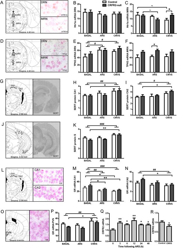
For example, they are activated by anxiogenic drugs [78], CRF-related peptides in vivo
[33,79] and in vitro [80], inescapable shock [70], noise stress [80], social defeat [81], the
avoidance task on the elevated T-maze [82], acoustic startle [83] and anxiety due to prior
experience of intimate partner violence [84]. In support of the specificity of these anxiety-
related effects on DRD/DRC serotonergic systems, in none of these studies were serotonergic
neurons in the adjacent ventrolateral part of the DRN activated. Elevation of 5-HIAA and 5-
HIAA:5-HT ratios in the DRC could be due to an organizational difference in 5-HT systems
as a consequence of CRFR2-null phenotype, or to differential activity that develops later in
life. In either case, DRC neurons appear to have altered baseline activity in adult CRFR2 null
mice, which may reflect a vulnerability to increased anxiety states.

Despite lower 5-HT content in efferent stress-related nuclei under unstressed conditions in
CRFR2-null mice, stress had a greater effect on their 5-HT content at 24 h, in keeping with
their stress-sensitive phenotype. This was particularly evident in LSI, which receives
significant 5-HT projections from the caudal DRN [85,86], in the subiculum and, to a lesser
extent, in the CeA. The subiculum is a key structure in inhibiting the hypothalamic-pituitary-
adrenal axis (HPAA) during termination of the stress response [87] and so altered function
here might relate to the higher responsiveness of the HPAA in CRFR2-null mice following
acute stress [50,62].

Not all anxiety-related nuclei examined showed such changes. We found no differences in the
LCMRglu of the bed nucleus of the stria terminalis (BNST) between control and CRFR2-null
mice at either baseline or in response to 8-OH-DPAT or DOI challenge. This was
unexpected, given the pivotal role of the BNST in the control of anxiety states [88]. However,
the serotonergic dysregulation in CRFR2-null mice may be downstream of the BNST which
projects strongly to the DRD/DRC region [89], where CRFR2 are abundant [12,13].
Overexpression of CRF in the BNST induces a decrease in CRFR2 binding selectively in the
DRD/DRC [90] and so it may be that the observed effects in CRFR2-null mice are primarily
mediated here.

5-HT firing activity is generally increased by stress [20] and negative feedback to the DRN
ultimately restores balance [66-69,71], as evidenced by essentially unchanged 5-HT and 5-
HIAA levels in control mice at 24 h following ARS. It has been previously reported that
CRFR2-null mice show greater sensitivity to 5-HT modulation of stress-induced behaviours
[64]. The pattern of increased responses in CRFR2-null mice to both 5-HT1AR and 5-HT2R
receptor agonists in areas expressing postsynaptic receptors is in keeping with this finding.
The LSI and BLA are key components of the limbic stress circuitry that were more
responsive to 5-HT1AR agonist in CRFR2-null mice. This might therefore relate to their
stress-sensitive phenotype and indicate a role for these structures in stress recovery.

The MRN has been implicated in mediating a delayed coping response following fear
behaviour induced by CRF in the DRN [35,91]. A delayed increase in 5-HT in the mPFC
mediated by CRFR2 in MRN is associated with cessation of intra-DRN CRF-induced
freezing behaviour [91] and is therefore proposed to mediate stress resilience effects [92]. In
CRFR2-null mice, the CRFR2-mediated surges in 5-HT neuronal firing from DRN and MRN
cannot occur, and unopposed CRFR1-mediated inhibition in DRN might further contribute to
this [15,18]. The normal 5-HT response in mPFC occurs one to two hours after intra-DRN
CRFR2 receptor activation [35,91], and we infer that the maladaptive state in CRFR2-mice
develops after the peak of this CRFR2-induced increase in mPFC 5-HT and by 24 h after the
stress. Thus, we propose that this delayed activity in efferent 5-HT neurons, which is critical
for successful adaptation to acute stress, is disrupted in CRFR2-null mice. The consequences
for CRFR2-null mice beyond 24 h are unknown, but unlike controls, 5-HT levels at this time
point are increased from basal levels in several limbic nuclei, indicating that homeostasis has
not been restored It is feasible that a lack of negative feedback within the 5-HT system due to
the failure of CRFR2-mediated 5-HT activity might contribute to this.

Therefore, we propose that a rapid and highly regulated increase in CRFR2 signalling in
response to acute stress, the resultant increase in efferent 5-HT activity and subsequent
negative feedback to restore homeostasis are important for a normal and successful coping
response. The delayed increase in 5-HT in mPMC is of key importance. Without this
orchestrated response, CRFR2-null mice do not respond to stress appropriately, and there is
prolonged anxiety that might account for their well-recognized anxiety phenotype. This
proposed model is presented in Figure 7. There is significant evidence for a role for the MRN
in stress recovery [91,92], and this model is consistent with our observation that LCMRglu is
lower under basal conditions in the MRN of CRFR2-null mice while the robust elevation of
TPH2 mRNA in CRFR2-null MRN might be a compensatory response to a lack of CRFR2
activation here. More detailed analysis of the dynamics of CRF and 5-HT processes in this
time frame and beyond in appropriate subregions of the raphe nuclei, and consideration of the
roles of other mediators of the stress response in CRFR2-null mice, are required to
substantiate this further.

Figure 7 Proposed model for development of prolonged anxiety following acute stress in
CRFR2-null mice. Following acute stress in control mice (A) CRF acting at CRFR1 in the
limbic forebrain produces immediate anxiety. High levels of CRF and potentially Ucn1
activate CRFR1 and more abundant CRFR2 in the DRN with a net effect to promote early
firing of efferent 5-HT neurons to limbic nuclei. Activation of CRFR2 in the MRN promotes
delayed 5-HT release in the mPFC at 1 to 2 h, which acts at 5-HT1AR to mediate successful
coping and anxiolysis by 24 h. Negative feedback in the 5-HT system restores homeostasis
by 24 h. (B) In CRFR2-null mice, the CRFR2-mediated increase in 5-HT firing cannot occur
and unopposed CRFR1 activity might inhibit 5-HT neuronal firing in limbic nuclei even
further. Absence of negative feedback within the 5-HT system contributes to the increased 5-
HT levels observed in limbic areas at 24 h. The temporal dynamics of the 5-HT system
following acute stress are dysregulated and homeostasis has not been restored. Crucially, the
delayed 5-HT activity in mPFC is disrupted and successful coping has not occurred, resulting
in prolonged anxiety.

Owing to the number of mediators involved in stress responses and the complex interactions
among them, other factors in addition to the serotonergic system are likely to be modified in
CRFR2-null mice in the hours following stress exposure, which might have implications for
the longer term. Indeed Ucn1 expression in the Edinger-Westphal nucleus and CRF in CeA
(but not the PVN) are increased in CRFR2 mice [50], which may be a developmental
compensatory change that is also responsible at least in part for their phenotype. Expression
of CRFR1 is, however, unaltered. We found that changes in serotonergic and corticosteroid
receptor gene expression in response to stress were generally greater in CRFR2-null mice,
again in keeping with their stress-sensitive behavioural phenotype. CRFR2-null mice have
normal basal HPAA activity, but higher responsiveness following acute stress [50,62].
Hence, changes such as the observed greater stress-induced increases in hippocampal SERT
levels in CRFR2-null mice may be mediated by glucocorticoids [93,94], adding potentially
further complexity to the relationship between CRFR2 and 5-HT function. Stress also
downregulated hippocampal GR mRNA to a greater degree in CRFR2-null mice, potentially
reflecting this expected HPAA hyperactivation. CRFR2-null mice also had lower basal GR
expression in the PVN, possibly reflecting chronically higher HPAA tone, and CVMS
unexpectedly increased this. Discordant regulation of GR expression in the hippocampus and
PVN has been reported previously [95,96], with upregulation of GR by stress suggested to
maintain glucocorticoid signalling to limit HPAA responses during prolonged stress. 5-HT
also regulates GR expression, and this may be mediated through TPH2 activity, in order to
regulate HPAA activity [97]. Both TPH2 and 5-HT1AR mRNAs in MRN were differentially
expressed in CRFR2-null mice. TPH2 mRNA levels in MRN were higher in CRFR2-null
mice, and there may be altered afferent control of DRN activity from here [98], suggesting
that the MRN should be more carefully considered in future studies of CRFR2 function.

Given this proposed role of CRFR2, we might expect expression to be regulated by stress
exposure. We found expression to increase, reaching a maximum at 3 to 12 h post-ARS and
subsequently declining, an effect similar to that seen for CRFR1 when acutely exposed to
ligand [99], while chronic stress decreased CRFR2 mRNA expression in this and a previous
study [100]. Others have observed lower CRFR2 expression in adult rats subjected to
maternal deprivation or in genetically stress-sensitive rodent strains [101,102], suggesting
that CRFR2 downregulation has the potential to be permanent in anxious or stress-sensitive
animals. The interesting exceptions are where CRFR2 is increased by chronically elevated
levels of CRF [103] or corticosterone [104], or in a model of maladaptive post-traumatic
stress disorder-like behaviour [105]. We hypothesize that while increased CRFR2 activity is
required for successful recovery from stress and subsequent downregulation is a normal
adaptive response associated with healthy coping, that ongoing hyperactivity of CRFR2
might be associated with a maladaptive stress response. The role of CRFR2 in mediating
learned helplessness in response to uncontrollable stress has implicated CRFR2 activity in the
development of maladaptive behavioural responses [38,79]. However equally, CRFR2
upregulation might be an appropriate secondary adaptation to a chronic stress. This issue
requires further investigation, to assess whether CRFR2 is a potential target in stress-related
psychiatric disorders.

In this study, 5-HT function in the lateral septum and subiculum, sites linked with anxiety as
well as the neural circuitry of reward and addiction [106-108], was particularly altered.
Dysregulated serotonergic function has long been linked to stress-related psychopathologies
[109,110] and direct effects of Ucns on CRFR2 in LSI have been observed in rodent models
of these disorders; hence, CRFR2 may play an important role in these processes [111-113].

Conclusions
While the role of CRFR2 in stress recovery was proposed some time ago [63,114], this study
provides new information regarding the mechanisms by which this may be mediated and
highlights the importance in the immediate post-stress period. This has implications for the
pathophysiology of psychiatric conditions associated with acute stress exposure, such as post-
traumatic stress disorder, reactive depression and relapse to substance abuse. As evidence
continues to emerge that CRFR2 may mediate its effects on stress primarily through 5-HT,
the potential for involvement in further mood disorders and ultimately for therapeutic
targeting is clear.
You can also read