Age affects the contribution of ipsilateral brain regions to movement kinematics
←
→
Page content transcription
If your browser does not render page correctly, please read the page content below
Received: 17 April 2019      Revised: 2 October 2019       Accepted: 4 October 2019
DOI: 10.1002/hbm.24829

RESEARCH ARTICLE

Age affects the contribution of ipsilateral brain regions
to movement kinematics

Caroline Tscherpel1,2 | Lukas Hensel1 | Katharina Lemberg1 | Jana Freytag1 |
Jochen Michely1,3 | Lukas J. Volz1 | Gereon R. Fink1,2 | Christian Grefkes1,2

1
 Medical Faculty, University of Cologne and
Department of Neurology, University Hospital         Abstract
Cologne, Cologne, Germany                            Healthy aging is accompanied by changes in brain activation patterns in the motor
2
 Cognitive Neuroscience, Institute of
                                                     system. In older subjects, unilateral hand movements typically rely on increased
Neuroscience and Medicine (INM-3), Research
Centre Jülich, Jülich, Germany                       recruitment of ipsilateral frontoparietal areas. While the two central concepts of
                                                     aging-related brain activity changes, “Hemispheric Asymmetry Reduction in Older
3
 Wellcome Trust Centre for Neuroimaging,
University College London, London, UK
                                                     Adults” (HAROLD), and “Posterior to Anterior Shift in Aging” (PASA), have initially
Correspondence                                       been suggested in the context of cognitive tasks and were attributed to compensa-
Christian Grefkes, Institute of Neuroscience
and Medicine - Cognitive Neuroscience                tion, current knowledge regarding the functional significance of increased motor
(INM-3), Research Centre Jülich, 52425 Jülich,       system activity remains scarce. We, therefore, used online interference transcranial
Germany.
Email: christian.grefkes@uk-koeln.de                 magnetic stimulation in young and older subjects to investigate the role of key
                                                     regions of the ipsilateral frontoparietal cortex, that is, (a) primary motor cortex
Funding information
Marga und Walter Boll-Stiftung; German               (M1), (b) dorsal premotor cortex (dPMC), and (c) anterior intraparietal sulcus (IPS)
Excellence Initiative; Universität zu Köln           in the control of hand movements of different motor demands. Our data suggest a
Emerging Groups Initiative; German Research
Foundation, Grant/Award Number: DFG GR               change of the functional roles of ipsilateral brain areas in healthy age with a
3285/2-1                                             reduced relevance of ipsilateral M1 and a shift of importance toward dPMC for
                                                     repetitive high-frequency movements. These results support the notion that mech-
                                                     anisms conceptualized in the models of “PASA” and “HAROLD” also apply to the
                                                     motor system.

                                                     KEYWORDS
                                                     aging, dorsal premotor cortex, movement kinematics, online TMS interference

1 | I N T RO D UC T I O N                                                           Parsons, 2014; Catalan, Honda, Weeks, Cohen, & Hallett, 1998;
                                                                                    Hummel, Kirsammer, & Gerloff, 2003; Verstynen, 2004).
Unilateral hand movements are primarily driven by a lateralized net-                     As in more complex tasks, the enhanced brain activity of the ipsi-
work of cortical and subcortical areas. However, besides contralateral              lateral motor system has frequently been encountered in association
sensorimotor areas (Porter & Lemon, 1995), also ipsilateral brain                   with age. Older subjects show more extensive, as well as more bilat-
regions contribute to the coordination of upper limb movements                      eral activation patterns (Heuninckx, 2005; Hutchinson, 2002;Mattay
(Cramer, Finklestein, Schaechter, Bush, & Rosen, 1999; Tinazzi &                    et al., 2002 ; Michely et al., 2018 ; Riecker et al., 2006). While at the
Zanette, 1998). Bilateral recruitment patterns have particularly been               behavioral level motor performance typically declines with increasing
observed in young healthy subjects for more complex motor tasks                     age (Hackel, Wolfe, Bang, & Canfield, 1992; Kolb, Forgie, Gibb,
with higher behavioral demands (Buetefisch, Revill, Shuster, Hines, &               Gorny, & Rowntree, 1998; Salthouse, 2000), ipsilateral brain activity

This is an open access article under the terms of the Creative Commons Attribution License, which permits use, distribution and reproduction in any medium,
provided the original work is properly cited.
© 2020 The Authors. Human Brain Mapping published by Wiley Periodicals, Inc.

640       wileyonlinelibrary.com/journal/hbm                                                                                   Hum Brain Mapp. 2020;41:640–655.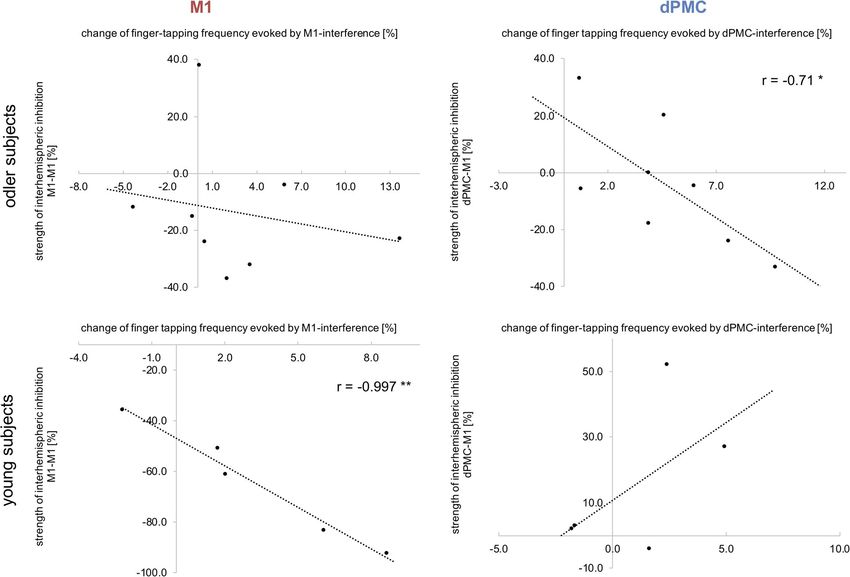
TSCHERPEL ET AL.                                                                                                                                  641

increases, comprising especially the primary motor cortex, premotor          Given the increases in frontoparietal activity reported for older sub-
and parietal areas (Mattay et al., 2002; Michely et al., 2018; Riecker       jects (Heuninckx, 2005; Mattay et al., 2002; Riecker et al., 2006), we
et al., 2006).                                                               hypothesized that TMS interference with ipsilateral frontoparietal
    Based on neuroimaging findings which have consistently shown             areas would primarily disturb motor performance in older subjects,
altered activation patterns during aging, two main models have been          consistent with a compensatory role of activity changes in these
conceptualized: a decrease in lateralization with increasing age             areas. Moreover, the different motor tasks were not only designed to
(“Hemispheric Asymmetry Reduction in Older Adults” (HAROLD) the-             recruit different motor effectors ranging from the entire arm for the
ory, (Cabeza et al., 1997; Cabeza, Anderson, Locantore, & McIntosh,          pointing tasks to the single index finger during the finger tapping task,
2002)) and a shift of activation from posterior to anterior regions          but also to differentially probe the involvement of the three brain
(“Posterior to Anterior Shift in Aging” (PASA) model, (Davis, Dennis,        regions of interest. Accordingly, primary motor cortex was supposed
Daselaar, Fleck, & Cabeza, 2008). While initially put forward in the         to be particularly relevant for fine-tuned repetitive finger movements
context of cognitive tasks, these models have been readily transferred       which strongly rely on the modulation of interhemispheric inhibition
to the motor system (Heuninckx, 2005; Hutchinson, 2002; Mattay               applicable to our tapping tasks (Hinder, 2012; Liuzzi, Hörniss,
et al., 2002; Michely et al., 2018; Riecker et al., 2006; Ward &             Zimerman, Gerloff, & Hummel, 2011), but may be supported by
Frackowiak, 2003).                                                           premotor regions in aging. The cortex in anterior intraparietal sulcus
    From a functional perspective, such changes observed during cog-         was assumed to be particularly involved during the pointing tasks
nitive tasks have mostly been associated with compensatory recruit-          since the IPS is known to be engaged in such visuomotor tasks
ment in order to counteract aging-related structural, functional, and        (Grefkes & Fink, 2005; Wang, Fink, Dafotakis, & Grefkes, 2009).
metabolic changes (Cabeza et al., 2002; Grady, 2012; Reuter-Lorenz &
Cappell, 2008) with the ultimate goal of maintaining performance.
    However, though intuitive, the functional significance of these          2 | MATERIALS AND METHODS
age-related activity changes associated with motor tasks remains to
be established (Seidler et al., 2010). Although the principle of compen-     2.1 | Main experiment
sation may also be relevant for the motor system (Mattay et al.,             2.1.1 | Subjects
2002), a majority of studies did not support this idea (Hutchinson,
2002; Langan et al., 2010; Riecker et al., 2006), since the authors          Thirty-two healthy subjects participated in this study: 15 young

could not link the degree of over-activation to improved motor per-          healthy subjects (five females, all subjects reported right-handedness

formance. Besides, altered brain activity may also represent dediffer-       according to the Edinburgh Handedness Inventory (EHI) (laterality

entiation (Bernard & Seidler, 2012; Langan et al., 2010; Seidler             score ≥ 75%), mean age 27.8 ± 3.5 SD years; range: 24–34 years) and

et al., 2010).                                                               17 older healthy subjects (four women, all right-handed, mean age

    Yet, the models of age-related changes and their functional signifi-     63.4 ± 9.2 SD years; range: 51–89 years) were recruited from our sub-
cance were primarily based on BOLD activity changes correlating with         ject database. Due to the procedure explained in the following, the
behavior. In this context, a significant limitation of functional neuroim-   recruitment was challenged and limited to subjects having an individ-
aging studies, including analyses of functional or effective connectiv-      ual MRI scan stored in the database. Post hoc calculation of power
ity, is that they cannot derive a causal role of a particular brain region   confirmed that although the given sample size is small, based on the
for performance. By contrast, transcranial magnetic stimulation (TMS)        detected effect sizes and given an alpha-error of 0.05, we achieved an
can be used to transiently interfere with neural activity of a brain area    observed power of 0.79 (G*Power 3.1). None of the subjects had a
during task performance (Gerloff et al., 1998; Pascual-Leone, Bartres-       history of neurological, psychiatric, orthopedic or rheumatic disease
Faz, & Keenan, 1999; Walsh & Cowey, 2000). By directly interfering           nor any contraindication to TMS (Rossi, Hallett, Rossini, & Pascual-
with the fine-tuned neural processing and thereby disturbing task per-       Leone, 2009). All participants gave informed written consent before
formance, online TMS interference allows insights into the causal role       entering the study, which was approved by the local ethics committee
of a specific area for a given task of interest.                             (number of ethical vote: 14–141) and the experiment was carried out
    We thus used neuronavigated, online TMS interference to eluci-           in accordance with the Declaration of Helsinki.
date the age-related role of ipsilateral brain regions in healthy aging.
As age-dependent changes have been frequently reported for ipsilat-
                                                                             2.1.2 | Experimental design
eral primary motor cortex (M1), dorsal premotor cortex (dPMC) and
intraparietal sulcus (IPS) (Mattay et al., 2002; Riecker et al., 2006;       We used a single-blinded, pseudorandomized mixed design. Accord-
Ward, Swayne, & Newton, 2008), we investigated their task-related            ingly, the order of stimulation sites was pseudorandomized per sub-
role in young and older healthy subjects while they performed three          ject before the experiment and balanced within subjects. The order of
motor task of different motor demands: (a) rapid alternating pointing        motor tasks was pseudorandomized across subjects as well. Short
between two fixed targets, (b) maximum finger tapping frequency,             bursts of repetitive TMS pulses (rTMS) were administered at a fre-
and (c) maximum hand tapping frequency. Movement kinematics were             quency of 10 Hz with a stimulation intensity of 90% of resting motor
recorded using a three-dimensional (3D) motion analyzer system.              threshold (see below) concurrent to task execution (Davare, Andres,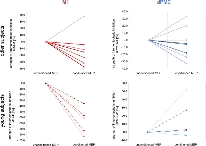
642                                                                                                                                  TSCHERPEL ET AL.

Cosnard, Thonnard, & Olivier, 2006; Gerloff et al., 1998; Volz et al.,       visual target, movement onsets were indicated by a brief acoustic
2017), starting with a visual go cue and lasting throughout the trial for    tone. Subjects were instructed to move as fast and as accurate as
2.0–2.5 s (20–25 pulses) depending on the given motor task (see              possible.
below).
      Trials within a given stimulation block were separated by a 3-s
                                                                             2.1.4 | Index finger tapping (II)
pause to minimize the likelihood of carry-over and lasting effects (see
below) (Rotenberg, Horvath, & Pascual-Leone, 2014). The software             The finger tapping task tested repetitive isolated finger movements
               ®
Presentation       (Version 9.9, Neurobehavioral Systems, Berkeley, CA,      which strongly rely on the modulation of interhemispheric interactions
http://www.neurobs.com) was used for both stimulus presentation              (Hinder, 2012; Liuzzi et al., 2011). Subjects were instructed to perform
and time-locked triggering of the TMS machine (Volz et al., 2017).           repetitive index finger tapping movements as fast and accurate as
      Three motor tasks of different behavioral demands were used to         possible with their right hand upon the appearance of a visual cue
probe the influence of the stimulation sites on motor performance            which was presented on a video screen controlled by the software
(Figure 1).                                                                  Presentation®. Subjects placed their right palm on a defined position
                                                                             on the table and performed repetitive vertical movements at the
                                                                             metacarpophalangeal joint of their index finger (Nowak et al., 2007). A
2.1.3 | Pointing task (I)
                                                                             dice with a height of 2.5 cm indicated the target amplitude (Figure 1).
The rapid pointing task engaged muscle groups of the entire arm              Each tapping trial lasted 2 s. As it is assumed that effects of high-
including the shoulder, and particularly required target accuracy.           frequency rTMS outlast the stimulation period for about half of the
Therefore, it strongly relied on neural systems for action-space repre-      duration of stimulation (Rotenberg et al., 2014), tapping trials were
sentations (Colby & Goldberg, 1999; Rizzolatti, Fogassi, & Gallese,          separated with a pause of 3 s to avoid rTMS carry-over effects and
1997), sensori- and visuomotor coordinate transformations, and               fatigue. Short finger tapping trials were used because the maximum
visuospatial attention (Grefkes & Fink, 2005; Wang et al., 2009). Sub-       finger tapping frequency is usually highest during the first few sec-
jects were asked to perform repetitive sagittal pointing movements           onds of a trial (Wang et al., 2009).
with their right index finger between two targets (Figure 1). For this
purpose, we installed an apparatus with two fixed targets. The dis-
                                                                             2.1.5 | Hand tapping (III)
tance between the pointing targets was 15 cm in the sagittal plane
and 3 cm in the vertical plane, which was visible throughout the entire      The hand tapping task was similar to the finger tapping task with
block of pointing movements (Figure 1).                                      respect to its cyclical, repetitive aspect. Moreover, it involved similar
      The difference in height ensured that participants lifted their arm,   motor components and, therefore, additionally served as an internal
thereby avoiding sliding movements on the table. Each trial lasted           control condition for effects seen in the finger tapping task. However,
2.5 s followed by a pause of 3 s. To prevent interference with the           the hand tapping task recruited more proximal arm and wrist muscle

F I G U R E 1 Motor task during online related TMS. (a) (I) Rapid pointing movements touching two spots with a distance of 15 cm as fast as
possible. (II) Index finger tapping with a height of 2.5 cm at maximum speed. (III) Hand tapping with a height of 7 cm at maximum speed. All motor
tasks were studied using a three-dimensional motion analyzer system based on ultrasound-emitting markers that were fixed to the dorsal side of
the distal interphalangeal joint of the right index finger (marker I), the dorsal side of the third metacarpophalangeal joint (marker II), and between
the styloid processes of ulna and radius (marker III). (b) Illustration of the kinematic data obtained for data analysis: traces of vertical movements
(z-direction) of marker I over time are shown for every motor task (I–III)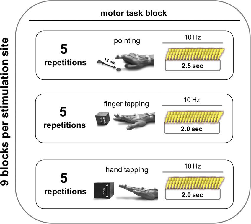
TSCHERPEL ET AL.                                                                                                                                     643

groups, which are considered to rely somewhat less on inter-                  depending on the given motor task (see above). A visual stop signal
hemispheric interactions compared to isolated index finger move-              displayed for 3 s indicated the end of a trial. Of note, for the pointing
ments (Aune, Ettema, & Vereijken, 2016; Harris-Love, Perez, Chen, &           task, start and stop of the trial were also indicated by a brief acoustic
Cohen, 2007; Sohn, Jung, Kaelin-Lang, & Hallett, 2003). Subjects were         tone so that the subjects could focus on the pointing apparatus.
asked to place their right hand on the defined position on the table              The entire experiment lasted about 60 min (15 min per stimulation
and, in response to the cue, perform repetitive flexion and extension         site). Before the TMS sessions, subjects were trained in all tasks until
of the wrist with a stretched hand at maximum speed (Nowak et al.,            they reached a stable performance.
2007). The target movement amplitude was indicated by a cube of
7 cm height (Figure 1). Similarly to (I), one tapping trial lasted 2 s with
                                                                              2.1.6 | 3D ultrasound movement recording
a pause interval of 3 s (all indicated by visual cues).
    The testing battery for each stimulation site consisted of nine           Motor performance was assessed using the Zebris CMS 20 kinematic
blocks, three repetitions of each type of movements. The order of             motion analyzer system (Zebris Medical Company, Isny, Germany)
these nine blocks per site was pseudorandomized. One block was                (Nowak et al., 2007; Wang et al., 2009). The system mainly consists of
composed of five consecutive repetitions of one movement trial                a measuring sensor, which captures with high spatial (0.1 mm) and
(i.e., 5 times of 2.0 s of finger tapping or hand tapping, or 2.5 s of        temporal (100 Hz) resolution the 3D positions of markers, fixed on
pointing) (Figure 2). This means that we acquired three blocks of five        the body parts to be examined, emitting ultrasonic pulses (diameter:
consecutively repetitions per task per stimulation site. Each trial of        5 mm, weight: 1 g) (Wang et al., 2009). For the current experiment,
movement was separated by a 3-s pause. Each block was separated               the 3D-tracking markers were attached to the dorsal side of the distal
by 10 s of rest. A break of 30–45 s for switching between the differ-         interphalangeal joint of the right index finger (marker I), the dorsal side
ent stimulation sites ensured a reliable location of each region using        of the third metacarpophalangeal joint (marker II), and between the
the neuronavigation system.                                                   styloid processes of ulna and radius (marker III) (Figure 1). The x-, y-,
    Written instructions were displayed for 3 s on a video screen, indi-      and z-directions of the position marker coordinates refer to the
cating the upcoming block of motor task (e.g., “pointing,” “finger tap-       medio-lateral, antero-posterior, and vertical directions with regard to
ping” or “hand tapping”). Upon the appearance of the visual go cue,           the subject performing the task. Kinematic data were continuously
instructing the subject to start the requested movements, the soft-           recorded throughout the entire experiment.

ware synchronously triggered the TMS machine to apply 10 Hz rTMS
throughout the duration of a trial for 2.0–2.5 s (20–25 pulses)               2.1.7 | Neuronavigated transcranial magnetic
                                                                              stimulation
                                                                              TMS was performed using a Magstim Super Rapid2 stimulator (The
                                                                              Magstim Co., Ltd, Whitland, United Kingdom) equipped with a 70 mm
                                                                              figure-of-eight air film coil. Throughout the TMS sessions, the position
                                                                              of the coil was tracked and recorded using a frameless computerized
                                                                              stereotaxic neuronavigation system (BrainSight V.2.0.7; Rogue
                                                                              Research Ltd; Montreal, Canada). For neuronavigation, the head of
                                                                              the subject was coregistered with the individual high-resolution ana-
                                                                              tomical MR image (MDEF sequence; voxel size: 1.0 × 1.0 × 1.0 mm3,
                                                                              FOV 256 mm, 176 sagittal slices, TR 2250 ms, TE 3.93 ms) via ana-
                                                                              tomical landmarks (see Nettekoven et al., 2014 for further details).
                                                                              The structural MRI images were available in the subject database.
                                                                                  The “motor hotspot” for ipsilateral M1 (i.e., right primary motor
                                                                              cortex) was defined as the coil position eliciting motor evoked poten-
                                                                              tials (MEP) of the highest amplitude in response to a TMS pulse
                                                                              applied tangentially to the skull in a 45 posterior–anterior current
                                                                              direction, thereby targeting the posterior wall of the precentral gyrus
                                                                              at the hand knob formation (Yousry et al., 1997). MEP amplitudes
F I G U R E 2 Study design. For each stimulation site, we acquired            were assessed with biphasic pulses. In addition, MEP amplitudes of
nine blocks for a given motor task. The order of stimulation sites was        the left first interosseous (FDI) muscle were measured using Ag/AgCl
pseudorandomized per subject and balanced within subjects. The                surface electrodes (Tyco Healthcare) in a belly-to-tendon montage.
order of motor tasks was also pseudorandomized across subjects.
                                                                              The EMG signal was amplified, filtered (0.5 Hz high pass and
One block consisted of five consecutive repetitions of one movement
                                                                              30–300 Hz bandpass), and digitized using a Powerlab 26T device and
trial (i.e., pointing, finger tapping, hand tapping). See methods for
more information                                                              the LabChart software package version 8.0 (AD Instruments,
                                                                              Australia).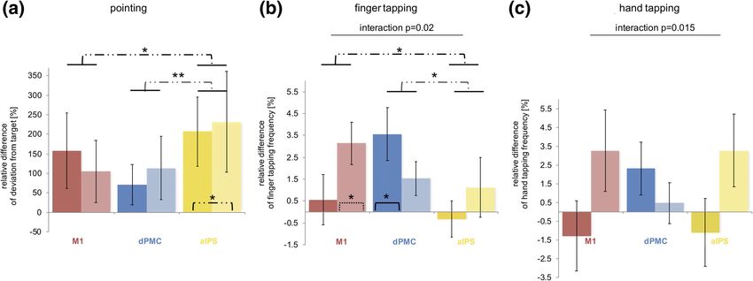
644                                                                                                                                      TSCHERPEL ET AL.

      The resting motor threshold (RMT) was defined using an algorithm         intensities, for a review see Fitzgerald, Fountain, & Daskalakis, 2006;
provided by the TMS Motor Threshold Assessment Tool (MTAT) 2.0                 Rossini et al., 2015).
(http://www.clinicalresearcher.org/software.html)      (Awiszus,   2003;           Furthermore, online TMS paradigms are more flexible with regard
Diekhoff et al., 2011). Using a maximum-likelihood procedure, the              to randomization of stimulation sites within a single session. High-
algorithm proposes stimulation intensities that are subsequently               frequency rTMS trains have widely been used before in order to inter-
tested regarding their ability to induce an EMG response higher than           fere with task performance (Davare, Andres, et al., 2006; Gerloff
50 μV, which is accordingly entered by the experimenter. The MTAT              et al., 1998; Lotze et al., 2006; Volz et al., 2017). In contrast to single
has been shown to accurately estimate motor thresholds using less              pulse TMS, 10 Hz rTMS trains increase the likelihood to effectively
stimuli than the standard 5-out-of-10 rule (Awiszus, 2003).                    and temporally accurately interfere with task performance.
      For motor task interference, 10 Hz rTMS was applied at 90% RMT               For online interference, the stimulator was controlled and trig-
during task execution (Volz et al., 2017). A subthreshold stimulation          gered by an in-house script using the software Presentation® to
intensity was chosen to prevent the induction of MEPs, which may               ensure reliable timing of TMS pulse applications (for further technical
irritate participants and impact on their task performance. Besides,           details see Volz et al., 2017). The stimulation was triggered by the
stimulating brain regions in the hemisphere ipsilateral to the moving          computer synchronously with the presentation of the go cue, and was
hand and areas beyond M1 additionally avoid the evocation of dis-              administered throughout the trial for 2.0–2.5 s (20–25 pulses)
tracting muscle activity. Moreover, by using an online TMS approach,           depending on the motor task (see above). Applying TMS throughout
we used the immediate effects of TMS interfering with physiological            the entire duration of the motor task implicated the possibility to
neural activity underneath the stimulation coil while subjects per-            intervene not only with movement execution, but also preparation or
formed a given task. The advantage of online TMS interference com-             sensory integration. However, it ensured an effective and comparable
pared to the more widely used “offline”-TMS approach is that by                interference for all motor tasks, all brain regions and both groups. Of
directly disturbing neural activity during task performance stimulation        note, the in-house script simultaneously presented the visual cues and
effects are immediately present and less prone to vary with respect to         triggered the TMS machine.
the magnitude and direction of after-effects as observed for “offline              The following stimulation sites were investigated all ipsilateral to
TMS” protocols (Hamada, Murase, Hasan, Balaratnam, & Rothwell,                 the right (dominant) hand (Figure 3): (a) right ipsilateral M1, (b) right
2013). Of note, TMS interference refers to the neurophysiological              ipsilateral dPMC, and (c) right ipsilateral IPS. Ipsilateral M1 was equiv-
mechanisms, that is, disturbing neural activity, rather to the behavioral      alent to the motor hotspot as defined above.
consequence. Besides, in contrast to the excitatory and inhibitory                 For targeting ipsilateral dPMC and IPS, we used coordinates based
effects of offline rTMS (Nettekoven et al., 2015), online TMS is con-          on recent activation likelihood estimation meta-analyses on hand
sidered to invariably interfere with the neural processing, that is,           motor fMRI activity (Hardwick, Rottschy, Miall, & Eickhoff, 2013;
inducing a virtual online lesion, and thereby disturb task performance         Rehme, Eickhoff, Rottschy, Fink, & Grefkes, 2012). For each subject,
(Rossi, 2004; Rossini et al., 2015; Walsh & Cowey, 2000). Importantly,         MNI (X/Y/Z) coordinates (dPMC: 38/6/62; IPS: 42/−40/50) were
there is no evidence that 10 Hz online interventions paradigms have a          warped into individual 3D-space of the anatomical MRI using Statisti-
lasting effect on cortical excitability, which might probably be due to        cal Parametric Mapping (SPM12, http://www.fil.ion.ucl.ac.uk/) and
the fact that 10 Hz stimulation applied for increasing cortical excitabil-     MRIcron (Neva, Brown, Mang, Francisco, & Boyd, 2017).
ity in offline-experiments are administered in a completely different              For interference with ipsilateral dPMC activity, the coil was held
stimulation protocol (longer stimulation trains, higher number of              tangentially to the skull in a 45 anterior–posterior position perpen-
pulses up to 1,000–2,000 in total, and often suprathreshold                    dicular to the course of the precentral sulcus. This position warranted

F I G U R E 3 Ipsilateral stimulation sites with coil orientations. (a) Right ipsilateral primary motor cortex (M1), (b) right ipsilateral dorsal premotor
cortex (dPMC), and (c) right ipsilateral anterior intraparietal sulcus (IPS)
TSCHERPEL ET AL. 645
optimal stimulation of dPMC neurons situated in the anterior wall of x-, y-, and z-directions and calculated as 3D Euclidean distances. Fur-
the precentral gyrus (close to the intersection with the superior fron- thermore, we calculated the deviation from the target by subtracting
tal sulcus). By contrast, for stimulation of ipsilateral anterior IPS, the the actual covered distance from the ideal one (which was 160 mm).
coil was held in a 90 latero-medial position, perpendicular to the We also considered the number of inversion of velocity (NIV), which
intraparietal sulcus, leading to a preferential stimulation of neurons describes the smoothness and rhythmicity of a repetitive movement
situated in the medial IPS wall, probably equivalent to macaque area (Amengual et al., 2013), along with all three dimensions.
MIP which is engaged in reaching movements and visuomotor trans-
formation (Colby, 1998; Grefkes & Fink, 2005; Grefkes, Ritzl, Zilles, &
2.1.10 | Index finger- and hand tapping task
Fink, 2004). Similar to M1, the stimulation intensity for ipsilateral
dPMC and IPS was applied at the same intensity, that is, 90% RMT For the tapping conditions, motor performance was quantified by
obtained at M1 for a given subject. Using the same stimulation inten- (a) tapping frequency (Hz), (b) vertical movement amplitude (mm), and
sity for each site ensures that sensory stimulation effects like acoustic (c) the number of inversion of velocity (NIV). The data used for all ana-
noise or tactile sensations remain comparable between stimulation lyses were obtained from the z-axis of marker I which was attached to
sites within a given subject. Hence, between-region differences do the index finger.
not result from differences in sensory stimulation interfering with
motor performance, but are most likely of neural origin. Furthermore,
2.1.11 | Statistical analysis
it ensured that stimulation effects were comparable between subjects
and groups. Statistical analyses were performed using the software package SPSS
Coil positions were logged into the neuronavigation software and (Statistical Package for the Social Science, version 23, IBM). To assess
maintained throughout the experiment for each stimulation site. age-related differences between the two groups for the resting motor
A stimulation over parieto-occipital vertex with the same intensity threshold and motor performance at baseline, we computed indepen-
served as control (sham) to account for unspecific stimulation effects dent two-sided t test. Of note, to isolate region-specific stimulation
like tactile and auditory sensation (Herwig, Cardenas-Morales, Con- effects, we initially calculated the ratio to the difference between
nemann, Kammer, & Schönfeldt-Lecuona, 2010). Importantly, to verum and sham relative to sham ([VERUM-SHAM]/SHAM), which
reduce possible cortical stimulation effects in the control condition, were then used for all further analyses. Stimulation effects for each
the coil was angled at 45 , touching the skull not with the center but task were evaluated using repeated measures analyses of variance
with the rim opposite to the handle. In this position, the coil to cortex (rm-ANOVA) including the within-subject factor STIMULATION SITE
distance is larger such that the electromagnetic field, if at all reaching (three levels: M1, dPMC, IPS) and the between-subject factor GROUP
the cortex, is substantially weaker and far outside the target range (two levels: young and old). Post hoc t tests were used to elucidate
while the typical skin sensation is preserved (Herwig et al., 2010). significant effects (p < .05). Furthermore, correlation analyses were
Using this procedure, a recent study reported no difference in the per- computed to reveal relationships between significant TMS effects and
ception of real and sham stimulation (Herwig et al., 2010). Likewise, baseline motor performance. Of note, due to our explicit interests on
no significant changes in neural activity and connectivity have been age-specific differences, we also computed linear correlations for both
detected using this coil position in other rTMS protocols (Nettekoven age groups separately.
et al., 2014). All t tests were Bonferroni-corrected for multiple comparisons. In
addition, we reported the effect sizes.
2.1.8 | Data analysis
2.2 | Post hoc control experiment
The kinematic data acquired during the TMS session was analyzed
offline using the software 3DAWin (Version 1.2, MedCom, Munich, After completing and analyzing all data of the main experiment, we
Germany) and the automated “segment analysis” tool of the 3DAWin decided to add a control experiment to validate and extend our con-
software. The researcher performing the analysis was blinded about clusions drawn from the main experiment concerning the age-related
the stimulated site. First, recording artifacts were identified by visual shift identified for ipsilateral primary motor cortex and dorsal
inspection of the time series and discarded from further analysis. premotor cortex. We were able to reassess a subgroup of subjects
Then, kinematic data were filtered with standard filter bandwidths (for which also participated in the main experiment (n = 13; five young
further technical details see (Nowak et al., 2007)), analyzed on a single healthy subjects (three females), mean age 29.4 ± 4.3 SD years; range:
trial level, and then averaged across trials. 25–34 years and eight older healthy subjects (one female), mean age
63.1 ± 5.7 SD years; range: 56–70 years) for a follow-up experiment
aiming at measuring interhemispheric inhibition (IHI) exerted by ipsi-
2.1.9 | Pointing task
lateral M1 and dPMC on contralateral M1.
For the pointing task, we analyzed the pointing frequency based on To assess IHI, we employed a paired-pulse stimulation technique
the movements in the sagittal (y-) direction of marker I. Of note, the using two Magstim 200 machines (Magstim Co., Ltd, Whitland, United
length of the pathways (mm) covered by the subject were analyzed in Kingdom), each equipped with a 70 mm figure-of-eight alpha coil646 TSCHERPEL ET AL.
(Ferbert et al., 1992). The test stimulus (TS) was delivered over the left when the EMG signal showed some level of muscle preactivity. Sub-
primary motor cortex. The conditioning stimulus (CS) was delivered to sequently, we excluded all trials of unconditioned MEPs with EMG
right, respectively ipsilateral, primary motor cortex or right dorsal response lower than 50 μV, and the corresponding conditioned MEP,
premotor cortex. The interstimulus interval between the conditioning from our analyses, since these trials cannot be considered as valid test
and the test stimulus was 10 ms (Ni et al., 2009; Rossini et al., 2015), stimuli. Together, these steps removed 4.3% of the data. Analyses
which is assumed to probe direct transcallosal connections (Chen, revealed that the data of conditioned and unconditioned MEPs for
2004; Chen, Yung, & Li, 2003). The coordinates of the ipsilateral TMS both stimulation sites were normally distributed (p > .1, Shapiro Wilk
targets (M1, dPMC) were identical to those used in the main experi- test). Therefore, IHI values for each stimulation site were compared
ment. The contralateral M1 coordinate was equivalent to the left using repeated measures analyses of variance (rm-ANOVA) including
motor hotspot as defined above. For dPMC-M1 stimulation, both coils the within-subject factor STIMULATION SITE (two levels: M1, dPMC)
were orientated as described in the main experiment for the and the between-subject factor GROUP (two levels: young, old). Post
corresponding stimulation sites. By contrast, for M1-M1 stimulation hoc two-sided t tests were used to reveal significant between-group
the coils were orientated in a latero-medial direction inducing a medi- effects (p < .05). In addition, Pearson correlations were computed to
ally directed current to avoid spatial overlapping of the two coils. Coil elucidate linear relationships between interhemispheric inhibition and
positions were logged into the neuronavigation software and significant TMS effects of ipsilateral M1 and dPMC.
maintained throughout the experiment for each stimulation site.
Stimulation intensities were determined separately for each hemi-
3 | RESULTS
sphere and orientation. The test stimuli were applied at the minimum
intensity required to evoke a MEP of 1 mV peak-to-peak amplitude.
3.1 | Cortical excitability
The conditioning stimuli were applied at 100% RMT (see above
for details on defining the RMT). Although M1-M1 interhemispheric Resting motor threshold did not significantly differ between the
inhibition is usually assessed with a suprathreshold CS, we decided young (53.1% MSO ± 11.9% SD) and the older subjects (48.7% MSO
for this CS-intensity because we primarily aimed at comparing ± 12.4% SD) (p = .28, d = 0.38, t(30) = 1.09).
M1-M1-IHI and dPMC-M1-IHI, and therefore sought to have compa-
rable stimulation parameters with similar tactile and acoustic effects
3.2 | Age difference of motor performance at
between stimulation sites (Bäumer et al., 2009; Bestmann et al., 2010;
baseline
Mochizuki, Huang, & Rothwell, 2004). Importantly, our pre-
experimental tests confirmed that a 100% RMT CS and a latero- We first compared behavioral data between “young” and “old” in the
medial coil orientation is sufficient to evoke significant M1-M1-IHI. control condition (sham) as an index of motor performance in the
For the experiment, 15 trials per stimulation site were recorded in absence of a specific neural perturbation (Nettekoven et al., 2014). As
an interleaved design. MEPs were recorded using surface EMG from expected, for the pointing task, we found a robust between-group dif-
the right FDI as described for the main experiment. IHI was calculated ference concerning the pointing frequency. Older subjects showed a
on an individual trial basis for each of the stimulation conditions significant slowing compared to younger subjects (p < .001, d = 0.86,
(dPMC-M1; M1-M1) separately, that is, by computing the ratio t(24.42) = 4.79) (Figure 4). Moreover, a between-group difference for
between the mean peak-to-peak amplitude of the conditioned and pointing NIV—a parameter for smoothness and rhythmicity of a
unconditioned MEPs (Ferbert et al., 1992; Mochizuki et al., 2004). We movement—was evident with reduced movement smoothness in the
first eliminated outliers which were attributable to erroneous trials older subjects (p = .02, d = 0.65, t(18.14) = 2.99). However, we found
F I G U R E 4 Between-group differences in motor performance in the sham condition. (a) Pointing frequency, (b) maximum finger tapping
frequency, (c) maximum hand tapping frequency, under the control condition (sham stimulation). Importantly, there were no significant
differences concerning task accuracy as assessed by NIV (not shown due to no significant effect); (**p ≤ .001, two-sided t test; error bars:
standard error of the mean)TSCHERPEL ET AL. 647
no between-group difference for pointing accuracy, as assessed by 3.3 | TMS effects on motor performance during the
absolute deviation from the target (p = 1.0, d = 0.21, t(30) = 0.61). pointing task
For both tapping conditions (i.e., finger tapping and hand tapping)
The analysis of the pointing task did not reveal any significant main or
we found a between-group difference with respect to tapping fre-
interaction effects for pointing frequency (repeated measures
quency, with a significantly slower tapping performance in older sub-
ANOVA: main effect STIMULATION SITE: F(2,60) = 0.33, p = .72,
jects compared to young subjects (finger tapping: frequency p = .002,
η2 = 0.01; interaction effect STIMULATION SITE × GROUP:
d = 0.67, t(30) = 3.71; hand tapping: frequency p < .001, d = 0.71,
F(2,60) = 0.52, p = .6, η2 = 0.02; main effect GROUP: F(1,30) = 3.34,
t(21.95) = 4.02) (Figure 4). By contrast, tapping smoothness and rhyth-
p = .08, η2 = 0.1). Hence, younger and older subjects showed no
micity as assessed by tapping NIV did not significantly differ between
region-specific task-effect.
groups (finger tapping: p = .22, d = 0.61, t(16.7) = 1.77; hand tapping:
However, for deviation from pointing target, we found a signifi-
p = .40, d = 0.48, t(30) = 1.31). In summary, we found a task-
cant main effect involving the factor STIMULATION SITE
independent deterioration of movement frequency as well as a
(F(2,60) = 3.59, p = .03, η2 = 0.11) (interaction effect STIMULATION
reduced rhythmicity during the pointing task in the group of old sub-
SITE × GROUP: F(2,60) = 0.547, p = .58, η2 = 0.02; main effect GROUP:
jects, consistent with the expected decline in motor performance with
F(1,30) = 0.002, p = .97, η2 = 0). Post hoc dependent two-sample t tests
higher age. revealed a significant effect for IPS-stimulation when comparing the
To rule out that fatigue or learning effects impacted upon motor three stimulation sites (IPS vs. dPMC: p = .01, d = 0.36, t(31) = −2.97;
performance, we compared means of motor performance of the first IPS vs. M1: p = .05, d = 0.22, t(31) = 1.80; M1 vs. dPMC: p = .43,
block to the ones of the last block and found no significant differ- d = 0.14, t(31) = 0.79), indicating that subjects showed a greater devia-
ence neither for young nor for older subjects (young subjects: tion from the given target upon TMS interference with ipsilateral IPS.
pointing accuracy: p = .30, d = 0.16, t(14) = 1.09; finger tapping fre- Importantly, this effect also significantly differed from the control
quency: p = .45, d = 0.07, t(14) = 0.78, hand tapping frequency: condition (IPS: p = .027, d = 0.51, t(31) = 2.90; M1: p = .12, d = 0.38,
p = .47, d = 0.06, t(14) = 1.57; older subjects: pointing accuracy: t(31) = 2.13; dPMC: p = .17, d = 0.35, t(31) = 1.98) (Figure 5).
p = .90, d = 0.02, t(16) = 0.13; finger tapping frequency: p = .52, In addition, a similar effect was evident with regard to NIV of the
d = 0.11, t(16) = 0.63; hand tapping frequency: p = .79, d = 0.04, pointing task. We found a significant main effect for STIMULATION
t(16) = 0.43). This also indicates that TMS intervention effects were SITE (F(2,60) = 4.64, p = .01, η2 = 0.13) but no main effect for GROUP
stable across the entire experiment, with no evidence for after- (F(1,30) = 2.04, p = .16, η2 = 0.06) or interaction for STIMULATION
effects or additive effects with higher number of administered TMS SITE × GROUP (F(2,60) = 0.243, p = .785, η2 = 0.01). Although subjects
pulses. tended to increase the NIV upon interference with IPS and dPMC
F I G U R E 5 TMS effects on motor performance at different stimulation sites. Dissociation with regard to age and stimulation site for (b) finger
tapping frequency: Young subjects (brighter columns and dash lines) only improved their finger tapping frequency upon interference with M1
(p = .018, one-sample two-sided t test), while older subjects (darker columns and solid lines) exclusively enhanced it during dPMC-stimulation
(p = .027, one-sample two-sided t test) (ANOVA: main effect [STIMULATION SITE] p = .039, interaction effect [STIMULATION SITE × GROUP]
p = .02, dependent t test: M1xIPS: p = .04; dPMCxM1: p = .39; dPMCxIPS: p = .03); and for (c) hand tapping frequency: Again, TMS over dPMC
improved tapping frequencies of older subjects while young subjects improved hand tapping frequency upon interference with M1 and IPS
(ANOVA: interaction effect [STIMULATION SITE × GROUP] p = .015). (a) By contrast, TMS upon IPS reduced all subjects' accuracy as the target
deviation increased irrespective of age (dependent t test: IPSxdPMC: p = .01; IPSxM1: p = .05; M1xdPMC: p = .43); (**p < .01, *p ≤ .05, asterisks
within columns mark results of one-sample t tests, error bars: standard error of the mean)648 TSCHERPEL ET AL.
compared to M1-stimulation (IPS vs. M1: p = .01, d = 0.51, t(31) = d = 0.50, t(14) = 1.92) and ipsilateral IPS (p = .43, d = 0.20, t(14) = 0.81).
−2.73; M1 vs. dPMC: p = .04, d = 0.42, t(31) = −2.11; IPS vs. dPMC: By contrast, older subjects showed an increase in tapping frequency
p = .37, d = 0.16, t(31) = −0.90), only interference with ipsilateral IPS during ipsilateral dPMC-interference (p = .027, d = 0.72, t(16) = 2.97),
tended to differ from the control condition (ipsilateral M1: p = .23, but did not upon TMS-induced disturbance of ipsilateral M1 and IPS
d = 0.02, t(31) = 1.21; ipsilateral dPMC: p = .21, d = 0.23, t(31) = 1.29; (M1: p = .63, d = 0.12, t(16) = 0.49; IPS: p = .71, d = 0.09, t(16) = 0.38)
ipsilateral IPS: p = .098, d = 0.30, t(31) = 1.71). (Figure 5).
Importantly, we did not find TMS effects for finger tapping ampli-
tude (main effect of STIMULATION SITE: F(2,60) = 0.31, p = .74,
3.4 | TMS effects on motor performance during the
η2 = 0.01; interaction effect for STIMULATION SITE × GROUP:
finger tapping task
F(2,60) = 1.01, p = .37, η2 = 0.03; main effect GROUP: F(1,30) = 0.59,
Comparing the sham-normalized TMS data, we found a significant p = .45, η2 = 0.02) or NIV of finger tapping (main effect of STIMULA-
main effect of STIMULATION SITE (F(2,60) = 3.43, p = .039, η2 = 0.10) TION SITE: F(2,60) = .367, p = .649, η2 = 0.01; interaction effect for
and a significant interaction effect for STIMULATION SITE × GROUP STIMULATION SITE × GROUP: F(2,60) = 0.858, p = .429, η2 = 0.03;
(F(2,56) = 3.98, p = .02, η = 0.12) for finger tapping frequency (main
2
main effect GROUP: F(1,30) = 1.97, p = .17, η2 = 0.06), indicating that
effect GROUP: F(1,30) = 0.31, p = .58, η2 = 0.1), indicating an age- the improvement in frequency was not accompanied by reduced tap-
dependent TMS effect for at least one of the three stimulation sites. ping amplitude or movement smoothness.
Post hoc t tests revealed a double dissociation with respect to region Furthermore, there was no significant correlation between base-
and age which further elucidated this interaction effect: For younger line finger tapping frequency and the TMS effect upon ipsilateral
subjects, we found a significant increase in frequency upon ipsilateral M1-interference (all p > .1). Yet, we found a significant linear relation-
M1-interference (p = .018, d = 0.84, t(14) = 3.25, one-sample two- ship between changes of finger tapping frequency evoked by ipsilat-
sided t test) but not for interference with ipsilateral dPMC (p = .22, eral dPMC-stimulation and baseline finger tapping (r = −.40, p = .04).
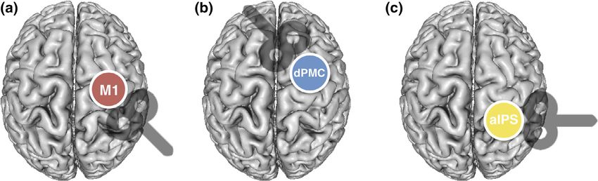
F I G U R E 6 Correlation analyses between strength of IHI and TMS effect. The strength of interhemispheric inhibition between both M1
correlated negatively with the change of finger tapping frequency evoked by TMS interference with ipsilateral M1 only in young healthy
individuals. By contrast, in older subjects IHI between ipsilateral dPMC and contralateral M1 correlated negatively with the effect of ipsilateral
dPMC-interference for finger tapping frequency; (*p < .05)TSCHERPEL ET AL. 649
However, when testing both age groups separately, we found that individuals (86.31% ± 23.44% SD; p = .31, d = 0.25, t(7) = 1.10). Con-
this correlation was primarily driven by the old subjects (young group: sistent with this result, we found a significant linear relationship
r = .08, p = .76; old group: r = −.66, p = .008). Further correlation ana- between individual changes of finger tapping frequency evoked by
lyses, especially with age, did not reveal any significant relationship. ipsilateral M1-stimulation and interhemispheric M1-M1 inhibition
Subsequently, we sought to link our findings for the finger tapping only in young subjects (young group: r = −.997, p = .002; old group:
task to interhemispheric inhibition effects. Repeated measures r = −.22, p = .60) (Figure 6). Although, for interhemispheric inhibition
ANOVA indicated a significant main effect for STIMULATION SITE from ipsilateral dPMC to contralateral M1, we found a nonsignificant
(F(1,11) = 19.94, p = .001, η2 = 0.64) and a significant interaction effect between-group difference (p = .14, d = 0.54, t(11) = 1.57), young sub-
for STIMULATION SITE × GROUP (F(1,11) = 12.41, p = .005, η2 = 0.53) jects featured on average facilitatory dPMC-influences in (116.1%
(main effect GROUP: F(1,11) = 4.01, p = .07, η2 = 0.27), thereby ± 23.4% SD), whereas older participants showed a decrease of facilita-
pointing to an age-dependent difference in interhemispheric inhibi- tion compared to young subjects with a trend toward inhibition
tion. A post hoc t test revealed a double dissociation for region and (94.0% ± 22.0% SD). Plotting the individual data revealed inhibitory
age, hence resembling TMS effects observed for finger tapping fre- influences in a relevant number of older subjects while none of the
quency: For interhemispheric inhibition from ipsilateral M1 to contra- young subjects showed inhibitory dPMC-M1 interactions (Figure 7).
lateral M1, we found a significant group difference between young Moreover, we found a significant correlation between the TMS
and old subjects (p = .002, d = 2.27, t(10) = 3.95). While young partici- effects during ipsilateral dPMC-stimulation in older but not in younger
pants showed strong M1-M1 IHI with significantly reduced condi- participants (young group: r = .30, p = .59; old group: r = −.71,
tioned MEPs (33.98% ± 22.87% SD; p = .016, d = 1.62, t(4) = 4.01, p = .049), indicating that in particular older individuals featuring stron-
two-sample t test between unconditioned MEPs and conditioned ger interhemispheric inhibition were more susceptible to interference
MEP), IHI in older subjects was diminished compared to young with ipsilateral dPMC (Figure 6). Further correlation analyses between
F I G U R E 7 Individual data of inhibition effects. The single subject data of interhemispheric inhibition between M1-M1 and dPMC-M1. Please
note, that older subjects featuring interhemispheric inhibitory influences are marked with color650 TSCHERPEL ET AL.
age and IHI did not reveal any significant relationship. Hence, the IHI et al., 2006; Hummel et al., 2003; Kim et al., 1993; Verstynen, 2004).
analyses support the findings for finger tapping performance with a In agreement with these findings, we here observed that interference
differential effect of age on the contribution of ipsilateral M1 with ipsilateral M1 significantly impacted upon both finger and hand
and dPMC. tapping tasks in young healthy subjects. Specifically, ipsilateral
M1-disruption led to higher tapping frequencies without changes in
tapping amplitudes or movement smoothness in both tasks,
3.5 | TMS effects on motor performance during the
suggesting an actual improvement of motor performance. Since the
hand tapping task
majority of previous online TMS studies have reported detrimental
For hand tapping frequency, no significant main effect was evident behavioral responses for interfering with ipsilateral M1 (Chen et al.,
for the factor STIMULATION SITE (F(2,60) = 0.06, p = .94, η = 0.002).
2
1997; Davare, Andres, et al., 2006; Foltys et al., 2001), our present
However, paralleling the findings of finger tapping, we found an inter- data—at least at first sight—seems to be at odds with these previous
action effect for STIMULATION SITE × GROUP (F(2,60) = 4.55, studies. However, the fact that we observed an increase of frequency
p = .015, η 2
= 0.13) (main effect GROUP: F(1,30) = 1.32, p = .26, upon ipsilateral M1 interference not only for the finger tapping but
η = 0.04). Likewise, hand tapping frequencies tended to be faster in
2 also for the hand tapping task, which served as an additional internal
younger subjects when interfering with ipsilateral M1 and, addition- control task, strengthen our data. Moreover, Davare and colleagues
ally, with ipsilateral IPS, while older subjects showed an increase in (Davare, Duque, Vandermeeren, Thonnard, & Olivier, 2006) have
tapping frequency under ipsilateral dPMC-stimulation. However, shown that disruption of ipsilateral M1 could either advance or delay
effects were much weaker as indicated by the nonsignificant post hoc muscle recruitment dependent on the timing of TMS interference,
one-sample t tests (young group: ipsilateral M1: p = .16, d = 0.39, adding additional support for differential effects of online TMS
t(14) = 1.50; ipsilateral dPMC: p = .68, d = 0.11, t(14) = 0.43, ipsilateral interference.
IPS: p = .11, d = 0.44, t(14) = 1.69) (old group: ipsilateral M1: p = .50, From a mechanistic perspective, online TMS applied to ipsilateral
d = 0.17, t(16) = 0.69; ipsilateral dPMC: p = .12, d = 0.40, t(16) = 1.63; M1 might have modulated interhemispheric inhibition between bilat-
ipsilateral IPS: p = .55, d = 0.15, t(16) = 0.61) (Figure 5). Likewise, nei- eral M1 which is in accordance with the significant relationship that
ther M1-M1 nor dPMC-M1 IHI correlated with hand tapping effects. we found between TMS effects of ipsilateral M1 stimulation and
interhemispheric inhibition exerted by ipsilateral M1 in young sub-
jects. In addition, TMS studies as well as connectivity analyses based
4 | DISCUSSION on task fMRI data have also provided converging evidence that each
M1 exerts reciprocal influences onto its contralateral homolog, which
We here applied online TMS interference to investigate the functional seems to be crucial for motor control and muscle recruitment
relevance of ipsilateral frontoparietal regions for motor performance (Di Lazzaro et al., 1999; Ferbert et al., 1992; Meyer et al., 1995).
in young and older subjects. At the behavioral level, older subjects Accordingly, the interhemispheric influences exerted from ipsilat-
featured a significant slowing in all investigated motor tasks. Impor- eral M1 onto the contralateral M1 have been shown to be initially
tantly, there was no group difference concerning movement accuracy, inhibitory at rest, to decrease progressively when approaching muscle
indicating that preserved movement accuracy was presumably contraction, and to reverse to facilitation before and during muscle
achieved via reduced movement speed. Interfering with neural activity contraction (Davare, Andres, et al., 2006; Duque et al., 2005; Murase,
during task performance yielded differential effects depending on age Duque, Mazzocchio, & Cohen, 2004). Therefore, depending on the
and stimulation site. A novel finding of our study is the differential rel- timing of TMS disruption relative to movement execution, it could
evance of ipsilateral M1 and ipsilateral dPMC on fast repetitive move- either advance or delay muscle recruitment (Davare, Duque, et al.,
ments depending on age, which was paralleled by age-related changes 2006). Accordingly, with respect to the present study, TMS interfer-
of IHI. In particular, the current findings are compatible with an age- ence seems to have affected the inhibitory role of M1 leading to a
related functional shift from ipsilateral M1 toward ipsilateral dPMC in more effective disinhibition of contralateral M1, thereby increasing
the attempt to maintain tapping performance at high frequencies in tapping frequency in young subjects (Davare, Andres, et al., 2006;
older subjects, while interfering with anterior superior parietal cortex Volz et al., 2017).
activity affected visuomotor performance independent of age. For older subjects, neuroimaging studies have typically revealed
increased activity in ipsilateral M1 compared to young individuals dur-
ing hand-motor tasks (Mattay et al., 2002; Riecker et al., 2006; Ward &
4.1 | HAROLD and the motor system: The role of
Frackowiak, 2003), consistent with an extension of the HAROLD
ipsilateral primary motor cortex in age
model into the motor domain. We could show that interhemispheric
In young adults, there is compelling evidence that ipsilateral M1 con- inhibition between both M1 is attenuated in older subjects compared
tributes to motor tasks of increasing complexity. Neuroimaging- as to young individuals, which is supported by a number of studies
well as TMS studies have revealed that tasks particularly requiring (Coppi et al., 2014; Talelli, Ewas, Waddingham, Rothwell, & Ward,
fine-tuned temporal regulation or high muscle selectivity involve ipsi- 2008; Talelli, Waddingham, Ewas, Rothwell, & Ward, 2008). Hence,
lateral M1 (Chen, Gerloff, Hallett, & Cohen, 1997; Davare, Andres, less hemispheric asymmetry might be a consequence of reducedTSCHERPEL ET AL. 651
interhemispheric inhibition effects leading to disinhibition of neural Highly similar to ipsilateral M1, also ipsilateral dPMC modulates
activity in ipsilateral M1 (Langan et al., 2010; Talelli, Greenwood, & the activity of contralateral M1 through inhibitory and facilitatory
Rothwell, 2006; Ward et al., 2008). As several authors could not link influences (Bäumer et al., 2009; Hinder, Fujiyama, & Summers, 2012;
over-activation of ipsilateral M1 to better motor performance, higher Koch et al., 2006; O'Shea, Sebastian, Boorman, Johansen-Berg, &
activity of this area might also represent unspecific disinhibition rather Rushworth, 2007). Most likely these effects are mediated on the ana-
than compensatory mechanisms (Riecker et al., 2006; Talelli, Ewas, tomical basis of direct commissural fibers from dorsal premotor cortex
et al., 2008). to contralateral M1, which have been confirmed in monkeys
Taken together, these findings are relevant for the interpretation (Boussaoud, Tanné-Gariépy, Wannier, & Rouiller, 2005; Jenny, 1979;
of our data, as unlike in younger subjects, we could not find a signifi- Marconi, Genovesio, Giannetti, Molinari, & Caminiti, 2003). This
cant functional involvement of ipsilateral M1 in the employed motor notion receives additional support since IHI with short interstimulus
tasks in older subjects. intervals is suggested to probe direct transcallosal pathways (Chen,
2004; Chen et al., 2003; Hinder et al., 2012).
Hinder and colleagues found that older subjects particularly rely
4.2 | PASA and the motor system: Premotor shift in on a stronger modulation of inhibition toward facilitation to release
aging the motor signal to the contralateral hand (Hinder et al., 2012; Levin,
PASA would predict an aging-associated shift from posterior to ante- Fujiyama, Boisgontier, Swinnen, & Summers, 2014). Although we
rior regions, not only in the contralateral but also ipsilateral hemi- were able to assess IHI only in a limited subgroup of the original
sphere (Davis et al., 2008; Michely et al., 2018), which is paralleled by cohort, we here found hints that, compared to young subjects, inter-
a key finding of our study—an age-related anterior shift within the hemispheric interactions between ipsilateral dPMC and contralateral
M1 of older participants were shifted toward inhibition. Therefore, a
motor network. The disruption of ipsilateral dPMC-activity affected
similar modulation stated for ipsilateral M1 in young subjects, namely
motor performance in older but not younger subjects, indicating a
a TMS-induced disruption of the inhibition exerted from ipsilateral
shift from ipsilateral M1 to premotor cortex during repetitive high-
dPMC onto contralateral M1, may have led to a more effective disin-
frequency movements.
hibition of contralateral M1 and mediated the improvement of motor
One might argue that the observed stimulation effects in dPMC in
performance in older subjects. This notion receives further support
the older group may result from stimulating adjacent M1 neurons, or
from the observed relationship between TMS effects of ipsilateral
unspecific facilitation effects due to the sensory input associated with
dPMC stimulation and IHI exerted by ipsilateral dPMC in older sub-
TMS (Duecker & Sack, 2013). However, since we observed a double
jects. Hence, during repetitive high-frequency movements, ipsilateral
dissociation between groups and brain areas, this hypothesis appears
dPMC presented a similar TMS profile in older subjects, analogous to
rather unlikely.
the response of younger subjects in ipsilateral M1. Furthermore, the
Given that neuroimaging studies have frequently demonstrated a
interhemispheric inhibition exerted from ipsilateral dPMC in older
greater activation of ipsilateral premotor regions during motor tasks
subjects revealed a similar relationship to the TMS effect of dPMC
with advancing age (Heuninckx, Wenderoth, & Swinnen, 2008;
interference, paralleling the results of interhemispheric inhibition and
Mattay et al., 2002; Riecker et al., 2006; Ward & Frackowiak, 2003)
TMS effect of ipsilateral M1 in young subjects.
and we here found an improvement of motor performance, one inter-
Notably, the IHI experiment bears some limitations such as the
pretation of the present data is an detrimental influence of ipsilateral
small sample size which reduces the generalizability of our data and
premotor cortex activity upon contralateral M1, which is released by
the fact that both experiments were not conducted on the same day.
TMS interference—similar to what has been described for neural
Nevertheless, the age-related differential findings fit the results
over-activity in the contralesional hemisphere in stroke patients
obtained in the main experiment and support our interpretation of an
(Grefkes et al., 2008; Murase et al., 2004; Rehme, Fink, Cramon, Y, & age-related functional shift for finger tapping performance.
Grefkes, 2011; Volz et al., 2017). So far, neuroimaging studies have revealed that premotor cortex
However, we would like to challenge this interpretation, espe- is more activated during motor tasks in older individuals (Heuninckx,
cially since we could also detect an improvement in task perfor- 2005; Heuninckx et al., 2008; Ward et al., 2008) and our data is com-
mance evoked by TMS in young healthy individuals in whom it patible with a causal involvement of dorsal premotor cortex in motor
seems rather unlikely that ipsilateral areas hold a detrimental role for tasks in older individuals. Given that we found a relationship between
motor performance. As the effects evoked by the stimulation are baseline motor performance and changes of finger tapping frequency
thought to be influenced by several factors like timing of stimulation evoked by ipsilateral premotor cortex stimulation, dorsal premotor
onset or stimulation intensity (Davare, Duque, et al., 2006; Foltys cortex seems to be particularly relevant in older subjects with poor
et al., 2001; Jahanshahi & Rothwell, 2000; Silvanto & Cattaneo, motor performance. Therefore, the age-related functional premotor
2017; Walsh & Cowey, 2000), the TMS-induced effects of a distinct shift, although potentially representing a reorganization due to struc-
region allow to draw conclusions about the causal and functional tural and biochemical changes in the motor system (Seidler et al.,
involvement of this region, but does not necessarily determine a 2015; Talelli, Waddingham, et al., 2008; Ward et al., 2008), could not
beneficial or maladaptive role. be considered as fully compensatory from a behavioral perspective.You can also read