Precise slow oscillation-spindle Coupling promotes Memory Consolidation in Younger and older Adults
←
→
Page content transcription
If your browser does not render page correctly, please read the page content below
www.nature.com/scientificreports

                        OPEN       Precise Slow Oscillation–Spindle
                                   Coupling Promotes Memory
                                   Consolidation in Younger and Older
Received: 19 October 2018
Accepted: 25 November 2018         Adults
Published: xx xx xxxx
                                   Beate E. Muehlroth1, Myriam C. Sander1, Yana Fandakova1, Thomas H. Grandy1, Björn Rasch2,
                                   Yee Lee Shing1,3 & Markus Werkle-Bergner 1
                                   Memory consolidation during sleep relies on the precisely timed interaction of rhythmic neural events.
                                   Here, we investigate differences in slow oscillations (SO; 0.5–1 Hz), sleep spindles (SP), and their
                                   coupling across the adult human lifespan and ask whether observed alterations relate to the ability
                                   to retain associative memories across sleep. We demonstrate that older adults do not show the fine-
                                   tuned coupling of fast SPs (12.5–16 Hz) to the SO peak present in younger adults but, instead, are
                                   characterized most by a slow SP power increase (9–12.5 Hz) at the end of the SO up-state. This slow
                                   SP power increase, typical for older adults, coincides with worse memory consolidation in young age
                                   already, whereas the tight precision of SO–fast SP coupling promotes memory consolidation across
                                   younger and older adults. Crucially, brain integrity in source regions of SO and SP generation, including
                                   the medial prefrontal cortex, thalamus, hippocampus and entorhinal cortex, reinforces this beneficial
                                   SO–SP coupling in old age. Our results reveal that cognitive functioning is not only determined by
                                   maintaining structural brain integrity across the adult lifespan, but also by the preservation of precisely
                                   timed neural interactions during sleep that enable the consolidation of declarative memories.

                                   Research in animals and younger adults yields converging evidence that newly acquired information is repro-
                                   cessed during deep non-rapid eye movement (NREM) sleep. According to the framework of system consoli-
                                   dation1,2 declarative memories are initially processed by a fast learning system in the hippocampus that binds
                                   information into a transient representation3. Only repeated reactivation of memories in the hippocampus then
                                   eventually allows transfer to a long-term store in the neocortex4–6. But how does the brain ensure precise commu-
                                   nication between cortical modules that allows for the reorganization of memory traces during sleep?
                                       During sleep, effective communication is enabled by the fine-tuned hierarchical succession of several brain
                                   rhythms governed by large-amplitude, low-frequency oscillations, so-called slow oscillations (SO; 0.5–1 Hz) that
                                   mainly emerge from prefrontal brain areas7–10. Via cortico–thalamic projections, neurons in the thalamic retic-
                                   ular nucleus are activated during the depolarized up-states of SOs resulting in the initiation of fast sleep spindles
                                   (SP; 12.5–16 Hz)11–14. These fast SPs, in turn, synchronize cortical activity in a precise spatiotemporal manner
                                   thereby establishing a correlated activation between the hippocampus and frontal brain regions15–18. As the
                                   excited neurons exhibit a massive influx of Ca2+, synaptic changes become facilitated and memory consolidation
                                   is enabled19,20. Further nesting of hippocampal high-frequency bursts, known as sharp-wave ripples, within the
                                   excitable troughs of fast SPs16,21,22 optimizes the hippocampo–neocortical dialogue. A second SP type, so-called
                                   slow SPs (9–12.5 Hz), occurs during a time window in which neural depolarization (i.e., the SO up-state) transi-
                                   tions into hyperpolarized down-states12,23,24. A specific role of slow SPs in the consolidation of memories is not
                                   yet established2,23,25.
                                       Typically, SOs and SPs are studied with regard to their independent contribution to consolidation mech-
                                   anisms. However, as system consolidation is assumed to rely on a tight coupling of SOs, SPs, and sharp-wave
                                   ripples4,26, recent studies have started to focus on the exact timing of brain rhythms11,23,24,27,28 and its functional

                                   1
                                    Center for Lifespan Psychology, Max Planck Institute for Human Development, Berlin, Germany. 2Department
                                   of Psychology, University of Fribourg, Fribourg, Switzerland. 3Department of Developmental Psychology, Goethe
                                   University Frankfurt, Frankfurt am Main, Germany. Correspondence and requests for materials should be addressed
                                   to B.E.M. (email: muehlroth@mpib-berlin.mpg.de) or M.W.-B. (email: werkle@mpib-berlin.mpg.de)

        Scientific Reports |   (2019) 9:1940 | https://doi.org/10.1038/s41598-018-36557-z                                                               1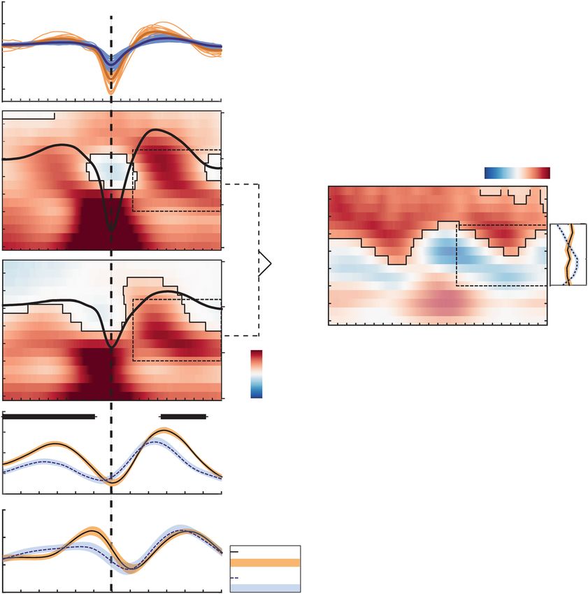
www.nature.com/scientificreports/

                           Figure 1. Study design. (a) Experimental procedure. The memory task at the core of the experiment consisted
                           of a learning phase and an immediate recall on Day 1 as well as a delayed recall approximately 24 hours later
                           (red boxes). Sleep was monitored in the nights before (PRE) and after (POST) learning using ambulatory
                           polysomnography (blue boxes). A prior adaptation night (dashed blue box) familiarized participants with the
                           sleep recordings. (b) Memory paradigm (cf. Fandakova et al.64). (A) During study, participants were instructed
                           to remember 440 (younger adults) or 280 scene–word pairs (older adults). (B) During the cued-recall and
                           feedback phase the scene was presented as a cue to recall the corresponding word. Irrespective of recall
                           accuracy, the original pair was presented again to allow for re-study. The whole cued-recall and feedback cycle
                           was performed once in younger and twice in older adults. (C) During final recall, scenes again served as cues to
                           recall the corresponding word, but no feedback was provided. (D) Delayed cued-recall took place approximately
                           24 hours later. Participants were presented with the scenes only and had to indicate if they still remembered the
                           associated word. Afterwards they had to select the corresponding second letter of the word to verify their true
                           memory of the associate.

                           significance for sleep-dependent memory consolidation23,28–31. Recent experiments in rats demonstrated that
                           reinforcing the coordinated pattern of SOs, SPs, and sharp-wave ripples boosts memory consolidation of object
                           locations29, whereas suppression of these patterns has disruptive effects on sleep-dependent memory consoli-
                           dation30. In humans, SO–SP coupling is enhanced by prior learning23,28 and is triggered by replaying previously
                           learned material during sleep32. Although first studies have highlighted the involvement of SO–SP coupling in
                           memory consolidation, particularly in humans, evidence for this link is still sparse. Further, it remains to be
                           established under which circumstances the coordination of SOs and SPs is most beneficial in supporting the
                           sleep-dependent reorganization and stabilization of memory contents.
                               SO–SP coupling, for instance, can be critically impaired in disrupted sleep. This is particularly common in
                           elderly adults who often lack the deepest NREM sleep stage, so-called slow-wave sleep33–35. In old age, SOs and
                           SPs themselves appear less often and with reduced amplitudes36–38. Age-related changes in brain structure possi-
                           bly contribute to the observed sleep alterations by impairing SO and SP generation and propagation37,39–42. This
                           may also impact SO–SP coupling itself, thereby resulting in impaired memory stabilization during sleep43. In
                           fact, several studies report age-related loss in overnight memory retention44–46, which can be linked to reduced
                           slow-wave sleep39,40 and a decline in SP occurrence47. In light of the growing interest in the interaction of SOs
                           and SPs, very recent evidence highlights that SO–SP coupling is indeed dispersed in elderly adults, resulting in
                           impaired overnight memory consolidation43. As this disturbed nesting of SOs and SPs may constitute a possible
                           target for clinical interventions48, the preconditions and the functional significance of SO–SP coupling across the
                           entire adult lifespan require investigation.
                               Following this rationale, it was our aim to examine age-related changes in the coupling of SOs and both
                           slow and fast SPs. We asked whether observed alterations can be explained by structural brain atrophy in source
                           regions of SO and SP generation and whether the resulting SO–SP dispersion negatively impacts the ability to
                           retain associative memories across sleep. In the present study, younger and older adults completed an associative
                           memory paradigm, consisting of a learning session of scene–word pairs on the first day and a delayed cued-recall
                           task on the following day (Fig. 1). During the nights before and after learning, sleep was monitored using ambu-
                           latory polysomnography (PSG). Structural brain integrity was assessed by voxel-based morphometry (VBM) of
                           structural magnetic resonance images (MRI). By comparing the PSG recordings of younger and older adults, we
                           identify age-related differences in the coordination of SOs and SPs. Using estimates of gray matter volume we

Scientific Reports |   (2019) 9:1940 | https://doi.org/10.1038/s41598-018-36557-z                                                           2
www.nature.com/scientificreports/

                                          Younger Adults – Median        Older Adults – Median
                                          [1st quartile; 3rd quartile]   [1st quartile; 3rd quartile]   z          p
                            TST (min)     457.00 [420.0; 508.5]          417.50 [370.75; 447.25]            2.60       0.009
                            WASO (min)       8.5 [1.75; 12.75]             20.5 [9.50; 32.25]           −3.56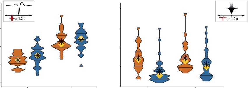
www.nature.com/scientificreports/
Figure 2. Fast and slow SPs coincide with SOs in both younger and older adults. Asterisks mark significance
after correction for multiple comparisons. (a) About one third of all fast SPs and more than half of all slow SPs
occur within an interval of ±1.2 s around the trough of a SO. (b) Only a small percentage of all appearing SOs
is coupled to the occurrence of SPs. SOs in older adults (blue) occur less frequently within an interval of ±1.2 s
around fast SP centers than in younger adults. YA: younger adults; OA: older adults; SO: slow oscillation; SP:
spindle.
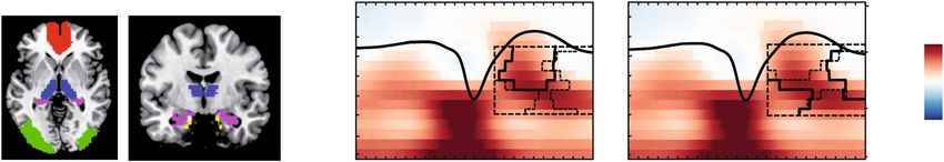
Figure 3. Temporal specificity of fast and slow SP occurrence. Peri-event time histograms of fast and slow SPs
co-occurring with frontal SOs are depicted for both age groups. Standard errors of the 100-ms bins are included
as black vertical lines. Reference distribution obtained after randomization of the data is shown in red. Red
asterisks indicate significantly increased SP occurrence contrasted with the reference distribution (cluster-based
permutation test, cluster α < 0.05, positive clusters only). Vertical dashed lines mark the SO trough. Average
SOs are shown for each age group. (a) Frontal slow SPs in younger adults globally peak at the up- to down-state
transition. (b) The frontal slow SP peak at the up- to down-state transition preceding the SO trough observed
in younger adults disappears in older adults. (c) In younger adults, fast SPs prominently peak during the SO
peak (SO peak at 480 ms, significant positive cluster: 200–700 ms). (d) In older adults, fast SPs are similarly
modulated during the SO up-state as observed in younger adults but their occurrence is maximized before the
SO has peaked (SO peak at 618 ms, significant positive cluster: 200–400 ms). YA: younger adults; OA: older
adults; SO: slow oscillation; SP: spindle.
SO–SP coupling differentially changes for fast and slow SPs in younger and older adults.
Having established the overall maintenance of a time-specific SO–SP coordination in older adults, we further
probed this pattern by comparing the oscillatory power in the SP frequency range for time segments with and
without SOs (cf. Materials and Methods). During SO trials (centered on the trough of the respective SO ± 1.2 s),
power in the SP frequency range indeed differed significantly from randomly selected intervals without SOs (for
all clusters: p < 0.001, Figs 4b and S2). This effect was present in both age groups and suggests a coupling of SPs
Scientific Reports | (2019) 9:1940 | https://doi.org/10.1038/s41598-018-36557-z 4
www.nature.com/scientificreports/

                           Figure 4. SO–SP coupling is differentially expressed in younger and older adults. (a) Average frontal SOs
                           are depicted for each younger (light orange) and older (light blue) individual. In older adults, the amplitude
                           of SOs is reduced and the frequency is decreased. (b) Differences in wavelet power for SO trials (respective
                           trough ± 1.2 s) compared to baseline trials without SOs are depicted (in t-score units, reference window for later
                           analyses outlined by dashed black line). Significant clusters (cluster-based permutation test, cluster α < 0.05) are
                           highlighted and outlined. The average frontal SO for each age group is shown in black (the scale in μ is indicated
                           on the right of each time–frequency graph). In both age groups, EEG activity is modulated as a function of
                           the SO phase. (c) Time–frequency t-map illustrating reliable age differences in SO-related power increases
                           compared to baseline trials (cluster p < 0.001). Significant age differences in fast SP power modulation are tied
                           to the SO up-states. The probabilities of frequency peaks (maximal t-values) across time (reference window
                           outlined by dashed black line) are shown in the line plot to the right. In older adults the probability of high fast
                           SP activity is significantly reduced during the SO up-state. In contrast, compared to younger adults, a strong
                           increase in slow SP frequencies is more likely (right line plot, all clusters: p ≤ 0.007, marked by asterisks). (d)
                           Temporal evolution of SO-specific power modulations (expressed in t-values) in the slow and fast SP range
                           extracted from the time–frequency maps above. In older adults, compared to younger adults, SO-specific fast
                           SP activity is significantly reduced during and following the SO peak (significant clusters marked in black). YA:
                           younger adults; OA: older adults; SO: slow oscillation; SP: spindle; TF: time–frequency.

                           to specific phases of the underlying SO. Nevertheless, each age group displayed a distinct modulation of the effect
                           (Figs 4b and S2).
                               In younger adults, we observed increases in fast SP power (12.5–16 Hz) during the SO up-state but not during
                           the down-state (Fig. 4b). Fast SP power increased most strongly during and shortly after the SO peak following
                           the down-state (400 to 700 ms, SO peak at 480 ms). Although this effect was visible across all chosen derivations
                           (Supplementary Figs S2 and S3), it was more pronounced over centroparietal sites. Frontal and central slow SP
                           activity (9–12.5 Hz) was increased during the whole SO interval, but peaked during the up- to down-state transi-
                           tion (−500 to 200 ms; Supplementary Figs S2 and S4).
                               In older adults, as expected, SOs themselves were expressed differently than in younger adults, with a
                           decreased amplitude and frequency (SO amplitude: z = 5.69, p < 0.001, MdYA = 123.54 μV, MdOA = 74.82 μV; SO
                           frequency: z = 2.68, p = 0.007, MdYA = 0.79 Hz, MdOA = 0.78 Hz, Fig. 4a). But SO-modulated EEG activity also

Scientific Reports |   (2019) 9:1940 | https://doi.org/10.1038/s41598-018-36557-z                                                            5
www.nature.com/scientificreports/
differed from younger adults (Fig. 4b–d). To identify age-specific characteristics of this global pattern we directly
compared the time–frequency patterns (i.e., the SO–baseline power differences) of both age groups (Fig. 4c).
Further, the temporal evolution of frontal slow SP (9–12.5 Hz) and central fast SP power modulation (12.5–16 Hz;
averaged t-values of the SO–baseline contrast within the respective frequency band) was extracted for both age
groups and compared by means of a cluster-based permutation test (Fig. 4d). Finally, as studies so far have indi-
cated that SP activity during the SO up-state is critical for memory consolidation28,32, we precisely determined in
which frequencies SP activity was modulated most strongly during the SO up-state (cf. Materials and Methods).
We thus compared the probability of frequency peaks between 9 and 16 Hz (maximal t-values of the SO–baseline
contrast) during the SO up-state (0.2–1.2 s after the SO trough) between younger and older adults (line subplot
to the right of Fig. 4c).
Over all derivations, power in higher frequencies (16–20 Hz) was globally reduced during SOs in older com-
pared to younger adults whereas fast SP power differences (12.5–16 Hz) were reduced during the SO up-states
only (all significant positive clusters: p ≤ 0.015, Figs 4c and S2). In older adults, the power peak over central sites
during the up-state was more stretched in time and shifted to lower frequencies (9–13 Hz, i.e., the slow SP fre-
quency band, Fig. 4b). Moreover, SO-specific fast SP activity was reduced during and following the positive SO
peak (SO peak at 618 ms; significant cluster: 459 ms–927 ms; cluster p = 0.013, Fig. 4d). In contrast, older adults
revealed a strong increase in slow SP activity during the SO up-state (Figs 3D and 4B). Both the age comparison of
the global time–frequency pattern (Fig. 4d) but also the age comparison of slow SP activity modulated during SOs
(Fig. 4d, lower panel) indicated a similar SO-specific slow SP power increase in both younger and older adults
(all clusters: p ≥ 0.073). Nevertheless, while younger adults showed an almost equal modulation of slow and fast
SP power during the SO up-state, older adults displayed a stronger emphasis on lower frequencies: compared to
younger adults, fast SP activity was significantly less and slow SP activity significantly more increased during the
SO up-state (all clusters: p ≤ 0.007, line subplot to the right of Fig. 4c).
To summarize, we found a characteristic SO–SP coupling pattern in younger adults that was marked by a
strong increase in fast SPs coupled to the SO peak as well as an increase in slow SPs during the up- to down-state
transition. This pattern changed significantly across the adult lifespan. Within this ‘aged’ SO–SP coupling, three
characteristics were striking: First, fast SP modulation was reduced overall in the elderly. Second, a reliable fast SP
increase comparable to that of younger adults was limited to the time period before the SO peak. Together with an
overall reduced frequency of SOs, fast SPs were thus no longer precisely tied to the SO peak. Finally, slow SPs were
similarly modulated in younger and older adults. Together, these changes result in the typical appearance of an
‘aged’ SO-SP coupling pattern with higher coupling in the slow SP frequency range at the end of the SO up-state.
Reduced memory retention is associated with dispersed SO–SP coordination in younger adults.
After establishing age-related changes in SO–SP coordination during the SO up-state, we asked how these altera-
tions relate to the ability to retain memories across sleep. As central fast SP activity tied to the SO up-state is crit-
ically involved in memory consolidation28,32, we focused our analyses on SP frequencies during the SO up-state
(9–16 Hz, 0.2–1.2 s) and on electrode Cz, where we observed a lack of SO–fast SP coupling in older adults (Figs 3
and 4; for frontal channels and Pz, see Supplementary Fig. S5). As a control, we ran the same analysis for a second
time window (−500 to 200 ms) during the SO up- to down-state transition that reflects SO-modulated slow SP
activity. Here, no significant associations with memory retention were observed after controlling for multiple
comparisons (cluster p = 0.169, Supplementary Fig. S6). To minimize biases in SP activity arising from potentially
preceding SOs23, all reported analyses were restricted to the up-state following the SO trough.
To quantify memory retention, we measured each participant’s ability to recall scene–word associations that
had been successfully encoded the day before. We calculated the percentage of correctly recalled items during
delayed recall relative to all items that were correctly retrieved during immediate recall before sleep. Whereas
younger and older adults did not differ with regard to their learning performance on Day 1 (t(59) = 1.62, p = 0.11;
MYA = 53.83%, SDYA = 20.80%; MOA = 45.80%, SDOA = 19.02%), memory consolidation was significantly
reduced in older adults. On Day 2, younger adults retained on average 90.21% of the previously learned pairs
(SDYA = 7.42%). However, older adults showed significantly worse memory retention across sleep (MOA = 71.13%,
SDOA = 13.33%, t(56) = 7.33, p < 0.001; Fig. 5a).
Correlational analyses between SO–baseline power differences (expressed in t-values) and memory reten-
tion were performed separately for each time–frequency point. Significant associations were identified by testing
against a bootstrapped reference distribution of the EEG–memory correlations. In this way, we could identify
significant correlation clusters that represent the association between memory retention and a specific pattern
of EEG activity modulated during the SO up-state (cf. Materials and Methods, Fig. 5b for a schematic depiction
of the performed correlation technique). Across both age groups, we identified a significant positive correlation
cluster (cluster p = 0.024, mean r = 0.36, Fig. 5c): more fast SP activity during the SO up-state, which is in general
more typical for SO–SP coupling in younger adults, was associated with better memory retention.
To further probe the association between memory retention and SO-modulated EEG activity, we then proceeded
to conduct the very same analysis within each age group separately. Here, we found a significant negative correlation
cluster in younger adults (cluster p = 0.028, mean r = −0.52, Fig. 5d): Younger adults showing a SO-specific power
modulation with less emphasis on fast SP activity during the SO peak but rather on lower frequencies (9–13 Hz)
during and following the SO peak showed worse overnight retention of memories. As depicted in the right panel
of Fig. 5d, the cluster extracted in younger adults overlapped with the typical SO-related activity pattern of older
adults that is marked by a shift to lower SP frequencies and a more stretched power peak sustained until the end of
the SO up-state. Younger adults displaying a coupling pattern typical for older adults tended to be worse at retaining
previously learned information overnight (cf. Supplementary Fig. S7). Although no significant correlation cluster
was extracted in older adults, the average correlation within the cluster identified in younger adults did not differ
Scientific Reports | (2019) 9:1940 | https://doi.org/10.1038/s41598-018-36557-z 6www.nature.com/scientificreports/
Figure 5. SO–SP coordination relates to memory retention. (a) Age-specific distribution of memory retention
in younger (orange) and older adults (blue). Older adults’ overnight memory retention is significantly reduced.
(b) Extraction of significant correlation clusters between SO-specific EEG activity and memory retention was
achieved by contrasting a test statistic (i.e., the correlation for each time–frequency point) against a reference
distribution of bootstrapped EEG–behavior correlations. (c) Left: Correlations between memory retention and
neuronal activity during SO up-states are shown for the whole sample (significant positive correlation cluster
highlighted and outlined). Middle: Maximal positive correlation between SO-related power modulations
(t-values) and memory retention in the cluster (least-squares fit line shown in black). Right: Correspondence
between the correlation cluster (outlined in black) and the age-specific time–frequency profiles (reference
window for the correlation analyses marked by dashed black line). Fast SP activity during the SO peak, that
is typically more expressed in younger adults, is related to better memory retention. (d) Left: In younger
adults, a significant negative correlation cluster (highlighted and outlined) was detected. Middle: Maximal
Scientific Reports | (2019) 9:1940 | https://doi.org/10.1038/s41598-018-36557-z 7www.nature.com/scientificreports/
negative correlation between SO-related power modulations (t-values) and memory retention in the cluster
extracted within YA. For completeness, the corresponding association within OA is depicted as well. Right:
Correspondence between the correlation cluster extracted within YA and the age-specific time–frequency
profiles. The significant cluster found in younger adults corresponds to the power increases typical for older
adults. Note. Scatter plots only serve illustrative purposes. Hence, no significance is stated in the respective
subplots. YA: younger adults; OA: older adults; SO: slow oscillation; SP: spindle; TF: time–frequency.
significantly between age groups (rOA = −0.11, z = −1.64, p = 0.10, Fig. 5d). This suggests that the same functional
association between SO-modulated SP activity and memory retention exists in both younger and older adults.
To conclude, we found evidence for an association between the coordination of SOs and SPs and memory
retention. In general, high levels of fast SP power during and following the positive SO peak were related to better
memory retention. Moreover, our results suggest that inter-individual differences in SO–SP coupling as maxi-
mized between age groups also drive differences in memory retention within age groups. Dispersed SO–SP cou-
pling, which was characteristic of older adults, already predicted worse memory retention among younger adults.
Structural brain integrity in old age promotes ‘youth-like’ SO–SP coupling. In line with previous
reports on the influence of age-related brain atrophy on the generation of sleep oscillations39,40, we finally asked
whether the identified age-specific modulation of SP activity during the SO up-state is associated with measures
of brain volume in specific regions of interest (ROI). As ROIs we primarily included brain regions that are con-
sidered source regions for the generation of SOs and SPs9,11 and proposed to be involved in memory processing
during sleep5 (Fig. 6a). To exclude any age-related confounds, measures of brain volume were derived by using
voxel-based morphometry (VBM) and correcting the extracted ROI-specific measures for total intracranial vol-
ume (TIV)50. Again, we focused our analysis on frequencies between 9 and 16 Hz and a time window between
0.2 and 1.2 s after the SO trough. Whereas the cluster-corrected correlation analysis (cf. Fig. 5b, Materials and
Methods) did not indicate any significant association between brain volume and EEG power in younger adults
(all clusters: p ≥ 0.073; Supplementary Fig. S8), we observed a very specific pattern in older adults.
In older adults, larger medial prefrontal cortex (mPFC) volume was associated with higher power in the fast
SP range during the SO peak (mean rmPFC = 0.43, cluster pmPFC = 0.018). Thalamus volume showed a similar posi-
tive relation with SP activity during the SO peak. This effect, though, was less precise with regard to the frequency
range (mean rthalamus = 0.45, cluster pthalamus = 0.013, see Fig. 6). For hippocampus, entorhinal, and occipital cortex
even broader effects were observed. Greater volume in these regions was not only associated with higher fast SP
power during the SO peak, but generally with more global neuronal activation during the SO up-state. In particu-
lar, the observed effect extended to slow SP frequencies at the end of the SO up-state (mean rhippocampus = 0.47, clus-
ter phippocampus = 0.004; both mean rentorhinal cortex = 0.46, cluster pentorhinal cortex = 0.013 and 0.031; mean roccipital lobe =
0.42 and 0.43, cluster p occipital lobe = 0.027 and 0.035).
To summarize, older adults with less age-related decline in mPFC and thalamus volume showed a more pre-
cise coupling between fast SPs and SOs time-locked to the SO peak – precisely the pattern observed in younger
adults. Less age-related decline in medial temporal lobe areas was moreover associated with overall enhanced
neuronal activity during the SO up-state. We thus conclude that structural brain integrity in old age promotes
‘youth-like’ SO–SP coupling.
Discussion
This study investigated whether the coupling of SOs and SPs during sleep changes across the adult lifespan and
how these alterations relate to the ability to retain associative memories across sleep. We demonstrate that older
adults do not display the central fast SP power increase time-locked to the SO peak that we observed in younger
adults. Instead, they exhibit overall reduced fast SP power modulations and a fast SP peak that is shifted before
the SO peak. Overall, the ‘aged’ SO–SP coupling pattern is most characterized by increases in slow SP power at
the end of the SO up-state. We find evidence that this increase in slow SP power, typical for older adults, coincides
with worse memory consolidation, whereas a ‘youth-like’ precision of fast SPs coupled to the SO peak promotes
memory consolidation across the entire adult lifespan. Moreover, greater structural integrity in brain regions
involved in the generation of SOs and SPs relates to an intact ‘youth-like’ SO–SP coupling in old age. Overall, our
results resonate well with a recent report by Helfrich and colleagues on age-related differences in SO–SP phase–
amplitude couplings and their functional significance for memory retention43. We extend these findings by apply-
ing analysis methods that are sensitive to age-related shifts in both the time and frequency domain and explicitly
base our analyses on the differentiation between slow and fast SPs. Together with Helfrich and co-authors43, we
are the first to demonstrate that an age-related dispersion of SO–SP coupling, as caused by structural brain atro-
phy, relates to impaired memory consolidation in old age.
Age-related dispersion of SO–SP coupling. Effective neural communication during NREM sleep is gov-
erned by SOs that, during their up-states, initiate the generation of thalamic fast SPs11. Here, we found that a gen-
erally constant pattern of SP–SO co-occurrence exists across adulthood, although SO and SPs did not co-occur
in a one-to-one fashion (Fig. 2). Our results imply that: (1) Although it appears that thalamic SP generation is
triggered by frontal SOs11,12, at least one other mechanism initiating SPs exists. (2) SO-induced SP initiation is not
a necessity, but rather a rare occurrence. Nevertheless, if SOs and SPs co-occur, they do so in a precisely timed
manner, as discussed next.
Consistent with a wide range of studies, we identified a precisely timed hierarchical structure of SOs and SPs
in younger adults with fast SPs appearing during the highly depolarized SO peak and slow SPs occurring at the
Scientific Reports | (2019) 9:1940 | https://doi.org/10.1038/s41598-018-36557-z 8www.nature.com/scientificreports/
Figure 6. Structural brain integrity in old age promotes ‘youth-like’ SO–SP coupling. (a) VBM measures are
extracted from ROIs to obtain individual values of brain volume (ROI masks overlaid in color). (b) In older
adults, greater volume in the mPFC and the thalamus is associated with increased fast SP activity at the peak of
the SO. Significant positive correlation clusters (controlled for multiple comparison using a cluster-corrected
correlation approach, cluster α = 0.05) are outlined by solid (mPFC) and dashed black lines (thalamus).
Underlying time–frequency profiles are only plotted for illustration (in t-score units, reference window for the
correlation analysis outlined by dashed black line). (c) Greater volume in the hippocampus (black solid line)
and entorhinal cortex (black dashed line) is positively correlated with more global increases in EEG activity
during the SO up-state. mPFC: medial prefrontal Cortex; VBM: voxel-based morphometry; ROI: region of
interest; SO: slow oscillation; SP: spindle.
up- to down-state transition21,23,24,27. SOs themselves change their appearance in old age and exhibit reduced
amplitudes and slower frequencies in older adults (Fig. 4a)35,37. Flatter SOs have previously been associated with
less synchronous neuronal switches between phases of depolarization and hyperpolarization38. As these phases
precisely time the generation of SP events themselves, SO–SP coordination should, as a consequence, disperse in
old age.
Indeed, consistent with recent reports43,48, in older adults EEG activity during SOs was differentially modu-
lated than in younger adults. (1) In line with previously reported reductions in central fast SPs in old age36,51, fast
SP increases during the SO up-state proved less pronounced in older adults. (2) If fast SPs appeared, they were
no longer precisely tied to the SO peak as observed in younger adults. Rather, they occurred before the up-state
reached its maximum. (3) Over central sites modulation of EEG activity during the SO up-state dispersed, the
power peak was stretched in time and shifted to lower frequencies (9–13 Hz). All things considered, we argue that
even in older adults a very specific pattern of SO–SP coupling exists, although its appearance changes compared
to younger adults, with more emphasis on lower frequencies occurring later during the SO up-state. Possible
causes and mechanisms of this ‘aged’ pattern will be discussed next.
Structural correlates of SO–SP coupling. The process of SO–SP coupling involves intact communi-
cation between different brain regions11. SOs, generated mainly in prefrontal areas, propagate throughout the
brain9 and potentially trigger the initiation of fast SPs in the thalamus11. Here, we indicate that in old age struc-
tural integrity in the mPFC accounts for a ‘youth-like’ SO–SP coupling (Fig. 6). This is in line with Helfrich and
colleagues (2018) who recently demonstrated that greater mPFC volume relates to a more precise coupling of
fast SPs to the SO peak43. Expanding on that observation, we show that additional brain regions are associated
with inter-individual differences in SO–SP coupling. Greater thalamus volume, the main source region of fast SP
generation13, related to more SP activity during the SO peak. The specificity of these effects strongly suggests that
structural integrity in both mPFC and thalamus is crucial for pacing a ‘youth-like’ SO–SP succession. In contrast,
hippocampus and entorhinal cortex volumes were linked to both fast SP activity coupled to the SO peak and
slow SP activity occurring at the up- to down-state transition. Both hippocampus and entorhinal cortex are key
players in the overnight consolidation of memories2. In particular, sharp-wave ripples are generated within the
hippocampus and thereby complete SO–SP coupling21 as the basis for successful memory reprocessing during
sleep29,30. Thus, we speculate that the association between structural integrity of the medial temporal lobe and a
successful coordination of oscillatory processes during sleep is indicative for sharp-wave ripple activity.
In our analyses, significant associations between SO–SP coupling and VBM measures were only found within
the older sample. Importantly, this does not imply that those brain areas do not contribute to an effective hier-
archical nesting of neuronal oscillations during sleep in younger adults. We rather suggest that brain integrity
in mPFC, thalamus and medial temporal lobe areas forms the basis for an effective ‘youth-like’ SO–SP coupling
independent of age. Under conditions of impaired structural integrity in these regions, as is the case in aging50,
the precise timing of SOs and SPs vanishes. This reasoning is in line with Nyberg and colleagues (2012) who argue
that the individuals who experience fewer age-related structural brain changes are more likely to show neural
activity patterns that resemble those of younger adults and that are associated with better performance52.
Functional significance of precise SO–SP coupling. Current theory holds that hippocampal–neo-
cortical communication during sleep is only enabled by the precise hierarchical coordination of SOs, SPs, and
hippocampal sharp-wave ripples53,54. This is achieved by locking fast SPs to the SO peak and further nesting
Scientific Reports | (2019) 9:1940 | https://doi.org/10.1038/s41598-018-36557-z 9www.nature.com/scientificreports/
sharp-wave ripples within the troughs of fast SPs21. Thus, memories initially dependent on a temporary store in
the hippocampus become more reliant on neocortical areas, where they are permanently stored4,26,55.
Here, we show that, across younger and older adults, ‘youth-like’ SO–SP coupling, that is marked by a tight
precision fast SPs to the SO peak, coincided with better memory retention (Fig. 5c). In contrast, a SO–SP cou-
pling pattern most characterized by an increase in slow SP power at the end of the SO up-state, as typically
displayed by older adults, was associated with worse memory retention in younger adults already (Fig. 5d). Our
results support previous evidence showing that the precisely timed interaction of neural rhythms indeed supports
sleep-dependent memory consolidation29–32,43,48. Brain activity is globally synchronized during the SO up-state
and maximized during the SO peak14. This constitutes a moment of effective interregional brain communica-
tion. Fast SPs occurring at this very moment induce a massive influx of Ca2+ into excited neurons. This enables
synaptic plasticity and promotes the stabilization and integration of memory representations56. As most recently
demonstrated, fast SPs are misaligned in older adults, resulting in worse memory retention43. But what is it that
makes the ‘aged’ SO–SP coupling detrimental?
First, the temporal dispersion of ‘aged’ SO–SP coordination in older adults is striking. In older adults, the
fast SP power increase during the SO up-state reaches its maximum before the SO peak (Figs 3 and 4). This
may be explained in two ways: (1) During flatter SOs, as observed in older adults, the switch between up- and
down-states is more variable35,37,38 and as a result SP initiation is also less precise. (2) The mechanism initiating
SPs might be similarly timed in younger and older adults, but as SOs are slower in older adults, fast SPs appear too
early during the SO up-state. Here, global brain synchrony has not reached its maximum yet and interregional
brain communication might not be optimal for reorganization of memories on a brain system level.
The second characteristic of ‘aged’ SO–SP coupling is a power shift to lower frequencies in the slow SP band at
the end of the SO up-state (Fig. 3). At young age already, this shift related to worse memory retention (Figs 5 and S7).
Recent evidence suggests that fast and slow SPs are generated via distinct mechanisms57,58 and have different func-
tional significances. While the occurrence of fast SPs probably reflects thalamo–cortical information processing
necessary for memory transfer, slow SPs may mirror cortico–cortical communication59. Slow SPs per se may not
be sufficient to enable effective memory consolidation during sleep2,23,25,32. When focusing our analysis on the
up- to down-state transition, where slow SPs preferentially occur23,24, no association with memory retention was
detected (Supplementary Fig. S6). To conclude, our results replicate the finding that a precise ‘youth-like’ SO-fast
SP coupling is beneficial for memory consolidation43,48. Vice versa,’aged’ coupling coincides with worse memory
consolidation already in young adulthood. This lines up with the concept of “brain maintenance”52 stating that
cognitive functioning is not only determined by maintained structural integrity across the adult lifespan, but also
by the preservation of functionally specific processes and networks52,60,61. Maintenance of ‘youth-like’ processing
during both encoding60 and memory retrieval61 has previously shown to relate to better long-term memory in
older adults. Here we complement this view by demonstrating that consolidation processes also benefit from
maintained brain mechanisms during sleep.
Materials and Methods
Participants and procedure. Participants. Thirty-four healthy younger adults (19–28 years) and 41
healthy older adults (63–74 years) participated in the experiment. Due to technical failures, data collection from
4 younger and 4 older adults could not be completed. One older adult had to be excluded due to an inciden-
tal MR finding. Accordingly, the final sample for our behavioral analysis consisted of 30 younger (Mage = 23.7
years, SDage = 2.6; 17 females) and 37 older adults (Mage = 68.92 years, SDage = 3.04; 16 females). For some par-
ticipants, parts of the neural data (PSG or MRI) were missing or of bad quality. Final PSG analyses were hence
conducted with 24 younger (Mage = 23.61 years, SDage = 2.55; 13 females) and 31 older adults (Mage = 68.63 years,
SDage = 3.10; 15 females), VBM analyses with 24 younger (Mage = 23.61 years, SDage = 2.55; 13 females) and 29
older adults (Mage = 68.64 years, SDage = 3.10; 14 females). The different samples did not differ with regard to any
behavioral measure.
All participants were right-handed native German speakers with no reported history of psychiatric or neu-
rological disease, or any use of psychoactive medication. To screen for cognitive impairments in the sample of
the elderly, all older adults completed the Mini-Mental State Examination (MMSE; M = 29.24, SD = 1.12, Range:
26–30)62 and passed a brief memory screening beforehand. General subjective sleep quality was controlled by
assessing the Pittsburgh Sleep Quality Index (PSQI)63 and did not show differences between the two age groups.
The study was approved by the Ethics Committee of the Deutsche Gesellschaft für Psychologie (DGPs) and, in
accordance with their guidelines, conducted at the Max Planck Institute for Human Development in Berlin. After
being informed about the complete study procedure, all participants gave written consent to their participation
in the experiment.
Experimental Procedures. The present data were derived from a series of studies investigating age-related
differences in the encoding, consolidation, and retrieval of associative memories (see Fandakova et al., 2018,
for the effects of age and memory quality on false memory retrieval64). Core of the experimental design was a
paired-associative scene–word memory paradigm, consisting of a learning session on the first day (Day 1) as well
as a delayed cued-recall task approximately 24 hours later (Day 2) (see Fig. 1, for illustration of the study proce-
dure). During the nights before and after learning (experimental nights PRE and POST) sleep was monitored at
participants’ home using ambulatory PSG. Prior to the first experimental night an adaptation night familiarized
the participants with the PSG procedure. Structural MRI data were collected on Day 2. Furthermore, EEG was
recorded during learning on Day 1. Additionally, functional MRI data were collected during delayed recall on
Day 2. Neither the EEG nor the fMRI data are included in the present report. Participants completed a short cog-
nitive screening prior to participation in the main study, but did not engage in any other behavioral task during
the course of the experimental procedure described above.
Scientific Reports | (2019) 9:1940 | https://doi.org/10.1038/s41598-018-36557-z 10www.nature.com/scientificreports/
Day 1 – Encoding and immediate cued-recall. During initial study, scene–word pairs were presented on
a black background for 4000 ms. Scene–word pairs were randomized combinations of indoor and outdoor scenes
and concrete nouns. Participants were instructed to remember the scene–word pair using a previously trained
imagery strategy and to indicate how well they were able to form an integrated image of the scene–word pair.
Cued-recall blocks followed immediately after the initial learning phase. Scenes served as cues for participants to
verbally recall the associated word. Recall time was not constrained. Independent of recall accuracy, the correct
scene–word pair was presented again for 3 seconds and participants were instructed to use this opportunity to
remember the word–scene pair by forming an integrated image of the two. Finally, participants completed a final
cued-recall test without feedback at the end of Day 1. This phase served as a measure of learning performance.
Importantly, task difficulty was adjusted between the age groups to achieve comparable recall success of approx-
imately 50% in each age group. This was done in two ways. First, younger adults learned 440 pairs, whereas
older adults learned 280 pairs on Day 1. Second, younger adults completed one cued-recall block with feedback,
whereas older adults completed two cued-recall blocks with feedback. Further details about the encoding task and
the stimulus set are described by Fandakova et al.64.
Day 2 – Delayed cued-recall. Delayed cued-recall of the scene–word pairs consisted of four blocks of
70 experimental trials each and was conducted inside a 3T MRI scanner. For the older age group, all of the 280
studied pairs were presented. For younger adults items were chosen with regard to their learning history. This
resulted in a selection of pairs, half of which had been recalled in the criterion cued-recall the day before. The 280
pictures were displayed for 3500 ms. Meanwhile participants had to decide via keypress if they still remembered
the corresponding word (“remembered” vs. “forgotten”). Afterwards, four letters were presented for 3500 ms.
One of these was the second letter of the correct word and had to be selected by pressing the corresponding key.
If the according word had been forgotten or none of the letters fitted their remembered answer word, participants
were told to choose one of the letters at random. Answers during delayed recall were only classified as correct if
participants both responded that they remembered the word and selected the right letter afterwards.
Sleep EEG data acquisition and analyses. Data acquisition. During the two experimental nights
before (PRE) and after learning (POST), sleep was recorded using an ambulatory PSG device (SOMNOscreen
plus; SOMNOmedics, Germany). Eight scalp electrodes were attached according to the international 10–20 sys-
tem for electrode positioning (Fz, F3, F4, C3, C4, Cz, Pz, Oz)65,66 along with two electrodes on the mastoids A1
and A2 that later served as the offline reference. All impedances were kept below 6 kΩ. Data were recorded using
Cz as the online reference for all EEG derivations and AFz as ground. Additionally, a bilateral electrooculogram
(EOG) was assessed. Two submental electromyogram channels (EMG) were positioned left and right inferior of
the labial angle and referenced against one chin electrode. Electrical activity of the heart was recorded using two
electrocardiogram channels (ECG). All electrodes were attached to a small recorder fixed with two straps either
to the front or back of the torso depending on the participant’s sleep habits. EEG channels were recorded between
0.2–75 Hz with a sampling rate of 128 Hz.
EEG pre-processing. Data was preprocessed using BrainVision Analyzer 2.1 (Brain Products, Germany). Here, all
EEG channels were re-referenced against the average of A1 and A2. Afterwards, sleep stages 1 and 2, slow-wave
sleep, REM sleep, awakenings, and body movements were visually scored in 30-second epochs according to the
standard criteria suggested by the American Academy for Sleep Medicine (AASM)49. Participants provided mark-
ers for switching the light off and on again, narrowing the time period to be analyzed. Visual determination of
sleep onset and offset took place when participants did not remember to set the markers (n = 5). Total sleep time
(TST) was calculated as time spent in stage 1, 2, SWS, and REM sleep. Wake after sleep onset (WASO) was defined
as the time participants were awake between sleep onset and final morning awakening.
Further analyses of the sleep EEG data were conducted using Matlab R2014b (Mathworks Inc., Sherbom,
MA) and the open-source toolbox Fieldtrip67. Bad EEG channels were visually rejected. For the remaining chan-
nels, artefact detection was implemented on 1-second long segments. Segments that were visually identified as
body movement or exhibited amplitude differences of more than 500 μV were marked as bad. To further exclude
segments that strongly deviated from the observed overall amplitude distribution, mean amplitude differences
for each segment were z-standardized within each channel. Segments with a z-score of more than 5 in any of the
channels were excluded.
Spindle detection. SPs were detected using an established automated algorithm23,24,68. For all NREM epochs
(i.e., stage 2, and slow-wave sleep), EEG data were band-pass filtered using a 6th-order Butterworth filter (in
forward and backward direction to prevent phase distortions) between 9 and 12.5 Hz for the detection of slow
SPs, respectively 12.5 and 16 Hz for fast SPs69,70. The root-mean-square (RMS) representation of the signal was
calculated at every sample point using a sliding window of 200 ms. Afterwards, the RMS signal was smoothed by
applying a moving average of 200 ms. To increase sensitivity and specificity of our algorithm71, we accounted for
individual differences in EEG amplitude by anchoring the SP identification on individually determined amplitude
thresholds. A potential SP was tagged if the amplitude of the smoothed RMS signal exceeded its mean by 1.5 SD
of the filtered signal for 0.5 to 3 seconds. SPs with boundaries closer than 0.25 seconds were eventually merged in
the following way: in each processing run, starting with the smallest boundary difference, two SPs were merged
if the resulting SP event remained within the time limit of 3 seconds. As soon as a SP was combined with another
one, it could not be merged with another SP in the very same run anymore. Only if all boundary differences were
revised and no further merging of putative SPs was possible, the new resulting SP events were processed in a next
run. Starting again with the smallest boundary difference, two putative SPs could be merged if the merged event
Scientific Reports | (2019) 9:1940 | https://doi.org/10.1038/s41598-018-36557-z 11www.nature.com/scientificreports/
remained within the required time limit. These runs were repeated until no further merging was possible. Finally,
only SPs not overlapping with previously marked artefact segments were considered SPs.
Slow oscillation detection. Detection of SOs at frontal electrodes was based on Mölle et al.27 and Ngo et al.72. For
all NREM epochs, EEG data was band-pass filtered between 0.2 and 4 Hz using a 6th-order Butterworth filter (in
forward and backward direction). The whole signal was then divided into negative and positive half-waves that
were separated by zero-crossings of the filtered signal. The combination of a negative half wave with the succeed-
ing positive half wave was considered a putative SO when its frequency was between 0.5 and 1 Hz. The amplitude
of each potential slow wave was calculated as the distance between the SO trough and positive peak, defined as
its maximal negative and positive potential, respectively. As in the SP detection, adaptive amplitude thresholds
were defined separately for each participant: Putative SOs exceeding a trough of 1.25 times the mean trough of all
putative SOs as well as an amplitude of 1.25 times the average amplitude of all potential SOs, were marked. Only
artefact-free SOs were considered in further analyses.
Statistical analysis. Statistical analyses were conducted using Matlab R2014b (Mathworks Inc., Sherbom, MA)
and the open-source toolbox Fieldtrip67 as well as RStudio 1.0.53 (RStudio, Inc., Boston, MA). Not all sleep varia-
bles used in our analysis followed a normal distribution. To identify age differences in the sleep architecture and
the expressed sleep oscillations between younger and older adults, non-parametric Mann-Whitney U Tests for
independent samples were calculated and median and quartile values of the variables were reported.
Temporal relation between detected SO and SP events: The coordination of SOs and SPs was evaluated for
NREM sleep as a combination of stage 2 and slow-wave sleep as SO–SP coupling has been demonstrated to be
stable across stage 2 and slow-wave sleep73. The general temporal relation between SO and SP events was calcu-
lated by determining the proportion of SPs whose center occurred in an interval of ±1.2 s around the trough of
the identified SOs, and the amount of SOs with SPs whose center occurred within ±1.2 s around the respective
trough of the oscillation. The time window of ±1.2 s was chosen to cover one whole SO cycle (0.5–1 Hz, i.e.,
1–2 s). The exact timing of SO and SP events was visualized by peri-event time histograms (PETHs) of fast and
slow SP centers (seed events) occurring within a time interval of ±1.2 s around each SO trough (target event).
Probabilities of seed event occurrence were summed within bins of 100 ms and normalized to add up to 100%.
Following the same pipeline, PETHs were also calculated for intervals of ±5 s to demonstrate the general spec-
ificity of SP appearance to the actual presence of SOs (Supplementary Figs S3 and S4). To test for the temporal
stability of the PETHs, we implemented a randomization procedure by randomly shuffling the temporal order
of the PETH bins 1000 times. The resulting surrogates were averaged for each individual and tested against the
original PETHs using dependent sample t-tests. Control for multiple comparisons was achieved by applying a
cluster-based permutation test with 5000 permutations74.
Time–frequency analyses: To describe the temporal association between SPs and SOs, we selected 6-second
long artefact-free trials from the EEG data, centered on the trough of the SOs ± 3 s. The longer time segments
were selected to prevent filter artefacts during later analysis steps (e.g., due to wavelet filtering). All analyses
described in the following were first conducted separately for SOs detected at the two frontal derivations F3 and
F4, and then averaged. Time–frequency analyses were performed at frontal electrodes as well as at electrodes Cz
and Pz. To achieve an appropriate baseline contrast allowing for the interpretation of SO specific power decreases
and increases, we matched every detected SO with a randomly chosen artefact-free time segment of 6 seconds
during the same sleep stage as the respective SO. To obtain time–frequency representations of trials with and
without SOs, we applied a Morlet-wavelet transformation (12 cycles) to the unfiltered EEG data of SO and base-
line trials between 5 Hz and 20 Hz in steps of 1 Hz and 2 ms. Trials with and without SOs were then contrasted
for each subject using independent-sample t-tests. The resulting t-maps reflect the increase/decrease in both the
fast and slow SP frequency range for trials with SOs, compared to trials without. Due to the high prevalence of
SOs during slow-wave sleep, the number of identified baseline trials sometimes differed from the number of SO
trials during this sleep stage. Thus, to calculate the within-subject contrasts, we drew 100 random sets of baseline
and SO trials while maintaining the ratio of stage 2 to slow-wave sleep trials (average trial number: 599 ± 225).
t-maps for all these random trial sets were averaged for each subject. These t-maps were further averaged across
F3 and F4 SOs for each participant before subjecting the results to group-level analyses. Separately for each age
group, t-maps were tested against zero using a cluster-based permutation test with 5000 permutations in a time
window of –1.2 to 1.2 s74.
Age comparison of SO–SP coupling: Age differences in SO-modulated SP activity were quantified in
three ways: (1) The time–frequency t-maps (SO vs. baseline trials) of both age groups were compared using a
cluster-based permutation test with 5000 permutations. (2) The time course of frontal slow SP (9–12.5 Hz) and
central fast SP power modulation (12.5–16 Hz) was extracted by averaging the t-values of the SO–baseline con-
trast within the respective frequency band. SO-specific SP power modulation was extracted at each time point
during the SO (SO trough ± 1.2 s) for both age groups and compared by means of a cluster-based permutation
test with 5000 permutations. (3) Finally, we determined the frequencies with strongest power modulation during
the SO up-state. We focused on a time–frequency window covering the whole SP range (9–16 Hz) and the entire
SO up-state (0.2–1.2 s). For each frequency, we determined the peak power modulation (i.e., the maximal t-values
of the SO–baseline contrast) during the SO up-state. For each subject, the maximum t-values across frequencies
were scaled between 0 (lowest t-value) and 1 (peak modulation). The resulting distribution reflects the probability
of observing the maximum power differences between SO and baseline trials at a given frequency during the SO
up-state. Age differences in the peak probability distribution were calculated using a cluster-based permutation
test with 5000 permutations.
EEG–behavior correlation: As we aimed to identify the functional significance of the detected SO–SP asso-
ciation during the SO up-state, we then correlated each participant’s individual time–frequency t-maps for the
Scientific Reports | (2019) 9:1940 | https://doi.org/10.1038/s41598-018-36557-z 12You can also read