ROLES OF PIRNAS IN TRANSPOSON AND PSEUDOGENE REGULATION OF GERMLINE MRNAS AND LNCRNAS - GENOME BIOLOGY
←
→
Page content transcription
If your browser does not render page correctly, please read the page content below
Wang and Lin Genome Biology        (2021) 22:27
https://doi.org/10.1186/s13059-020-02221-x

 REVIEW                                                                                                                                          Open Access

Roles of piRNAs in transposon and
pseudogene regulation of germline mRNAs
and lncRNAs
Chen Wang1 and Haifan Lin2*

* Correspondence: haifan.lin@yale.
edu                                     Abstract
2
 Yale Stem Cell Center and
Department of Cell Biology, Yale        PIWI proteins, a subfamily of PAZ/PIWI Domain family RNA-binding proteins, are best
University School of Medicine, New      known for their function in silencing transposons and germline development by
Haven, CT 06519, USA                    partnering with small noncoding RNAs called PIWI-interacting RNAs (piRNAs).
Full list of author information is
available at the end of the article     However, recent studies have revealed multifaceted roles of the PIWI-piRNA pathway
                                        in regulating the expression of other major classes of RNAs in germ cells. In this
                                        review, we summarize how PIWI proteins and piRNAs regulate the expression of
                                        many disparate RNAs, describing a highly complex global genomic regulatory
                                        relationship at the RNA level through which piRNAs functionally connect all major
                                        constituents of the genome in the germline.

                                      Introduction
                                      PIWI proteins represent one of the two subfamilies of the PAZ/PIWI Domain (PPD)
                                      protein family, with the other subfamily termed Ago proteins. All PPD family proteins
                                      contain a variable N-terminal domain followed by a highly conserved PAZ domain,
                                      which together with the MID domain binds a small RNA [1, 2] (Fig. 1a, b). The C-ter-
                                      minal PIWI domain resembles RNase H and in some PPD family proteins are capable of
                                      cleaving target RNAs [26–28]. Ago subfamily proteins are present in most types of cells.
                                      They bind to microRNAs (miRNAs) and small interfering RNAs (siRNAs), both of which
                                      are 21-nucleotide in length and are produced from double-stranded precursors via a
                                      Dicer-dependent process [29]. In contrast, PIWI subfamily proteins are mostly expressed
                                      in the germline, even though in most arthropod species, PIWI proteins are expressed in
                                      the soma as well [30–32]. PIWI proteins bind to piRNAs that are generally 24–32 nucleo-
                                      tides in length and are also enriched in the germline [33–36].
                                        PiRNAs in most organisms are processed from long single-stranded precursors in a
                                      Dicer-independent manner (Fig. 1c), except for C. elegans, in which piRNAs (21 U-
                                      RNAs) are produced from short, single-stranded precursors (~ 26 nt capped tran-
                                      scripts) in a Dicer-independent manner [15, 37] (Fig. 1d). The long single-stranded
                                      precursors RNAs were transcribed from loci termed piRNA clusters in the genome
                                      © The Author(s). 2021 Open Access This article is licensed under a Creative Commons Attribution 4.0 International License, which
                                      permits use, sharing, adaptation, distribution and reproduction in any medium or format, as long as you give appropriate credit to
                                      the original author(s) and the source, provide a link to the Creative Commons licence, and indicate if changes were made. The
                                      images or other third party material in this article are included in the article's Creative Commons licence, unless indicated otherwise
                                      in a credit line to the material. If material is not included in the article's Creative Commons licence and your intended use is not
                                      permitted by statutory regulation or exceeds the permitted use, you will need to obtain permission directly from the copyright
                                      holder. To view a copy of this licence, visit http://creativecommons.org/licenses/by/4.0/. The Creative Commons Public Domain
                                      Dedication waiver (http://creativecommons.org/publicdomain/zero/1.0/) applies to the data made available in this article, unless
                                      otherwise stated in a credit line to the data.
Wang and Lin Genome Biology      (2021) 22:27                                                                                  Page 2 of 21

                 Fig. 1 Overview of the PIWI-piRNA pathway. a Structures of the fruitfly Piwi and silkworm Siwi [2, 3]. Piwi
                 and Siwi are highly conserved and shared eight regions that form four common domains. The N-terminal
                 domain, PAZ domain, MID domain, and PIWI domain and interspersed by L0, L1, and L2 sequences. The
                 nuclear localization signal (NLS) in the N domain of Piwi enables the nuclear localization. Siwi does not
                 contain an NLS and is a cytoplasmic protein. b The first 3D structure of a Piwi protein-Siwi by Matsumoto
                 et al. [2]. Siwi is divided into two lobes. L0, N, L1, PAZ, and L2 domains form the N-PAZ lobe, whereas L0,
                 L2, MID, and PIWI domains form the MID-PIWI lobe. These two lobes give rise to a nucleic acid-binding
                 channel. c Biogenesis of piRNAs in Drosophila. piRNA precursor is transcribed from piRNA clusters and
                 exported from nucleus. 5′ end monophosphorylated piRNA precursors are bond by Piwi protein loaded
                 with initiator piRNAs and processed into pre-piRNA and pre-pre-piRNA. Pre-pre-piRNAs are phased by
                 Zucchini endonuclease (Zuc) [4–6] while pre-piRNAs are trimmed by Nibbler to produce mature piRNAs [7,
                 8]. Finally, the 3′ end of piRNAs are 2′O-methylated by the Hen1 methylase [9–12]. After 2-O-methylation,
                 the mature piRNA-Piwi complex further initiates ping-pong piRNA biogenesis facilitated by the other two
                 Piwi proteins Ago3 and Aub [13, 14]. d Biogenesis of piRNAs in C. elegans [15]. piRNA precursors in C.
                 elegans are short RNAs with a length of 25–27 nt. The processed piRNA precursors were also trimmed and
                 2′O-methylated by respective nucleases to generate mature piRNAs. e Prevalent models for Piwi-mediated
                 transcriptional silencing of transposons. In the nucleus of fruitfly (upper panel), the Piwi-piRNA complex
                 binds to a nascent transposon transcript and recruits Panoramix (Panx) and Asterix (Arx) mediator complex
                 to the vicinity of the target chromatin region. Panoramix further interacts with Nxf2 and Nxt1 and which
                 recruits histone methytransferase, dSetDB1(dSET) to methylate the Lysine residue at the 9th position of
                 histone 3, which establishes a repressive chromatin state to suppress transposon expression [16–22]. In
                 mice (lower panel), piRNA-loaded MIWI2 associates with TDRD9, DNMT3L, DNMT3a, and DNMT3a2 in
                 transfected 293 T cells. The complex is guided by piRNA and deposits DNA methylation via DNMT3a2 [23].
                 However, the latest reports indicated that SPOCD1 links MIWI2 to Dnmt3a and Dnmat3L and the complex
                 also contain TEX15 [24, 25]. Therefore, how exactly these proteins interact with each other remain unknown

                and processed through ping-pong cycles that post-transcriptionally amplifies piRNAs
                with overlapping complementarity by accelerating their production from precursors by
                the alternative action of their associated PIWI proteins as follows [13, 26, 33–36]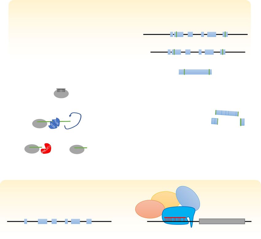
Wang and Lin Genome Biology   (2021) 22:27                                                                  Page 3 of 21

                (Fig. 1c). The transcribed long precursors are initially processed into 5′ end monopho-
                sphorylated RNAs by PIWI protein loaded with initiator piRNAs. The processed pre-
                cursors (pre-pre-piRNAs) are further cleaved by PIWI proteins to generate responder
                pre-piRNAs from the 5′ ends of pre-pre-piRNAs. These pre-piRNAs are trimmed by
                exonucleases to generate functional piRNAs [38–41] and give rises to mature responder
                piRNAs that recognize the complementary strands of long precursors and initiated the
                ping-pong cycles. Meanwhile, 5′ end monophosphorylated precursor RNAs are frag-
                mented into a string of phased trailing pre-RNAs by PIWI and PIWI-coupled-proteins
                on the mitochondrial outer membrane [4–6]. Importantly, piRNAs are symbolically 2′-
                O-methytlated at the 3′ end by S-adenosylmethionine-dependent methyltransferases
                [9–12]. Perhaps the two best-known functions of PIWI proteins and piRNAs are trans-
                poson silencing and fertility [42]. However, many recent studies have started to reveal a
                much broader role of the PIWI-piRNA pathway in meditating the regulation of major
                constituents of the genome, which is the focus of this review.

                The function of PIWI proteins and piRNAs in transposon silencing and
                fertility: an update
                Before reviewing the new functions of the PIWI-piRNA pathway, here we provide an
                update on its function in transposon silencing and fertility, which also serves as needed
                background information of the rest of the review. Transposon silencing has been widely
                regarded as a requirement for fertility. However, this relationship has not been sup-
                ported by definitive evidence. In contrast, these two functions are separable at least in
                Drosophila [43] and mice [44].
                  Transposon silencing is achieved by repression the expression of retrotransposon
                RNAs at both transcriptional and posttranscriptional levels [45]. Transcriptional silen-
                cing is mediated by nuclear PIWI proteins such as PIWI in Drosophila and MIWI2
                (a.k.a. PIWIL4) in mice, whereas posttranscriptional silencing is mediated by cytoplas-
                mic PIWI proteins such as Aubergine (Aub) and Ago3 in Drosophila or MIWI (a.k.a.
                PIWIL1) and MILI (a.k.a. PIWIL2) in mice. A prevalent model of transcriptional silen-
                cing in Drosophila is that the PIWI-piRNA complex binds to a nascent transposon
                transcript (Fig. 1e upper panel). The complex interacts with mediator proteins Asterix
                and Panoramix (a.k.a. Silencio). Furthermore, Panoramix interacts with Nxf2 (Nuclear
                Export Factor 2) and Nxt1 (Nuclear Transport Factor 2 Like Export Factor 1) which re-
                cruit a histone methytransferase, dSetDB1 (a.k.a. Eggless), to methylate Lysine 9 residue
                in histone 3 (H3K9). This promotes the repressive chromatin state [16–22, 46–48]. Al-
                ternative models such as the Drosophila Piwi protein directly recruiting Heterochroma-
                tin Protein 1a to initiate heterochromatinization or recruiting the linker histone H1 to
                induce repressed chromatin state have also been proposed [49–51]. The above three
                models do not mutually exclude each other and might indeed co-exist in the cell. They
                share two common features: (1) transcriptional repression requires the specific binding
                of a PIWI-piRNA complex to a nascent RNA at the target site to recruit epigenetic/
                chromatin factors but does not require the PIWI slicer activity [52, 53]; (2) transcrip-
                tional repression occurs by modifying chromatin structure.
                  In mice, piRNA-dependent transcriptional silencing in the germline is also achieved
                by methylation of DNA through a recruitment scheme that is less known but similar to
                that in Drosophila [23, 54–60] (Fig. 1e, lower panel). For example, Tudor Domain-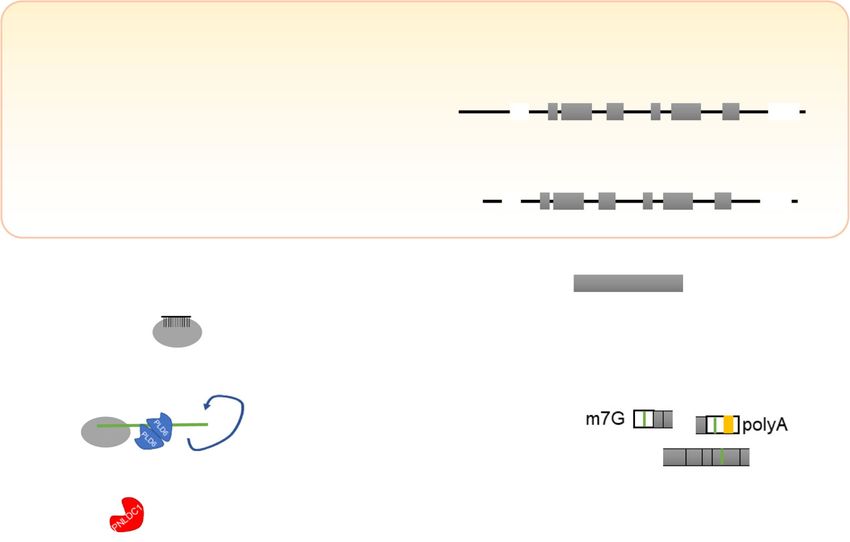
Wang and Lin Genome Biology    (2021) 22:27                                                                  Page 4 of 21

                Containing 9 (TDRD9) complexes with MIWI2 and suppresses transposons in a
                piRNA-dependent manner [61]. MIWI2 is guided by piRNA to the nascent transcript
                of specific genomic region to mediate the methylation of the target DNA [60]. MIWI2
                in turn appears to directly recruit with DNMT3A, DNMT3A2, AND DNMT3L, and
                TDRD9 to achieve methylation, since MIWI2 seems to directly interact with these pro-
                teins in co-expression and immunoprecipitation assay in a 293 T cell [23]. However, a
                most recent report indicates that a nuclear protein, SPOCD1, interacts with MIWI2 as
                well as with DMNT3A, DNMT3L, and components of NURD and BAF chromatin re-
                modeling complexes during de novo DNA methylation [24]. Among the DNA methyl-
                ases, DNMT3C functions as one of the key methyltransferases that protects male germ
                cells from transposon activity [62]. Another nuclear protein, TEX15, also interacts with
                MIWI2 in the process [25]. A separate report indicates that TEX15 interacts with MILI,
                and the genome of Tex15 mutant is hypomethylated similar to Mili and Dnmt3c but
                not Miwi2 mutants [63]. Therefore, the above have revealed some of the key players in
                the PIWI-piRNA-mediated mechanism in the de novo DNA methylation, but substan-
                tial work is still needed to delineate their exact interactions and relative contributions
                to DNA methylation.
                   In addition, a recent study showed that the Ubiquitin-like, Containing PHD and
                RING finger Domains 1 (UHRF1) protein may interact with MIWI and MILI to
                deposit DNA methylation or to further recruit PRMT5 to methylate arginine of
                histones [64]. This study demonstrated the requirement of UHRF1 in histone and
                DNA methylation. However, the co-immunoprecipitation data did not convincingly
                show the UHRF1-MIWI/MILI interaction. In addition, MIWI and MILI are in the
                cytoplasm yet UHRF1 is in the nucleus. Thus, the nature of MIWI/MILI inter-
                action with UHRF1 remains to be further explored. Finally, the zinc finger protein
                MORC1 promotes DNA methylation of transposons but not protein-coding genes
                in male germ cells during the period of global de novo methylation [65].Whether/
                how these proteins work together to methylate the genome of male germ cells
                awaits further investigation.
                   Post-transcriptional silencing occurs in the cytoplasm, where a PIWI-piRNA complex
                binds to a piRNA-complementary transposon RNAs and cleaves the RNA by the slicer
                activity of the PIWI protein (Fig. 1c). This silencing meanwhile fulfills piRNA biogen-
                esis. Both transcriptional and post-transcriptional silencing of transposons by the
                piRNA pathway has been well-reviewed (e.g., [66]); thus, this topic will not be further
                covered in our review.
                   In contrast to transposon silencing, little is known about whether/how PIWI proteins
                and piRNAs regulate gene expression and other major types of RNAs in the germline.
                In this review, we summarize recent research on this less-investigated topic. Recent
                progress has led to surprising discoveries that transposons actively regulate the expres-
                sion of mRNAs and long noncoding RNAs (lncRNAs), mediated by the PIWI-piRNA
                pathway. Moreover, pseudogenes also regulate the expression of their cognate mRNAs,
                also mediated by the PIWI-piRNA pathway. Finally, the PIWI-piRNA pathway even
                regulates the expression of (sub-) telomeric and (peri-) centromeric transcripts. These
                surprising findings have begun to reveal genome-wide functions of the PIWI-piRNA
                pathway in functionally links the major classes of genomic sequences and protein-
                coding genes at the RNA level.
Wang and Lin Genome Biology (2021) 22:27 Page 5 of 21
Regulation of mRNAs by transposons
A recently discovered function of the piRNA pathway in the cytoplasm is to mediate
the regulation of mRNAs by transposons (Fig. 2). This discovery came as a surprise at
multiple levels—from realizing that many mRNAs contain transposon sequences to
learning that transposons can use these sequences as Trojan’s horses to regulate target
mRNAs, and to revealing the essential role of PIWI proteins and piRNAs in this
regulation.
Many mRNAs in mammals contain transposon sequences in the 3′ untranslated region
The first hint of transposons regulation towards mRNAs came from observations
that some expressed retrotransposon sequences, especially short interspersed nu-
clear element (SINE) and long interspersed nuclear element (LINE) families,
mapped to the 3′ untranslated regions (UTRs) of mRNAs in the mammalian gen-
ome [67, 68] (Fig. 2). For example, 27.7% of mouse and 28.5% of human mRNAs
contain at least one retrotransposon fragment, often a SINE, that is predominantly
in the 3′ UTR of mRNAs (Table 1), while a few others are in 5’UTR and even
fewer are in protein-coding sequences (CDS) [68]. In Drosophila, densities of puta-
tive piRNA target sites in both the 5′ UTR and especially the 3′ UTR regions are
also higher than that in coding sequences [69], even though this bias is not as pro-
nounced as in mammalian genomes. The paucity of the transposon insertions in
CDS likely results from evolutionary selection, because an insertion in CDS dis-
rupts a protein’s sequence and often its function, and thus, it is eliminated from
the gene pool. However, there are frequent instances in which retrotransposon in-
sertions reduce host mRNA expression [67, 68], indicating a potential role of the
inserted transposon sequence in regulating mRNA expression.
Transposon sequences in mRNAs are Trojan’s horses that lead to mRNA degradation
Recent studies indicate that transposon sequences in mRNAs are the targeting sites
of transposon-derived piRNAs that degrade these mRNAs [68] (Fig. 2a). MIWI pro-
motes the degradation of the mRNA from a mCherry reporter containing a B1 or
B2 SINE sequence at its 3′ UTR [68]. This RNA-degradation function depends on
its slicer function. RNA immunoprecipitation (RIP) against MIWI and MILI
followed by RT-PCR, as well as high-throughput sequencing of RNA isolated by
crosslinking immunoprecipitation (HITS-CLIP) in the adult mouse testis showed
that MIWI and MILI directly bind to mRNAs [76, 77]. Additionally, MIWI binds
to mRNAs as a mRNA ribonucleoprotein complex in the testis [77, 78]. MIWI
CLIP-seq indicates that piRNAs preferably bind to the 3′ UTR of mRNAs in
mouse elongating spermatids [72, 76]. Moreover, a group of meiotic mRNAs were
significantly upregulated in Miwi−/− mice round spermatids, and the degradation of
meiotic mRNA exhibited piRNA generation signatures [68, 79]. All these findings
indicate that PIWI proteins and their associated piRNAs might be directly involved
in regulating the stability of target mRNAs.
The involvement of transposon-derived piRNAs in regulating their target mRNAs is
further supported by experiments that investigate the effect of deleting piRNAs or their
target sequences on mRNA stability. In mouse late spermatocytes deficient in Miwi orWang and Lin Genome Biology (2021) 22:27 Page 6 of 21
Fig. 2 piRNA mediates mRNA regulation by transposons. a piRNA precursors are transcribed from piRNA
clusters that include retrotransposon sequences (the green box). Mature piRNAs (short green lines)
associated with Piwi proteins guide the Piwi-piRNA complex to complementary transposon sequences
predominately in 3′ UTR and occasionally to 5′ UTR or CDS of specific mRNAs [67–69]. The targeted mRNAs
are degraded through Piwi-slicing and other mechanisms (see Fig. 3). Alternatively, piRNAs are generated
from mRNA that contains transposon sequences (the orange box) in the 3′ UTR. These piRNAs (orange
short lines) in turn target corresponding mRNAs and mediate their degradation [73–75]. b The piRNA-target
RNA pairing rule as exemplified by C. elegans studies. Other organisms appear to follow a similar rule
Table 1 Classification of retrotransposon sequence in mRNA 3′ UTR
Retrotransposon [67] Human 3’ UTR Mouse 3’ UTR
DNA 6.6% 2.9%
LINE.L1 4.7% 2.8%
LINE.L2 4.2% 2.5%
LTR.ERV1 6.4% 2.9%
LTR.ERVK 0.7% 2.6%
LTR.ERVL 4.5% 4.3%
LTR.MALR 5.2% 3.8%
SINE.Alu 5.7% 4.7%
SINE.B2 N.A. 5.0%
SINE.B4 N.A. 4.8%
SINE.MIR 2.7% 6.3%Wang and Lin Genome Biology (2021) 22:27 Page 7 of 21
Mov10l1, another gene involved in piRNA biogenesis, piRNA biogenesis is deficient,
and the expression levels of a large number of protein-coding genes are altered [68]. In
addition, depletion of the retrotransposon sequences in either the 3′ UTR of prelid1 or
the piRNA cluster that produces the targeting piRNAs results in a six-fold up-
regulation of the prelid1 mRNA level in late spermatocytes. Furthermore, this MIWI-
and MOV10L1-mediated regulation of mRNAs requires the slicer activity of MIWI [68,
80]. Most recently, it was reported that deleting a specific piRNA cluster, pi6, led to up-
regulating of mRNAs related to sperm function, in addition to affecting piRNA-piRNA
precursor interactions [81]. This provides an additional example for piRNA function in
regulating mRNAs and in spermatogenesis.
The suppression of retrotransposon-containing mRNAs in mouse primordial ovarian
follicles also requires MILI and other piRNA pathway components such as mouse Vasa
homolog (MVH) and TDRD9 [82]. In Drosophila, piRNAs suppress the Stellate repeat
mRNA to ensure spermatogenesis, even though this regulation was initially reported as
a siRNA-mediated mechanism [83]. These piRNAs, however, cannot repress the
Stellate mRNA from closely related Drosophila species, thus introducing reproductive
isolation [84]. All these observations demonstrate a direct role of PIWI proteins and
their partner piRNAs in regulating mRNAs.
PIWI-piRNAs targeting rules
The slicing of piRNA-targeted mRNA mostly occurs at the 10th position from the 5′ end
of the targeting piRNAs [13, 68]. In-depth analysis of the cleavage site in the 3′ UTR of
Tdrd1 mRNA with respect to its targeting piRNAs reveal that most piRNAs are aligned
antisense to the mRNA sequence with their 5′ nucleotide being 10 nucleotides away from
the cleavage site, but several percent of piRNAs are aligned with their 5′ nucleotide 11–
20 nucleotides away [68]. This could indicate that the cleavage site for these piRNA-
complementary mRNAs is downstream of the 10th position of piRNAs.
Despite the relatively clear information on the cleavage site as supported by in vitro,
in vivo, and structural analyses, the piRNA targeting rule is still elusive and increasing
effort has been put into revealing this puzzle. In C. elegans and mice, piRNA binding to
target RNA is sequence-dependent but not completely sequence-specific, with position
2–8 as the seed sequence and 14–22 nucleotides are also important for piRNA target-
ing [69, 72, 85, 86], reminiscent of the microRNA seed sequence for target recognition
(Fig. 2b). Further attempts in ectopic expression of human piRNAs in mouse testes
show that perfect matching in nucleotides 2–11 of piRNA is required for mRNA target-
ing [79]. In a different study with deep RNA sequencing that allowed up to four mis-
matches at random positions in a piRNA, all of the 172 mRNAs that were significantly
upregulated in Piwil1−/− testes contain transposon piRNA target sites in their 3′ UTR,
with such a transposon sequence experimentally demonstrated to be responsible for
MIWI-piRNA-mediated degradation [68]. Notably, these 172 mRNAs are quite differ-
ent from the MIWI-target RNAs identified by Vourekas et al. (2016) using iCLIP. This
likely reflects that iCLIP approach revealed stable binding of PIWI to target mRNAs
that do not lead to mRNA degradation but possibly other regulatory effects, in contrast
to RNA sequencing that revealed the degraded products of MIWI binding and cleavage
of another set of target mRNAs (i.e., the mRNA degradome).Wang and Lin Genome Biology (2021) 22:27 Page 8 of 21
In Drosophila, mutating 1, 2, 3 nucleotides of a given piRNA in different positions
did not abolish the binding ability of Piwi to its target nascent RNA at the genomic site,
but reduce its binding affinity proportional to the number of mutations [50]. These ob-
servations also support the notion that piRNA targeting is sequence-dependent but not
completely sequence-specific.
mRNAs also produce piRNAs
The source of piRNAs is sometimes mRNAs. In Drosophila ovaries, transposons
that are integrated in the 3′ UTR of actively transcribed genes induce piRNA pro-
duction, which in-turn suppresses corresponding gene expression [73, 74] (Fig. 2a,
right part). For example, in the Drosophila ovarian germline, proto-oncogene c-Fos
is repressed post-transcriptionally in germline stem cells (GSCs) by Piwi protein.
As a by-product of this repression, piRNAs are produced from the transposon ele-
ments in the 3′ UTR of c-Fos mRNA [74]. This repression is critical for GSC self-
renewal and differentiation. In the Drosophila ovarian somatic cell (OSC) line, the
3′ UTR of c-Fos mRNA alone is sufficient to introduce gene suppression in a
Piwi-dependent manner [74]. Similarly, in this OSC line, the 3′ UTR of a protein-
coding gene traffic jam produces piRNAs to repress the expression of another
protein-coding gene, fasciclin III [75]. However, in this case, the traffic jam piRNAs
are not transposon-originated piRNAs.
piRNAs can target the protein-coding sequence of mRNA for its degradation
A few publications have reported that piRNAs can also target the protein-coding
sequence (CDS) of a mRNA to degrade the mRNA. For instance, in the Drosophila
embryo, Aub directly binds to the CDS and the 3′ UTR of maternal mRNAs in a
piRNA-dependent manner to facilitate the decay of these maternal mRNAs [87]. The
Aub-piRNA complex is also associated with CCR4 in Drosophila GSCs and represses
the expression of Casitas B-cell lymphoma (Cbl) mRNA [88]. This mRNA contains tar-
get sites for transposon-derived piRNA in both 5′ UTR and 3′ UTR, and these target
sites overlap with Aub CLIP hits. In Caenorhabditis, a sex-chromosome-derived piRNA
suppresses the expression of xol-1 (XO Lethal), the master sex determination gene, via
targeting the CDS region of the xol-1 mRNA [89]. In the silkworm, a Feminizer-derived
piRNA targets the CDS of the Masculinizer mRNA and determines the sex of the silk-
worm [90]. In human breast cancer cell lines, a piRNA called piR-FTH1 shows an in-
verse correlation with the expression of Fth1 (ferritin heavy chain 1) [91]. MILI and
MIWI2 bind to piR-FTH1 to repress the Fth1 mRNA via targeting the CDS region of
Fth1 [91]. These observations from diverse and independent processes, each of which
with biological significance, indicate that transposon sequences in CDS are equally ef-
fective in regulating mRNA stability. Thus, the paucity of piRNA target sites in the
CDS is likely only due to their disruptive effect on protein-coding capacity but not a
lower efficiency for piRNA-targeted degradation.
Finally, the insertion of transposon sequences into mRNAs as piRNA target se-
quences appears to be a dynamic process at least for certain cell types. The transposon
landscape in cultured Drosophila ovarian somatic sheet (OSS) cells differs from that of
cultured Drosophila OSCs [92], indicating that factors even as culture conditions mayWang and Lin Genome Biology (2021) 22:27 Page 9 of 21
select for transposon insertions that are in favor of the regulation of mRNA expression
that meets the needs of the given condition.
Protein machinery involved in mRNA degradation by transposon-derived piRNAs
A few studies have further suggested that the PIWI-piRNA regulation of mRNAs
involves canonical mRNA decay machineries. In the Drosophila germline, Aub and
Ago3 colocalize with the mRNA degradation proteins Decapping Protein 1/2
(DCP1/2), Maternal expression at 31B (Me31B), and Pacman (PCM) in the nuage,
a perinuclear riboprotein assembly, forming a complex (the pi-body) in a piRNA-
dependent manner that facilitates mRNA degradation [70] (Fig. 3a). In early Dros-
ophila embryos, the key piRNA pathway components Piwi, Aub, Ago3, Armitage
(Armi), Spindle-E (Spn-E), and Squash (Squ) are all involved in the deadenylation
and decay of maternal Nanos (Nos) mRNA through a piRNA targeting region in
the nos 3′ UTR [71]. Aub is known to achieve so by forming a complex with
RNA-binding protein Smaug (Smg) and the deadenylase CCR4 at the sites of
piRNA–mRNA interaction (Fig. 3b). The deadenylation machinery further recog-
nizes the retrotransposon sequences in the nos 3′ UTR and executes deadenylation
within 4 h [71]. In mouse spermatogenic cells, the piRNA-induced silencing com-
plex (pi-RISC) contains MIWI and CAF1 (Chromatin Assembly Factor 1), a key
catalytic subunit of the CCR4-NOT deadenylase complex [72] (Fig. 3c). Thus, the
piRNA mediated mRNA degradation is not only achieved by MIWI slicing of the
target mRNAs but also by concurrent deadenylation, and possibly decapping, of
mRNAs.
piRNAs activate the translation of mRNAs
The homology between PIWI proteins and eIF2C implies the possibility that they might
promote translation, as first proposed for Aub in promoting translation of nanos
mRNA in Drosophila embryos [93]. This function of Aub has been clearly demon-
strated by a most recent study [94]. In the Drosophila early embryo, Aub physically in-
teracts with eIF3d and the poly(A)-binding protein (PABP) to activate translation of
nanos mRNA in the germ plasm (Fig. 4a).
PIWI proteins and piRNAs have also been implicated in promoting translation in
mammalian systems. MILI is required for germline stem cell self-renewal in the mouse
testis [96]. It forms a stable RNA-independent complex with eIF3a and associates with
the m7G cap-binding complex that contains eIF4E and eIF4G in spermatogonia and
spermatocytes [96]. In 7 dpp spermatogonia (most of which are germline stem cells),
the Mili mutation has no significant effect on the cellular mRNA level but significantly
reduces the rate of protein synthesis. Thus, MILI appears to positively regulate transla-
tion for germline stem cell self-renewal.
The MIWI-piRNA complex also associates with the capping complex, including
cap-binding protein eIF4E, and regulates translation in the mouse spermatocytes
and spermatids [97]. Recently, it has been further reported that, in mouse sperma-
tids, a fraction of piRNAs associate with MIWI and recognize ARE (AU-rich elem-
ent)-containing target mRNAs through imperfect base pairing in the 3′ UTR of
target mRNAs [95]. ARE-binding protein HuR loaded with target mRNA and theWang and Lin Genome Biology (2021) 22:27 Page 10 of 21
Fig. 3 The piRNA-Piwi complex recruits canonical mRNA processing and localization machineries to
regulate mRNAs. a In the Drosophila germline, the mRNA de-capping complex, which includes PCM, Me31B,
DCP1, and DCP2 proteins, is recruited by the piRNA-Piwi complex to HeT-A mRNA to promote the decay of
HeT-A mRNA [70]. b In Drosophila embryos, the piRNA-Piwi complex recruits the Smg protein and CCR4
complex to the 3′ UTR of nos mRNA to facilitate the degradation of nos mRNA [71]. c In the mouse testis,
the piRNA-Piwi complex interacts with Caf1 bound to the 3′ UTRs of target mRNAs to execute mRNA
degradation through the slicer activity of Piwi [72]
translation initiation factor eIF3f form a complex with piRNA and MIWI to further
activate the translation of mRNAs required for acrosome formation during sperm-
atid development (Fig. 4b). These lines of evidence illustrate a sophisticated regula-
tory nexus of PIWI-piRNA complex in modulating the transcriptome at
posttranscriptional level. Despite the above findings, the function of PIWI proteins
in translational regulation has not been extensively studied and deserves more
attention.
piRNAs regulate the localization of mRNAs
In addition to working with the mRNA decay machinery, the PIWI-piRNA complex
has also been reported to be involved in the subcellular localization of mRNAs. For ex-
ample, in Drosophila, the Aub-piRNA complex facilitates the localization of critical
germ cell mRNAs to the germ plasm [98]. This function is achieved by working with
Wispy, a germline-specific non-canonical poly(A) polymerase. During Drosophila oo-
genesis, Aub participates in the formation of the polar granule that localizes germline-
specific mRNAs.Wang and Lin Genome Biology (2021) 22:27 Page 11 of 21
Fig. 4 The piRNA-Piwi complex mediates transcriptional activation of target mRNAs. a In Drosophila germ
plasm, Aub interacts with PABP and eIF3 subunits to allow unconventional translation and recruits Wispy
poly(A) polymerase to facilitates polyadenylation which also promotes translation [94]. This process is
dependent on Cup binding to eIF4E. b In mouse spermatids, the piRNA-PIWIL1 complex binds to eIF3f and
the 3′ UTRs of ARE-containing target mRNAs, which triggers mRNA looping [95]. HuR binds to the ARE
sequences and mediates the association of other translation initiation factors, including eIF4G and PABPC1,
to activate translation
Regulation of lncRNAs by transposons
Recent studies also indicate that transposon sequences in lncRNAs are the target-
ing sites of transposon-derived piRNAs that identify these lncRNAs for degrad-
ation. Global characterization of transposon distribution in the human genome
revealed that the vast majority of lncRNAs (83.4%) overlap with at least one trans-
poson, in contrast to protein-coding sequences—only 6.2% of which overlap with
transposons [99]. In the human genome, 75% of lncRNA transcripts contain an
exon that originates at least partially from a transposon. Different types of transpo-
sons are differentially represented in lncRNA loci, and transposon landscapes differ
among various tissue types [99]. LncRNAs that overlap with transposons are less
expressed as compared to those that do not overlap with a transposon, indicating a
suppressive role of transposon insertion [99].Wang and Lin Genome Biology (2021) 22:27 Page 12 of 21
Transposons repress lncRNA expression via the PIWI-piRNA pathway
The involvement of piRNAs in transposon regulation of lncRNAs has been demon-
strated in multiple organisms. In mouse spermatocytes, retrotransposon sequences,
mostly SINE, are distributed across the entire length of approximately 1500 lncRNAs
that are upregulated in Miwi−/− testis, which represents ~ 25% of the expressed
lncRNAs in these cells [68]. This distribution is in contrast to their predominant
localization to the 3′ UTR of mRNAs. The transposon-containing lncRNAs become
overexpressed in Miwi and Mov10l1 mutants, implying that the transposon regulation
of lncRNA expression is via the piRNA pathway [68] similar to its role in mediating
transposon regulation of mRNAs (Fig. 5a). Presumably, the PIWI-piRNA complex slices
the target lncRNAs in a way similar to their slicing of mRNAs. However, this remains
to be directly demonstrated.
Other regulatory relationships between transposons, lncRNAs, and piRNAs
The piRNA regulation of lncRNA expression can be manifested in different ways. Some
piRNA clusters that are expressed during the pachytene stage originate from testis-
specific lncRNAs, indicating that lncRNAs themselves can be precursors of piRNAs
[14, 101]. Although not as much is known about the relationship among transposons,
lncRNAs, and piRNAs in the primordial germ cells, in their precursor cells in humans,
i.e., human embryonic stem cells and induced pluripotent stem cells (iPSCs), the
presence of transposon families such as HERVH (human endogenous retrovirus H)
elements in lncRNAs is correlated with high expression levels of host lncRNAs,
implicating that HERVH sequences positively regulate the expression of lncRNAs in a
cell-type-specific manner [99], even though it is not known whether such regulation oc-
curs at the transcriptional or post-transcriptional level. In Drosophila OSS and OSC
cells, transposon insertions around lncRNA stimulate the expression of corresponding
lncRNAs in a Piwi-dependent manner [92], implying a possible role of piRNA in tran-
scriptional regulation of lncRNA expression.
In addition, some lncRNAs can produce piRNAs to promote gene expression at the
transcriptional level. In a human breast cancer cell line (MCF7), a lncRNA called
Growth Arrest Specific 5 (GAS5) facilitates the transcription of TRAIL [transcription of
tumor necrosis factor (TNF)-related apoptosis-inducing ligand] mRNA. This is through
GAS5-derived piRNAs that are associated with PIWI proteins [100]. These piRNA-
PIWI complexes then recruit WDR5 and the COMPASS complex, a key complex for
epigenetic modification, to the promoter of the TRAIL gene to modify the epigenetic
status of the gene, which facilitates TRAIL transcription. This indicates an important
role of PIWI-piRNA complex in epigenetic regulation of gene expressions (Fig. 5b). In
Tetrahymena, piRNAs that are derived from noncoding RNA loci bind to Tetrahymena
PIWI protein Twi8p and its target lncRNAs, which leads to a decrease in the level of
the targeted lncRNAs [102]. These roles are similar to what has been found in mamma-
lian systems.
The regulation of mRNAs by pseudogenes
There are 14,000 pseudogenes in the human genome [103]. Analysis of human sperm
small RNAs reveals that some piRNA clusters are located within pseudogenes [104].Wang and Lin Genome Biology (2021) 22:27 Page 13 of 21
Fig. 5 piRNAs mediate transposon regulation of lncRNAs. a piRNA mediates post-transcriptional regulation
of mRNAs. piRNA precursors are transcribed from piRNA clusters that contain transposon sequences and are
processed into mature piRNAs. Mature piRNAs are then loaded onto Piwi proteins and guide the Piwi
complex to target lncRNAs that contain complementary transposon sequences to degrade these lncRNAs
[68]. b piRNA mediates transcriptional regulation of mRNAs. In a human breast cancer cell line (MCF7), the
Growth Arrest Specific 5 (GAS5) lncRNA produces piRNAs that are associated with Piwi proteins [100]. This
piRNA-Piwi complex then recruits WDR5 and the COMPASS epigenetic complex to the promoter of the
TRAIL (transcription of tumor necrosis factor (TNF)-related apoptosis-inducing ligand) gene to modify its
epigenetic status to promote its transcription
piRNAs derived from pseudogenes are predicted to target protein-coding cognate
genes. In mouse late spermatocytes, at least 14 genes are significantly regulated by their
pseudogenes [68]. Thus, pseudogene regulation of mRNAs via piRNAs may be a signifi-
cant mechanism in the germline that has been largely ignored.
Recent studies indicate that piRNA derived from pseudogenes can regulate the
expression of their cognate mRNAs. The first clear demonstration of such a
function comes from a study of the mouse late spermatocyte. The mRNA level of
the Stambp gene is dramatically increased in Miwi−/− and Mov10l1−/− late sper-
matocytes, indicating that the piRNA pathway may be involved in preventing the
over-expression of Stambp mRNA [68] (Fig. 6a). However, the Stambp mRNA
complements to PIWIL1-associated piRNAs that do not map to transposons but
uniquely to its pseudogene, Stambp-ps1 [68]. Indeed, knocking out Stambp-ps1 ex-
pression abolishes the Stambp-ps1-derived piRNAs and causes drastic increases inWang and Lin Genome Biology (2021) 22:27 Page 14 of 21
Fig. 6 piRNAs mediate pseudogene regulation of cognate mRNAs. a Anti-sense transcripts of pseudogenes
serve as precursors of piRNAs. These pseudogene-derived piRNAs form Piwi-piRNA complex that binds to
complementary sequences in the cognate mRNAs to promote their degradation [68]. b In human breast
cancer cell lines, the Piwi-piR-36712 complex degrades the RNA of pseudogene SEPW1P, which releases
SEPW1P-RNA-bound miR-7 and miR-324 to bind to cognate SEPW mRNA, promoting its degradation and
inhibiting its translation [105]. The reduction of SEPW protein weakens its repression towards p53 and p21,
allowing p53 and p21 to release Slug suppression towards E cadherin expression, which blocks
cancer progression
the Stambp mRNA level, illustrating the importance of a pseudogene in the regula-
tion of its cognate active gene via the piRNA [68].
Conversely, pseudogenes can also be regulated by piRNAs. In human breast cancer
cell lines MCF7 and ZR75–1, a piRNA called piR-36,712 directly binds to pseudogene
SEPW1P RNA and reduces its level [105] (Fig. 6b). This releases its bound miRNAs
miR-7 miR-324, allowing more miR-7 miR-324 to target the mRNA of its cognate gene
SEPW to reduce its stability and translation. The reduced SEWP level in turn weakens
its inhibition towards p53 and p21, allowing p53 and p21 to reduce the suppression of
E cadherin expression by a zinc finger protein called Slug, which blocks cancerWang and Lin Genome Biology (2021) 22:27 Page 15 of 21
development [105]. This type of regulation may be specific to cancer cells because piR-
NAs are not detected in normal somatic cells, such as breast epithelial cells,
Pseudogene-derived piRNAs have been identified across the evolutionary tree. In
Tetrahymena, pseudogene-derived small RNAs have been identified that have bind-
ing preference to Twi2 (Tetrahymena PIWI2) but not to Twi7 or Twi8 [106]. In
the marmoset testis, piRNA clusters contain processed pseudogenes that are
antisense-oriented relative to their cognate genes so they can produce piRNAs that
target their cognate genes (cf. Fig. 6aa) [107]. In addition, pig piRNA clusters are
also highly enriched for exon sequences of pseudogenes [108]. In vertebrate ge-
nomes, endogenous bornavirus-like nucleoprotein elements (EBLNs) represent a
special type of genomic sequences derived from ancient bornaviral nucleoprotein
mRNA via retrotransposition. Human EBLNs are actively transcribed pseudogenes
that give rise to piRNAs with a possible role in interfering with ancient bornaviral
infection [109]. Thus, regulation by pseudogene-derived piRNAs is likely a con-
served regulatory pathway.
Diverse functions of satellite-repeat derived piRNAs
Satellite-repeated derived piRNAs negatively regulates mRNA stability
In Drosophila, it has been shown that pericentromeric piRNAs, specifically, AT-chX
piRNAs and Su(Ste) piRNAs, also suppresses the vasa mRNA of a different species
of Drosophila during interspecies mating to ensure reproductive isolation [84].
Most recently, it was shown that a large number of piRNAs were derived from a
satellite repeat in both the soma tissues and germline of the mosquito Aedes
aegypti [110]. Two of them, tapiR1 and tapiR2 (tandem repeat-associated piRNA1
and 2), are associated with Aedes aegypti PIWI protein Piwi4. The resulting Piwi4-
piRNA complex silence target RNAs (both mRNA and lncRNA) via a sequence-
specific recognition rule reminiscent of microRNA seed sequence. Notably, the
piRNA-generating satellite repeats were highly conserved across mosquito species
for approximately 200 million years and are very similar to that of other higher or-
ganisms. Hence, there might be an evolutionarily conserved mechanism for post-
transcriptional regulation mediated by PIWI-satellite piRNA complex.
Subtelomeric piRNAs regulate telomeric function
Remarkably, the piRNAs are even involved in regulating telomeres and centromeres.
Telomeric and centromeric piRNAs have been reported in various organisms. Shortly
after piRNAs were characterized, piRNAs were mapped to subtelomeric regions of the
Drosophila genome. One of these piRNAs, the 3R-TAS1 piRNA, regulates the epigen-
etic state of the target subtelomeric sequence [111] and is essential for germline stem
cell maintenance [112]. Telomeric piRNAs in Drosophila are required for the depos-
ition of Heterochromatin Protein 1 (HP1) and its homolog Rhino as well as epigenetic
suppression of telomeric retrotransposon [113, 114]. Loss of piRNA pathway proteins
results in the significant upregulation of telomeric retroelement transcripts and down-
regulation of telomeric piRNAs [115–118]. HP1a, a partner of Piwi protein [49, 111], is
required for the generation of piRNAs that map to telomeres and peri-centromeres in
the Drosophila germline [119]. The nuclear CCR-NOT complex is required for theWang and Lin Genome Biology (2021) 22:27 Page 16 of 21
degradation of telomeric transcripts in a Piwi-dependent manner [120]. In addition,
Aub, Armi, and Ago3 mutations reduce telomeric piRNAs production and disrupt the
binding between telomere and telomere protection complex [117, 121]. All these find-
ings together indicate an essential role of the piRNA pathway in subtelomeric and telo-
meric function in Drosophila.
Interestingly, the prevalence of subtelomeric piRNA clusters is significantly higher in
the genomes of Drosophila in the wilderness than in standardized laboratory Drosoph-
ila stocks [122]. Since a well-established telomeric function in Drosophila is to suppress
telomeric retrotransposons, this difference may reflect another function of telomeric
piRNAs in the suppression of telomeric retrotransposons, which might be more needed
by flies in the wilderness that are possibly more ridden with retrotransposons.
Because mammalian telomeres are composed of simple repeats instead of telomeric
retroelement, the telomeric function of piRNA in Drosophila is unlikely to be con-
served. However, subtelomeric sequence and structure is highly conserved from Dros-
ophila to mammals. Hence, some other important function of the subtelomere, such as
repressing and regulating nearby euchromatic gene expression (position effect), might
be conserved in higher organisms.
In Tetrahymena, telomeric repeat-derived small RNAs selectively bind to Twi10
[106]. In Caenorhabditis, perfect telomeric small RNAs are extremely rare among small
RNAs that are immunoprecipitated with Ago proteins; however, there are significant
numbers of telomeric small RNAs with mismatches were associated with Ago proteins
[123]. These small RNAs might be involved in telomere protection in a way similar to
that in Drosophila.
Pericentromeric and centromeric piRNAs regulate centromere function
In Drosophila, pericentromeric satellite repeats are known to be crucial for centromere
function and give rise to piRNAs [13, 28, 124] that are critical for the suppression of
satellite RNA expression [28, 125, 126]. The involvement of piRNAs in telomeres and
centromeres is likely to be crucial for the genome integrity and segregation during
germ cell division. A clear requirement of the Piwi-piRNA pathway for chromosome
segregation is illustrated by a most recent study in mice, where MIWI prevents aneu-
ploidy during meiosis by piRNA-guided cleavage of excess major and minor satellite
RNAs [127]. This allows normal assembly of homologous kinetochore pairs. Overex-
pression of these satellite RNAs in wildtype meiotic cells also causes aneuploidy. Dicer
facilitates the degradation of MIWI cleavage products and other double-strand satellite
RNAs. These findings, supported by another latest study on Dicer [128], start to reveal
a direct role of PIWI proteins and satellite RNAs in chromosome segregation during
meiosis.
Conclusion
A eukaryotic genome is composed of protein-coding genes, transposons, pseudogenes,
centromeres, telomeres, and other repeat sequences. Recent studies, mostly in the
germline, start to reveal important roles of piRNAs in functionally linking these major
constituents of the genome. piRNAs can be derived from transposon RNAs, pseudo-
gene RNAs, mRNAs, lncRNAs, and telomeric/centromeric repeat RNAs andWang and Lin Genome Biology (2021) 22:27 Page 17 of 21
subsequently partner with PIWI proteins to recognize complementary sequences in
these RNAs. These observations together start to reveal a complex network of regula-
tion mediated by piRNAs that unifies the genome at the post-transcriptional level. This
is perhaps the first regulatory network of any kind that executes genome-wide regula-
tion in any cell type. Future investigations on this complex network at the mechanistic
level will further reveal how this network interacts with other canonical RNA regula-
tion mechanisms to define the genomic function in germ cells and shed light on such
function in other cell types.
Acknowledgements
We thank Christopher Antos, Ting Lu, Shou Shi, Yuanyuan Gong, and Sanhong Liu for critical reading of the
manuscript.
Authors’ contributions
H.L. conceived the topic and the outline of the review. C.W. and H.L. wrote the review. Both authors read and
approved the final manuscript.
Funding
C.W. is supported by ShanghiTech University, and H.L. is funded by NIH R37HD42012.
Competing interests
The authors declare no competing interests.
Glossary
Transposon:
A DNA segment that can undergo transposition into a different site in the genome.
Retrotransposons:
Transposons that transcribe itself into a RNA intermediate for reverse transcription to produce double-stranded
DNA for transposition (insertion).
Pseudogene:
A DNA segment that is an imperfect copy of a functional gene, often including a (sometime altered) promoter.
Most pseudogenes are transcribed but not translated due to mutations in protein-coding sequences.
lncRNA:
Long non-coding RNAs, which are conventionally defined as RNAs that are at least 200-nucleotide in length but
do not contain a protein-coding sequence.
Dicer:
A type of RNase III family endoribonucleases that cleave double-stranded RNA into short double-stranded RNA
fragments of mostly 21–22 nucleotides.
Centromere:
The region of a chromosome, containing highly repetitive DNA sequence, that serves as the attachment site for
microtubules of the spindle during cell division.
Pericentromere:
The chromosomal regions flanking the centromere that contains highly repetitive DNA and are heterochromatic.
Telomere:
The two end-regions of a chromosome that protect the chromosome from shortening after DNA replication or
from fusion to other chromosomes.
Subtelomere:
The two regions of a chromosome that is right next to the telomere, containing of highly repetitive DNA
sequences and are heterochromatic.
Pachytene:
A mid-stage of the prophase of meiosis I, following zygotene, characterized by the presence of shortened and
thickened paired chromosomes, with the two chromatids clearly separate.
Author details
1
Shanghai Institute for Advanced Immunochemical Studies, ShanghaiTech University, Shanghai 201210, China. 2Yale
Stem Cell Center and Department of Cell Biology, Yale University School of Medicine, New Haven, CT 06519, USA.
Received: 5 August 2020 Accepted: 7 December 2020
References
1. Song JJ, Smith SK, Hannon GJ, Joshua-Tor L. Crystal structure of Argonaute and its implications for RISC slicer activity.
Science. 2004;305:1434–7.
2. Matsumoto N, Nishimasu H, Sakakibara K, Nishida KM, Hirano T, Ishitani R, Siomi H, Siomi MC, Nureki O. Crystal structure
of silkworm PIWI-clade Argonaute Siwi bound to piRNA. Cell. 2016;167:484–97 e489.
3. Yamaguchi S, Oe A, Nishida KM, Yamashita K, Kajiya A, Hirano S, Matsumoto N, Dohmae N, Ishitani R, Saito K, et al.
Crystal structure of Drosophila Piwi. Nat Commun. 2020;11:858.Wang and Lin Genome Biology (2021) 22:27 Page 18 of 21
4. Homolka D, Pandey RR, Goriaux C, Brasset E, Vaury C, Sachidanandam R, Fauvarque MO, Pillai RS. PIWI slicing and RNA
elements in precursors instruct directional primary piRNA biogenesis. Cell Rep. 2015;12:418–28.
5. Mohn F, Handler D, Brennecke J. Noncoding RNA. piRNA-guided slicing specifies transcripts for Zucchini-dependent,
phased piRNA biogenesis. Science. 2015;348:812–7.
6. Han BW, Wang W, Li C, Weng Z, Zamore PD. Noncoding RNA. piRNA-guided transposon cleavage initiates Zucchini-
dependent, phased piRNA production. Science. 2015;348:817–21.
7. Hayashi R, Schnabl J, Handler D, Mohn F, Ameres SL, Brennecke J. Genetic and mechanistic diversity of piRNA 3'-end
formation. Nature. 2016;539:588–92.
8. Wang H, Ma Z, Niu K, Xiao Y, Wu X, Pan C, Zhao Y, Wang K, Zhang Y, Liu N. Antagonistic roles of nibbler and Hen1 in
modulating piRNA 3' ends in Drosophila. Development. 2016;143:530–9.
9. Horwich MD, Li C, Matranga C, Vagin V, Farley G, Wang P, Zamore PD. The Drosophila RNA methyltransferase, DmHen1,
modifies germline piRNAs and single-stranded siRNAs in RISC. Curr Biol. 2007;17:1265–72.
10. Saito K, Sakaguchi Y, Suzuki T, Suzuki T, Siomi H, Siomi MC. Pimet, the Drosophila homolog of HEN1, mediates 2'-O-
methylation of Piwi- interacting RNAs at their 3' ends. Genes Dev. 2007;21:1603–8.
11. Ohara T, Sakaguchi Y, Suzuki T, Ueda H, Miyauchi K, Suzuki T. The 3' termini of mouse Piwi-interacting RNAs are 2'-O-
methylated. Nat Struct Mol Biol. 2007;14:349–50.
12. Kirino Y, Mourelatos Z. Mouse Piwi-interacting RNAs are 2'-O-methylated at their 3' termini. Nat Struct Mol Biol.
2007;14:347–8.
13. Brennecke J, Aravin AA, Stark A, Dus M, Kellis M, Sachidanandam R, Hannon GJ. Discrete small RNA-generating loci as
master regulators of transposon activity in Drosophila. Cell. 2007;128:1089–103.
14. Gainetdinov I, Skvortsova Y, Kondratieva S, Funikov S, Azhikina T. Two modes of targeting transposable elements by
piRNA pathway in human testis. RNA. 2017;23:1614–25.
15. Gu W, Lee HC, Chaves D, Youngman EM, Pazour GJ, Conte D Jr, Mello CC. CapSeq and CIP-TAP identify Pol II start sites
and reveal capped small RNAs as C. elegans piRNA precursors. Cell. 2012;151:1488–500.
16. Wang SH, Elgin SC. Drosophila Piwi functions downstream of piRNA production mediating a chromatin-based
transposon silencing mechanism in female germ line. Proc Natl Acad Sci U S A. 2011;108:21164–9.
17. Muerdter F, Guzzardo PM, Gillis J, Luo Y, Yu Y, Chen C, Fekete R, Hannon GJ. A genome-wide RNAi screen draws a
genetic framework for transposon control and primary piRNA biogenesis in Drosophila. Mol Cell. 2013;50:736–48.
18. Sienski G, Donertas D, Brennecke J. Transcriptional silencing of transposons by Piwi and maelstrom and its impact on
chromatin state and gene expression. Cell. 2012;151:964–80.
19. Le Thomas A, Rogers AK, Webster A, Marinov GK, Liao SE, Perkins EM, Hur JK, Aravin AA, Toth KF. Piwi induces piRNA-
guided transcriptional silencing and establishment of a repressive chromatin state. Genes Dev. 2013;27:390–9.
20. Ohtani H, Iwasaki YW, Shibuya A, Siomi H, Siomi MC, Saito K. DmGTSF1 is necessary for Piwi-piRISC-mediated
transcriptional transposon silencing in the Drosophila ovary. Genes Dev. 2013;27:1656–61.
21. Sienski G, Batki J, Senti KA, Donertas D, Tirian L, Meixner K, Brennecke J. Silencio/CG9754 connects the Piwi-piRNA
complex to the cellular heterochromatin machinery. Genes Dev. 2015;29:2258–71.
22. Yu Y, Gu J, Jin Y, Luo Y, Preall JB, Ma J, Czech B, Hannon GJ. Panoramix enforces piRNA-dependent cotranscriptional
silencing. Science. 2015;350:339–42.
23. Kojima-Kita K, Kuramochi-Miyagawa S, Nagamori I, Ogonuki N, Ogura A, Hasuwa H, Akazawa T, Inoue N, Nakano T.
MIWI2 as an effector of DNA methylation and gene silencing in embryonic male germ cells. Cell Rep. 2016;16:2819–28.
24. Zoch A, Auchynnikava T, Berrens RV, Kabayama Y, Schopp T, Heep M, Vasiliauskaite L, Perez-Rico YA, Cook AG, Shkumatava
A, et al. SPOCD1 is an essential executor of piRNA-directed de novo DNA methylation. Nature. 2020;584:635–9.
25. Schopp T, Zoch A, Berrens RV, Auchynnikava T, Kabayama Y, Vasiliauskaite L, Rappsilber J, Allshire RC, O'Carroll D. TEX15
is an essential executor of MIWI2-directed transposon DNA methylation and silencing. Nat Commun. 2020;11:3739.
26. Gunawardane LS, Saito K, Nishida KM, Miyoshi K, Kawamura Y, Nagami T, Siomi H, Siomi MC. A slicer-mediated
mechanism for repeat-associated siRNA 5' end formation in Drosophila. Science. 2007;315:1587–90.
27. Okamura K, Ishizuka A, Siomi H, Siomi MC. Distinct roles for Argonaute proteins in small RNA-directed RNA cleavage
pathways. Genes Dev. 2004;18:1655–66.
28. Vagin VV, Sigova A, Li C, Seitz H, Gvozdev V, Zamore PD. A distinct small RNA pathway silences selfish genetic elements
in the germline. Science. 2006;313:320–4.
29. Kim VN, Han J, Siomi MC. Biogenesis of small RNAs in animals. Nat Rev Mol Cell Biol. 2009;10:126–39.
30. Teixeira FK, Okuniewska M, Malone CD, Coux RX, Rio DC, Lehmann R. piRNA-mediated regulation of transposon
alternative splicing in the soma and germ line. Nature. 2017;552:268–72.
31. Lewis SH, Quarles KA, Yang Y, Tanguy M, Frezal L, Smith SA, Sharma PP, Cordaux R, Gilbert C, Giraud I, et al.
Pan-arthropod analysis reveals somatic piRNAs as an ancestral defence against transposable elements. Nat Ecol
Evol. 2018;2:174–81.
32. Ross RJ, Weiner MM, Lin H. PIWI proteins and PIWI-interacting RNAs in the soma. Nature. 2014;505:353–9.
33. Aravin A, Gaidatzis D, Pfeffer S, Lagos-Quintana M, Landgraf P, Iovino N, Morris P, Brownstein MJ, Kuramochi-Miyagawa
S, Nakano T, et al. A novel class of small RNAs bind to MILI protein in mouse testes. Nature. 2006;442:203–7.
34. Girard A, Sachidanandam R, Hannon GJ, Carmell MA. A germline-specific class of small RNAs binds mammalian Piwi
proteins. Nature. 2006;442:199–202.
35. Grivna ST, Beyret E, Wang Z, Lin H. A novel class of small RNAs in mouse spermatogenic cells. Genes Dev. 2006;20:1709–14.
36. Lau NC, Seto AG, Kim J, Kuramochi-Miyagawa S, Nakano T, Bartel DP, Kingston RE. Characterization of the piRNA
complex from rat testes. Science. 2006;313:363–7.
37. Ruby JG, Jan C, Player C, Axtell MJ, Lee W, Nusbaum C, Ge H, Bartel DP. Large-scale sequencing reveals 21U-RNAs and
additional microRNAs and endogenous siRNAs in C. elegans. Cell. 2006;127:1193–207.
38. Saxe JP, Chen M, Zhao H, Lin H. Tdrkh is essential for spermatogenesis and participates in primary piRNA biogenesis in
the germline. EMBO J. 2013;32:1869–85.
39. Kawaoka S, Izumi N, Katsuma S, Tomari Y. 3' end formation of PIWI-interacting RNAs in vitro. Mol Cell. 2011;43:1015–22.
40. Tang W, Tu S, Lee HC, Weng Z, Mello CC. The RNase PARN-1 trims piRNA 3' ends to promote transcriptome surveillance
in C. elegans. Cell. 2016;164:974–84.Wang and Lin Genome Biology (2021) 22:27 Page 19 of 21
41. Izumi N, Shoji K, Sakaguchi Y, Honda S, Kirino Y, Suzuki T, Katsuma S, Tomari Y. Identification and functional analysis of
the pre-piRNA 3' trimmer in silkworms. Cell. 2016;164:962–73.
42. Juliano C, Wang J, Lin H. Uniting germline and stem cells: the function of Piwi proteins and the piRNA pathway in
diverse organisms. Annu Rev Genet. 2011;45:447–69.
43. Klenov MS, Sokolova OA, Yakushev EY, Stolyarenko AD, Mikhaleva EA, Lavrov SA, Gvozdev VA. Separation of stem cell
maintenance and transposon silencing functions of Piwi protein. Proc Natl Acad Sci U S A. 2011;108:18760–5.
44. Xu M, You Y, Hunsicker P, Hori T, Small C, Griswold MD, Hecht NB. Mice deficient for a small cluster of Piwi-interacting
RNAs implicate Piwi-interacting RNAs in transposon control. Biol Reprod. 2008;79:51–7.
45. Zhang F, Wang J, Xu J, Zhang Z, Koppetsch BS, Schultz N, Vreven T, Meignin C, Davis I, Zamore PD, et al. UAP56 couples
piRNA clusters to the perinuclear transposon silencing machinery. Cell. 2012;151:871–84.
46. Fabry MH, Ciabrelli F, Munafo M, Eastwood EL, Kneuss E, Falciatori I, Falconio FA, Hannon GJ, Czech B: piRNA-guided co-
transcriptional silencing coopts nuclear export factors. eLife 2019;8:e47999.
47. Batki J, Schnabl J, Wang J, Handler D, Andreev VI, Stieger CE, Novatchkova M, Lampersberger L, Kauneckaite K, Xie W,
et al. The nascent RNA binding complex SFiNX licenses piRNA-guided heterochromatin formation. Nat Struct Mol Biol.
2019;26:720–31.
48. Murano K, Iwasaki YW, Ishizu H, Mashiko A, Shibuya A, Kondo S, Adachi S, Suzuki S, Saito K, Natsume T, et al. Nuclear
RNA export factor variant initiates piRNA-guided co-transcriptional silencing. EMBO J. 2019;38:e102870.
49. Brower-Toland B, Findley SD, Jiang L, Liu L, Yin H, Dus M, Zhou P, Elgin SC, Lin H. Drosophila PIWI associates with
chromatin and interacts directly with HP1a. Genes Dev. 2007;21:2300–11.
50. Huang XA, Yin H, Sweeney S, Raha D, Snyder M, Lin H. A major epigenetic programming mechanism guided by piRNAs.
Dev Cell. 2013;24:502–16.
51. Iwasaki YW, Murano K, Ishizu H, Shibuya A, Iyoda Y, Siomi MC, Siomi H, Saito K. Piwi modulates chromatin accessibility
by regulating multiple factors including histone H1 to repress transposons. Mol Cell. 2016;63:408–19.
52. De Fazio S, Bartonicek N, Di Giacomo M, Abreu-Goodger C, Sankar A, Funaya C, Antony C, Moreira PN, Enright
AJ, O'Carroll D. The endonuclease activity of Mili fuels piRNA amplification that silences LINE1 elements. Nature.
2011;480:259–63.
53. Darricarrere N, Liu N, Watanabe T, Lin H. Function of Piwi, a nuclear Piwi/Argonaute protein, is independent of its slicer
activity. Proc Natl Acad Sci U S A. 2013;110:1297–302.
54. Aravin AA, Sachidanandam R, Bourc'his D, Schaefer C, Pezic D, Toth KF, Bestor T, Hannon GJ. A piRNA pathway primed
by individual transposons is linked to de novo DNA methylation in mice. Mol Cell. 2008;31:785–99.
55. Aravin AA, Bourc'his D. Small RNA guides for de novo DNA methylation in mammalian germ cells. Genes Dev.
2008;22:970–5.
56. Molaro A, Falciatori I, Hodges E, Aravin AA, Marran K, Rafii S, McCombie WR, Smith AD, Hannon GJ. Two waves of de
novo methylation during mouse germ cell development. Genes Dev. 2014;28:1544–9.
57. Ishiuchi T, Torres-Padilla ME. LINEing germ and embryonic stem cells' silencing of retrotransposons. Genes Dev.
2014;28:1381–3.
58. Manakov SA, Pezic D, Marinov GK, Pastor WA, Sachidanandam R, Aravin AA. MIWI2 and MILI have differential effects on
piRNA biogenesis and DNA methylation. Cell Rep. 2015;12:1234–43.
59. Nagamori I, Kobayashi H, Shiromoto Y, Nishimura T, Kuramochi-Miyagawa S, Kono T, Nakano T. Comprehensive DNA
methylation analysis of retrotransposons in male germ cells. Cell Rep. 2015;12:1541–7.
60. Watanabe T, Cui X, Yuan Z, Qi H, Lin H. MIWI2 targets RNAs transcribed from piRNA-dependent regions to drive DNA
methylation in mouse prospermatogonia. EMBO J. 2018;37:e95329.
61. Shoji M, Tanaka T, Hosokawa M, Reuter M, Stark A, Kato Y, Kondoh G, Okawa K, Chujo T, Suzuki T, et al. The
TDRD9-MIWI2 complex is essential for piRNA-mediated retrotransposon silencing in the mouse male germline.
Dev Cell. 2009;17:775–87.
62. Barau J, Teissandier A, Zamudio N, Roy S, Nalesso V, Herault Y, Guillou F, Bourc'his D. The DNA methyltransferase
DNMT3C protects male germ cells from transposon activity. Science. 2016;354:909–12.
63. Yang F, Lan Y, Pandey RR, Homolka D, Berger SL, Pillai RS, Bartolomei MS, Wang PJ. TEX15 associates with MILI and
silences transposable elements in male germ cells. Genes Dev. 2020;34:745–50.
64. Dong J, Wang X, Cao C, Wen Y, Sakashita A, Chen S, Zhang J, Zhang Y, Zhou L, Luo M, et al. UHRF1 suppresses
retrotransposons and cooperates with PRMT5 and PIWI proteins in male germ cells. Nat Commun. 2019;10:4705.
65. Pastor WA, Stroud H, Nee K, Liu W, Pezic D, Manakov S, Lee SA, Moissiard G, Zamudio N, Bourc'his D, et al. MORC1
represses transposable elements in the mouse male germline. Nat Commun. 2014;5:5795.
66. Ozata DM, Gainetdinov I, Zoch A, O'Carroll D, Zamore PD. PIWI-interacting RNAs: small RNAs with big functions. Nat Rev
Genet. 2019;20:89–108.
67. Faulkner GJ, Kimura Y, Daub CO, Wani S, Plessy C, Irvine KM, Schroder K, Cloonan N, Steptoe AL, Lassmann T, et al. The
regulated retrotransposon transcriptome of mammalian cells. Nat Genet. 2009;41:563–71.
68. Watanabe T, Cheng EC, Zhong M, Lin H. Retrotransposons and pseudogenes regulate mRNAs and lncRNAs via the
piRNA pathway in the germline. Genome Res. 2015;25:368–80.
69. Vourekas A, Alexiou P, Vrettos N, Maragkakis M, Mourelatos Z. Sequence-dependent but not sequence-specific piRNA
adhesion traps mRNAs to the germ plasm. Nature. 2016;531:390–4.
70. Lim AK, Tao L, Kai T. piRNAs mediate posttranscriptional retroelement silencing and localization to pi-bodies in the
Drosophila germline. J Cell Biol. 2009;186:333–42.
71. Rouget C, Papin C, Boureux A, Meunier AC, Franco B, Robine N, Lai EC, Pelisson A, Simonelig M. Maternal mRNA
deadenylation and decay by the piRNA pathway in the early Drosophila embryo. Nature. 2010;467:1128–32.
72. Gou LT, Dai P, Yang JH, Xue Y, Hu YP, Zhou Y, Kang JY, Wang X, Li H, Hua MM, et al. Pachytene piRNAs instruct massive
mRNA elimination during late spermiogenesis. Cell Res. 2014;24:680–700.
73. Shpiz S, Ryazansky S, Olovnikov I, Abramov Y, Kalmykova A. Euchromatic transposon insertions trigger production of
novel Pi- and endo-siRNAs at the target sites in the drosophila germline. PLoS Genet. 2014;10:e1004138.
74. Klein JD, Qu C, Yang X, Fan Y, Tang C, Peng JC. c-Fos repression by Piwi regulates Drosophila ovarian germline
formation and tissue morphogenesis. PLoS Genet. 2016;12:e1006281.You can also read