First insight into the proteome landscape of the porcine short posterior ciliary arteries: Key signalling pathways maintaining physiologic functions
←
→
Page content transcription
If your browser does not render page correctly, please read the page content below
www.nature.com/scientificreports

              OPEN            First insight into the proteome
                              landscape of the porcine short
                              posterior ciliary arteries: Key
received: 06 September 2016
accepted: 07 November 2016    signalling pathways maintaining
                              physiologic functions
Published: 06 December 2016

                              Caroline Manicam, Natarajan Perumal, Norbert Pfeiffer, Franz H. Grus & Adrian Gericke

                              Short posterior ciliary arteries (sPCA) provide the major blood supply to the optic nerve head. Emerging
                              evidence has linked structural and functional anomalies of sPCA to the pathogenesis of several ocular
                              disorders that cause varying degrees of visual loss, particularly anterior ischaemic optic neuropathy
                              and glaucoma. Although the functional relevance of this vascular bed is well-recognized, the proteome
                              of sPCA remains uncharacterized. Since the porcine ocular system closely resembles that of the
                              human’s and is increasingly employed in translational ophthalmic research, this study characterized
                              the proteome of porcine sPCA employing the mass spectrometry-based proteomics strategy. A total of
                              1742 proteins and 10527 peptides were identified in the porcine sPCA. The major biological processes
                              involved in the maintenance of physiological functions of the sPCA included redox and metabolic
                              processes, and cytoskeleton organization. These proteins were further clustered into diverse signalling
                              pathways that regulate vasoactivity of sPCA, namely the tight junction, α- and β-adrenoceptor, 14-3-3,
                              nitric oxide synthase and endothelin-1 -mediated signalling pathways. This study provides the first
                              insight into the complex mechanisms dictating the vast protein repertoire in normal vascular physiology
                              of the porcine sPCA. It is envisioned that our findings will serve as important benchmarks for future
                              studies of sPCA.

                              Short posterior ciliary arteries (sPCA) are the major blood suppliers to the optic nerve head (ONH). In humans,
                              the sPCA arise from the medial branching of the ophthalmic artery, which emerges from the internal carotid
                              artery1–3. Circulatory insufficiency in these retrobulbar blood vessels constitute one of the crucial contributing
                              factors to the pathogenesis of several vision threatening ocular disorders, especially anterior ischaemic optic neu-
                              ropathy (AION) and glaucomatous optic neuropathy (GON)4–8. The global burden of visual impairment due to
                              glaucoma is projected to escalate by the year 2040, with almost 111.8 million of the world population affected by
                              this second leading cause of blindness and 11.1 million of these are estimated to be bilaterally blind9–12 Likewise,
                              up to 82 in 100 000 individuals are estimated to suffer from the most common type of AION, non-arteritic ante-
                              rior ischemic optic neuropathy (NAION) annually13,14. Over the years, mounting evidence has associated these
                              debilitating ocular diseases with hemodynamic alterations in the ONH15–17 Although elevated intraocular pres-
                              sure (IOP) has been identified as the primary disease factor for glaucoma, its pathogenesis still remains a topic of
                              much challenge in ophthalmic research owing to myriad other risk factors that may eventually cause optic nerve
                              and retinal dysfunctions, regardless of the IOP5,18,19. It has been suggested that treatment options which increase
                              ONH perfusion may facilitate clinical management of GON more effectively20.
                                  The study of the pathogenesis of GON has made some progress in recent years with the use of animal mod-
                              els and genomic tools21,22. Although molecular technologies have profoundly facilitated the discovery of gene
                              expression profiles in disease conditions, it is the effectors i.e. proteins that are the major players which regulate
                              normal physiological functions. Therefore, proteomics, the protein cognate of genomics, has emerged as a power-
                              ful tool to characterize the protein expressions, post-translational modifications and to identify candidate disease

                              Department of Ophthalmology, University Medical Centre of the Johannes Gutenberg University Mainz, Mainz,
                              Germany. Correspondence and requests for materials should be addressed to C.M. (email: cmjc_82@yahoo.com)

Scientific Reports | 6:38298 | DOI: 10.1038/srep38298                                                                                             1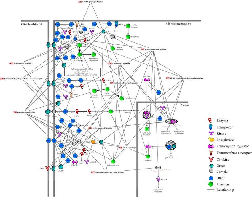
www.nature.com/scientificreports/

                              Figure 1. The porcine short posterior ciliary arteries. Photograph showing the short posterior ciliary arteries
                              that enter the eye globe close to the optic nerve.

                              biomarkers in pathological states compared to normal condition. Nevertheless, there is a paucity of information
                              on the cellular signalling mechanisms underlying perturbed ocular microcirculation. A confounding challenge
                              in ophthalmic research is the limited availability of human tissue samples for analysis. Therefore, it is important
                              to employ samples from animal models that closely resemble those of human’s for highly translational results. In
                              light of this, porcine ocular tissues were used in this study due to the high phylogenetic and morphological sim-
                              ilarities to the human eye23. For decades, the pig has become an animal model of choice in biomedical research
                              to study various human pathologies, including vascular functions in disease conditions compared to healthy
                              controls24,25. Additionally, the pig eye is commonly used in vision research and is also a validated animal model
                              to study glaucoma26.
                                  Considering the functional relevance and importance of the sPCA in the perfusion of ONH and, the
                              dearth of studies investigating the molecular regulators that maintain physiological functions in ocular blood
                              vessels, this is the first study to characterize the fundamental cellular signalling mechanisms employing the
                              mass-spectrometry-based proteomics approach. It is projected that the findings emerging from this study will
                              provide an in-depth mechanistic insight into the complex cell signalling pathways that orchestrate circulatory
                              functions in the sPCA and furnish vital information at the protein level. Finally, the methodology employed in
                              this investigation, particularly catered for optimum protein extraction and proteome characterization of ocular
                              blood vessels, is envisaged to be instrumental for future studies utilizing other ocular vascular beds.

                              Results
                              Identification of porcine sPCA proteins.             This study endeavoured to profile the proteome of porcine
                              sPCA (Fig. 1). The experimental workflow overview of this study is depicted as a schematic representation in
                              Fig. 2. A total of 1742 proteins and 10 527 peptides were identified with a false discovery rate (FDR) of less than
                              1% after removal of reverse hits (Supplementary Table S1). The protein profiles of this vascular bed resolved in
                              one-dimensional gel electrophoresis (1DE) is as depicted in Fig. 3a. With the approach of intensity-based abso-
                              lute quantification (iBAQ), the relative abundance of the identified proteins was estimated. Of the 1742 proteins
                              identified, the five most abundant proteins comprise 50% of the proteome, while 38 most abundant proteins
                              accounted for 75% of the porcine sPCA proteome (Fig. 3b, Table 1).

                              Proteomic profiles and functional annotations of sPCA.                 The classification of the identified sPCA
                              proteins was performed employing the Ingenuity Pathways Analysis software (IPA, Ingenuity QIAGEN Redwood
                              City, CA) (www.qiagen.com/ingenuity), DAVID tool (version 6.7) (http://david.abcc.ncifcrf.gov/home.jsp) and
                              Protein ANalysis THrough Evolutionary Relationships (PANTHER, http://pantherdb.org) functional anno-
                              tation tools. First, the analysis of the major Gene Ontology Cellular Component terms (GOCCs) employing
                              IPA demonstrated that the top four subcellular localizations of the sPCA proteins corresponded to cytoplasm
                              (56%), while almost 14% and 10% of the total proteins are from the nucleus and plasma membrane, respectively.
                              Only a small percentage (9%) of the total proteins was localized in the extracellular space, while the remaining
                              11% comprises unknown and other proteins, as shown in Supplementary Fig. S1. Next, to assess the biological

Scientific Reports | 6:38298 | DOI: 10.1038/srep38298                                                                                          2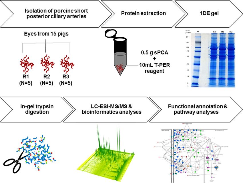
www.nature.com/scientificreports/

                              Figure 2. Workflow overview for mapping of the porcine sPCA proteome. Freshly isolated porcine sPCA
                              were pooled equally into three biological replicates, represented by R1, R2 and R3. Samples were extracted with
                              T-PER tissue protein extraction reagent and subjected to 1DE gel electrophoresis. The protein bands were sliced
                              and trypsin-digested prior to proteomic analysis by LC-MS/MS. Finally, the emerging datasets were subjected
                              to robust bioinformatics analyses and functional annotations of the significant gene ontology (GO) terms and
                              pathways employing various tools to produce a comprehensive map of the normal porcine sPCA proteome.

                              Figure 3. Proteomics analysis employing the 1DE & LC-ESI-MS/MS strategy reveals the inherent
                              characteristics of the porcine sPCA. (a) Representative protein profiles of the sPCA resolved in 1DE gel after
                              colloidal blue staining. (b) Quantitative analysis employing iBAQ shows the cumulative relative abundance of
                              identified proteins, ranked from the highest to the lowest abundance.

                              relevance of the identified proteins, enrichment analysis employing DAVID was performed. These proteins were
                              classified into 626 distinct categories of GO Biological Process (GOBPs) terms, with 227 significant processes
                              (p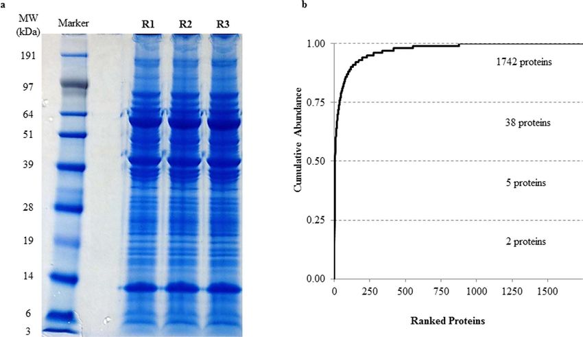
www.nature.com/scientificreports/
Sequence
Rank Protein Name Gene Name Peptides Coverage (%) iBAQ
1 Hemoglobin subunit beta HBB 17 84.37 4.09E+09
2 Hemoglobin subunit alpha HBA 12 87.03 3.00E+09
3 Uncharacterized protein LOC100302368 8 45.50 2.65E+09
4 Cardiac muscle alpha actin 1 ACTC1 33 81.23 2.23E+09
5 Serum albumin ALB 69 82.37 1.96E+09
6 Ig lambda chain C region N/A 7 92.40 5.77E+08
7 Uncharacterized protein LUM 15 47.30 5.16E+08
8 Transgelin TAGLN 20 76.27 4.78E+08
9 Uncharacterized protein (Fragment) N/A 5 80.70 4.06E+08
10 Protease serine 1 (Fragment) PRSS1 1 23.80 3.74E+08
11 Actin, cytoplasmic 2 ACTG1 29 80.00 3.63E+08
12 Decorin DCN 21 58.97 3.11E+08
13 IgG heavy chain IGHG 18 51.33 2.66E+08
14 IgG heavy chain IGHG 17 45.97 2.33E+08
15 Galectin LGALS1 9 75.80 2.17E+08
16 Serotransferrin TF 57 78.70 2.16E+08
17 Tropomyosin 1 (Alpha), isoform CRA TPM1 33 61.43 2.03E+08
18 Osteoglycin/mimecan OGN 16 44.50 1.86E+08
19 Vimentin VIM 35 71.83 1.77E+08
20 S-arrestin SAG 27 82.80 1.67E+08
21 Protein S100-A1 S100A1 2 38.30 1.63E+08
22 Uncharacterized protein CA2 20 71.63 1.59E+08
23 Creatine kinase B-type CKB 13 75.00 1.50E+08
24 Glyceraldehyde-3-phosphate dehydrogenase GAPDH 20 70.60 1.47E+08
25 Protein S100 S100B 3 40.20 1.41E+08
26 Triosephosphate isomerase TPI1 18 83.63 1.29E+08
27 Actin alpha 2 ACTA2 33 81.23 1.27E+08
28 Phosphatidylethanolamine-binding protein 1 PEBP1 15 79.10 1.26E+08
29 Cysteine and glycine-rich protein 1 CSRP1 11 56.00 1.24E+08
30 Synaptotagmin-7 SYT7 1 6.60 1.23E+08
31 Annexin A2 ANXA2 28 72.60 1.18E+08
32 Uncharacterized protein TPM2 29 64.30 1.16E+08
33 Creatine kinase M-type CKM 22 59.67 1.12E+08
34 Uncharacterized protein ENO1 18 55.83 1.09E+08
35 Myosin light chain 1/3, skeletal muscle isoform MYL1 16 78.30 1.07E+08
36 Annexin ANXA5 24 74.60 1.06E+08
37 Glutathione S-transferase GSTP1 11 61.23 1.03E+08
38 Filamin-A FLNA 116 56.27 1.03E+08
Table 1. Top 38 most abundant proteins in the porcine sPCA.
were responsible for electron transport chain and the remaining clusters were implicated in protein folding,
translational elongation, oxidative phosphorylation, glucose metabolism and glycolysis. In the category of GO
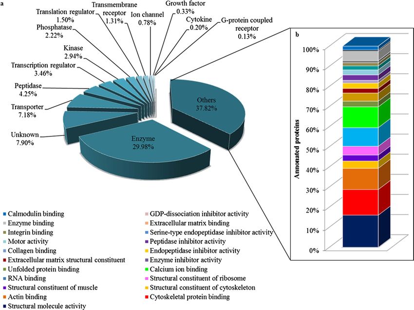
molecular types analysed employing IPA, almost 30% of the total proteins function as enzymes, transporters
(7.18%), peptidases (4.25%), transcription regulators (3.46%), kinases (2.94%) and phosphatases (2.22%), while
an approximately 38% proteins were assigned into the category of other molecular types, as depicted in Fig. 5a.
The molecular identity of these ‘other’ proteins were further dissected employing the DAVID analysis tool and the
highly significant (p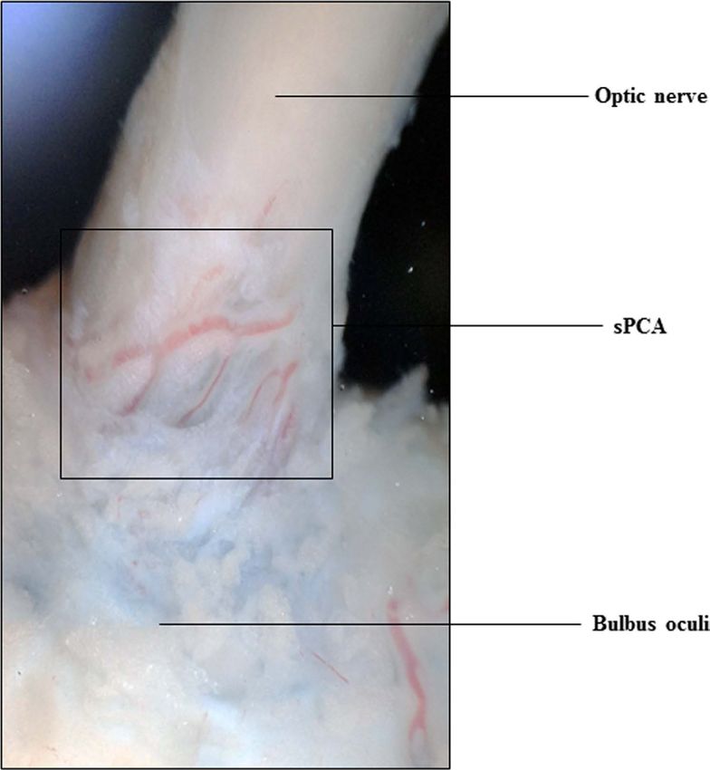
www.nature.com/scientificreports/

                              Figure 4. Functional classification of the sPCA proteome associated with over-represented GOBP terms.
                              Top twenty significantly (p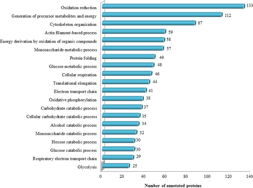
www.nature.com/scientificreports/

                              Figure 6. Functional classification of the sPCA proteome according to enriched canonical pathways. Eleven
                              functional and disease pathways were significantly (p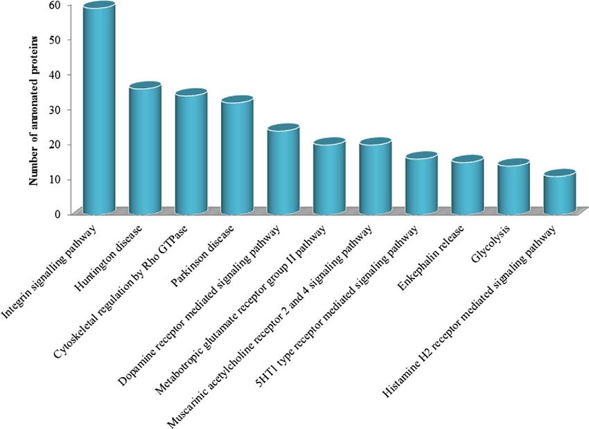
www.nature.com/scientificreports/

                              Figure 8. Protein interaction networks of merged vasoactive pathways in the sPCA. The significantly
                              (p
www.nature.com/scientificreports/
clinical settings, there is an upsurge of interest in the ocular research community in the role played by these trans-
membrane glycoproteins in the pathogenesis of glaucomatous optic neuropathy (GON)34–36.
On the other hand, integrins are also closely associated with Rho family GTPase in modulating junctional
integrity in vascular endothelial cells and in signal transduction for cytoskeletal organization37–39. Vascular per-
meability is tightly regulated under physiological conditions by intercellular tight junctions and vascular endothe-
lial growth factor (VEGF), which work in concert with Rho-GTPases to form semipermeable paracellular barriers
that control passage of solutes and also maintain cell polarity40–42. Correspondingly, the findings from the cur-
rent study have also demonstrated that the tight junction and VEGF signalling pathways were implicated in the
maintenance of vasoactivity of the sPCA (Fig. 7). Apart from regulating diverse cellular functions to maintain
physiological integrity, the Rho family GTPases are intimately implicated in maintaining vascular homeostasis43.
In blood vessels, the Rho/Rho-associated protein kinase (ROCK) pathway is expressed abundantly in the vascular
smooth muscle and is known to regulate vasoreactivity44,45. Noteworthy in this context is that this family of pro-
teins is recognized as key signalling molecules that regulate arterial blood pressure and are important mediators
involved in maintaining vascular tone in pathophysiological conditions, such as hypertension46,47. Hence, any
alterations in the hemodynamic balance and level of activation of the Rho protein family members may compro-
mise the vascular integrity and lead to various vasculopathies43,48. It is well-known that the ROCK inhibitors are
able to relax vascular smooth muscle cells and may enhance ocular blood flow49. As such, the Rho/ROCK signal-
ling pathway has emerged as a promising druggable target to treat GON because one of the major causative fac-
tors of blindness in glaucoma is recognized to arise from perturbed perfusion at the level of optic nerve head50–52.
It is intriguing that apart from identification of proteins involved in the organization of cytoskeleton and
maintenance of cellular functions, two other clusters of proteins involved in neurodegenerative disease signal-
ling pathways, the Huntington and Parkinson’s disease, were also identified with high significance in the por-
cine sPCA. A large proportion of the proteins identified in the Parkinson’s disease pathway consisted of 14-3-3,
proteasomes and to a lesser extent, heat shock protein (HSP) 70 kDa and synucleins (Supplementary Table S4).
The regulation of 14-3-3 family of proteins is tightly controlled in normal cellular milieu and implicated in a
broad spectrum of both general and specialized signalling pathways, indicating their pivotal roles in health and
disease53,54. Albeit their ‘global’ roles in cell cycle and programmed cell death, as well as in protein trafficking in
multiple tissue types, these proteins are found most abundantly in neurons of the brain and are associated with
the pathogenesis of neurodegenerative disorders, namely Parkinson’s disease55–57. Consistent with this, a study
by Yacoubian et al. employing cellular and animal models of Parkinson’s disease has demonstrated that binding
impairment of multiple isoforms of 14-3-3 to α-synuclein led to toxicity in dopaminergic neurons58. Interestingly,
the 14-3-3-mediated signalling was found to have significant role(s) in the vasomotricity of sPCA in the present
study, although the exact mechanism(s) how this signalling network functions in this vascular bed remains to be
determined. Basal levels of 14-3-3 isoforms were also detected in other arterial beds such as in the healthy human
coronary arteries, with potential function in vascular smooth muscle activation and response to arterial injury56.
Conversely in disease conditions, for example in diabetic animals with occlusion of the middle cerebral artery,
the expression of 14-3-3 proteins was diminished and this exacerbated brain damage59. It is well-recognized that
the plasticity of the vascular smooth muscle cells is a pivotal adaptation mechanism to physiological and envi-
ronmental changes, including cell proliferation, extracellular matrix organization and apoptosis60,61. Since one of
the primary functions of this protein family is to antagonize apoptotic signals and thereby, inhibit cell death62,
the expression levels of the 14-3-3 proteins in porcine sPCA in the current study conceivably reflect its functional
relevance in the maintenance of vascular integrity in normal physiological state. It is noteworthy that in the ocular
system, abnormalities of 14-3-3-mediated signalling are related to the pathogenesis of glaucoma63.
A plethora of proteasome isoforms were also found in the Parkinson’s disease pathway in the porcine sPCA.
This multifaceted ubiquitous proteinase governs a wide array of intracellular functions, with particular function
in degradation of damaged proteins64,65. In a study employing normal animals, the inhibition of proteasome
system proved lethal and led to the development of vasculopathy, where altered microvascular permeability and
cardiac apoptosis were observed, to name a few detrimental effects of proteasome dysfunction66. Additionally,
angiogenesis is regulated through proteasome and any deleterious changes in its functional activity will affect
the formation of blood vessels67,68. On the other hand, given the vital regulatory role of redox signalling in
various vascular smooth muscle cell patho(biology), also as evidenced by the significantly high expression of
proteins involved in the oxidation-reduction activities in the porcine sPCA (Fig. 4), it is not wrong to conceptu-
alize the importance of interplay between the ubiquitin-proteasome system and cellular redox processes69. The
oxido-reductive signalling plays integral roles in vascular remodelling processes, importantly in the regulation of
cytoskeletal dynamics and modulation of smooth muscle cell differentiation27,70. Concomitant identification of
actin-related complexes and tubulins in the Huntington disease pathway in the current exploratory study comes
as no surprise as these are arguably among the most overwhelmingly expressed proteins in cells, which assemble
to construct complex networks of cytoskeleton71,72, and this is in agreement with the significant detection of the
actin-cytoskeleton processes in the porcine sPCA (Fig. 4). It goes without saying that any cytoskeletal aberrations
are therefore, responsible for cellular contractile dysfunction73.
Interestingly, the proteomic findings emerging from the present study on the pathways involved in the vas-
oactivity of the porcine sPCA not only provide novel insight at the protein level, but are also in agreement with
previous in vivo and in vitro studies carried out with pharmacological approaches. In retrospect, studies have
demonstrated that the α1-adrenergic and 5-HT receptors are responsible for mediating vasoconstriction, while
the β-adrenergic receptors mediated vasodilation in the human posterior ciliary arteries74,75. This corroborates
with the detection of both α- and β-adrenergic, and 5-HT receptor-mediated signalling pathways with high sig-
nificance in the current study. Likewise, the endothelin-1 (ET-1) signalling was also significantly detected in this
investigation and this protein signalling has been associated with the pathogenesis of glaucoma, regardless of the
intraocular pressure. Perturbances in the blood flow velocity were found primarily in the retrobulbar vasculature
Scientific Reports | 6:38298 | DOI: 10.1038/srep38298 8www.nature.com/scientificreports/
of glaucoma patients, with most pronounced changes in the sPCA7. ET-1 signalling is not only implicated in
the regulation of smooth contraction, but also functions as an important mediator in inflammation76,77. On the
other hand, neuronal mediators of vasoactivity comprising the muscarinic acetylcholine receptor (Chrm) 2 and
4 signalling pathways were also detected in this ocular vascular bed in the present investigation. Both recep-
tor subtypes selectively activate the G-proteins of the Gi/Go family78. Studies carried out with Chrm 2−/− mice
strongly suggest that Chrm 2 is the predominant autoreceptor that mediates cortical and hippocampal release
of acetylcholine79,80. Since the sPCA are autonomically innervated, acetylcholine released from the cholinergeic
nerves seemed to modulate nitroxidergic nerve functions in porcine ciliary arteries74,75. Presynaptic Chrm 2 was
also identified in the mouse cerebral arteries and was implicated in neurogenic vasoconstriction via inhibition
of nitric oxide synthase (NOS)81. Consistent with this mechanism, several NOS pathways were also found to be
significantly expressed in the porcine sPCA in this study. Besides, this Chrm subtype was also known to exert
anti-apoptotic effects on cardiomyocytes and mediate antinociception78,82.
Taken together, the vascular integrity of the porcine sPCA is maintained by a plethora of highly dynamic
protein networks that ensure proper vessel functionality and physiology, as evidenced by this study. For the first
time, the expanding paradigm of exploratory proteomics has provided a sophisticated and unbiased approach to
unravel the biological processes governing fundamental cell functions in a retrobulbar vascular bed at the protein
level. However, a limitation of the present study is that the proteins identified from the sPCA cannot be particu-
larly ascribed to the different layers that make up the blood vessels i.e. the adventitia, vascular smooth muscle and
endothelial cells. It would be therefore interesting to further elucidate, compare and contrast the proteome of the
major layers of the sPCA in future investigations utilizing the proteomic and bioinformatics platform established
in this study. In conclusion, the findings emerging from our study are envisaged to serve as important benchmark
and database for future studies employing porcine sPCA.
Materials and Methods
Animals. All experiments were conducted in adherence to the Association for Research in Vision and
Ophthalmology (ARVO) Statement for the Use of Animals in Ophthalmic and Vision Research and by insti-
tutional guidelines. The study was conducted and approved at the Department of Ophthalmology, University
Medical Centre Mainz. Fresh eyes from pigs (Piétrain breed) approximately 3–6 months old were obtained from
a local abattoir immediately post-mortem. The enucleated eyes attached with optic nerve and extraocular tissues
were transported to the laboratory in ice-cold Krebs-Henseleit buffer at pH 7.4 with the following ionic compo-
sition in mM: 118.3 NaCl, 4.7 KCl, 2.5 CaCl2, 1.2 MgSO4, 1.2 KH2PO4, 25 NaHCO3, and 11 glucose (Carl Roth
GmbH, Karlsruhe, Germany).
Protein extraction from sPCA. The porcine ciliary arterial branches are divided into the short and long
posterior ciliary arteries, as described elsewhere83–85. Therefore, to standardize the isolation procedure, the arte-
rial branch used in the current study was restricted to the paraoptic sPCA that travel along the optic nerve to
enter the eye globe, as shown in Fig. 1. The luminal diameter of the isolated vessels is between 130 and 250 μm.
Branches of the sPCA were carefully dissected and cleaned of connective tissues using fine-point tweezers under
a dissecting microscope. Thereafter, the isolated blood vessels were gently rinsed in ice-cold phosphate buffered
saline (PBS) to remove any blood contaminants and were used for proteomic analysis. The sPCA from five pigs
were pooled (which constituted one biologic replicate) and a total of three biological replicates were used in this
study. Protein extraction from the porcine sPCA was carried out employing T-PER Tissue Protein Extraction
Reagent (Thermo Scientific Inc., Waltham, MA, USA) according to the manufacturer’s instruction with slight
modifications. Briefly, the isolated sPCA were weighed immediately after isolation and cleaning, followed by
protein extraction with 10 ml T-PER reagent added to 0.5 g samples. Subsequently, the vascular samples in T-PER
reagent were homogenized employing the BBY24M Bullet Blender Storm (Next Advance Inc., Averill Park,
NY, USA) and centrifuged at 10,000 × g for 5 minutes to pellet the tissue debris. The supernatant was collected
and subjected to sample cleaning employing the Amicon Ultra 0.5 mL centrifugal filters with 3 K cutoff (Merck
Millipore, Carrigtwohill, Ireland). The protein concentration of the obtained eluate was determined using BCA
Protein Assay Kit (Pierce, Rockford, IL).
1DE. The sPCA eluates were subjected to 1DE (50 μg sample per well) using 4–12% Bis-Tris Gels (Invitrogen,
Karlsruhe, Germany) with MOPS running buffer under reducing conditions for 60 min with a constant voltage
of 150 V, according to the manufacturer’s instructions. The SeeBlue Plus 2 (Invitrogen, Karlsruhe, Germany)
pre-stained protein standard was used as a molecular mass marker. Next, the gels were stained with Colloidal
Blue Staining Kit (Invitrogen, Karlsruhe, Germany) according to the manufacturer’s instructions. Then, pro-
tein bands (sliced into 39 bands per replicate) were cut into small pieces and subjected to dehydration utilizing
neat acetonitrile prior to disulfide bonds cleavage with 10 mM 1,4-Dithiothreitol (DTT) in 100 mM ammonium
bicarbonate (NH4HCO3) and alkylation with 55 mM iodoacetamide (IAA) in 100 mM NH4HCO3. The reduced
and alkylated protein mixtures were digested with sequence grade-modified trypsin (Promega, Madison, USA)
resuspended in 10 mM NH4HCO3 and 10% acetonitrile for 16 h at 37 °C. Subsequently, proteolysis was quenched
by acidification of the reaction mixtures with 100 μl of extraction buffer composed of 1: 2 (vol/vol) of 5% formic
acid: acetonitrile and, incubated for 15 min at 37 °C in a shaker. The supernatant containing peptides was collected
and the remaining peptides in the gel pieces were extracted with two 20 minutes washes in extraction buffer. The
supernatants were pooled and concentrated to dryness in SpeedVac (Eppendorf, Darmstadt, Germany) prior
to storage at −20 °C86,87. Next, the peptides recovered from the in-gel digestion were subjected to purification
employing ZipTip C18 columns (Millipore, Billerica, MA, USA) according to the manufacturer’s instructions.
This peptide-purification procedure was repeated four times for each sample and the combined eluate was dried
in the SpeedVac, dissolved in 10 μL of 0.1% trifluoroacetic acid (TFA) solution prior to LC-MS/MS analysis.
Scientific Reports | 6:38298 | DOI: 10.1038/srep38298 9www.nature.com/scientificreports/
LC-ESI-MS/MS. The LC-ESI-LTQ-Orbitrap MS system is well established in our laboratory and details of
this system are described in detail elsewhere86,88,89. Briefly, solvent A which consisted of LC-MS grade water with
0.1% (v/v) formic acid, and solvent B consisting of LC-MS grade acetonitrile with 0.1% (v/v) formic acid were
utilized. The gradient was run for 90 min per gel spot as follows; 0–50 min: 10–35% B, 50–70 min: 35–55% B,
70–75 min: 55–90% B, 75–80 min: 90% B, 80–83 min: 90–10% B und 83–90 min: 10% B. Continuum mass spectra
data were acquired on an ESI-LTQ-Orbitrap-XL MS (Thermo Scientific, Bremen, Germany) and the general
mass spectrometric conditions were as follows: positive-ion electrospray ionization mode, spray, capillary and
the tube lens voltages were set at 4.5 kV, 48 V and 120 V, respectively, and the heated capillary temperature was
set at 275 °C. The LTQ-Orbitrap was operated in a data-dependent mode of acquisition to automatically switch
between Orbitrap-MS and LTQ-MS/MS acquisition. Survey full scan MS spectra (from m/z 300 to 2000) were
acquired in the Orbitrap with a resolution of 30000 at m/z 400 and a target automatic gain control (AGC) setting
of 1.0 × 10⁶ ions. The lock mass option was enabled in MS mode and the polydimethylcyclosiloxane (PCM) m/z
445.120025 ions were used for internal recalibration in real time90. The five most intense precursor ions were
sequentially isolated for fragmentation in the LTQ with a collision-induced dissociation (CID) fragmentation,
the normalized collision energy (NCE) was set to 35% with activation time of 30 ms with repeat count of 3 and
dynamic exclusion duration of 600 s. The resulting fragment ions were recorded in the LTQ.
Bioinformatics and Gene Ontology (GO) functional annotation analysis. The acquired continuum
MS datasets were analysed by MaxQuant (version 1.5.2.8, http://www.maxquant.org/) computational proteomics
platform and its built-in Andromeda search engine for peptide and protein identification, with iBAQ algorithm
enabled91–94. The tandem MS spectra were searched against UniProtSP/TrEMBL (Homo sapiens and Sus scrofa
database (date: 1st July 2016) using standard settings with peptide mass tolerance of ±30 ppm, fragment mass
tolerance of ±0.5 Da, with ≥6 amino acid residues and only “unique plus razor peptides” that belong to a protein
were chosen91. For limiting a certain number of peak matches by chance, a target-decoy-based FDR for peptide
and protein identification was set to 0.01. Carbamidomethylation of cysteine was set as a fixed modification, while
protein N-terminal acetylation and oxidation of methionine were defined as variable modifications, enzyme:
trypsin and maximum number of missed cleavages: 2. The output of the generated “proteingroups.txt” data from
the MaxQuant analysis was utilized for subsequent functional annotation and pathway analysis.
The GO functional annotation analysis of identified proteins was carried out with three different bioinformat-
ics tools. These analyses allowed elucidation of the different functions and processes in which the identified and
validated proteins would be putatively involved. Three independent sets of ontology were used in the annotation:
“the molecular function”, “the biological processes”, in which the proteins participate, and their “cellular compo-
nent”. Proteins without similarity to database entries were not considered for collation. First, the IPA was used
for interpreting the GOCC terms, molecule types and PPI networks associated with the identified proteins. Top
canonical pathways involving in the vasoactivity of the identified sPCA proteins were presented, along with a
p-value calculated using Fisher’s exact test. Next, the molecular interactions networks between proteins associated
with top diseases and functions were reported by the PANTHER classification system (version 11.0, released on
the 15th July 2016), which includes 13096 protein families with 78442 functionally distinct protein subfamilies95.
Finally, DAVID tool was used for interpreting the GOBP and GOMF terms of the identified sPCA proteins that
could not be specifically dissected employing IPA96,97. The protein list was uploaded into DAVID and searched
for enrichment for GOBP and GOMF terms, and the results were filtered based on threshold count ≥2 and P
valueswww.nature.com/scientificreports/
18. Buckley, C. H., Hadoke, P. W. F. & OBrien, C. J. Use of isolated ocular arteries in vitro to define the pathology of vascular changes in
glaucoma. Brit J Ophthalmol 81, 599–607 (1997).
19. Nita, M. & Grzybowski, A. The Role of the Reactive Oxygen Species and Oxidative Stress in the Pathomechanism of the Age-Related
Ocular Diseases and Other Pathologies of the Anterior and Posterior Eye Segments in Adults. Oxid Med Cell Longev 2016, 1–23
(2016).
20. Schmidt, K. G., Klingmuller, V., Gouveia, S. M., Osborne, N. N. & Pillunat, L. E. Short posterior ciliary artery, central retinal artery,
and choroidal hemodynamics in brimonidine-treated primary open-angle glaucoma patients. Am J Ophthalmol 136, 1038–1048
(2003).
21. Fernandes, K. A. et al. Using genetic mouse models to gain insight into glaucoma: Past results and future possibilities. Exp Eye Res
141, 42–56 (2015).
22. Nickells, R. W. & Pelzel, H. R. Tools and resources for analyzing gene expression changes in glaucomatous neurodegeneration. Exp
Eye Res 141, 99–110 (2015).
23. Ruiz-Ederra, J. et al. The pig eye as a novel model of glaucoma. Exp Eye Res 81, 561–569 (2005).
24. Lunney, J. K. Advances in swine biomedical model genomics. Int J Biol Sci 3, 179–184 (2007).
25. Prather, R. S., Lorson, M., Ross, J. W., Whyte, J. J. & Walters, E. Genetically engineered pig models for human diseases. Annu Rev
Anim Biosci 1, 203–219 (2013).
26. Sanchez, I., Martin, R., Ussa, F. & Fernandez-Bueno, I. The parameters of the porcine eyeball. Graefes Arch Clin Exp Ophthalmol 249,
475–482 (2011).
27. Galougahi, K. K., Ashley, E. A. & Ali, Z. A. Redox regulation of vascular remodeling. Cell Mol Life Sci 73, 349–363 (2016).
28. Regent, A. et al. Proteomic analysis of vascular smooth muscle cells in physiological condition and in pulmonary arterial
hypertension: Toward contractile versus synthetic phenotypes. Proteomics 16, 2637–2649 (2016).
29. Harburger, D. S. & Calderwood, D. A. Integrin signalling at a glance. J Cell Sci 122, 159–163 (2009).
30. Morrison, J. C. Integrins in the optic nerve head: potential roles in glaucomatous optic neuropathy (an American Ophthalmological
Society thesis). Trans Am Ophthalmol Soc 104, 453–477 (2006).
31. Giancotti, F. G. & Ruoslahti, E. Integrin signaling. Science 285, 1028–1032 (1999).
32. Mousa, S. A. Integrins as novel drug discovery targets: potential therapeutic and diagnostic implications. Emerging Therapeutic
Targets 4, 143–153 (2000).
33. Lehoux, S. & Tedgui, A. Cellular mechanics and gene expression in blood vessels. J Biomech 36, 631–643 (2003).
34. Atan, N. A. D., Yekta, R. F., Nejad, M. R. & Nikzamir, A. Pathway and Network Analysis in Primary Open Angle Glaucoma. JPS 5,
92–101 (2014).
35. Elner, S. G. & Elner, V. M. The integrin superfamily and the eye. Invest Ophthalmol Vis Sci 37, 696–701 (1996).
36. Filla, M. S., Faralli, J. A., Peotter, J. L. & Peters, D. M. The role of integrins in glaucoma. Exp Eye Res (2016).
37. Clark, E. A. & Brugge, J. S. Integrins and signal transduction pathways: the road taken. Science 268, 233–239 (1995).
38. Clark, E. A., King, W. G., Brugge, J. S., Symons, M. & Hynes, R. O. Integrin-mediated signals regulated by members of the rho family
of GTPases. J Cell Biol 142, 573–586 (1998).
39. Park-Windhol, C. & D’Amore, P. A. Disorders of Vascular Permeability. Annu Rev Pathol 11, 251–281 (2016).
40. Balda, M. S. & Matter, K. Tight junctions as regulators of tissue remodelling. Curr Opin Cell Biol 42, 94–101 (2016).
41. Sawada, N. et al. Tight junctions and human diseases. Med Electron Microsc 36, 147–156 (2003).
42. Terry, S., Nie, M., Matter, K. & Balda, M. S. Rho signaling and tight junction functions. Physiology (Bethesda) 25, 16–26 (2010).
43. Rolfe, B. E., Worth, N. F., World, C. J., Campbell, J. H. & Campbell, G. R. Rho and vascular disease. Atherosclerosis 183, 1–16 (2005).
44. Hirata, K. et al. Involvement of rho p21 in the GTP-enhanced calcium ion sensitivity of smooth muscle contraction. J Biol Chem 267,
8719–8722 (1992).
45. Worth, N. F., Campbell, G. R., Campbell, J. H. & Rolfe, B. E. Rho expression and activation in vascular smooth muscle cells. Cell
Motil Cytoskeleton 59, 189–200 (2004).
46. Bond, L. M., Sellers, J. R. & McKerracher, L. Rho kinase as a target for cerebral vascular disorders. Future Med Chem 7, 1039–1053
(2015).
47. Hervé, J. C. & Bourmeyster, N. Rho GTPases at the crossroad of signaling networks in mammals. Small GTPases 6, 43–48 (2015).
48. Nunes, K. P., Rigsby, C. S. & Webb, R. C. RhoA/Rho-kinase and vascular diseases: what is the link? Cell Mol Life Sci 67, 3823–3836
(2010).
49. Wang, J., Liu, X. & Zhong, Y. Rho/Rho-associated kinase pathway in glaucoma (Review). Int J Oncol 43, 1357–1367 (2013).
50. Anderson, D. R. Introductory comments on blood flow autoregulation in the optic nerve head and vascular risk factors in glaucoma.
Surv Ophthalmol 43 Suppl 1, S5–9 (1999).
51. Chung, H. S. et al. Vascular aspects in the pathophysiology of glaucomatous optic neuropathy. Surv Ophthalmol 43 Suppl 1, S43–50
(1999).
52. Flammer, J. et al. The impact of ocular blood flow in glaucoma. Prog Retin Eye Res 21, 359–393 (2002).
53. Bridges, D. & Moorhead, G. B. 14-3-3 proteins: a number of functions for a numbered protein. Sci STKE 2004, re10 (2004).
54. Zhao, J., Meyerkord, C. L., Du, Y., Khuri, F. R. & Fu, H. 14-3-3 proteins as potential therapeutic targets. Semin Cell Dev Biol. 22,
705–712 (2011).
55. Aghazadeh, Y. & Papadopoulos, V. The role of the 14-3-3 protein family in health, disease, and drug development. Drug Discov
Today 21, 278–287 (2016).
56. Autieri, M. V. Inducible expression of the signal transduction protein 14-3-3γin injured arteries and stimulated human vascular
smooth muscle cells. Exp Mol Pathol 76, 99–107 (2004).
57. Jin, J. et al. Proteomic, functional, and domain-based analysis of in vivo 14-3-3 binding proteins involved in cytoskeletal regulation
and cellular organization. Curr Biol 14, 1436–1450 (2004).
58. Yacoubian, T. A. et al. Differential neuroprotective effects of 14-3-3 proteins in models of Parkinson’s disease. Cell Death Dis 1, e2,
doi: 10.1038/cddis.2009.4 (2010).
59. Jeon, S. J., Sung, J. H. & Koh, P. O. Hyperglycemia decreases expression of 14-3-3 proteins in an animal model of stroke. Neurosci Lett
626, 13–18 (2016).
60. Demasi, M. & Laurindo, F. R. Physiological and pathological role of the ubiquitin-proteasome system in the vascular smooth muscle
cell. Cardiovasc Res 95, 183–193 (2012).
61. Majesky, M. W. Developmental basis of vascular smooth muscle diversity. Arterioscler Thromb Vasc Biol 27, 1248–1258 (2007).
62. Yang, X., Luo, C., Cai, J., Pierce, W. M. & Tezel, G. Phosphorylation-dependent interaction with 14-3-3 in the regulation of bad
trafficking in retinal ganglion cells. Invest Ophthalmol Vis Sci 49, 2483–2494 (2008).
63. Wang, D. Y. et al. Global gene expression changes in rat retinal ganglion cells in experimental glaucoma. Invest Ophthalmol Vis Sci
51, 4084–4095 (2010).
64. Elliott, P. J. & Ross, J. S. The proteasome: a new target for novel drug therapies. Am J Clin Pathol 116, 637–646 (2001).
65. McNaught, K. S., Belizaire, R., Isacson, O., Jenner, P. & Olanow, C. W. Altered proteasomal function in sporadic Parkinson’s disease.
Exp Neurol 179, 38–46 (2003).
66. Herrmann, J. et al. Primary proteasome inhibition results in cardiac dysfunction. Eur J Heart Fail 15, 614–623 (2013).
67. Oikawa, T. et al. The proteasome is involved in angiogenesis. Biochem Bioph Res Co 246, 243–248 (1998).
Scientific Reports | 6:38298 | DOI: 10.1038/srep38298 11www.nature.com/scientificreports/
68. Walsh, D. A. & Pearson, C. I. Angiogenesis in the pathogenesis of inflammatory joint and lung diseases. Arthritis Res 3, 147–153
(2001).
69. Fernandes, D. C., Manoel, A. H., Wosniak, J. Jr. & Laurindo, F. R. Protein disulfide isomerase overexpression in vascular smooth
muscle cells induces spontaneous preemptive NADPH oxidase activation and Nox1 mRNA expression: effects of nitrosothiol
exposure. Arch Biochem Biophys 484, 197–204 (2009).
70. Gellert, M., Hanschmann, E. M., Lepka, K., Berndt, C. & Lillig, C. H. Redox regulation of cytoskeletal dynamics during
differentiation and de-differentiation. Biochim Biophys Acta 1850, 1575–1587 (2015).
71. Meinke, P., Makarov, A., Thanh, P., Sadurska, D. & Schirmer, E. Nucleoskeleton dynamics and functions in health and disease. Cell
Health Cytoskelet 7, 55–69 (2015).
72. Zou, C., La Bonte, L. R., Pavlov, V. I. & Stahl, G. L. Murine hyperglycemic vasculopathy and cardiomyopathy: whole-genome gene
expression analysis predicts cellular targets and regulatory networks influenced by mannose binding lectin. Front Immunol 3, 1–9,
doi: 10.3389/fimmu.2012.00015 (2012).
73. Hein, S., Kostin, S., Heling, A., Maeno, Y. & Schaper, J. The role of the cytoskeleton in heart failure. Cardiovasc Res 45, 273–278
(2000).
74. Orgul, S., Gugleta, K. & Flammer, J. Physiology of perfusion as it relates to the optic nerve head. Surv Ophthalmol 43, S17–S26
(1999).
75. Yu, D. Y. et al. Agonist response of human isolated posterior ciliary artery. Invest Ophthalmol Vis Sci 33, 48–54 (1992).
76. Pache, M. et al. Extraocular blood flow and endothelin-1 plasma levels in patients with multiple sclerosis. Eur Neurol 49, 164–168
(2003).
77. Zouki, C., Baron, C., Fournier, A. & Filep, J. G. Endothelin-1 enhances neutrophil adhesion to human coronary artery endothelial
cells: role of ETA receptors and platelet-activating factor. Brit J Pharmacol 127, 969–979 (1999).
78. Wess, J., Eglen, R. M. & Gautam, D. Muscarinic acetylcholine receptors: mutant mice provide new insights for drug development.
Nat Rev Drug Discov 6, 721–733 (2007).
79. Tzavara, E. T. et al. Dysregulated hippocampal acetylcholine neurotransmission and impaired cognition in M2, M4 and M2/M4
muscarinic receptor knockout mice. Mol Psychiatry 8, 673–679 (2003).
80. Zhang, W. L. et al. Characterization of central inhibitory muscarinic autoreceptors by the use of muscarinic acetylcholine receptor
knock-out mice. J Neurosci 22, 1709–1717 (2002).
81. Badhwar, A., Stanimirovic, D. B., Hamel, E. & Haqqani, A. S. The proteome of mouse cerebral arteries. J Cereb Blood Flow Metab 34,
1033–1046 (2014).
82. Miao, Y. et al. Acetylcholine Inhibits Tumor Necrosis Factor αActivated Endoplasmic Reticulum Apoptotic Pathway via EGFR‐
PI3K Signaling in Cardiomyocytes. J Cell Physiol 230, 767–774 (2015).
83. Haefliger, I. O., Flammer, J. & Luscher, T. F. Heterogeneity of endothelium-dependent regulation in ophthalmic and ciliary arteries.
Invest Ophthalmol Vis Sci 34, 1722–1730 (1993).
84. Ninomiya, H. & Inomata, T. Microvascular anatomy of the pig eye: scanning electron microscopy of vascular corrosion casts. J Vet
Med Sci 68, 1149–1154 (2006).
85. Yao, K., Tschudi, M., Flammer, J. & Luscher, T. F. Endothelium-dependent regulation of vascular tone of the porcine ophthalmic
artery. Invest Ophthalmol Vis Sci 32, 1791–1798 (1991).
86. Perumal, N., Funke, S., Pfeiffer, N. & Grus, F. H. Characterization of lacrimal proline‐rich protein 4 (PRR4) in human tear proteome.
Proteomics 14, 1698–1709 (2014).
87. Shevchenko, A., Tomas, H., Havli, J., Olsen, J. V. & Mann, M. In-gel digestion for mass spectrometric characterization of proteins
and proteomes. Nat Protoc 1, 2856–2860 (2006).
88. Perumal, N., Funke, S., Wolters, D., Pfeiffer, N. & Grus, F. H. Characterization of human reflex tear proteome reveals high expression
of lacrimal proline-rich protein 4 (PRR4). Proteomics 15, 3370–3381 (2015).
89. Perumal, N., Funke, S., Pfeiffer, N. & Grus, F. H. Proteomics analysis of human tears from aqueous-deficient and evaporative dry eye
patients. Sci Rep 6, 29629, doi: 10.1038/srep29629 (2016).
90. Olsen, J. V. et al. Parts per million mass accuracy on an Orbitrap mass spectrometer via lock mass injection into a C-trap. Mol Cell
Proteomics 4, 2010–2021 (2005).
91. Cox, J. & Mann, M. MaxQuant enables high peptide identification rates, individualized p.p.b.-range mass accuracies and proteome-
wide protein quantification. Nat Biotechnol 26, 1367–1372 (2008).
92. Luber, C. A. et al. Quantitative proteomics reveals subset-specific viral recognition in dendritic cells. Immunity 32, 279–289 (2010).
93. Cox, J. et al. Accurate Proteome-wide Label-free Quantification by Delayed Normalization and Maximal Peptide Ratio Extraction,
Termed MaxLFQ. Mol Cell Proteomics 13, 2513–2526 (2014).
94. Cox, J. et al. Andromeda: A Peptide Search Engine Integrated into the MaxQuant Environment. J Proteome Res 10, 1794–1805
(2011).
95. Mi, H., Poudel, S., Muruganujan, A., Casagrande, J. T. & Thomas, P. D. PANTHER version 10: expanded protein families and
functions, and analysis tools. Nucleic Acids Res 44, D336–342 (2016).
96. Huang, D. W., Sherman, B. T. & Lempicki, R. A. Systematic and integrative analysis of large gene lists using DAVID bioinformatics
resources. Nat Protoc 4, 44–57 (2009).
97. Huang, D. W., Sherman, B. T. & Lempicki, R. A. Bioinformatics enrichment tools: paths toward the comprehensive functional
analysis of large gene lists. Nucleic Acids Res 37, 1–13 (2009).
Acknowledgements
Dr. Manicam’s work is supported by the Internal University Research Funding (Stufe 1) from the University
Medical Centre of the Johannes Gutenberg University Mainz and supported in part by a grant from the Ernst und
Berta Grimmke-Stiftung.
Author Contributions
C.M. conceived and designed the study, performed the experiments, analysed and interpreted the data, and
wrote the manuscript. N. Perumal designed the study, analysed and interpreted the data, and helped to write
the manuscript. FHG contributed essential materials and analysis tools, and helped draft the manuscript. N.
Pfeiffer contributed essential materials and analysis tools, and helped draft the manuscript. A.G. participated in
the design of the study, contributed essential materials and tools, and helped draft the manuscript. All authors
were involved in the discussion of results, critical reading and final approval of the manuscript.
Additional Information
Supplementary information accompanies this paper at http://www.nature.com/srep
Competing financial interests: The authors declare no competing financial interests.
Scientific Reports | 6:38298 | DOI: 10.1038/srep38298 12www.nature.com/scientificreports/
How to cite this article: Manicam, C. et al. First insight into the proteome landscape of the porcine short
posterior ciliary arteries: Key signalling pathways maintaining physiologic functions. Sci. Rep. 6, 38298;
doi: 10.1038/srep38298 (2016).
Publisher's note: Springer Nature remains neutral with regard to jurisdictional claims in published maps and
institutional affiliations.
This work is licensed under a Creative Commons Attribution 4.0 International License. The images
or other third party material in this article are included in the article’s Creative Commons license,
unless indicated otherwise in the credit line; if the material is not included under the Creative Commons license,
users will need to obtain permission from the license holder to reproduce the material. To view a copy of this
license, visit http://creativecommons.org/licenses/by/4.0/
© The Author(s) 2016
Scientific Reports | 6:38298 | DOI: 10.1038/srep38298 13You can also read