Curcumenol mitigates chondrocyte inflammation by inhibiting the NF κB and MAPK pathways, and ameliorates DMM induced OA in mice
←
→
Page content transcription
If your browser does not render page correctly, please read the page content below
INTERNATIONAL JOURNAL OF MOlecular medicine 48: 192, 2021

                    Curcumenol mitigates chondrocyte inflammation
                     by inhibiting the NF‑κB and MAPK pathways,
                      and ameliorates DMM‑induced OA in mice
                  XIAO YANG*, YIFAN ZHOU*, ZHIQIAN CHEN, CHEN CHEN, CHEN HAN, XUNLIN LI,
                     HAIJUN TIAN, XIAOFEI CHENG, KAI ZHANG, TANGJUN ZHOU and JIE ZHAO

               Shanghai Key Laboratory of Orthopedic Implants, Department of Orthopedics, Ninth People's Hospital,
                         Shanghai Jiaotong University School of Medicine, Shanghai 200011, P.R. China

                                        Received May 25, 2021; Accepted August 9, 2021

                                                  DOI: 10.3892/ijmm.2021.5025

Abstract. At present, an increasing number of individuals are       in ATDC5 chondrocytes and primary mice chondrocytes, and
affected by osteoarthritis (OA), resulting in a heavy socio‑        also ameliorated OA in a DMM‑induced mouse model.
economic burden. OA in knee joints is caused by the release
of inflammatory cytokines and subsequent biomechanical              Introduction
and structural deterioration. To determine its anti‑inflam‑
matory function, the current study investigated the use of          Osteoarthritis (OA) is a degenerative joint disorder causing
the plant‑derived medicine, curcumenol, in OA treatment.            disability in the elderly population worldwide (1), and is esti‑
Curcumenol was not cytotoxic to ATDC5 chondrocytes and              mated to potentially affect ~400 million individuals in China
primary chondrocytes, as determined using a cell viability test.    by 2030 (2). OA has caused a heavy socioeconomic burden,
When these cells were treated with TNF‑α and IL‑1β to induce        which costs almost 2.5% of the gross domestic product of
inflammation, curcumenol treatment inhibited the progres‑           developed countries (3). Generally speaking, joints (especially
sion of inflammation by inactivating the NF‑κ B and MAPK            the knee joint) are flexible, act as a functional motif to withstand
signaling pathways, as well as decreasing the expression levels     compressive forces, and allow for a multi‑directional range of
of MMP3 (as indicated by reverse transcription‑quantitative         motion for movement. Joints are composed of articular carti‑
PCR and western blotting). Moreover, to analyze metabolic           lage, subchondral bone and synovium, which are all severely
and catabolic status in high‑density and pellet culture, cata‑      compromised by OA, but especially the cartilage. However,
lytic changes and the degradation of the extracellular matrix       the etiology of OA is complicated, particularly that which is
induced by TNF‑α and IL‑1β, were evaluated by alcian blue           associated with anatomic hip dysplasia or joint morphology (4),
staining. These catalytic deteriorations were ameliorated by        or that caused by immune factors, including rheumatoid
curcumenol. Using curcumenol in disease management, the             arthritis (5). Thus, the management of OA is currently focused
mechanical and metabolic disruption of cartilage caused in          on pain relief and functional reconstruction. These conven‑
the destabilization of medial meniscus (DMM) model was              tional strategies include oral non‑steroidal anti‑inflammatory
prevented in vivo. Thus, curcumenol mitigated inflammation          drugs (6), intra‑articular injection of glucocorticoids (2) or
                                                                    hyaluronic acid (7), and surgical methods such as arthroscopic
                                                                    management and total arthroplasty. Stem cell injection
                                                                    therapy has gained significant attention in basal research and
                                                                    clinical trials (8); however, based on challenges such as cell
Correspondence to: Dr Jie Zhao or Dr Tangjun Zhou, Shanghai         leakage, osteogenic transformation of mesenchymal stem cells
Key Laboratory of Orthopedic Implants, Department of Orthopedics,   and other safety concerns, long‑term complications and the
Ninth People's Hospital, Shanghai Jiaotong University School of     cost‑effectiveness of the procedure should be addressed (9,10).
Medicine, 639 Zhizaoju Road, Shanghai 200011, P.R. China            Therefore, from the prospective of the underlying molecular
E‑mail: profzhaojie@126.com
                                                                    mechanism of OA progression, inhibiting inflammatory
E‑mail: zhoutangjun@outlook.com
                                                                    pathways, such as the NF‑κ B or MAPK cascades, may help to
*
    Contributed equally                                             alleviate the progression of joint degeneration.
                                                                        Curcumenol is a bioactive ingredient isolated from edible
Abbreviations: OA, osteoarthritis; DMM, destabilization of medial   rhizome of Curcuma zedoaria (zedoary, Zingiberaceae),
meniscus; ECM, extracellular matrix                                 which is an important constituent of Chinese Traditional
                                                                    Medicine (11,12). Subsequently, curcumenol was found
Key words: curcumenol, OA, NF‑κ B pathway, MAPK pathway,            to be one of the primary constituents in numerous other
DMM mouse model                                                     plants, such as various Piper species, Torilis japonica and
                                                                    Neolitsea pallens (13,14). Such plants exhibit various functions,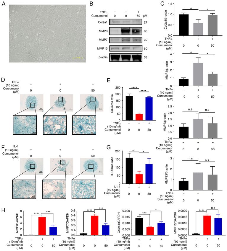
2                 YANG et al: CURCUMENOL INHIBITS INFLAMMATION in vitro AND MITIGATES ARTHRITIS in vivo

including anti‑inflammatory, hepatoprotective, neuroprotec‑       Inc.) and 1% insulin‑transferrin‑selenium (ITS) solution
tive and antioxidant activities (15,16). Thus, curcumenol may     at 37˚C with 5% CO2.
be considered a potential option for treating inflammation.
    The aim of the present study was to investigate the           ATDC5 cell culture. Mouse ATDC5 immortalized chon‑
potential use of curcumenol to treat OA in vitro and in vivo.     drocytes (17) were purchased from Shaanxi Fuheng (FH)
First, the inhibitory effect of curcumenol on the NF‑κ B and      Biotechnology Co., Ltd., and were maintained in DMEM/F12
MAPK pathways was determined in ATDC5 chondrocytes                supplemented with 5% FBS and 1% penicillin‑streptomycin
and primary chondrocytes in vitro, after which the rescue         (Gibco; Thermo Fisher Scientific, Inc.) at 37˚C with 5% CO2.
function of curcumenol in destabilization of medial meniscus
(DMM)‑induced knee joint OA in mice was evaluated in vivo.        RNA extraction and reverse transcription‑quantitative PCR
                                                                  (qPCR). ATDC5 and primary chondrocytes were stimulated
Materials and methods                                             with TNF‑ α and IL‑1β (both 10 ng/ml) with or without
                                                                  curcumenol (50 µM), and the control group was cultured in
Reagents. Curcumenol was purchased from Selleck Chemicals,        medium with 1:2,000 DMSO. After 24 h at 37˚C, total RNA
and according to the manufacturer's protocol, was isolated        was isolated from cells using TRIzol® reagent (Thermo Fisher
from Curcuma zedoary, with the following characteristics:         Scientific, Inc.) according to the manufacturer's protocol.
High performance liquid chromatography, purity=99.89%;            Extracted RNA was reverse transcribed to first strand cDNA
nuclear magnetic resonance, consistent structure. Then,           using the cDNA Synthesis kit (Takara Bio, Inc.). qPCR was
10 mg curcumenol was dissolved in 0.4268 ml DMSO                  conducted using the TB Green Premix Ex Taq kit (Takara Bio,
(Sigma‑Aldrich; Merck KGaA) to a concentration of 100 mM,         Inc.) on an Applied Biosystems QuantStudio 6 Flex Real‑Time
and stored at ‑20˚C. Recombinant TNF‑α (PeproTech China)          PCR system (Applied Biosystems; Thermo Fisher Scientific,
and IL‑1β (R&D Systems, Inc.) were dissolved in sterile PBS       Inc.) per the following conditions: Denaturation at 95˚C for
containing 0.1% BSA (Beyotime Institute of Biotechnology) to      30 sec; 40 cycles of 95˚C for 3 sec and 60˚C for 34 sec; and then
a concentration of 10 µg/ml.                                      95˚C for 15 sec, 60˚C for 60 sec and finally, 95˚C for 15 sec.
    Primary antibodies against IKKα (cat. no. D3W6N;              Primers were designed using NCBI BLAST (18), the sequences
rabbit monoclonal), phosphorylated (p)‑IKKα/β (Ser176/180;        of which are provided in Table I. Target gene expression levels
cat. no. 16A6; rabbit monoclonal), P65 (cat. no. D14E12; rabbit   were determined using the 2‑ΔΔCq method (19), with GAPDH as
monoclonal), p‑P65 (Ser536; cat. no. 93H1; rabbit monoclonal),    the internal reference control.
Iκ Bα (cat. no. L35A5; mouse monoclonal), p‑Iκ Bα (Ser32;
cat. no. 14D4; rabbit monoclonal), Akt (cat. no. 11E7; rabbit     Cell viability analysis. Cell viability was evaluated using the
monoclonal), p‑Akt (Ser473; cat. no. D9E; rabbit monoclonal),     Cell Counting Kit‑8 (CCK‑8; Dojindo Molecular Laboratories,
SAPK/JNK (cat. no. 9252; rabbit monoclonal), p‑SAPK/JNK           Inc.). ATDC5 chondrocytes were seeded into a 96‑well plate
(Thr183/Tyr185, G9; cat. no. 81E11; rabbit monoclonal), P38       at a density of 3x10³ cells/well. The next day, the cells were
(cat. no. D13E1; rabbit monoclonal), p‑P38 (Thr180/Tyr182;        treated with increasing concentrations of curcumenol (12.5,
cat. no. D13.14.4E; rabbit monoclonal), p44/42 (cat. no. 137F5;   25, 50 and 100 µM, dissolved in DMSO) for 24, 48 and 72 h
rabbit monoclonal), p‑p44/42 (Thr202/Tyr204; cat. no. D3F9;       at 37˚C; the control group was cultured in medium containing
rabbit monoclonal) and β‑actin (cat. no. D6A8; rabbit mono‑       1:1,000 DMSO. Media were refreshed every 2 days.
clonal) were purchased from Cell Signaling Technology,            Subsequently, the cells were incubated with fresh complete
Inc. Primary antibodies against collagen type II α 1 chain        media containing 10 µl CCK‑8 reagent, for 2 h at 37˚C.
(Col2a1; cat. no. ab188570; rabbit monoclonal), MMP3              Complete medium containing CCK‑8 reagent, with no cells
(cat. no. ab52915; rabbit monoclonal), MMP7 (cat. no. ab5706;     or untreated cells, were used as the blank and mock controls,
rabbit monoclonal) and MMP13 (cat. no. ab51072; rabbit            respectively. Absorbance at 450 nm (mean optical density;
monoclonal) were obtained from Abcam.                             OD) was measured using an Infinite M200 Pro multimode
                                                                  microplate reader (Tecan Group, Ltd.).
Isolation and culture of primary mouse chondrocytes. For
each isolation process, three 5‑day‑old mice (weight, 2‑4 g;      High‑density culture and pellet culture. To assess chondrogenic
Shanghai Lab, Animal Research Center Co., Ltd.; housed            differentiation, 1.5x10⁵ ATDC5 or primary chondrocytes were
under pathogen‑free conditions at 26‑28˚C and 50‑65%              resuspended in 10 µl incomplete MEM/F12 (Gibco; Thermo
humidity with a 12‑h day/night cycle.) were sacrificed via        Fisher Scientific, Inc.) and seeded as micromasses in the
decapitation and immersed in 75% ethanol for 10 min. Both         bottom of a 24‑well plate. The cells were allowed to adhere for
of the lower limbs were dissected, and the skin removed, and      1 h at 37˚C, after which 0.5 ml MEM/F12 containing 10 ng/ml
the whole knee joint was extracted with the synovial and          ITS and 2% FBS were added. After 24 h at 37˚C, the cells
muscle tissue stripped. These six cartilage samples were cut      were stimulated with TNF‑α and IL‑1β (both 10 ng/ml) with
into pieces (0.5‑1 mm) and then soaked in 1% collagenase          or without curcumenol (50 µM), and the control groups were
II solution for 2 h at 37˚C, followed by centrifugation (in       cultured in a medium with DMSO only (1:2,000) for 9 days
300 x g, 37˚C for 5 min) and resuspension in complete medium      at 37˚C. All media were refreshed every other day, and after
(DMEM/F12 with 5% FBS, 1% penicillin‑streptomycin). The           9 days the micromasses were stained with alcian blue for 24 h
primary chondrocytes were cultured in DMEM/F12 (Gibco;            at room temperature (RT).
Thermo Fisher Scientific, Inc.) supplemented with 5% FBS,             For pellet culture, 1.5x10⁷ ATDC5 were pelleted in 15‑ml
1% penicillin‑streptomycin (Gibco; Thermo Fisher Scientific,      centrifuge tubes (200 x g, 37˚C for 5 min) supplemented with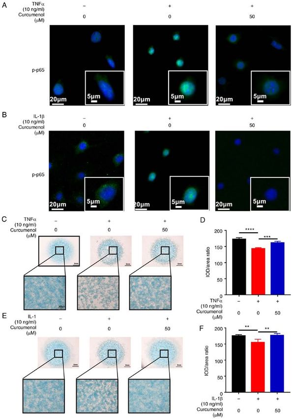
INTERNATIONAL JOURNAL OF MOlecular medicine 48: 192, 2021                                         3

Table I. PCR primer information.

Gene                     Accession number	Description                                             Sequence (5'‑3')

MMP3    NM_010809.2                                       Forward	CCCTGCAACCGTGAAGAAGA
		                                                        Reverse   GACAGCATCCACCCTTGAGT
MMP7    NM_010810.5                                       Forward	CCCTGTTCTGCTTTGTGTGTC
		                                                        Reverse   AGGGGGAGAGTTTTCCAGTCA
MMP13   NM_008607.2                                       Forward   AGAAGTGTGACCCAGCCCTA
		                                                        Reverse   GGTCACGGGATGGATGTTCA
ADAMTS4 NM_172845.3                                       Forward   GAGTCCCATTTCCCGCAGA
		                                                        Reverse   GCAGGTAGCGCTTTAACCCT
ADAMTS5 NM_011782.2                                       Forward   GAGAACCCTGCAAAACAGCC
		                                                        Reverse   AACCATACAAGTGCCTTTTCTCT
Col2a1  NM_053593.2                                       Forward   GTGTGACACTGGGAATGTCCTCT
		                                                        Reverse   TGGCCCTAATTTTCCACTGGC
GAPDH   NM_008084.3                                       Forward	CGACTTCAACAGCAACTCCCACTCTTCC
		                                                        Reverse   TGGGTGGTCCAGGGTTTCTTACTCCTT

ADAMTS, ADAM metallopeptidase with thrombospondin type 1 motif.

mesenchymal stem cell chondrogenic differentiation medium         and Col2a1 protein expression. For preventive analysis of the
(Cyagen Biosciences, Inc.). After 48 h at 37˚C, the ATDC5 and     NF‑κ B and MAPK pathways, ATDC5 and primary chon‑
primary chondrocyte pellets were stimulated with TNF‑α and        drocytes were pretreated with increasing concentrations of
IL‑1β (both 10 ng/ml) with or without curcumenol (50 µM),         curcumenol (6.25, 12.5, 25 and 50 µM, dissolved in DMSO)
and the control group was cultured in medium containing           for 2 h at 37˚C, and then stimulated with TNF‑α and IL‑1β for
DMSO only (1:2,000) for 21 days at 37˚C. The media were           10 min at 37˚C, then total cellular proteins were extracted. For
refreshed every 3 days. After 21 days of culture, the pellets     reactive analysis of the NF‑κ B and MAPK pathways, ATDC5
were collected and fixed at RT in 4% paraformaldehyde (PFA)       chondrocytes were pretreated with serum‑free medium for
for 5 h, and then embedded in optimal cutting temperature         2 h at 37˚C and then stimulated with TNF‑α with or without
compound (Sakura Finetek USA, Inc.). The samples were then        curcumenol for 10 min at 37˚C; total cellular proteins were
stored at ‑80˚C overnight and cut to a 20‑µm thickness using a    then extracted.
freezing microtome (Leica Microsystems GmbH).                         Cultured cells were lysed using RIPA lysis buffer supple‑
    Digital images were captured under a light microscope at      mented with phosphatase and protease inhibitors (Roche
a x7.8 magnification (Leica DM4000 B; Leica Microsystems          Diagnostics). The protein was quantified by BCA assay
GmbH). Alcian blue staining intensity was analyzed using          (Thermo Fisher Scientific, Inc.) and then equal quantities
Image Pro Plus 6.0 software (20) to evaluate the ratio of         of extracted protein (20‑30 µg) were separated via 10 or
integrated (I)OD (expressed as the IOD/area for each sample).     12.5% SDS‑PAGE and electroblotted onto 0.22‑µm PVDF
                                                                  membranes (MilliporeSigma). The membranes were blocked
Senescence assays. The senescence of primary chondro‑             with 5% BSA‑PBS (Beyotime Institute of Biotechnology) at
cytes was analyzed using the Senescence β ‑Galactosidase          room temperature for 1 h, and then incubated with primary
Staining kit (Beyotime Institute of Biotechnology). Primary       antibodies against IKKα, phosphorylated (p)‑IKKα /β, P65,
chondrocytes were seeded into a 12‑well plate at a density of     p‑P65, IκBα, p‑IκBα, Akt, p‑Akt, SAPK/JNK, p‑SAPK/JNK,
4x10⁵ cells/well, following stimulation with TNF‑α and IL‑1β      P38, p‑P38, p44/42, p‑p44/42 and β‑actin overnight (≥16 h)
(10 ng/ml each) with or without curcumenol (50 µM). After         at 4˚C. The membranes were washed with TBS‑0.1% Tween20
24 h at 37˚C, the cells were fixed with Beyotime Fixative         (TBST) and subsequently incubated with anti‑rabbit IgG (H+L)
Solution for 15 min at room temperature, and then incubated       secondary antibody (cat. no. 5151; DyLight™ 800 4X PEG
with Beyotime β‑Galactosidase Staining buffer at 37˚C over‑       Conjugate; Cell Signaling Technology, Inc.; 1:5,000) for 1 h at
night. Digital images were captured under a light microscope      room temperature in the dark. After washing in TBST, protein
at x10x and x20 magnification (Leica DM4000 B; Leica              immunoreactivity was detected using the Odyssey Fluorescence
Microsystems GmbH) and the percentage of positive cells was       Imaging system (LI‑COR Biosciences). Semi‑quantitative
calculated.                                                       analysis of protein band intensity was conducted using ImageJ
                                                                  V1.8.0 software (National Institutes of Health) and normalized
Western blot analysis. ATDC5 and primary chondrocytes             to the internal loading control, β‑actin.
were stimulated with TNF‑α and IL‑1β (10 ng/ml each) with
or without curcumenol (50 µM). After 24 h at 37˚C, total          Animals and surgical procedures. All animal experiments
cellular proteins were extracted for detection of MMP family      were approved by the Institutional Animal Care and Ethics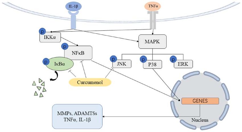
4                   YANG et al: CURCUMENOL INHIBITS INFLAMMATION in vitro AND MITIGATES ARTHRITIS in vivo

Committee of Ninth People's Hospital, Shanghai Jiaotong                      For immunofluorescence assessment, ATDC5 cells were
University School of Medicine (Shanghai, China), and                     cultured on slides added to a 6‑well plate. At 10% confluence,
performed in accordance with the principles and procedures               the cells were stimulated with TNF‑α and IL‑1β for 20 min
of the National Institutes of Health Guide for the Care and              at 37˚C, with or without curcumenol pretreatment for 2 h
Use of Laboratory Animals, and the Guidelines for Animal                 at 37˚C. Then the slides were fixed with 4% PFA at RT for
Treatment of Shanghai Jiaotong University. A total of 18                 48 h, and then immersed in PBS (pH 7.4) and washed three
8‑week‑old male C57/BL mice (weight, 18‑22 g; Shanghai                   times for 5 min each. Auto‑fluorescence quencher was added to
Lab, Animal Research Center Co., Ltd.) were housed under                 the sections for 5 min, which were then blocked with blocking
pathogen‑free conditions at 26‑28˚C and 50‑65% humidity                  buffer (Cell Signaling Technology, Inc.) for 30 min at RT. The
with a 12‑h day/night cycle. Animals were fed standard rodent            slides were subsequently incubated with primary antibodies
chow and had access to fresh water ad libitum. Before the                in a wet box at 4˚C overnight. Anti‑pp65 primary antibody
surgical procedures, mice were anesthetized by intraperi‑                was used at a 1:100 dilution. The following day, the slides
toneal injection of pentobarbital sodium (50 mg/kg of body               were washed with PBS and incubated with N Alexa Fluor
weight). In the control group (n=6; underwent sham surgery               594‑conjugated secondary antibody (cat. no. 8889; anti‑rabbit;
and were treated with corn oil; however, during research, one            1:500; Cell Signaling Technology, Inc.) for 50 min at RT in
mouse was lost from the control group), the fur on the skin was          the dark. Subsequently, the slides were washed with PBS and
shaved, a 0.5‑cm incision was made near the right knee joint,            then incubated with DAPI solution (Sigma‑Aldrich; Merck
and the ligamentum patellae was exposed and stretched. The               KGaA) for 10 min at RT in the dark to stain cell nuclei. After
remaining 12 mice were assigned to the DMM group (n=6;                   a final wash with PBS, the samples were air‑dried and sealed
underwent DMM surgery and were treated with corn oil; 1                  with anti‑fluorescence quenching tablets. Digital fluorescence
mouse was removed from the DMM group to equalize the                     images were captured under a Leica DM4000 B epifluores‑
numbers) and the curcumenol group (n=6; underwent DMM                    cence microscope (Leica Microsystems GmbH) at a x10 and
surgery and were treated with curcumenol; one was removed                x20 magnification, and IOD measurements were obtained
from curcumenol group to equalize the numbers). After                    using Image Pro Plus 6.0 software (Media Cybernetics, Inc.).
exposure, the medial collateral ligaments were transected,                   For tissue staining, the sections were de‑paraffinized in
and the medial meniscus of the tibia was partially removed               graded xylene, rehydrated in graded alcohol solutions, and
using a 5‑mm blade micro‑surgical knife (Beyotime Institute              then incubated in antigen retrieval buffer (Roche Diagnostics)
of Biotechnology) (21). After the operation, the incisions were          at 37˚C for 30 min. After cooling to RT, the sections were
sutured and the mice were initially treated two days after               immersed in PBS (pH 7.4) and washed three times for
surgery, and then for another 2 months with intraperitoneal              5 min each, and then processed as slides as aforemen‑
injections of curcumenol (50 mg curcumenol pre‑dissolved in              tioned. Anti‑TNF‑α (cat. no. ab183218; Abcam), anti‑IL‑1β
1 ml DMSO and then diluted in 100 ml corn oil) for the curc‑             (cat. no. ab234437; Abcam) and anti‑Col2a1 (cat. no. AF0135;
umenol group, and corn oil (cat. no. C8267; Sigma‑Aldrich;               Affinity) primary antibodies were used at a 1:100 dilution.
Merck KGaA; 1 ml DMSO diluted in 100 ml corn oil) for the
control and DMM groups, at 4 mg/kg/time twice a week. At                 Immunohistochemistry. Fixed lower limb samples were
the end of the experimental period, all mice were sacrificed by          embedded in paraffin as aforementioned, and cut into slices
cervical dislocation and the right lower limbs were extracted,           (8 µm), then subjected to immunohistochemistry using a kit
cleaned of soft tissues, stretched and fixed in 4% PFA at RT             (cat. no. G1215‑200T; Wuhan Servicebio Technology Co., Ltd.)
for 48 h.                                                                according to the manufacturer's instructions. Briefly, tissue
                                                                         sections were incubated with rabbit anti‑TNF‑α (cat. no. ab9579;
Histology and immunofluorescence staining. Fixed lower limb              Abcam), anti‑IL‑1β (cat. no. ab283818; Abcam) and anti‑Col2a1
samples were embedded in paraffin and subjected to histo‑                (cat. no. ab34712; Abcam) overnight at 4˚C (1:100 dilution). The
logical sectioning (5‑µm thickness). For histological assessment,        following day, the slides were washed with PBS and incubated
paraffin‑embedded tissue sections were processed for Safranin            with goat anti‑mouse/rabbit IgG HRP‑polymer (cat. no. 91196;
O‑Fast Green and hematoxylin and eosin (H&E) staining                    anti‑rabbit; 1:500; Cell Signaling Technology, Inc.) for 30 min
(Servicebio) at RT for 2‑5 min, in accordance with the manufac‑          at RT using 3,3'‑diaminobenzidin as the chromogen. Digital
turer's instructions. Sections were examined for tissue thickness,       images were captured under a Leica DM4000 B microscope
which was quantified by measuring the Safranin O‑positive                at x10 and x20 magnification, and positively‑stained cell
thickness in the center of the medial tibial plateau (22,23), and the    measurements were obtained using Image Pro Plus 6.0 software.
OA Research Society Internationall histological (OARSI) score
system: 0, normal; 0.5, loss of Safranin‑O without structural            Radiographic analysis. Digital X‑ray imaging of the right
changes; 1, small fibrillations without loss of cartilage; 2, vertical   lower limbs was conducted per the manufacturer's instruc‑
clefts down to the layer immediately below the superficial layer         tions (24) in the anteroposterior axis with a 21 lp/mm detector
and some loss of surface lamina; 3, vertical clefts/erosion to the       that provides up to x5 geometric magnification (Faxitron
calcified cartilage extending to 75% of the articular surface.                                           groups were analyzed by one‑way ANOVA with Tukey's post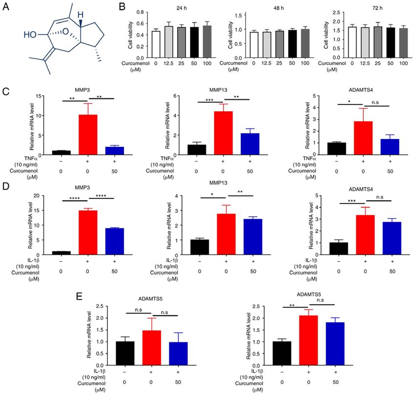
INTERNATIONAL JOURNAL OF MOlecular medicine 48: 192, 2021                                                           5

Figure 1. Curcumenol is not cytotoxic to ATDC5 chondrocytes, and inhibits TNF‑α and IL‑1β‑induced MMP family upregulation in vitro. (A) Chemical struc‑
ture of curcumenol. (B) Cell Counting Kit‑8 assay results of ATDC5 chondrocytes stimulated with curcumenol at different concentrations (0, 12.5, 25, 50 and
100 µM) and over different time periods (24‑72 h). (C) RT‑qPCR analysis of relative mRNA expression levels of MMP3, MMP13 and ADAMTS4 in ATDC5
chondrocytes with TNF‑α (10 ng/ml) or/and curcumenol (50 µM) administration. (D) RT‑qPCR analysis of relative mRNA expression levels of MMP3, MMP13
and ADAMTS4 in ATDC5 chondrocytes with IL‑1β (10 ng/ml) or/and curcumenol (50 µM) administration for 24 h. (E) RT‑qPCR analysis of relative mRNA
expression levels of ADAMTS5 in ATDC5 chondrocytes with TNF‑α (10 ng/ml) and IL‑1β (10 ng/ml) with or without curcumenol (50 µM) administration.
All data are presented as the mean ± SD from three experiments. *P
6                 YANG et al: CURCUMENOL INHIBITS INFLAMMATION in vitro AND MITIGATES ARTHRITIS in vivo

effects of curcumenol on ATDC5 chondrocytes, TNF‑ α                    In pellet culture, the ATDC5 pellets of the control group
and IL‑1β (10 ng/ml, 24 h) were used to activate the inflam‑       showed abundant ECM, while in the TNF‑α and IL‑1β groups,
matory response. The expression of the MMP3, MMP13,                the pellets were shrunken and the ECM was degraded, with
ADAM metallopeptidase with thrombospondin type 1 motif 4           decreased Safranine O staining. Moreover, curcumenol was
(ADAMTS4) and ADAMTS5 genes was increased following                able to rescue this disruption, recovering the ECM to near
TNF‑α (Fig. 1C and E) and IL‑1β (Fig. 1D and E) stimulation.       normal status (Fig. S2A and C), with the ratio of Safranin
However, following treatment with 50 µM curcumenol, the            O‑Fast Green decreased in the TNF‑α and IL‑1β group, and
expression levels of these genes were significantly decreased;     increased in the curcumenol group (Fig. S2B and D). Therefore,
ADAMTS gene expression was also downregulated, but not             curcumenol effectively altered catabolism status following
to a significant degree (Fig. 1C‑E). Western blotting was          deterioration by inflammatory cytokines, and partially rescued
conducted to determine the effects of curcumenol on protein        micromass and pellet damage.
expression levels in ATDC5 chondrocytes. After stimulation
with TNF‑ α and IL‑1β, MMP3 expression was increased,              Curcumenol exerts an anti‑inflammatory effect on primary
and 50 µM curcumenol effectively mitigated the upregulation        chondrocytes by inhibiting the NF‑κ B and MAPK pathway
of MMP3 protein (Figs. S1E‑H). Moreover, Col2a1 expres‑            in vitro. Considering the prospect of using curcumenol in
sion decreased following inflammation induction, and was           clinical practice, the current study aimed to isolate mouse
subsequently increased by curcumenol treatment, though not         primary chondrocytes to further confirm the anti‑inflamma‑
significantly so. In conclusion, curcumenol safely and effec‑      tory function of curcumenol (Fig. 4A). Curcumenol effectively
tively inhibited the TNF‑α‑ and IL‑1β‑induced upregulation of      mitigated the ECM degradation induced by TNF‑α and IL‑1β
MMP family proteins in ATDC5 chondrocytes.                         (Fig. 4D‑G). Moreover, the TNF‑α‑ and IL‑1β‑induced senes‑
                                                                   cence of primary chondrocytes was rescued by curcumenol
Curcumenol mitigates TNF‑α and IL‑1β induced inflammation          treatment (Fig. S3A‑D). Following stimulation with inflam‑
in ATDC5 cells by inhibiting the phosphorylation of NF‑κ B and     matory cytokines, the number of cells stained with β ‑gal
MAPK pathway components. To further investigate the under‑         (blue stain) increased, suggesting that these cells aged under
lying mechanisms by which curcumenol inhibits inflammation,        stress; however, with curcumenol treatment number of aging
ATDC5 cells were treated with various curcumenol concentra‑        cells decreased compared with the TNF‑ α or IL‑1β group.
tions following stimulation with TNF‑α and IL‑1β. For NF‑κB        The results demonstrated that TNF‑ α stimulation activated
pathway analysis, the levels of p‑IKKα, p‑P65 and p‑IκBα were      the classical inflammation (NF‑κ B and MAPK) pathways,
significantly increased 10 min after TNF‑α and IL‑1β stimula‑      and immediately increased the phosphorylation of IKKα,
tion (Fig. 2A and E). Although curcumenol had little effect on     P65, Iκ Bα, Akt, SAPK/JNK, P44/P42 and P38. However,
p‑IKKα, it effectively decreased the upregulation of p‑P65 and     after pre‑treatment with curcumenol, the phosphorylation
p‑Iκ Bα. Moreover, curcumenol increased the total inflamma‑        of P65, Iκ Bα and SAPK/JNK was significantly inhibited
tion‑induced protein expression level of IκBα (Fig. 2A and E).     (Fig. S3E and F). Furthermore, the degradation of Iκ Bα was
The quantification analysis revealed a significant rescue effect   rescued by curcumenol treatment (Fig. S3E). After these
of curcumenol on p‑P65 and p‑Iκ Bα, following both TNF‑α           pathways were inhibited with curcumenol, the catabolic
(Fig. 2B‑D) and IL‑1β (Fig. 2F‑H) stimulation.                     MMP family genes were similarly downregulated compared
    For MAPK pathway analysis (10 min is the only timepoint        with the inflammation‑induced cells alone (Fig. 4H), as
used in this study), curcumenol effectively inhibited the phos‑    shown by the reduced expression of MMP3, MMP7 and
phorylation of SAPK/JNK, but showed a minimal inhibitory           MMP13 (Fig. 4B and C), which further confirmed the current
effect on ERK and P38 phosphorylation in a short time period       hypothesis. With regards to the chondrogenic marker Col2a1,
(Figs. S1A and S1C). Quantification also revealed a significant    curcumenol significantly rescued its downregulation following
rescue effect of curcumenol on p‑SAPK/JNK, but not on              inflammatory stimulation (Fig. 4B, C and H). Based on the
p‑ERK/ERK and p‑p38/p38 (Fig. S1B and S1D). Based on               aforementioned results, curcumenol was not only functional
immunofluorescence analysis, after stimulation with TNF‑α          in ATDC5 cells, but also exerted anti‑inflammatory effects in
and IL‑1β, P65 was phosphorylated and translocated into the        primary chondrocytes.
cell nucleus within 20 min, but curcumenol treatment was able
to effectively block the phosphorylation and translocation of      Curcumenol alleviates DMM‑induced OA in mice by inhibiting
P65 (Fig. 3A and B). The results indicated that in ATDC5 chon‑     TNF‑ α expression. In addition to its preventative effects
drocytes, curcumenol exerted an inhibitory effect on NF‑κ B        in vitro, curcumenol also significantly inhibited the phosphory‑
and MAPK pathway activation induced by TNF‑α and IL‑1β.            lation of P65, Iκ Bα and SAPK/JNK (Fig. S4A and B). Thus,
                                                                   to further investigate the possibility of clinical curcumenol
Curcumenol modifies TNF‑ α and IL‑1β ‑induced catabolic            use, a DMM‑induced OA model was established in mice, and
status in high‑density culture and pellet culture. The dynamic     intraperitoneally‑injected curcumenol was used to mitigate
status between catabolism and metabolism was analyzed              this degeneration (the exact number of mice in every group
using alcian blue staining of high‑density and pellet cultures.    was 6; however, during the research, 1 mouse was lost from
ATDC5 chondrocytes were cultured at high‑density culture,          the control group, and 1 was removed from each of the other
and TNF‑α and IL‑1β stimulation was found to disrupt the           two groups to equalize the numbers). As presented in Fig. 5A,
extracellular matrix (ECM) of the micromass (Fig. 3C and E).       DMM‑induced OA was severe, with additional osteophyte
However, the damage to the ECM was significantly reversed          formation and the collapse of the joint space. As shown by
by curcumenol treatment (Fig. 3D and F).                           the H&E and Safranine O‑Fast green staining, curcumenol
INTERNATIONAL JOURNAL OF MOlecular medicine 48: 192, 2021 7 Figure 2. Curcumenol inhibits TNF‑α and IL‑1β‑induced phosphorylation of NF‑κ B pathway in ATDC5 cells. (A‑D) Western blot analysis of p‑IKKα, IKKα, p‑P65, P65, p‑Iκ Bα and Iκ Bα expression in ATDC5 chondrocytes stimulated with TNF‑α (10 ng/ml) for 10 min (E‑H) Western blot analysis of p‑IKKα, IKKα, p‑P65, P65, p‑Iκ Bα and Iκ Bα expression in ATDC5 chondrocytes stimulated with IL‑1β (10 ng/ml) for 10 min. Cells were pretreated with 0, 6.25, 12.5, 25 and 50 µM curcumenol. Grey scale values were generated using β‑actin as the internal reference. All data are presented as mean ± SD from three experiments. * P
8                    YANG et al: CURCUMENOL INHIBITS INFLAMMATION in vitro AND MITIGATES ARTHRITIS in vivo

Figure 3. Curcumenol modifies catabolism status induced by TNF‑α and IL‑1β in high‑density culture. Immunofluorescence analysis for phosphorylation
and translocation of P65 in ATDC5 chondrocytes pretreated with 50 µM curcumenol and stimulated with (A) TNF‑α (10 ng/ml) or (B) IL‑1β (10 ng/ml) for
20 min. (C and D) Alcian blue staining of ATDC5 chondrocytes using high‑density culture, stimulated with TNF‑α (10 ng/ml) or/and curcumenol (50 µM) for
9 days. (E and F) Alcian blue staining of ATDC5 chondrocytes using high‑density culture stimulated with IL‑1β (10 ng/ml) or/and curcumenol (50 µM) for
9 days. Ratio of IOD/area was analyzed using Image Pro Plus 6.0. All data are presented as mean ± SD from three experiments. **P
INTERNATIONAL JOURNAL OF MOlecular medicine 48: 192, 2021                                                            9

Figure 4. Curcumenol exerts an anti‑inflammatory effect on primary chondrocytes by inhibiting MMP family protein expression, rescuing high‑density culture
in vitro. (A) Successful isolation of primary chondrocytes from mice. (B and C) Western blot analysis of Col2a1, MMP3, MMP7 and MMP13 expression in
primary chondrocytes stimulated with TNF‑α (10 ng/ml) or/and 50 µM curcumenol for 24 h. Grey scale values were generated using β‑actin as the internal
reference. (D and E) Alcian blue staining of primary chondrocytes using high‑density culture stimulated with TNF‑α (10 ng/ml) or/and curcumenol (50 µM)
for 9 days. (F and G) Alcian blue staining of primary chondrocytes using high‑density culture stimulated with IL‑1β (10 ng/ml) or/and curcumenol (50 µM) for
9 days. Ratio of IOD/area was analyzed using Image Pro Plus 6.0. (H) Reverse transcription‑quantitative PCR analysis of the relative mRNA expression levels
of MMP3, MMP7, MMP13 and Col2a1 in primary chondrocytes with TNF‑α (10 ng/ml) or/and curcumenol (50 µM) treatment for 24 h. All data are presented
as mean ± SD from three experiments. *P
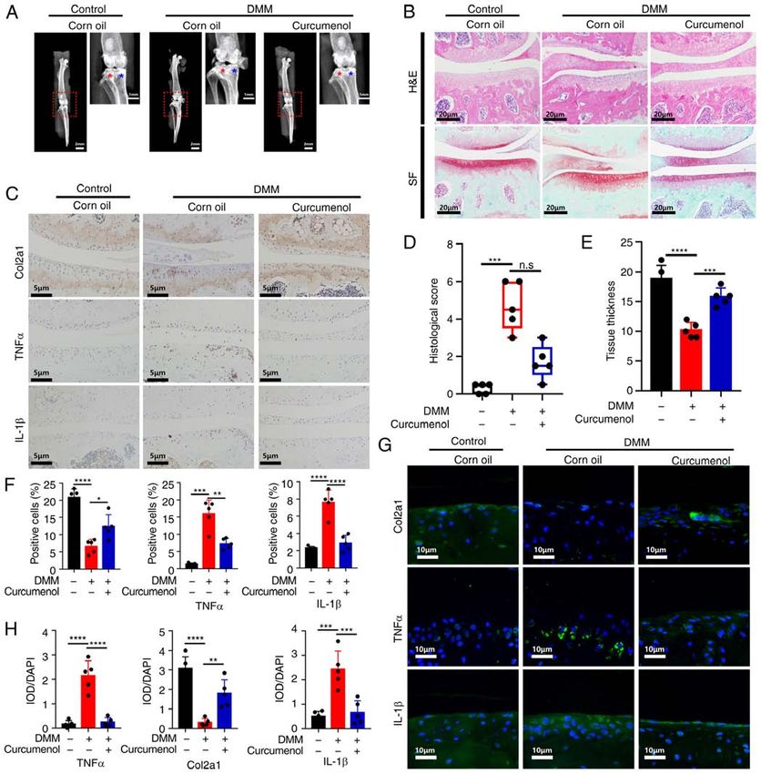
10                    YANG et al: CURCUMENOL INHIBITS INFLAMMATION in vitro AND MITIGATES ARTHRITIS in vivo

Figure 5. Curcumenol treats DMM‑induced osteoarthritis in mice by inhibiting TNF‑α and IL‑1β in vivo. (A) X‑ray of lower limbs. (B) Safranin O‑Fast Green
and hematoxylin & eosin staining of paraffin sections of the medial chondrocyte end plate of knee joints at a coronal position. (C) Immunohistochemical
analysis of Col2a1, TNF‑α and IL‑1β expression in knee joints at a coronal position. (D and E) Quantification of histological score and tissue thickness in
the sections described in (B). (F) Quantification of positive cells in sections described in (C). (G and H) Immunofluorescence analysis of Col2a1, TNF‑α and
IL‑1β expression in knee joints at a coronal position. Data are presented as mean ± SD using one‑way ANOVA with a Tukey's post hoc test; ordinal data were
analyzed by Kruskal‑Wallis test and Dunn's post hoc test from three experiments. *P
INTERNATIONAL JOURNAL OF MOlecular medicine 48: 192, 2021                                                        11

Figure 6. Curcumenol inhibits the phosphorylation and activation of the NF‑κ B and MAPK pathways induced by TNF‑α and IL‑1β, and prevents inflammatory
cascade reactions in chondrocytes in vitro. Moreover, curcumenol serves as a protector in destabilization of medial meniscus‑induced osteoarthritis model
mice by inhibiting TNF‑α‑ and IL‑1β‑activated inflammation in vivo. ADAMTS, ADAM metallopeptidase with thrombospondin type 1 motif.

OA (27,28), but the side‑effects of these drugs, such as hepatic              of inflammatory products, catabolic enzymes and apoptotic
damage and gastrointestinal injury, are concerns for their                    mediators (42‑45). Curcumenol blocks the phosphorylation
long‑term usage (29). Due their reduced side‑effects, abun‑                   and translocation of NF‑κ B, and inhibits the phosphoryla‑
dant production capacities and anti‑inflammatory functions,                   tion of Iκ Bα, to block the anti‑inflammatory function. With
plant‑derived traditional medicines have gained increasing                    regards to the MAPK pathway, curcumenol prevents the
attention in previous years (30‑32). Therefore, the present                   phosphorylation of JNK, which belongs to a large family
study investigated whether traditional medicines could be used                of serine/threonine kinases and crosslinks with numerous
to ameliorate the symptoms and slow the progression of OA.                    developmental pathways, such as Hippo signaling (46),
    Curcumenol is a bioactive compound isolated from edible                   promoting proteoglycan metabolism and inhibiting the
rhizome of Curcuma zedoaria, with potential anti‑inflamma‑                    production of catabolic enzymes and inflammatory media‑
tory effects (33). Curcumenol belongs to the Curcuma genus,                   tors in chondrocytes (47) and the nucleus pulposus (48).
and one of the most well‑known members is Curcuma longa, of                   After TNF‑ α stimulation, the expression levels of MMPs
which the bioactive extraction curcumin is used to treat syno‑                are increased and the production of ECM is inhibited, which
vitis experienced by patients with knee OA (34). Considering                  promotes the ECM to change to a catabolic and degradable
the broad application range of the Curcuma genus, and the                     status (49,50). This imbalance causes the senescence of
role of proinflammatory cytokines such as TNF‑α and IL‑1β                     primary chondrocytes, followed by instability and cartilage
in the pathophysiology of OA (35), the current study aimed                    loss in joints (51). In the present study, curcumenol was used
to investigate the anti‑inflammatory effects of curcumenol to                 to successfully inhibit the phosphorylation of Iκ Bα, NF‑κ B
treat TNF‑α‑ and IL‑1β‑induced inflammation in ATDC5 and                      P65 subunit and SAPK/JNK (Fig. 6). At the same time, the
primary chondrocytes. The present results also demonstrated                   nuclear translocation of p‑P65 was blocked, which lead to
that curcumenol ameliorated the effects of OA in the knee                     a subsequent decrease in MMP expression and the rescue
joints of DMM‑model mice in vivo.                                             of type 2 collagen in ATDC5 and primary chondrocytes.
    DMM surgery was used to generate a mechanical OA                          Thus, it was suggested that inhibiting the function of these
model. During joint degeneration, especially that of the carti‑               cytokines may be beneficial in the DMM‑induced OA model.
lage, the levels of some inflammatory cytokines, (including                       The DMM‑induced OA model in mice is an effective
TNF‑ α and IL‑1β) are increased, and subsequently cause a                     animal model first established in 2007 (21), whereby removing
progressive, cell‑mediated cascade of molecular and struc‑                    the medial meniscus can pathologically disrupt the stability
tural deterioration (36,37). TNF‑ α and IL‑1β exert their                     status of normal knee joints, which can restrict the range of
detrimental functions via multiple important intracellular                    motion (52). Using the widely recognized DMM‑induced
cascades, such as the NF‑κ B and MAPK pathways (38,39),                       model, the present study demonstrated the rescue effect of
In general, TNF‑α and IL‑1β transmit inflammatory signals                     curcumenol in OA of the knee joint, as it effectively mitigated
via their receptors and activate the phosphorylation of the                   inflammation in the cartilage of the tibia and femur, as well
IKK complex (40), which then phosphorylates Iκ Bα (41).                       as preventing joint space collapse and osteophyte formation.
Subsequently, Iκ Bα is unbound and phosphorylates NF‑κ B,                     The mechanism underlying this damage was consistent with
which translocates into the nucleus to initiate the transcription             the in vitro data. The immunohistochemistry results revealed
12 YANG et al: CURCUMENOL INHIBITS INFLAMMATION in vitro AND MITIGATES ARTHRITIS in vivo
an increase in TNF‑ α and IL‑1β expression in the DMM Availability of data and materials
group, which triggered the subsequent detrimental molecular
cascade and degeneration. In the curcumenol‑treated group, The datasets used and/or analyzed during the current study are
TNF‑α and IL‑1β expression was downregulated and Col2a1 available from the corresponding author on reasonable request.
expression was restored, demonstrating the curative effect of
curcumenol in DMM‑induced OA in vivo. Another important Authors' contributions
consideration is how to reconstitute and administer curcum‑
enol, as it is insoluble in water. Therefore, DMSO was used JZ and TZ guided the study, making substantial contributions
in the present study to pre‑dissolve curcumenol, which was to conception and design. XY and YZ performed the experi‑
then diluted in non‑pharmaceutical grade corn oil, which ments. XY, YZ, CH and XL interpreted the data and drafted
showed little adverse effect during intraperitoneal injection the manuscript. KZ, ZC and CC performed the statistical
in mice (53,54). With regard to the administration method, analysis and reviewed the manuscript critically for important
intraperitoneal injection was selected rather than intraarticular intellectual content. HT and XC confirmed the authenticity
administration, based on the following: i) Intraperitoneal of all the raw data. All authors read and approved the final
injection of corn oil is widely used in mouse models and the manuscript.
only administration method of curcumenol in mice appears to
be intraperitoneal injection (55); and ii) there is a possibility Ethics approval and informed consent
of cartilage injury (56,57) and difficulties associated with
injecting corn oil via multiple micro‑injections into the knee Animal Ethics approval was received from the Institutional
joint. Animal Ethics Review Board of Shanghai Ninth People's
The current study confirmed the efficiency and safety of Hospital, Shanghai Jiao Tong University School of Medicine
curcumenol, but there are still some concerns and limitations (approval no. SH9H‑2020‑A559‑1). Written informed consent
to our studies. For efficiency, the systemic distribution of curc‑ was obtained from all participants.
umenol after intraperitoneal injection was not assessed, thus
there was a lack of direct evidence that the optimal concentra‑ Patient consent for publication
tion of curcumenol reached the joint. In subsequent studies,
the concentration of curcumenol in in the blood will first be Not applicable.
assessed, and then preliminary investigations of curcumenol
distribution in the knee joint tissues will be conducted. For Competing interests
safety, although the intraperitoneal injection of curcumenol
is relatively safe with corn oil (53), treatment‑induced injury The authors declare that they have no competing interests.
was not evaluated. In subsequent studies, relevant experi‑
ments will be conducted, such as the evaluation of paraffin References
sections of lung, liver, heart, kidney and spleen, to further
confirm the safety of curcumenol in vivo. Furthermore, the 1. Glyn‑Jones S, Palmer AJ, Agricola R, Price AJ, Vincent TL,
administration method was via intraperitoneal injection, Weinans H and Carr AJ: Osteoarthritis. Lancet 386: 376‑387,
2015.
which in clinical use, may be more difficult when treating 2. Zhang Z, Huang C, Jiang Q, Zheng Y, Liu Y, Liu S, Chen Y,
patients. Therefore, the efficiency and safety of oral admin‑ Mei Y, Ding C, Chen M, et al: Guidelines for the diagnosis and
istration in DMM‑induced osteoarthritis or type II collagen treatment of osteoarthritis in China (2019 edition). Ann Transl
Med 8: 1213, 2020.
(UC‑II) diminished deterioration of articular cartilage will be 3. Hiligsmann M, Cooper C, Arden N, Boers M, Branco JC,
investigated in mice (58,59). Luisa Brandi M, Bruyère O, Guillemin F, Hochberg MC,
In conclusion, the current study presented a novel Hunter DJ, et al: Health economics in the field of osteoarthritis:
An expert's consensus paper from the European society for
plant‑derived bioactive medicine, curcumenol, which was clinical and economic aspects of osteoporosis and osteoarthritis
demonstrated to serve as a potential anti‑inflammatory agent (ESCEO). Semin Arthritis Rheum 43: 303‑313, 2013.
for the management of OA. The low cytotoxicity, reduced 4. Ag r icola R, Heijbo er M P, Roz e R H, Reijma n M,
Bierma‑Zeinstra SMA, Verhaar JAN, Weinans H and Waarsing JH:
side‑effects and high production capacity are also considerable Pincer deformity does not lead to osteoarthritis of the hip whereas
advantages of future curcumenol use in the clinic. acetabular dysplasia does: Acetabular coverage and develop‑
ment of osteoarthritis in a nationwide prospective cohort study
(CHECK). Osteoarthritis Cartilage 21: 1514‑1521, 2013.
Acknowledgements 5. Tsuchiya H, Ota M, Sumitomo S, Ishigaki K, Suzuki A, Sakata T,
Tsuchida Y, Inui H, Hirose J, Kochi Y, et al: Parsing multiomics
Not applicable. landscape of activated synovial fibroblasts highlights drug targets
linked to genetic risk of rheumatoid arthritis. Ann Rheum Dis:
Nov 2, 2020 (Epub ahead of print).
Funding 6. Myers J, Wielage RC, Han B, Price K, Gahn J, Paget MA
and Happich M: The efficacy of duloxetine, non‑steroidal
The present study was supported by grants from the National anti‑inflammatory drugs, and opioids in osteoarthritis: A system‑
atic literature review and meta‑analysis. BMC Musculoskelet
Natural Science Foundation of China (grant nos. 81871790, Disord 15: 76, 2014.
81572768 and 81972136) and the fundamental research 7. McAlindon TE, Bannuru RR, Sullivan MC, Arden NK,
program funding of Ninth People's Hospital Affiliated to Berenbaum F, Bierma‑Zeinstra SM, Hawker GA, Henrotin Y,
Hunter DJ, Kawaguchi H, et al: OARSI guidelines for the
Shanghai Jiao Tong university School of Medicine (grant non‑surgical management of knee osteoarthritis. Osteoarthritis
no. JYZZ003G). Cartilage 22: 363‑388, 2014.INTERNATIONAL JOURNAL OF MOlecular medicine 48: 192, 2021 13
8. Pas HI, Winters M, Haisma HJ, Koenis MJ, Tol JL and Moen MH: 31. Liu Y, Deng SJ, Zhang Z, Gu Y, Xia SN, Bao XY, Cao X and
Stem cell injections in knee osteoarthritis: A systematic review Xu Y: 6‑Gingerol attenuates microglia‑mediated neuroinflam‑
of the literature. Br J Sports Med 51: 1125‑1133, 2017. mation and ischemic brain injuries through Akt‑mTOR‑STAT3
9. Hached F, Vinatier C, Le Visage C, Gondé H, Guicheux J, signaling pathway. Eur J Pharmacol 883: 173294, 2020.
Grimandi G and Billon‑Chabaud A: Biomaterial‑assisted cell 32. Li Y, Lin S, Liu P, Huang J, Qiu J, Wen Z, Yuan J, Qiu H, Liu Y,
therapy in osteoarthritis: From mesenchymal stem cells to cell Liu Q, et al: Carnosol suppresses RANKL‑induced osteoclas‑
encapsulation. Best Pract Res Clin Rheumatol 31: 730‑745, 2017. togenesis and attenuates titanium particles‑induced osteolysis.
10. Wang X, Liao T, Wan C, Yang X, Zhao J, Fu R, Yao Z, Huang Y, J Cell Physiol 236: 1950‑1966, 2021.
Shi Y, Chang G, et al: Efficient generation of human primordial 33. Lo JY, Kamarudin MNA, Hamdi OAA, Awang K and Kadir HA:
germ cell‑like cells from pluripotent stem cells in a methylcellu‑ Curcumenol isolated from curcuma zedoaria suppresses
lose‑based 3D system at large scale. PeerJ 6: e6143, 2019. Akt‑mediated NF‑ κ B activation and p38 MAPK signaling
11. Hikino H, Sakurai Y, Numabe S and Takemoto T: Structure of pathway in LPS‑stimulated BV‑2 microglial cells. Food Funct 6:
curcumenol. Chem Pharm Bull (Tokyo) 16: 39‑42, 1968. 3550‑3559, 2015.
12. Xu J, Ji F, Kang J, Wang H, Li S, Jin DQ, Zhang Q, Sun H and Guo Y: 34. Wang Z, Jones G, Winzenberg T, Cai G, Laslett LL, Aitken D,
Absolute configurations and NO inhibitory activities of terpenoids Hopper I, Singh A, Jones R, Fripp J, et al: Effectiveness of
from curcuma longa. J Agric Food Chem 63: 5805‑5812, 2015. extract for the treatment of symptoms and effusion‑synovitis of
13. Assis A, Brito V, Bittencourt M, Silva L, Oliveira F and knee osteoarthritis : A randomized trial. Ann Intern Med 173:
Oliveira R: Essential oils composition of four Piper species from 861‑869, 2020.
Brazil. J Essential Oil Res 25: 203‑209, 2013. 35. Kapoor M, Martel‑Pelletier J, Lajeunesse D, Pelletier JP and
14. Saikia AK, Sarma SK, Strano T and Ruberto G: Essential oil Fahmi H: Role of proinflammatory cytokines in the pathophysi‑
from piper pedicellatum C. DC. Collected in North‑East India. ology of osteoarthritis. Nat Rev Rheumatol 7: 33‑42, 2011.
J Essential Oil Bearing Plants 18: 314‑319, 2015. 36. Rodriguez‑Trillo A, Mosquera N, Pena C, Rivas‑Tobío F,
15. Sun DX, Fang ZZ, Zhang YY, Cao YF, Yang L and Yin J: Mera‑Varela A, Gonzalez A and Conde C: Non‑Canonical
Inhibitory effects of curcumenol on human liver cytochrome WNT5A signaling through RYK contributes to aggressive
P450 enzymes. Phytother Res 24: 1213‑1216, 2010. phenotype of the rheumatoid fibroblast‑like synoviocytes. Front
16. Pintatum A, Maneerat W, Logie E, Tuenter E, Sakavitsi ME, Immunol 11: 555245, 2020.
Pieters L, Berghe WV, Sripisut T, Deachathai S and Laphookhieo S: 37. Zhao X, Meng F, Hu S, Yang Z, Huang H, Pang R, Wen X, Kang Y
In Vitro anti‑inflammatory, anti‑oxidant, and cytotoxic activities and Zhang Z: The synovium attenuates cartilage degeneration
of four species and the isolation of compounds from rhizome. in KOA through activation of the Smad2/3‑Runx1 cascade and
Biomolecules 10: 799, 2020. chondrogenesis‑related miRNAs. Mol Ther Nucleic Acids 22:
17. Oh CD, Im HJ, Suh J, Chee A, An H and Chen D: Rho‑associated 832‑845, 2020.
kinase inhibitor immortalizes rat nucleus pulposus and annulus 38. Baker RG, Hayden MS and Ghosh S: NF‑κ B, inflammation, and
fibrosus cells: Establishment of intervertebral disc cell lines with metabolic disease. Cell Metab 13: 11‑22, 2011.
novel approaches. Spine (Plila Pa 1976) 41: E255‑E261, 2016. 39. Moqbel SAA, Xu K, Chen Z, Xu L, He Y, Wu Z, Ma C, Ran J,
18. Johnson M, Zaretskaya I, Raytselis Y, Merezhuk Y, McGinnis S Wu L and Xiong Y: Tectorigenin alleviates inflammation,
and Madden TL: NCBI BLAST: A better web interface. Nucleic apoptosis, and ossification in rat tendon‑derived stem cells
Acids Res 36 (Web Server issue): W5‑W9, 2008. modulating NF‑Kappa B and MAPK pathways. Front Cell Dev
19. Livak KJ and Schmittgen TD: Analysis of relative gene expres‑ Biol 8: 568894, 2020.
sion data using real‑time quantitative PCR and the 2(‑Delta Delta 40. Zhang Q, Lenardo MJ and Baltimore D: 30 Years of NF‑κ B: A
C(T)) method. Methods 25: 402‑408, 2001. blossoming of relevance to human pathobiology. Cell 168: 37‑57,
20. Shi JW, Zhang TT, Liu W, Yang J, Lin XL, Jia JS, Shen HF, 2017.
Wang SC, Li J, Zhao WT, et al: Direct conversion of pig fibro‑ 41. Zhongyi S, Sai Z, Chao L and Jiwei T: Effects of nuclear factor
blasts to chondrocyte‑like cells by c‑Myc. Cell Death Discov 5: kappa B signaling pathway in human intervertebral disc degen‑
55, 2019. eration. Spine (Phila Pa 1976) 40: 224‑232, 2015.
21. Glasson SS, Blanchet TJ and Morris EA: The surgical destabiliza‑ 42. Baldwin AS: The NF‑kappa B and I kappa B proteins: New
tion of the medial meniscus (DMM) model of osteoarthritis in the discoveries and insights. Annu Rev Immunol 14: 649‑683, 1996.
129/SvEv mouse. Osteoarthritis Cartilage 15: 1061‑1069, 2007. 43. Sun Z, Yin Z, Liu C, Liang H, Jiang M and Tian J: IL‑1β promotes
22. Wang M, Sampson ER, Jin H, Li J, Ke QH, Im HJ and Chen D: ADAMTS enzyme‑mediated aggrecan degradation through NF‑κB
MMP13 is a critical target gene during the progression of osteo‑ in human intervertebral disc. J Orthop Surg Res 10: 159, 2015.
arthritis. Arthritis Res Ther 15: R5, 2013. 44. Tu J, Li W, Zhang Y, Wu X, Song Y, Kang L, Liu W, Wang K,
23. Loeser RF, Kelley KL, Armstrong A, Collins JA, Diekman BO Li S, Hua W and Yang C: Simvastatin inhibits IL‑1β ‑induced
and Carlson CS: Deletion of JNK enhances senescence in joint apoptosis and extracellular matrix degradation by suppressing
tissues and increases the severity of age‑related osteoarthritis in the NF‑kB and MAPK pathways in nucleus pulposus cells.
mice. Arthritis Rheumatol 72: 1679‑1688, 2020. Inflammation 40: 725‑734, 2017.
24. Sohara Y, Shimada H, Scadeng M, Pollack H, Yamada S, Ye W, 45. Chen J, Garssen J and Redegeld F: The efficacy of bortezomib in
Reynolds CP and DeClerck YA: Lytic bone lesions in human human multiple myeloma cells is enhanced by combination with
neuroblastoma xenograft involve osteoclast recruitment and are omega‑3 fatty acids DHA and EPA: Timing is essential. Clin
inhibited by bisphosphonate. Cancer Res 63: 3026‑3031, 2003. Nutr 40: 1942‑1953, 2021.
25. Sharma L: Osteoarthritis of the knee. N Engl J Med 384: 51‑59, 46. Pham TH, Hagenbeek TJ, Lee HJ, Li J, Rose CM, Lin E, Yu M,
2021. Martin SE, Piskol R, Lacap JA, et al: Machine learning and
26. Thorlund JB, Juhl CB, Roos EM and Lohmander LS: chemico‑genomics approach defines and predicts cross‑talk of
Arthroscopic surgery for degenerative knee: Systematic review Hippo and MAPK pathways. Cancer Discov 11: 778‑793, 2021.
and meta‑analysis of benefits and harms. BMJ 350: h2747, 2015. 47. Cao C, Wu F, Niu X, Hu X, Cheng J, Zhang Y, Li C, Duan X, Fu X,
27. Rasmussen‑Barr E, Held U, Grooten WJA, Roelofs PDDM, Zhang J, et al: Cadherin‑11 cooperates with inflammatory factors
Koes BW, van Tulder MW and Wertli MM: Nonsteroidal to promote the migration and invasion of fibroblast‑like synovio‑
anti‑inflammatory drugs for sciatica: An updated cochrane cytes in pigmented villonodular synovitis. Theranostics 10:
review. Spine (Phila Pa 1976) 42: 586‑594, 2017. 10573‑10588, 2020.
28. Derry S, Wiffen PJ, Kalso EA, Bell RF, Aldington D, Phillips T, 48. Séguin CA, Bojarski M, Pilliar RM, Roughley PJ and Kandel RA:
Gaskell H and Moore RA: Topical analgesics for acute and Differential regulation of matrix degrading enzymes in a
chronic pain in adults‑an overview of cochrane reviews. TNFalpha‑induced model of nucleus pulposus tissue degenera‑
Cochrane Database Syst Rev 5: CD008609, 2017. tion. Matrix Biol 25: 409‑418, 2006.
29. Marmon P, Owen SF and Margiotta‑Casaluci L: Pharma 49. Wang G, Chen S, Xie Z, Shen S, Xu W, Chen W, Li X, Wu Y,
cology‑informed prediction of the risk posed to fish by mixtures Li L, Liu B, et al: TGFβ attenuates cartilage extracellular matrix
of non‑steroidal anti‑inflammatory drugs (NSAIDs) in the envi‑ degradation via enhancing FBXO6‑mediated MMP14 ubiquiti‑
ronment. Environ Int 146: 106222, 2021. nation. Ann Rheum Dis 79: 1111‑1120, 2020.
30. Chen J, Xuan J, Gu YT, Shi KS, Xie JJ, Chen JX, Zheng ZM, 50. He L, He T, Xing J, Zhou Q, Fan L, Liu C, Chen Y, Wu D,
Chen Y, Chen XB, Wu YS, et al: Celastrol reduces IL‑1β induced Tian Z, Liu B and Rong L: Bone marrow mesenchymal stem
matrix catabolism, oxidative stress and inflammation in human cell‑derived exosomes protect cartilage damage and relieve knee
nucleus pulposus cells and attenuates rat intervertebral disc osteoarthritis pain in a rat model of osteoarthritis. Stem Cell Res
degeneration in vivo. Biomed Pharmacother 91: 208‑219, 2017. Ther 11: 276, 2020.14 YANG et al: CURCUMENOL INHIBITS INFLAMMATION in vitro AND MITIGATES ARTHRITIS in vivo
51. Inoue R, Ishibashi Y, Tsuda E, Yamamoto Y, Matsuzaka M, 56. Kompel AJ, Roemer FW, Murakami AM, Diaz LE, Crema MD
Takahashi I, Danjo K, Umeda T, Nakaji S and Toh S: Knee and Guermazi A: Intra‑articular corticosteroid injections in the
osteoarthritis, knee joint pain and aging in relation to increasing hip and knee: Perhaps not as safe as we thought? Radiology 293:
serum hyaluronan level in the Japanese population. Osteoarthritis 656‑663, 2019.
Cartilage 19: 51‑57, 2011. 57. Mehta PN and Ghadially FN: Articular cartilage in corn
52. Lamo‑Espinosa JM, Blanco JF, Sánchez M, Moreno V, oil‑induced lipoarthrosis. Ann Rheum Dis 32: 75‑82, 1973.
Granero‑Moltó F, Sánchez‑Guijo F, Crespo‑Cullel I, Mora G, 58. Bagi CM, Berryman ER, Teo S and Lane NE: Oral administra‑
Vicente DDS, Pompei‑Fernández O, et al: Phase II multicenter tion of undenatured native chicken type II collagen (UC‑II)
randomized controlled clinical trial on the efficacy of intra‑artic‑ diminished deterioration of articular cartilage in a rat model
ular injection of autologous bone marrow mesenchymal stem of osteoarthritis (OA). Osteoarthritis Cartilage 25: 2080‑2090,
cells with platelet rich plasma for the treatment of knee osteoar‑ 2017.
thritis. J Transl Med 18: 356, 2020. 59. Runhaar J, Rozendaal RM, van Middelkoop M, Bijlsma HJW,
53. Hubbard JS, Chen PH and Boyd KL: Effects of repeated Doherty M, Dziedzic KS, Lohmander LS, McAlindon T,
intraperitoneal injection of pharmaceutical‑grade and nonphar‑ Zhang W and Zeinstra SB: Subgroup analyses of the effective‑
maceutical‑grade corn oil in female C57BL/6J mice. J Am Assoc ness of oral glucosamine for knee and hip osteoarthritis: A
Lab Anim Sci 56: 779‑785, 2017. systematic review and individual patient data meta‑analysis from
54. Alsina‑Sanchis E, Mülfarth R, Moll I, Mogler C, Rodriguez‑Vita J the OA trial bank. Ann Rheum Dis 76: 1862‑1869, 2017.
and Fischer A: Intraperitoneal oil application causes local
inflammation with depletion of resident peritoneal macrophages.
Mol Cancer Res 19: 288‑300, 2021. This work is licensed under a Creative Commons
55. Wang S, Ma Q, Xie Z, Shen Y, Zheng B, Jiang C, Yuan P, Attribution-NonCommercial-NoDerivatives 4.0
An Q, Fan S and Jie Z: An antioxidant sesquiterpene inhibits
International (CC BY-NC-ND 4.0) License.
osteoclastogenesis via blocking IPMK/TRAF6 and counteracts
OVX‑induced osteoporosis in mice. J Bone Miner Res: May 6,
2021 (Epub ahead of print).You can also read