PROTEIN EXPRESSION AND PURIFICATION - DIVA PORTAL
←
→
Page content transcription
If your browser does not render page correctly, please read the page content below
Protein Expression and Purification 186 (2021) 105919
Contents lists available at ScienceDirect
Protein Expression and Purification
journal homepage: www.elsevier.com/locate/yprep
Milligram scale expression, refolding, and purification of Bombyx mori
cocoonase using a recombinant E. coli system
Chanrith Phoeurk a, b, Ameeq Ul Mushtaq a, Per Rogne a, *, Magnus Wolf-Watz a, *
a
Department of Chemistry, Umeå University, Umeå, Sweden
b
Department of Bio-Engineering, Royal University of Phnom Penh, Phnom Penh, Cambodia
A R T I C L E I N F O A B S T R A C T
Keywords: Silk is one of the most versatile biomaterials with signature properties of outstanding mechanical strength and
Silk moth (Bombyx mori) flexibility. A potential avenue for developing more environmentally friendly silk production is to make use of the
Cocoonase silk moth (Bombyx mori) cocoonase, this will at the same time increase the possibility for using the byproduct,
Serine protease
sericin, as a raw material for other applications. Cocoonase is a serine protease utilized by the silk moth to soften
Refolding
Escherichia coli
the cocoon to enable its escape after completed metamorphosis. Cocoonase selectively degrades the glue protein
of the cocoon, sericin, without affecting the silk-fiber made of the protein fibroin. Cocoonase can be recombi
nantly produced in E. coli, however, it is exclusively found as insoluble inclusion bodies. To solve this problem
and to be able to utilize the benefits associated with an E. coli based expression system, we have developed a
protocol that enables the production of soluble and functional protease in the milligram/liter scale. The core of
the protocol is refolding of the protein in a buffer with a redox potential that is optimized for formation of native
and intramolecular di-sulfide bridges. The redox potential was balanced with defined concentrations of reduced
and oxidized glutathione. This E. coli based production protocol will, in addition to structure determination, also
enable modification of cocoonase both in terms of catalytic function and stability. These factors will be valuable
components in the development of alternate silk production methodology.
1. Introduction the cocoon [4]. Cocoonase has a high potential for improving the
effectivity of degumming, while at the same time reducing the envi
Silks are a general term for widely different protein polymers spun by ronmental impact of the silk industry.
a range of different arthropods with a range of functions [1]. Textile silk The cocoon from B. mori contains a number of proteins. Sericin make
is extracted from the cocoon of the silkworm Bombyx mori (B. mori). up approximately one fourth of the weight of the cocoon. However, most
With a global production of more than 150,000 tons a year, from 1 abundant are the fibroins, the water-insoluble core of the silk fiber that
million tons of fresh cocoons, silk is one of the most valuable natural is processed into the silk-threads, which makes up about two-thirds [5,6]
fibers in the world [2], with an annual production value of more than 3 of the weight of the cocoon. In addition to these two main proteins, two
billion dollars in 2019 (fao.org/faostat/, FAO 2020). so-called seroins (BmSER 1 and BmSER 2) and two protease inhibitors
One crucial step in the process of turning the cocoon into fabric is the (BmSPI 1 and BmSPI 2) have been isolated from the cocoon [7]. These
removal of the protein glue, sericin that holds the fibroin fibers together last four proteins are involved in protecting the pupae against diseases
in the cocoon, this process is called degumming [3]. Degumming is [8,9]. Fibroin is made up of three different polypeptides, the heavy
heavily energy consuming and it is of interest to develop “green” chain, the light chain, and a glycoprotein called P25 [10]. The heavy and
degumming methods that are more environmentally friendly. In nature, the light chains are bound to each other with disulfide bonds while P25
the silk moth uses the protease cocoonase to degrade sericin to soften the is associated via non-covalent interactions [10].
cocoon. This is essential for the fully developed moth to be able to escape Sericin is a group of proteins encoded by four different genes, sericin
Abbreviations: B. mori, Bombyx mori; BmCoc, Bombyx mori cocoonase; BPTI, Bovine pancreatic trypsin inhibitor; CD, Circular dichroism; CSE, Cocoon shell extract;
HSQC, Heteronuclear single quantum coherence; SBTI, Soybean trypsin inhibitor; TEV, Tobacco etch virus; TLCK, N-α-p-Tosyl-L-lysine chloromethyl ketone; TROSY,
Transverse relaxation optimized spectroscopy.
* Corresponding authors. Umeå University, Department of Chemistry, SE-901 87, Umeå, Sweden.
E-mail addresses: per.rogne@umu.se (P. Rogne), magnus.wolf-watz@umu.se (M. Wolf-Watz).
https://doi.org/10.1016/j.pep.2021.105919
Received 19 March 2021; Received in revised form 28 April 2021; Accepted 20 May 2021
Available online 24 May 2021
1046-5928/© 2021 The Authors. Published by Elsevier Inc. This is an open access article under the CC BY license (http://creativecommons.org/licenses/by/4.0/).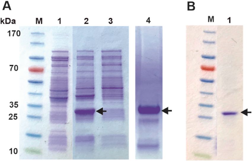
C. Phoeurk et al. Protein Expression and Purification 186 (2021) 105919
1 through 4 [11]. Sericin 1 and 3 are the main cocoon sericin, the other resonance (NMR) spectroscopy. Effective NMR spectroscopy on proteins
sericin covers silk fibers that have other functions such as attaching the larger than 10 kDa requires proteins that are labeled with either 13C,
15
larvae or cocoon to leaves or tree branches [12]. The sericin genes un N, 2H, or a combination of these isotopes [27]. This can be obtained by
dergo alternative splicing to give rise to differently sized polypeptides in growing the expression cells in a media that is enriched with the desired
the range of 150–400 kDa [11]. However, other reports suggest sericin isotopes. E. coli can grow on minimal media where the sources of ni
polypeptides with as low mass as 10 kDa [13] and these smaller peptides trogen and carbon can be closely controlled. In the commonly used M9
may be a result of the different methods used to extract sericin from the media nitrogen and carbon is supplied in the form of ammonium chlo
cocoon. Presently, sericin are mostly discarded as waste during silk fiber ride (15NH4Cl) and 13C enriched glucose, respectively. These chemicals
production [2]. However, sericin has several important applications are available, relatively affordable, enriched with the sought-after
where the medical industry has, for example, shown interest due to its isotopes.
mitogenic properties for wound healing and bone tissue engineering The disadvantage with using an E. coli-based expression system to
[14]. The cosmetic industry is also a potential application, in moistur express eukaryotic proteins is the lack of “folding system” which can
izers, for example [14,15]. result in the expressed proteins being misfolded and aggregated as so-
Degumming is one of the steps in the standard processing approach called inclusion bodies. The problem is especially significant for pro
of the cocoons into silk fibers. Degumming can be accomplished by teins containing intramolecular di-sulfide bridges. BmCoc contains six
soaking the cocoons in hot water, however, this is a lengthy process that cysteine residues that might form up to three such bridges. If the cys
causes damage to the fibroin [2]. Most industrial silk processes use soaps teines are solvent-exposed, either due to misfolding or during the folding
in an alkaline water solution to hydrolyze and remove the sericin [3,16]. process, inter-molecular di-sulfide bridges can form, permanently trap
These methods do, however, require large quantities of heat and water ping the expressed proteins in a misfolded state. In order to refold active
and are therefore resulting in a significant environmental impact. In protease from inclusion bodies, they must be solubilized by unfolding
addition, the waste products can pollute the environment of the silk and refolded into their native fold. A significant challenge to a refolding
processing site [3]. protocol is to develop a method that favors the formation of native di-
An attractive route to make the degumming process more environ sulfide bridges while dis-favoring non-native di-sulfide bridges and
mentally friendly is to utilize enzymatic degumming [17]. In enzymatic higher-order oligomers.
degumming, sericin is hydrolyzed and removed by using proteases. Here we present a robust and reproducible method to recombinantly
Proteases like alcalase, savinase [18], and bromelain [19] have been produce large (mg/liter) amounts of functional BmCoc in E. coli by
explored for this role. Enzymatic reactions are generally conducted at unfolding and refolding of protein expressed as inclusion bodies. The
ambient temperatures and in non-toxic solutions directly reducing both core of the protocol is that refolding is performed in an oxygen-free
the energy consumption and the pollution problem. When using pro environment and with a redox potential that is optimized for the for
teases, it is important to consider the specificity of the enzymes since the mation of native di-sulfide bridges. This favorable redox potential is
fiber is also made of protein (fibroin) and is, generally, susceptible to accomplished with a balance of reduced and oxidized glutathione.
degradation. Cocoonase has been shown to out-perform commercially Two different versions of BmCoc were produced, one including a
available alcalase by providing a more complete sericin removal while at short N-terminal peptide (GAM-BmCoc) and one with a native N-ter
the same time causing less damage to the silk fiber [20]. Increased un minal (En-BmCoc). The GAM peptide is used to reduce the auto-
derstanding of the properties of this enzyme will open avenues to using proteolytic activity of cocoonase, giving a more stable enzyme for
cocoonase in the silk industry and as a product of the knowledge the structural investigations. Quantitative analysis of the purified protein
industrial potential of the enzyme might be increased by biotechno demonstrated that the enzyme is active and monomeric. Additionally,
logical improvements of its properties, i.e., increased stability or the GAM variant is stable over extended periods of time. En-BmCoc was
activity. shown to effectively degrade sericin on solid cocoon particles but dis
B. mori cocoonase (BmCoc) is a serine protease, similar to trypsin, played self-cleavage when incubated over longer time periods. In the
and is produced as an inactive zymogen by the silk moth during the pupa manuscript we refer to the two variants (GAM-BmCoc and En-BmCoc)
state [21]. The zymogen is exported and stored as a dry deposit on the when appropriate, or alternatively BmCoc when joint properties of the
head of the moth [21]. At the time of break-out of the silk moth, the two variants are discussed. The results represent a significant step for
deposited enzyme is dissolved in a buffer produced by the moth and ward and will enable both fundamental biochemical and structural in
activated by cleavage of a 34 amino acid residues long N-terminal vestigations, as well as the initial steps toward green production of silk
peptide to its active form [20]. The cleavage of the zymogen into the fibers.
active BmCoc can be accomplished by different proteases like trypsin or
subtilisin [21]. However, the main route of activation is cleavage by 2. Materials and methods
already activated BmCoc [22] in a self-propelling mechanism.
BmCoc can be extracted from its natural source, and early in 2.1. Cloning of BmCoc into the pET-His vector
vestigations [23–25] relied upon this method. However, the amounts of
enzymes that can be obtained are very small, a protocol published by Using the amino acid sequence for BmCoc (NCBI accession ID:
Fukumori et al. [25] showed that less than 1 μg of the pure enzyme could BAJ46146) the corresponding cDNA, codon optimized for E. coli
be obtained from one silk moth. expression [28], was synthesized (Eurofins Genomics, Germany). The
To efficiently obtain protein on a scale necessary for in depth bio BmCoc cDNA was cloned into either (1) a pETHis-1a vector, providing a
physical investigations, including structure determination, a recombi tobacco etch virus (TEV) protease cleavable N-terminal poly histidine
nant system is required. Using a yeast system based on Pichia pastoris (P. tag (His-tag) or (2) a pET-NHis vector providing a N-terminal His-tag
pastoris), Unajak and co-workers [26] were able to express and purify with an enterokinase cleavable linker (introduced by using PCR). Both
BmCoc with a yield of 5.4 mg/l media. However, an expression system vectors were kindly provided by Günther Stier, EMBL, Heidelberg,
based on Escherichia coli (E. coli) has several advantages compared to a Germany. The TEV protease cleaves the recognition sequence
system based on yeast (eukaryotic cells). The cloning and trans (ENLYFQG) between the glutamine (Q) and the glycine (G) resulting in a
formation of E. coli cells are quick and easy in comparison and there are primary sequence with the non-native N-terminal residue “G”. In addi
a multitude of developed expression vectors. The cells grow quickly on tion, the restriction enzyme NcoI is used to cleave the vector in order to
minimal media, which is important if isotopically labeled proteins are insert the gene and the recognition site for NcoI (CCATGG) introduces
required. Isotopically labeled proteins are for example necessary when codons for alanine (A) and methionine (M) between the TEV cleavage
properties on the atomic scale are studied using nuclear magnetic site and the N-terminal of the protein. Since TEV cleavage would not
2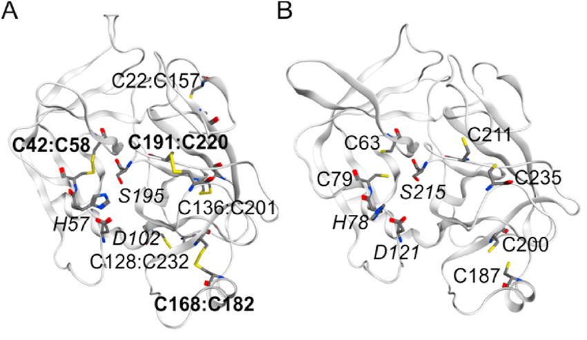
C. Phoeurk et al. Protein Expression and Purification 186 (2021) 105919
produce a native N-terminal, the inclusion of these two extra amino acid BmCoc and En-BmCoc respectively.
residues was not considered problematic. This version of BmCoc will be
referred to as GAM-BmCoc. Since enterokinase cleaves C-terminally of 2.4. Removal of His-tag
its recognition site (DDDDK), a native N-terminal is obtained, this
version will be called En-BmCoc. For large parts of the expression, To remove the His-tag, the GAM-BmCoc solution (with the TEV
refolding, purification, and characterization the two variants protease cleavage site) was buffer exchanged into 0.5 mM ethyl
(GAM-BmCoc and En-BmCoc) are treated equally, in these cases, they enediaminetetraacetic acid (EDTA), 1 mM DTT, 50 mM Tris-HCl, pH 8.0.
are called BmCoc. TEV protease was added in the ratio of 1:50 (w/w) and incubated
overnight at room temperature. Proteolytic removal of the His-tag was
2.2. Expression and refolding of recombinant BmCoc verified with SDS-PAGE. The buffer was exchanged, by repeated con
centration in a 15 ml Amicon® Ultra 10K centrifugal filter (Merck
The recombinant plasmids were transformed into BL21 (DE3) E. coli Millipore Ltd., Ireland) and dilution, and loaded on the Ni-NTA gravity
cells by heat shock and grown overnight on lysogeny broth (LB) -plates flow column equilibrated with 50 mM Tris-HCl, 500 mM NaCl, 5 mM
supplemented with 50 μg/ml kanamycin. Transformed cells were picked imidazole, pH 7.5 to remove the His-tagged TEV protease that is of equal
and grown at 37 ◦ C in 15 ml LB containing 50 μg/ml kanamycin over- size to the cocoonase, 27 kDa and 24 kDa, respectively. This step also
night. One milliliter of the over-night culture was used to inoculate removes the cleaved off His-tag.
20 ml of fresh LB (50 μg/ml kanamycin) that was subsequently cultured For the En-BmCoc, the buffer was exchanged to 50 mM Tris-HCl, pH
at 37 ◦ C for 2 h. The cells were harvested by gentle centrifugation and 7.75. The protein was activated by auto-proteolytic (i.e. an intermo
resuspended in 1 L of M9 medium (see Supplementary Table 1 for lecular cleavage process in the absence of enterokinase) cleavage of the
complete recipe). The cells were grown to an optical density at 600 nm linker by incubating the solution at 37 ◦ C for 15–17 h. This can be
(OD600) in the interval 0.6–0.7 and then induced with 0.75 mM iso rationalized with that the linker contains a lysine residue that together
propyl β-d-1-thiogalactopyranoside (IPTG) and further grown at 37 ◦ C with arginines are known targets of certain serine proteases The cleaved
for an additional 20–21 h. The cells were harvested by centrifugation at protein was polished with a second round of SEC to separate it from His-
3,300 *g for 30 min and the pellet was resuspended in 100 mM Tris tag residues. Finally, both GAM-BmCoc and En-BmCoc containing so
(hydroxymethyl) aminomethane hydrochloride (Tris-HCl) at pH 7.5. lutions were buffer exchanged to 20 mM NaPi, 50 mM NaCl, pH 6.0 for
Cell lysis was accomplished by using a Branson 450 Digital Sonifier NMR experimental analysis. Purified BmCoc, was stored at − 20 ◦ C.
(BRANSON Ultrasonics Corporation, USA). The lysate was centrifuged at
21,000 *g for 45 min, and the overexpressed recombinant BmCoc was 2.5. Circular dichroism (CD) spectroscopy
found exclusively in the pellet fraction.
To refold and recover the recombinant BmCoc from inclusion bodies, Far-UV CD spectra were recorded in the range of 195–250 nm at
a previously described protocol was modified [29]. The pellet was dis 20 ◦ C using a Jasco J-715 spectropolarimeter (Jasco inc, Japan). The
solved in 12 ml of 6 M guanidine hydrochloride (GuHCl), 100 mM protein concentration was 20 μM in 10 mM sodium phosphate buffer at
Tris-HCl at pH 7.5 containing 30 mM dithiothreitol (DTT) and was pH 7.0. The spectra were acquired using a 0.1 cm quartz cuvette, a
incubated for 2 h at 37 ◦ C. The dissolved pellet solution was centrifuged bandwidth of 2 nm, and the data were recorded and averaged based on
at 14,000 *g for 10 min to remove undissolved cell debris. 300 ml of ten repetitions. Background correction was performed by subtracting
refolding buffer (0.9 M GuHCl, 100 mM Tris-HCl at pH 7.5) was pre the buffer signal. Thermal unfolding experiments were conducted using
pared and deoxygenated by purging the solution with nitrogen gas with the same sample conditions by following the CD signal at 205 nm
stirring for an hour in a cold room. Reduced and oxidized glutathione wavelength. The temperature was raised from 20 ◦ C to 75 ◦ C at a rate of
was added to the deoxygenized refolding buffer to a final concentration 1 ◦ C per minute. The melting point (TM) and enthalpy of unfolding
of 5 mM and 1 mM, respectively. The soluble, reduced and unfolded (ΔHU) was quantified by non-linearly fitting equation (1) [30] to the
protein solution was rapidly diluted into the refolding buffer with a acquired data the Origin software (OriginLab, USA)
volume ratio of one to thirty. This dilution lowers the DTT concentration ( )
to below 1 mM, lower than the reduced and oxidized glutathione. The R *
ΔHU 1 1
TM − T
bottle with the refolding solution was rapidly closed and sealed with (CDn + mn T) + (CDd + md T)*e
CD(T) = ( ) (1)
parafilm; and the refolding reaction was allowed to proceed for 16–18 h
R *
ΔHU 1 1
at 6.5 ◦ C with gentle stirring. Finally, the refolded sample was diluted
TM − T
1+e
two-fold in 50 mM Tris at pH 7.5, 500 mM NaCl with 10 mM imidazole.
Precipitated protein were removed by centrifugation at 33,000 *g for 20 Where CD (T) is the signal as a function of the temperature, T, CDn is the
min followed by filtering with a 0.45 μm filter. CD signal of the native protein at 0 K, mn is the change of the native CD
signal with temperature, CDd and md are the corresponding parameters
2.3. Purification of His-tagged BmCoc for the denatured state and R is the gas constant.
Refolded BmCoc was applied to a Ni-NTA gravity flow column (Ni 2.6. Protein NMR spectroscopy
Sepharose™ 6 Fast Flow, GE Healthcare, USA) equilibrated with 50 mM
Tris-HCl at pH 7.5, 500 mM NaCl, 5 mM imidazole. After washing the Protein NMR spectra were acquired on a Bruker Avance III HD 600
column with the equilibration buffer, the protein was eluted with 50 mM MHz spectrometer equipped with a 5 mm BBO cryoprobe and a Sam
Tris-HCl, 500 mM NaCl, 400 mM imidazole at pH 7.5. The eluted BmCoc pleJet sample changer, using a 15N labeled GAM-BmCoc sample at a
containing fractions were analyzed with SDS-PAGE, and concentrated concentration of 0.8 mM and containing 5% D2O (v/v). 1H–15N-TROSY-
using a 15 ml Amicon® Ultra 10K centrifugal filter (Merck Millipore HSQC spectra were acquired in 20 mM sodium phosphate, 50 mM NaCl,
Ltd., Ireland) for the following purification step by size exclusion pH 6.0 at 308 K. The pulse programs were obtained from the Topspin
chromatography (SEC). SEC was performed using a HiLoad™ 16/600 3.6.2 library. Two-dimensional 1H–15N TROSY experiments were ac
Superdex™ 75 prep grade column (GE Healthcare, USA) equilibrated quired with 8 scans, time-domain sizes of 256(15N) × 2048(1H) complex
with 50 mM NaCl, 2 mM DTT, 30 mM sodium phosphate buffer at pH points and sweep widths of 8417.51 Hz and 1946.30 Hz along the 1H
7.0. The purity and concentration of protein fractions were determined and 15N dimensions, respectively. All NMR experimental data were
with SDS-PAGE and the absorption at 280 nm using an extinction co processed and analyzed using the TopSpin 3.6.2 program (Bruker Bio
efficient of 24785 M− 1 cm− 1 and 21805 M− 1 cm− 1 for His-tag GAM- spin, Switzerland).
3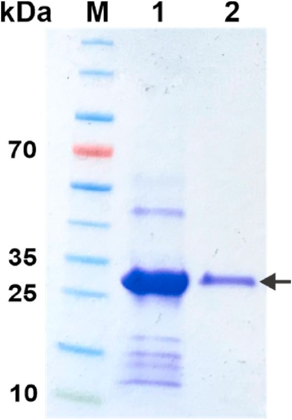
C. Phoeurk et al. Protein Expression and Purification 186 (2021) 105919
NMR data for the qualitative protein stability assessment were ac residues (GAM-BmCoc). GAM-BmCoc serves as a proxy of the inacti
quired on a Bruker Avance III HD 850 MHz spectrometer equipped with vated zymogen and was designed to enable structural studies since it is
a z-gradient cryoprobe, using a 1 mM GAM-BmCoc sample containing expected to have a lower likelihood of self-cleavage. Bombyx mori
5% D2O (v/v). 3 mM protease inhibitor, phenylmethyl sulfonyl fluoride cocoonase is a serine protease and it displays strong sequence similarity
(PMSF) was added to prevent the self-degradation of protein. 1H–15N- to trypsin with 41% sequence identity. Trypsin contains six intra
TROSY-HSQC spectra were recorded in identical conditions and pa molecular di-sulfide bridges [31] (Fig. 1A). In order to determine the
rameters other than the sweep widths of 10204 Hz and 2757 Hz along number of putative native di-sulfide bridges in BmCoc a structural ho
the 1H and 15N dimensions, respectively. mology model of BmCoc was created using Robetta modeling [32] with
trypsin (PDB ID 2FI5) as template (Fig. 1B). From the model, we iden
2.7. Activity of cocoonase from NMR and model substrates tified three putative di-sulfides that all are conserved in relation to
trypsin. In BmCoc the di-sulfides are formed from the following pairs of
The enzymatic activities of GAM-BmCoc and En-BmCoc were cysteines: C63:C79, C187:C200 and C211:C235, corresponding to the
determined by measuring the initial rate of the enzyme-catalyzed hy conserved cysteine pairs of trypsin: C42:C58, C168:C182 and C191:
drolysis reaction of three individual model substrates: (1) Z-L-Lys-ONp C220 in trypsin. Thus in development of a protocol for production of
hydrochloride, (2) Nα-benzoyl-L-arginine 4-nitroanilide hydrochloride BmCoc care must be taken such that the three expected native and
(Sigma Aldrich, USA), and (3) Sericin3 peptide (acetylated-SDEKSSQS- intramolecular di-sulfides are properly formed. These di-sulfides poses a
NHMe, synthesized by GenScript Biotech, Netherlands). For each challenge to protein production in E. coli since its cytoplasm has a
experiment 2 mM substrate and between 50 nM and 5 μM BmCoc were reducing redox potential.
used. All experiments were carried out in a 20 mM sodium phosphate
buffer at pH 6.0, supplemented with 50 mM NaCl and 5% D2O (v/v). 3.1. Sub-cloning of BmCoc into pET plasmids
Trypsin from bovine pancreas (Sigma Aldrich, USA) was used as a
control. Quantitative NMR data of enzyme kinetic assays were acquired In order to have complete control of the gene, the BmCoc gene cor
on a Bruker Avance III HD 600 MHz spectrometer equipped with a 5 mm responding to the active enzyme (residues 35–260) were synthesized
BBO cryoprobe and a SampleJet sample changer. One-dimensional 1H (Eurofins Genomics, Germany) with an N-terminal His-tag and the two
NMR experiment was first performed on the sample containing only different linkers, TEV and enterokinase cleavable respectively (Fig. 2).
substrate(s) for resonance identification. One dimensional (1D) 1H NMR To improve the expression, the genes were codon optimized for E. coli
experiment with excitation sculpting (es) for water suppression [33]. The genes was cloned into a pET vector (pET His 1a) under control
(Topspin3.6 zgesgp pulse program) was used. Each 1D proton NMR of the T7 promotor.
spectrum was recorded in the same buffer conditions at 298 K, and
collected with a spectral width of 16.02 ppm (9615.385 Hz) over 24998 3.2. Production and refolding of BmCoc
points yielding an acquisition time of 1.3 s per experiment. A relaxation
delay of 1 s was used between each scan and 112 of scans were averaged The two versions showed a similar behavior during expression,
for the signal. This experiment was assembled to collect FIDs continu refolding, and initial purification. Significant differences did not occur
ously in a time series for example, a total of 50 such experiments (50 × until the His-tag removal step.
4.52 min = 226 min) were performed. The plasmids were transformed into E. coli BL21(DE3) cells using the
heat-shock procedure. Transformed cell colonies were picked and the
2.8. In situ, cocoon assay cells were cultivated in LB-media to an optical density at 600 nm
(OD600) of 0.7 before induction with IPTG at a final concentration of
Bombyx mori cocoons were obtained from the Silk Center, Royal 0.75 mM. After 20 h of expression at 37 ◦ C, SDS-PAGE analysis on the
University of Phnom Penh, Cambodia. The pupas were removed from whole cells showed that both recombinant GAM-BmCoc (Fig. 3A) and
the cocoons by cutting the cocoons with a clean pair of scissors. All En-BmCoc was highly overexpressed. In addition we demonstrated a
handling of the cocoons was performed at room temperature. Seventy
milligram of silkworm cocoon was weighed and cut into approximately
0.5–1 mm pieces and extensively washed with the reaction buffer (20
mM sodium phosphate, 50 mM NaCl at pH 6.0). Thereafter the cocoon
pieces were suspended in 5 ml reaction buffer and incubated with either
1 μM or 10 μM En-BmCoc at 25 ◦ C for 24 h with shaking at 170 rpm.
Released peptide fragments were observed by one-dimensional 1H NMR
spectra acquired at 298 K. The experiment was designed with a negative
control (no enzyme added) and a positive control (by adding 1 μM
trypsin). The principle of the experiment is that while the cocoon is a
macroscopic object and does therefore not give rise to observable NMR
signals, released peptide fragments that tumble fast in solution give rise
to observable signals. This principle is demonstrated by the absence of
observable signals in the negative control while there exist numerous
observable resonances after incubation with both En-BmCoc and
trypsin.
Fig. 1. Di-sulfides and catalytic triad in BmCoc from a homology model
3. Results and discussion based on trypsin. (A) Structure of trypsin (PDB ID 2FI5) with the six di-sulfide
bonds indicated (named as residueX:residueY), the bonds indicated in bold
have conserved counterparts in BmCoc, together with the catalytic triad (H57,
To fulfill the demands of structural studies and industrial applica
D102 and S195, italic). (B) Homology model of BmCoc obtained by using the
tions, recombinant BmCoc production is required. Therefore, we have Robetta server [32]. The cysteine residues involved in putative di-sulfide
developed a robust and reproducible protocol for production of milli bridges are labeled together with the catalytic triad (H78, D121 and S215).
gram quantities of BmCoc in E. coli. We have focused on two variants of The resulting sequence alignment from the comparative modelling is shown in
the enzyme one with a completely native and activated sequence (En- Supplementary Fig. 1. The figure was created using MOE (Chemical computing
BmCoc) and one with three non-native and N-terminal amino acid group, Canada).
4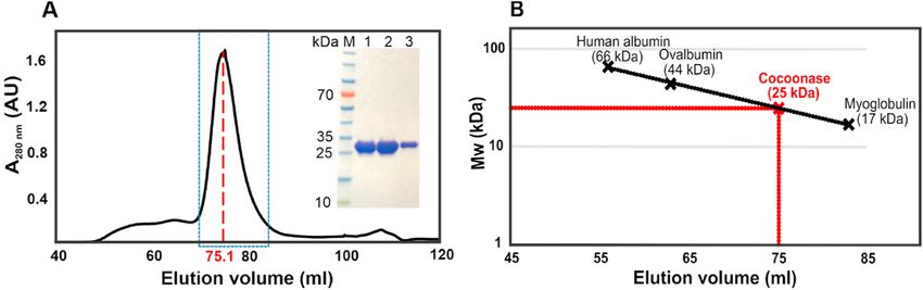
C. Phoeurk et al. Protein Expression and Purification 186 (2021) 105919
pure cocoonase (Fig. 3B). The efficacy of the refolding step was assessed
by comparing the absorbance of the eluted monomer peak to dimer/
oligomer peaks in the size exclusion chromatography (SEC) to around
90%.
3.3. Purification of refolded BmCoc
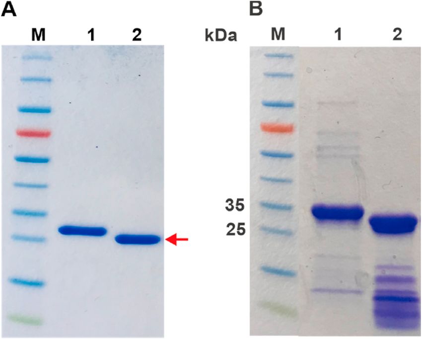
The refolded BmCoc was first purified in a batch-mode by virtue of
Fig. 2. Bombyx mori cocoonase plasmid constructs. Schematic illustration of
the N-terminal His-tag by loading the supernatant from the refolding
the two plasmid constructs utilized for production in E. coli. BmCoc, was cloned
with an N-terminal His-tag with either (A) a TEV protease- or (B) an
step onto a nickel affinity column. After washing the column with the
enterokinase-cleavage site to facilitate removal the tag. Cleavage with the TEV binding buffer, bound proteins were eluted with 400 mM imidazole. The
protease leaves the non-native sequence GAM sequence N-terminal to the eluated fractions were qualitatively analyzed with SDS-PAGE gel indi
native isoleucine 35 of activated BmCoc. On the other hand, cleavage at the cating reasonably pure GAM-BmCoc (Fig. 4), from a single purification
enterokinase site produces the correct and native activated enzyme with an N- step. En-BmCoc exhibited a similar purification pattern.
terminus corresponding to ILE35. The protein was further purified by size exclusion chromatography
(SEC) using a HiLoad™ 16/600 Superdex™ 75, The GAM-BmCoc con
taining chromatography peak is symmetric with an apparent Gaussian
shape, which is indicative of mono-disperse material (Fig. 5A). SDS-
PAGE analysis of the fractions in the main peak show a single protein
band (Fig. 5A, inset) indicating that the protein is purified to homoge
netity. To determine the aggregation status of the eluted GAM-BmCoc
we turned to quantitative analysis of the SEC chromatogram and using
the empiric linear relationship between log(Mw) and elution volume
(Ve) for spherical particles [35]. On basis of a calibration curve gener
ated with the proteins human albumin (66 kDa), Ovalbumin (44 kDa)
and Myoglobulin (17 kDa), and the Ve of GAM-BmCoc (75.1 ml) we
computed an apparent molecular weight of 25 kDa for GAM-BmCoc
(Fig. 5B). En-BmCoc shows a similar behavior to GAM-BmCoc with Ve
of 75.5 ml and an estimated molecular weight of 25 kDa. Since these
quantified molecular weights are virtually similar to the weight
computed from the primary sequence (24 kDa), it is evident that both
versions of refolded BmCoc are monomeric in solution. This is a signif
icant result since it indicates that only internal di-sulfides have been
Fig. 3. Production and solubilization of GAM-BmCoc. Both production and
a successful solubilization of insoluble GAM-BmCoc was analyzed with SDS- formed during refolding.
PAGE. (A) Lane 1: Approximately 400 μg of cells prior to induction, Lane 2: For GAM-BmCoc the N-terminal His-tag was removed by adding TEV
approximately 400 μg of cells 20 h after induction with IPTG, Lane 3: soluble protease in ratio of 1:50 (w/w). After 15–17 h incubation at room
fraction of approximately 400 μg of cells after lysis, Lane 4: insoluble fraction of temperature, the cleavage was analyzed with SDS-PAGE (Fig. 6A)
approximately 2 mg of cells after lysis. GAM-BmCoc (25 kDa) production was showing that the protein was completely cleaved. The protease and the
induced with 0.75 mM IPTG and 20 h of overexpression. (B) Lane 1: 12 μl out of remaining His-tag were removed using a Ni-column by collecting the
600 ml (1:50000) of the soluble fraction after the refolding step. The arrow
shows the protein band at the size of His-tagged GAM-BmCoc, indicating that
GAM-BmCoc stays soluble in a native buffer after the refolding step. En-BmCoc
showed similar expression and solubilization characteristics. The migration of
BmCoc is indicated with an arrow. PageRulerTM prestained protein ladder was
used as marker in both A and B (lanes M).
high level of overexpression also in M9 minimal media supplemented
with either 15N or 15N/13C, this is relevant since it enables future
structure determination and also quantification enzyme dynamics with
solution state NMR spectroscopy [34]. After cell lysis by sonication, the
expressed protein, with the expected molecular weight of 26 kDa, was
absent from the soluble fraction and present exclusively as inclusion
bodies in the cell debris pellet (Fig. 3A). The insoluble proteins from the
inclusion bodies were resolubilized by resuspending the pellet in 6 M
GuHCl at 37 ◦ C for 2 h. Di-sulfide bridges were reduced by adding DTT to
a final concentration of 30 mM. Both versions of BmCoc were success
fully refolded by diluting the unfolded protein 30 times (v/v) in a buffer
containing 5 mM reduced and 1 mM oxidized L-glutathione. This ratio of
reduced vs. oxidized glutathione is optimized in order to keep the redox
balance environment such that native and internal di-sulfide bridges are
favored while intermolecular di-sulfide bridges or non-native and sol
vent exposed bridges would not form. The protocol is based on a pre Fig. 4. Analysis of His-tag purification. The initial purification step and
viously established methods that was developed for the Human Cationic subsequent removal of the His-tag was analyzed with SDS-PAGE. Analysis of
Trypsinogen [29]. The soluble material from this refolding step was his-tagged GAM-BmCoc fractions eluted with 400 mM imidazole, first and
analyzed on a SDS-PAGE gel showing a high concentration of almost second 5 ml fractions. The black arrow indicates the His-tagged GAM-BmCoc.
Lane M: 3 μl PageRuler™ prestained protein ladder.
5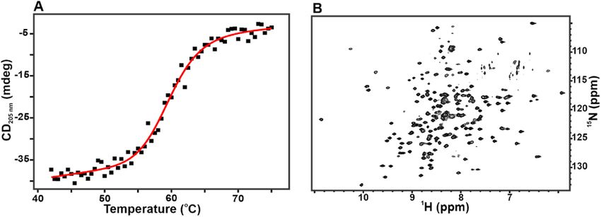
C. Phoeurk et al. Protein Expression and Purification 186 (2021) 105919
Fig. 5. GAM-BmCoc is monomeric in
solution. The apparent molecular
weight of GAM-BmCoc was quantified
with quantitative size exclusion chro
matography (SEC) (A) SEC profile of
refolded GAM-BmCoc. Three fractions
from the main peak, eluted at 75.1 ml
(highlighted with the box), was
analyzed by SDS page gel (insert). (B)
The apparent molecular weight was
determined by comparing the elution
volume to three standard proteins. The
elution volumes of the standard proteins
were obtained from the manufacturer of
the Superdex 75 pg 16/600 pre-packed
column. A plot of log(Mw) of the stan
dard proteins versus elution volume (dotted line) rendered a linear correlation that was used as a standard curve to determine the size of GAM-BmCoc. The size of
GAM-BmCoc was determined from the elution volume (75.1 ml) to be 25 kDa. Since the molecular weight computed from the primary sequence is 24 kDa it is evident
that GAM-BmCoc is monomeric in solution.
h. We observe that active BmCoc itself can cleave the His-tagged pre
cursor protein and therefore a chain reaction is initiated after buffer
exchange by autolyzed BmCoc molecules (Fig. 6B). Therefore, entero
kinase is not required for removal of the tag. The cleaved protein was
polished with another round of SEC to separate from His-tag residues
and degraded protein. For En-BmCoc, the final yield was about, 5 mg/l
15
N in labeled M9 media, a bit lower than GAM-BmCoc, 8 mg/l. A
problem with active En-BmCoc is that it also displays auto-cleavage
resulting in degradation of the native enzyme, as evident by the
smaller peptides seen on the SDS-PAGE gel of the cleavage (Fig. 6B).
This process is however slow enough to allow quantification of activity
as discussed below (see below). Taken together the developed protocols
enables the production of large amounts of both unlabeled but also of
isotopically enriched GAM/En BmCoc.
3.4. Biophysical characterization of BmCoc
Although the SEC analysis provides an indication that the enzyme is
not only monomeric but also properly folded, we wanted to test this
feature further and on the molecular level with an extended biophysical
Fig. 6. His-Tag removal from BmCoc. The His-tag was removed by either TEV
characterization. First, we quantified the thermal unfolding of the
protease, from the GAM-BmCoc (A), or by autolysis, from En-BmCoc (B). The
enzyme by monitoring the CD signal at 205 nm in response to a tem
SDS-PAGE gel shows the protein before (lane A1 and B1) and the protein after
cleavage (A2 and B2). The change in migration indicates that the molecular perature scan (Fig. 7A). The temperature dependency of the CD signal
weight has be reduced by the removal of the His-Tag. The autolysis of En- displays a cooperative transition over a narrow temperature interval.
BmCoc does not only remove the His-tag (too small to be visible on the gel) This behavior is a solid indication that the protein has secondary
but also degrades BmCoc itself as evident by the low molecular weight bands structure at ambient temperatures, while it is unfolded at temperatures
visible on the gel. well above the infliction point (i.e. the melting temperature, TM). The
main driving force in thermal unfolding of a protein is the difference in
flow-through fraction. The final yield obtained from this construct were enthalpy (ΔHU) between folded and unfolded states. A fit of the CD data
15 mg/l, 8 mg/l, and 7 mg/l respectively from three different culture to equation (1) revealed that the ΔHU for unfolding of His-tag GAM-
media (LB, 15N labeled M9, and double 15N & 13C labeled M9 media) BmCoc was 390 ± 50 kJ mol− 1. The absolute magnitude of the unfolding
(Table 1). ΔHu is comparable to other proteins of similar size, for instance ade
After the SEC purification step, En-BmCoc protein, was buffer nylate kinase from E. coli with a size of 24 kDa has a ΔHU of 570 ± 20 kJ
exchanged to 50 mM Tris-HCl, pH 7.75 and incubated at 37 ◦ C for 15–17 mol− 1 [36]. The quantitative analysis of the CD data also provided the
TM that was found to be 60 ± 0.5 ◦ C. Thus the TM is well above the
temperature where cocoonase is active in its natural environments. The
Table 1
CD signal in the far-UV region is predominantly sensitive to secondary
Summary of protein yields during purification of recombinant Bombyx mori
structure elements [37] and to take the characterization further and to
cocoonase (GAM-BmCoc).
the level of individual amino acid residues, we turned to protein NMR
Step Total protein (mg) from 1 L cell culture
spectroscopy on 15N enriched material. The 1H–15N TROSY-HSQC NMR
LB media 15
N-M9 mediaa 13
C-M9 mediab spectra of 15N labeled GAM-BmCoc, was well dispersed in both 1H and
15
Ni-NTA affinity chromatography 47 11 N/A N dimensions, with approximately 200 backbone amide resonances
Size exclusion chromatography 18 9.5 9.1 highly resolved (Fig. 7B). This well-dispersed NMR spectrum is strongly
Ni-NTA affinity chromatography 15 8.0 7.0 indicative of a homogenous, folded globular protein. The enzyme was
N/A = not checked. shown to be active (see below) and hence the fold must be correct. This
a 15
N labeling medium. is the first heteronuclear 2D spectrum of Bombyx mori cocoonase pub
b
Double15N and13C labeling medium. lished, reflecting on that we have solved the difficulties to obtain
6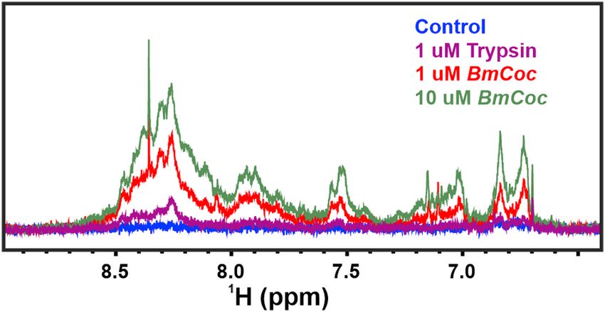
C. Phoeurk et al. Protein Expression and Purification 186 (2021) 105919
Fig. 7. The refolded BmCoc is a fol
ded protein. (A) The thermal unfolding
curve of His-tagged BmCoc shows a
cooperative unfolding indicating a well
folded protein. The unfolding tempera
ture (Tm) and the enthalpy of unfolding
(ΔH) were determined to 60 ± 0.5 ◦ C
and − 390 ± 50 kJ mol− 1 respectively.
The unfolding was monitored by circu
lar dichroism (CD) at 205 nm and the
data was fitted to a two-state reaction
(equation (1)). The sample contained
20 μM of His-tagged GAM-BmCoc in 10
mM NaPi buffer at pH 7.0. (B) The well
dispersed TROSY-HSQC NMR spectrum
of 15N labeled GAM-BmCoc is indicative
of a folded protein. The spectrum was
recorded on a 0.8 mM GAM-BmCoc
sample in a 20 mM NaPi buffer at pH 6.0 supplemented with 50 mM NaCl, at 308K and 600 MHz.
1
sufficient amount for structural studies of cocoonase. H NMR at 25 ◦ C. The assay is a modification of our previously estab
Since En-BmCoc showed signs of self-degradation (Fig. 6B), we used lished assay based on 31P NMR designed for nucleoside phosphate
NMR spectroscopy to investigate this. To this end the magnitude of the catalysis [42]. In short, the assay provides kcat values by determining the
En-BmCoc signals were monitored over time and we did observe spectral initial slopes of the changes in either substrate or product concentra
changes that indicated self-degradation (Supplementary Fig. 2A). tions, determined by the intensities of the NMR resonances (Supple
However, the degradation is sufficiently slow to allow quantitative mentary Fig. 3). Since NMR is a quantitative technique, the intensity of
analysis of enzymatic activity (see below). On the other hand, stability resonances are directly correlated to the concentrations, so the
over extended periods is essential for structural studies with NMR, and maximum reaction velocity (Vmax) was determined as the initial slopes
we, therefore, investigated the inhibitory effect of different known of the peak intensities as a function of time after enzyme addition. The
protease inhibitors. In the apo state, En-BmCoc showed an extensive kcat, values were determined from the maximum reaction rate divided by
degradation within 22 h at 35 ◦ C; therefore, it is necessary, in order to the concentration of the enzyme, i.e kcat = Vmax/[E]tot (Table 2). The
obtain the long-term stability needed for the acquisition of structural design of experiment included trypsin as a positive control for bench
data to use an effective protease inhibitor. Unajak and co-workers [26] marking of quantified kcat values. With Z-L-Lys-ONp was used as a
reported that PMSF, a serine protease specific inhibitor, could strongly substrate, the kcat value of the active En-BmCoc (20 s− 1) is equal to
inhibit the protease activity of recombinant BmCoc. Our results showed trypsin within the experimental uncertainty. On the other hand, the kcat
that PMSF provided only a limited inhibitory effect. Although cocoonase of En-BmCoc is 10 fold lower than trypsin for cleavage of L-BAPNA
belongs to the same family of serine protease as trypsin and chymo substrate indicating that cocoonase have a higher specificity for a lysine
trypsin, the inhibitory effects by a wide range of serine protease in peptide compared to trypsin. This results indicates that BmCoc has a
hibitors are known to be relatively low [25]. Previous studies has preference for cleavage after lysine over arginine. Although the activity
showed that the inhibitory effect of a range of trypsin inhibitors, such as of En-BmCoc is comparable to trypsin for the model compounds, the kcat
bovine pancreatic trypsin inhibitor (BPTI), soybean trypsin inhibitor for cleavage of the peptide derived from the natural sericin substrate is
(SBTI), cocoon shell extract (CSE), and N-α-p-Tosyl-L-lysine chlor significantly slower (kcat is 0.25 s− 1). With this somewhat unexpected
omethyl ketone (TLCK), was lower for BmCoc compared to trypsin [25]. observation we decided to develop an in-situ assay for Sericin degra
Zhao and co-workers found that PMSF could inhibit the total larval dation of cocoons (see below).
mid-gut proteases, high-alkaline trypsin, low-alkaline trypsin and The enzymatic activities are so far discussed for native, En-BmCoc. It
chymotrypsin [38]. Diisopropyl fluorophosphate was proved as a potent is known that the activation mechanism of trypsin is dependent on the
inhibitor of cocoonase [24]; however, it is about 200 times less sensitive insertion of the N-terminal residue (Isoleucine 16 in BmCoc and
with cocoonase than chymotrypsin [39,40]. Based on these previous isoleucine 35 in trypsin) to enable indirect contacts with the active site
investigations, it is not surprising that PMSF has a limited inhibitory [43]. This feature is not possible for the zymogen where the N-terminal
effect on cocoonase. peptide blocks insertion of Ile16/Ile35. Even though we have not studied
We have also used the cOmplete™ protease inhibitor cocktail the zymogen per se we reasoned that the GAM-BmCoc could acts as
(Roche, USA) containing both reversible and irreversible protease in proxy for the zymogen since the N-terminal is non-native and likely
hibitors to increase the stability of BmCoc (Supplementary Fig. 2B). The hinders Ile35 to insert into the body of the enzyme. The kcat value of
inhibitor stabilizes the enzyme for at least 3 days; which will enable the GAM-BmCoc was low with all substrates; for instance, the hydrolysis of
time consuming NMR experiments required for structure determination.
Table 2
3.5. Enzymatic activity Enzyme kinetics constant (kcat, turnover number) of BmCoc and trypsin using
three different synthesized peptides.
As mentioned above BmCoc and trypsin have a high sequence sim Substrate, kcat (s− 1)
ilarity (41% identity), therefore it was postulated that BmCoc would Z-L-Lys- Nα-Benzoyl-L-arginine 4- Sericin3
Enzyme
have a similar substrate selectivity as for trypsin, cleaving the peptides ONp nitroanilide peptide
on the C-terminal side of lysine and arginine amino acid residues GAM- 0.11 ± 0.01 0.01 ± 0.001 0.03 ± 0.002
(MEROPS database [41]). In this study, two lysine peptides (Z-L-Ly BmCoc
s-ONp and a peptide derived from Sericin3) and one arginine peptide En-BmCoc 20.0 ± 0.51 0.04 ± 0.01 0.25 ± 0.02
(Nα-Benzoyl-L-arginine 4-nitroanilide hydrochloride, L-BAPNA) were Trypsin 20.0 ± 0.23 0.36 ± 0.05 0.56 ± 0.03
designed and used as model substrates to determine the activity of The kcat values are mean values of two independent experiments with four
En-BmCoc. Real-time enzyme kinetics were monitored by quantitative technical repetitions.
7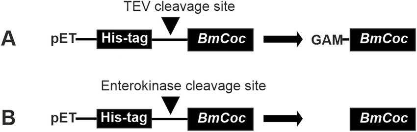
C. Phoeurk et al. Protein Expression and Purification 186 (2021) 105919
Z-L-Lys-ONp catalyzed by this GAM-BmCoc was approximately 180
times slower than En-BmCoc (Table 2). This results indicates that leav
ing GAM residues at the N-terminal significantly interferes the activity of
BmCoc and that the determined kcat values is an approximation of the
activity of the zymogen. The quantification of activity with model sub
strates demonstrated that BmCoc is an active enzyme which is the ul
timate proof that our refolding protocol is functional.
3.6. In-situ assay for softening of cocoons
Although the experiments with model substrates are useful, the main
goal of our research is to study the efficacy of En-BmCoc towards its
natural substrate, i.e. the sericin proteins embedded in cocoons. GAM- Fig. 8. In situ assay of cocoon softening by BmCoc. Proteolytic activity of
BmCoc was not tested because it showed low activity against the pep BmCoc on B. mori cocoons was detected with 1H NMR spectroscopy. The 600
tides. In its natural environment, BmCoc is activated by flushing of MHz 1H NMR spectra of peptide fragments released from the cocoon by the
excreted solvent over the solid BmCoc deposit on the head of the moth. proteolytic activity of fully active En-BmCoc and trypsin (control) are shown.
This wetting mechanism enables the direct contact between the enzyme The proteolytic reaction was occurring for 24 h and thereafter NMR spectra
and the surface of the cocoon. In order to mimic these circumstances in a were acquired for the soluble fraction. Proteolytic cleavage was performed on
cocoons in 20 mM NaPi, 50 mM NaCl buffer, pH 6.0 and measured at 25 ◦ C. The
laboratory setting, we developed a NMR detected experiment where
color coding of the spectra is as follows: red; 1 μM BmCoc, green; 10 μM BmCoc,
recombinant En-BmCoc was added to cocoon fragments that were pre-
purple; 1 μM trypsin, and blue; the negative control without enzyme.
wetted with buffer. Pieces of cocoon were suspended and pre-
incubated in a buffer consisting of 20 mM NaPi, 50 mM NaCl at pH
reduced yield of active enzyme. In the light of the results with the non-
6.0 for 24 h at 25 ◦ C. In order to define the time equal to zero point a 1H
active, non-auto-proteolytic GAM variant, there might be a possibility to
1D NMR spectrum was recorded before adding the enzyme. To initiate
develop a variant with an N-terminal that is active but not auto-
hydrolysis of sericin either 1 μM or 10 μM of En-BmCoc were added to
proteolytic.
70 mg of cocoon. As a control, 1 μM trypsin was added in a separate
An E. coli based system has a number of benefits in the character
experiment. The principle of detection in the experiment is that while
ization and potential industrial use of cocoonase. It is fast and gives a
the intact cocoon is too large to yield detectable NMR signals [27],
generally high yield, in addition, E. coli based systems are well studied
released peptide fragments tumble fast enough to be readily detected.
and there are tools available for efficient genetic manipulation. For
The hydrolyzed products released into the solution were analyzed by
biophysical characterization using NMR, 15N and/or 13C labeled pro
serial 1H NMR spectra up to 24 h after the enzyme was added. Reso
teins are necessary, and an E. coli based system makes this feasible. This
nances in the amide proton region, between 6.7 and 8.7 ppm, origi
is because E. coli can be effectively grown in minimal media where
nating from the peptides released upon sericin break-down of the cocoon
carbon and nitrogen are provided in the form of single chemicals. In M9
were monitored (Fig. 8). The spectra showed that En-BmCoc had a
medium, commonly used for E. coli cultivation, carbon and nitrogen are
sizeable activity in hydrolyzing the sericin, which is notable since the
provided as glucose and ammonium chloride, respectively. Uniformly
action on a sericin model peptide was very slow (Table 2). The amount
labeled variants of these chemicals are commercially available, at a
of the released sericin peptides scaled with enzyme concentration indi
relatively affordable price. Using labeled minimal media, the yield was
cating a Michaelis-Menten like behavior. It should be noted that
not as good as for expression in rich, LB, media, but sufficient for NMR
although we perform a quantitative detection of released peptides it is
analysis.
not possible to provide a meaningful Vmax or kcat since the substrate is a
BmCoc has large potential to improve the environmental profile and
solid particle. In addition, trypsin displayed detectable cleavage hy
increase the economic benefit of the silk industry. A more gentle
drolysis of the cocoon but in comparison to En-BmCoc the activity of
approach, using enzymes, to silk degumming might at the same time
trypsin is considerably lower. Thus, we can conclude that although the
provide the medical and cosmetic industries with a potential raw ma
kcat for cleavage of model peptides is similar, BmCoc has evolved with an
terial in the leftover sericins. Other enzymes than BmCoc have been used
increased efficacy towards sericin embedded in the cocoon substrate in
for enzymatic degumming [18–20], however they have not shown the
comparison to trypsin. These differences may be encoded in the surface
same selectivity and efficiency [20]. Our investigation showed that
properties of BmCoc and therefore it is important to solve its three
trypsin was as efficient in cleaving sericin peptides as BmCoc, however,
dimensional structure to enable identification of the physiochemical
it was less effective when applied to the natural substrate, the cocoon.
mechanism of the increased efficacy of BmCoc, The fact that our
With the help of our new expression and purification system, the mo
recombinantly produced and refolded BmCoc is active on the natural
lecular properties of the BmCoc can be determined and also optimized,
substrate opens the door for studies not only limited to the enzyme itself
and using the well-developed tools for E. coli genetics the possibility of
but also to the properties of the de-gummed silk fiber with for instance
exploring these properties.
electron microscopy [26].
5. Conclusions Author statement
Here we describe a stable and reproducible method for the produc Chanrith Phoeurk: Overall experimental work and writing of
tion of large amounts of recombinant Bombyx mori cocoonase (BmCoc). manuscript.
By combining refolding under carefully controlled redox conditions with Ameeq Ul Mushtaq: Recording and analysis of NMR experiments.
efficient E. coli expression, we were able to express and purify fully Per Rogne: Assistance in experimental work and writing of
active BmCoc. Using this protocol, a yield of up to 15 mg of functional manuscript.
BmCoc per liter media was obtained. The function of the enzyme was Magnus Wolf-Watz: Conceptualizing and writing of the manuscript.
demonstrated for model substrates, but also and importantly for Bombyx
mori cocoons. A biophysical characterization revealed that the enzyme is Acknowledgements
stable, monomeric and well folded. However, a still unsolved problem
with our method is the auto-proteolysis of the enzyme resulting in We thank The Protein Expertise Platform at Umeå University for
8
C. Phoeurk et al. Protein Expression and Purification 186 (2021) 105919
providing reagents and expertise for protein overexpression and purifi [19] U. Ninpetch, M. Tsukada, A. Promboon, Mechanical properties of silk fabric
degummed with bromelain, J. Eng. Fibers Fabr. 10 (2015) 69–78.
cation. NMR experiments were performed at “NMR for life”, Swedish
[20] P. Rodbumrer, D. Arthan, U. Uyen, J. Yuvaniyama, J. Svasti, P.
NMR centre at Umeå University. This study was financially supported by Y. Wongsaengchantra, Functional expression of a Bombyx mori cocoonase:
grants from The Swedish Research Council (2017–04203), the Kempe potential application for silk degumming, Acta Biochim. Biophys. Sin. 44 (2012)
Foundation (JCK-1417), and the Sweden-RUPP Bilateral Program 974–983.
[21] E. Berger, F.C. Kafatos, R.L. Felsted, J.H. Law, Cocoonase 3. Purification,
(11599). The Silk Center, Royal University of Phnom Penh, Cambodia is preliminary characterization, and activation of zymogen of an insect protease,
acknowledged for Bombyx mori cocoons. J. Biol. Chem. 246 (1971) 4131–4137.
[22] R.L. Felsted, J.H. Law, A.K. Sinha, M.S. Jolly, Properties of the Antheraea mylitta
cocoonase, Comp. Biochem. Physiol. B Comp. Biochem. 44 (1973) 595–609.
Appendix A. Supplementary data [23] F.C. Kafatos, C.M. Williams, Enzymatic mechanism for escape of certain moths
from their cocoons, Science 146 (1964) 538–540.
Supplementary data to this article can be found online at https://doi. [24] F.C. Kafatos, A.M. Tartakoff, J.H. Law, Cocoonase 1. Preliminary characterization
of a proteolytic enzyme from silk moths, J. Biol. Chem. 242 (1967) 1477–1487.
org/10.1016/j.pep.2021.105919. [25] H. Fukumori, S. Teshiba, Y. Shigeoka, K. Yamamoto, Y. Banno, Y. Aso, Purification
and characterization of cocoonase from the silkworm Bombyx mori, Biosci.
References Biotechnol. Biochem. 78 (2014) 202–211.
[26] S. Unajak, S. Aroonluke, A. Promboon, An active recombinant cocoonase from the
silkworm Bombyx mori: bleaching, degumming and sericin degrading activities,
[1] G.H. Altman, F. Diaz, C. Jakuba, T. Calabro, R.L. Horan, J. Chen, H. Lu,
J. Sci. Food Agric. 95 (2015) 1179–1189.
J. Richmond, D.L. Kaplan, Silk-based biomaterials, Biomaterials 24 (2003)
[27] J. Cavanagh, W.J. Fairbrother, A.G. Palmer III, N.J. Skelton, Protein NMR
401–416.
Spectroscopy, Academic Press inc., San Diego, USA, 1996.
[2] P. Aramwit, T. Siritientong, T. Srichana, Potential applications of silk sericin, a
[28] J. Bogomolovas, B. Simon, M. Sattler, G. Stier, Screening of fusion partners for high
natural protein from textile industry by-products, Waste Manag. Res. 30 (2012)
yield expression and purification of bioactive viscotoxins, Protein Expr. Purif. 64
217–224.
(2009) 16–23.
[3] G. Freddi, R. Mossotti, R. Innocenti, Degumming of silk fabric with several
[29] L. Szilagyi, E. Kenesi, G. Katona, G. Kaslik, G. Juhasz, L. Graf, Comparative in vitro
proteases, J. Biotechnol. 106 (2003) 101–112.
studies on native and recombinant human cationic trypsins - Cathepsin B is a
[4] T. Gai, X. Tong, M. Han, C. Li, C. Fang, Y. Zou, H. Hu, H. Xiang, Z. Xiang, C. Lu,
possible pathological activator of trypsinogen in pancreatitis, J. Biol. Chem. 276
F. Dai, Cocoonase is indispensable for Lepidoptera insects breaking the sealed
(2001) 24574–24580.
cocoon, PLoS Genet. 16 (2020), e1009004.
[30] L. Swint, A.D. Robertson, Thermodynamics of unfolding for Turkey ovomucoid 3rd
[5] N.M. Mahmoodi, F. Moghimi, M. Arami, F. Mazaheri, Silk degumming using
domain- thermal and chemical denaturation, Protein Sci. 2 (1993) 2037–2049.
microwave irradiation as an environmentally friendly surface modification
[31] E. Zakharova, M.P. Horvath, D.P. Goldenberg, Functional and structural roles of
method, Fibers Polym. 11 (2010) 234–240.
the cys14–cys38 Disulfide of bovine pancreatic trypsin inhibitor, J. Mol. Biol. 382
[6] S.C. Kundu, B. Kundu, S. Talukdar, S. Bano, S. Nayak, J. Kundu, B.B. Mandal,
(2008) 998–1013.
N. Bhardwaj, M. Botlagunta, B.C. Dash, C. Acharya, A.K. Ghosh, Nonmulberry silk
[32] Y. Song, F. DiMaio, R.Y.-R. Wang, D. Kim, C. Miles, T.J. Brunette, J. Thompson,
biopolymers, Biopolymers 97 (2012) 455–467.
D. Baker, High-resolution comparative modeling with RosettaCM, Structure 21
[7] X. Nirmala, K. Mita, V. Vanisree, M. Žurovec, F. Sehnal, Identification of four small
(2013) 1735–1742.
molecular mass proteins in the silk of Bombyx mori, Insect Mol. Biol. 10 (2001)
[33] N.A. Burgess-Brown, S. Sharma, F. Sobott, C. Loenarz, U. Oppermann, O. Gileadi,
437–445.
Codon optimization can improve expression of human genes in Escherichia coli: a
[8] Z. Dong, P. Zhao, C. Wang, Y. Zhang, J. Chen, X. Wang, Y. Lin, Q. Xia, Comparative
multi-gene study, Protein Expr. Purif. 59 (2008) 94–102.
proteomics reveal diverse functions and dynamic changes of Bombyx mori silk
[34] M. Kovermann, P. Rogne, M. Wolf-Watz, Protein dynamics and function from
proteins spun from different development stages, J. Proteome Res. 12 (2013)
solution state NMR spectroscopy, Q. Rev. Biophys. 49 (2016).
5213–5222.
[35] D.G. Rhodes, R.E. Bossio, T.M. Laue, Determination of size, molecular weight, and
[9] C.P. Singh, R.L. Vaishna, A. Kakkar, K.P. Arunkumar, J. Nagaraju, Characterization
presence of subunits, in: second ed., in: R.R. Burgess, M.P. Deutscher (Eds.), Guide
of antiviral and antibacterial activity of Bombyx mori seroin proteins, Cell
to Protein Purification, vol. 463, Elsevier Academic Press Inc, San Diego, 2009,
Microbiol. 16 (2014) 1354–1365.
pp. 691–723.
[10] S. Inoue, K. Tanaka, F. Arisaka, S. Kimura, K. Ohtomo, S. Mizuno, Silk fibroin of
[36] U. Olsson, M. Wolf-Watz, Overlap between folding and functional energy
Bombyx mori is secreted, assembling a high molecular mass elementary unit
landscapes for adenylate kinase conformational change, Nat. Commun. 1 (2010).
consisting of H-chain, L-chain, and P25, with a 6 : 6 : 1 molar ratio, J. Biol. Chem.
[37] K.E. van Holde, W.C. Johnson, P.S. Ho, Principles of Physical Biochemistry,
275 (2000) 40517–40528.
Prentice Hall, Upper Saddle River, New Jersey, USA, 1998.
[11] Z. Dong, K. Guo, X. Zhang, T. Zhang, Y. Zhang, S. Ma, H. Chang, M. Tang, L. An,
[38] A. Zhao, Y. Li, C. Leng, P. Wang, Y. Li, Inhibitory effect of protease inhibitors on
Q. Xia, P. Zhao, Identification of Bombyx mori sericin 4 protein as a new biological
larval midgut protease activities and the performance of Plutella xylostella
adhesive, Int. J. Biol. Macromol. 132 (2019) 1121–1130.
(Lepidoptera: Plutellidae), Front. Physiol. 9 (2019).
[12] Y. Takasu, T. Hata, K. Uchino, Q. Zhang, Identification of Ser2 proteins as major
[39] E.F. Jansen, A.K. Balls, The inhibition of b- and y-chymotrypsin and trypsin by
sericin components in the non-cocoon silk of Bombyx mori, Insect Biochem. Mol.
diisopropyl fluorophosphate, J. Biol. Chem. 194 (1952) 721–727.
Biol. 40 (2010) 339–344.
[40] E.F. Jansen, M.D.F. Nutting, R. Jang, A.K. Balls, Inhibition of the proteinase and
[13] Y.-Y. Jo, H. Kweon, J.-H. Oh, Sericin for tissue engineering, Appl. Sci. 10 (2020).
esterase activities of trypsin and chymotrypsin by diisopropyl fluorophosphate:
[14] L. Lamboni, M. Gauthier, G. Yang, Q. Wang, Silk sericin: a versatile material for
crystallization of inhibited chymotrypsin, J. Biol. Chem. 179 (1949) 189–199.
tissue engineering and drug delivery, Biotechnol. Adv. 33 (2015) 1855–1867.
[41] N.D. Rawlings, A.J. Barrett, P.D. Thomas, X. Huang, A. Bateman, R.D. Finn, The
[15] S.C. Kundu, B.C. Dash, R. Dash, D.L. Kaplan, Natural protective glue protein,
MEROPS database of proteolytic enzymes, their substrates and inhibitors in 2017
sericin bioengineered by silkworms: potential for biomedical and biotechnological
and a comparison with peptidases in the PANTHER database, Nucleic Acids Res. 46
applications, Prog. Polym. Sci. 33 (2008) 998–1012.
(2017) D624–D632.
[16] M.N. Padamwar, A.P. Pawar, Silk sericin and its applications: a review, J. Sci. Ind.
[42] P. Rogne, T. Sparrman, I. Anugwom, J.-P. Mikkola, M. Wolf-Watz, Realtime 31P
Res. 63 (2004) 323–329.
NMR investigation on the catalytic behavior of the enzyme adenylate kinase in the
[17] S. Suwannaphan, E. Fufeungsombut, A. Promboon, P. Chim-anage, A serine
matrix of a switchable ionic liquid, ChemSusChem 8 (2015) 3764–3768.
protease from newly isolated Bacillus sp. for efficient silk degumming, sericin
[43] J.P. Abita, M. Delaage, M. Lazdunski, Mechanism of activation of trypsinogen- role
degrading and colour bleaching activities, Int. Biodeterior. Biodegrad. 117 (2017)
of 4 N-terminal apartyl residues, Eur. J. Biochem. 8 (1969) 314–324.
141–149.
[18] J. Kim, M. Kwon, S. Kim, Biological Degumming of silk fabrics with proteolytic
enzymes, J. Nat. Fibers 13 (2016) 629–639.
9
You can also read