Effects of eight neuropsychiatric copy number variants on human brain structure
←
→
Page content transcription
If your browser does not render page correctly, please read the page content below
Translational Psychiatry                                                                                                                                  www.nature.com/tp

ARTICLE                  OPEN

Effects of eight neuropsychiatric copy number variants on
human brain structure
Claudia Modenato 1,74, Kuldeep Kumar 2,74, Clara Moreau2, Sandra Martin-Brevet1, Guillaume Huguet 2, Catherine Schramm2,
Martineau Jean-Louis2, Charles-Olivier Martin2, Nadine Younis2, Petra Tamer2, Elise Douard2, Fanny Thébault-Dagher2, Valérie Côté2,
Audrey-Rose Charlebois2, Florence Deguire2, Anne M. Maillard3, Borja Rodriguez-Herreros3, Aurèlie Pain3, Sonia Richetin3, 16p11.2
European Consortium*, Simons Searchlight Consortium*, Lester Melie-Garcia 4, Leila Kushan5, Ana I. Silva 6,7,
Marianne B. M. van den Bree 7,8,9, David E. J. Linden6,7,9, Michael J. Owen 7,8, Jeremy Hall 7,8,9, Sarah Lippé2, Mallar Chakravarty10,
                                                                                                      ✉
Danilo Bzdok11,12, Carrie E. Bearden 5, Bogdan Draganski 1,13,75 and Sébastien Jacquemont 2,75

© The Author(s) 2021

    Many copy number variants (CNVs) confer risk for the same range of neurodevelopmental symptoms and psychiatric conditions
    including autism and schizophrenia. Yet, to date neuroimaging studies have typically been carried out one mutation at a time,
    showing that CNVs have large effects on brain anatomy. Here, we aimed to characterize and quantify the distinct brain
    morphometry effects and latent dimensions across 8 neuropsychiatric CNVs. We analyzed T1-weighted MRI data from clinically and
    non-clinically ascertained CNV carriers (deletion/duplication) at the 1q21.1 (n = 39/28), 16p11.2 (n = 87/78), 22q11.2 (n = 75/30),
    and 15q11.2 (n = 72/76) loci as well as 1296 non-carriers (controls). Case-control contrasts of all examined genomic loci
    demonstrated effects on brain anatomy, with deletions and duplications showing mirror effects at the global and regional levels.
    Although CNVs mainly showed distinct brain patterns, principal component analysis (PCA) loaded subsets of CNVs on two latent
    brain dimensions, which explained 32 and 29% of the variance of the 8 Cohen’s d maps. The cingulate gyrus, insula, supplementary
    motor cortex, and cerebellum were identified by PCA and multi-view pattern learning as top regions contributing to latent
    dimension shared across subsets of CNVs. The large proportion of distinct CNV effects on brain morphology may explain the small
    neuroimaging effect sizes reported in polygenic psychiatric conditions. Nevertheless, latent gene brain morphology dimensions will
    help subgroup the rapidly expanding landscape of neuropsychiatric variants and dissect the heterogeneity of idiopathic conditions.

    Translational Psychiatry (2021)11:399 ; https://doi.org/10.1038/s41398-021-01490-9

INTRODUCTION                                                                                increases in risk for SZ have been documented for the 22q11.2
Genomic copy number variants (CNVs) are deletions or duplica-                               deletion (30 to 40-fold) followed by 16p11.2 duplication (10-fold),
tions of DNA segments of more than 1000 base pairs. Rare CNVs                               1q21.1 deletion and 15q11.2 deletion (1.5-fold) [2]. ASD risk is
with large effects have been associated with a range of often                               highest for 16p11.2 deletions and duplications (10-fold) followed by
overlapping developmental psychiatric phenotypes and condi-                                 1q21.1 duplications and 22q11.2 duplications (3 to 4-fold) [1, 2, 10–
tions, including autism spectrum disorder (ASD) and schizophrenia                           13]. The nature and specificity of CNV effects on cognitive and
(SZ) [1–4]. A looming question in psychiatric genetics pertains to                          behavioral traits is an area of intense investigation. All CNVs studied
the underlying basis of polygenicity: How do different variants                             to date affect cognition to varying degrees and a broad range of
lead to risk for the same psychiatric condition?                                            cognitive functions [14, 15]. A recent study found that the range of
   Some of the most frequent risk factors for neuropsychiatric                              affected traits was broadly similar for 13 CNVs at 8 loci and specific
disorders identified in pediatric clinics include CNVs at the 22q11.2,                       genotypes accounted for a low proportion of phenotypic variance
16p11.2, 1q21.1, and 15q11.2 genomic loci [5, 6]. They affect the                           [3]. These variants are therefore opportunities to investigate brain
dosage of 60, 29, 12 and 4 genes, respectively [7–9]. The largest                           phenotypes conferring high-risk for mental illness.

1
 LREN - Department of Clinical Neurosciences, Centre Hospitalier Universitaire Vaudois and University of Lausanne, Lausanne, Switzerland. 2Centre de recherche CHU Sainte-
Justine and University of Montréal, Montréal, Canada. 3Service des Troubles du Spectre de l’Autisme et apparentés, Centre Hospitalier Universitaire Vaudois and University of
Lausanne, Lausanne, Switzerland. 4Applied Signal Processing Group (ASPG), Swiss Federal Institute Lausanne (EPFL), Lausanne, Switzerland. 5Semel Institute for Neuroscience and
Human Behavior, Departments of Psychiatry and Biobehavioral Sciences and Psychology, UCLA, Los Angeles, USA. 6School for Mental Health and Neuroscience, Maastricht
University, Maastricht, Netherlands. 7MRC Centre for Neuropsychiatric Genetics and Genomics, Cardiff University, Cardiff, UK. 8Division of Psychological Medicine and Clinical
Neurosciences, School of Medicine, Cardiff University, Cardiff, UK. 9Neuroscience and Mental Health Research Institute, Cardiff University, Cardiff, UK. 10Douglas Research Centre,
McGill University, Montréal, QC, Canada. 11Department of Biomedical Engineering, McConnell Brain Imaging Centre; Montreal Neurological Institute, McGill University, Montréal,
QC, Canada. 12Mila - Quebec Artificial Intelligence Institute, Montréal, QC, Canada. 13Neurology Department, Max-Planck-Institute for Human Cognitive and Brain Sciences,
Leipzig, Germany. 74These authors contributed equally: Claudia Modenato, Kuldeep Kumar. 75These authors contributed equally: Bogdan Draganski, Sébastien Jacquemont.
✉email: sebastien.jacquemont@umontreal.ca

Received: 12 February 2021 Revised: 3 June 2021 Accepted: 16 June 2021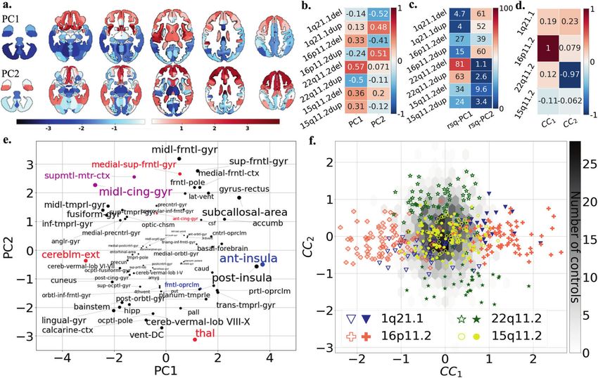
C. Modenato et al.
                  2
                         Neuroimaging studies have only been performed for a few                   Data quality check
                      CNVs. Robust effects on total and regional brain volumes, cortical           All data included in the analysis were quality checked by the same
                      thickness (CT) and surface area (SA), have been reported in                  researcher (CM). A total of 107 structural brain scans from carriers and
                      22q11.2 [12, 13, 16], 16p11.2 BP4-5 [17–19], and 15q11.2 CNVs                controls were excluded from further analysis based on visual inspection
                      [20–23]. Opposing effects on global and-or regional brain volumes            that identified significant artifacts compromising accurate tissue classifica-
                                                                                                   tion and boundary detection (Supplementary materials).
                      between deletions and duplications were observed for 16p11.2
                      [19], 22q11.2 [16], 1q21.1 [24] and 15q11.2 [20] loci (hereafter
                      referred to as “mirror effects”).                                            MRI data processing
                         Neuroanatomical alterations associated with 16p11.2 and                   Data for Voxel-Based Morphometry were preprocessed and analyzed with
                                                                                                   SPM12 (http://www.fil.ion.ucl.ac.uk/spm/software/spm12/) [33–35] running
                      22q11.2 show overlap with those observed in idiopathic ASD
                                                                                                   under     MATLAB       R2018b      (https://www.mathworks.com/products/
                      and SZ [17–19, 21, 25]. Finally, most of the effects are observed            new_products/release2018b.html). For surface-based feature extraction,
                      irrespective of psychiatric diagnoses and symptoms [12], suggest-            we used FreeSurfer 5.3.0 (http://surfer.nmr.mgh.harvard.edu [36,37,]).
                      ing that the final clinical outcome may result from the effect of             Quality control was performed using standardized ENIGMA quality control
                      CNVs and additional factors.                                                 procedures (http://enigma.ini.usc.edu/protocols/imaging-protocols/).
                         Neuroimaging studies across genomic variants are scarce. An
                      investigation of 49 unaffected carriers of SZ-associated CNVs                Statistical analysis for global brain measures
                      across five genomic loci in the UK biobank showed smaller                     Global brain aggregate measures (TIV, total gray matter (GM) volume, total
                      volumes of the thalamus, hippocampus, and nucleus accumbens                  SA, and mean CT) were adjusted for age, age2, and sex as fixed effects and
                      [26]. Functional connectivity similarities have also been demon-             scanning site as random factor. Non-clinically ascertained subjects from
                      strated between 16p11.2 and 22q11.2 deletions as well as with                the UKBB are on average 30 years older than the clinically ascertained
                      idiopathic ASD and SZ [27]. Alternatively, a recent study suggests a         subjects. Because of this age difference we used age matched control
                      relatively distinct association between neuroimaging alterations             groups for univariate analysis. Global measure z-scores for each CNV for
                      and six different CNVs [28].                                                 clinically and non-clinically ascertained CNVs were calculated using 331
                                                                                                   and 965 controls, respectively. All statistical analyses were performed in R,
                         In this study, we aimed to characterize shared and distinct
                                                                                                   version 3.4.4 (https://www.r-project.org/), or in MatlabR2018b.
                      neuroanatomical alterations associated with eight CNVs at four
1234567890();,:

                      genomic loci. We analyzed high-resolution structural brain scans
                      from the largest multi-site dataset of CNV carriers (n = 484, of             Voxel-based measures and statistical analyses
                      which 87 have not yet been published) and controls (n = 1296) to             We performed whole-brain voxel-based analysis testing for voxel-wise
                      date. Different approaches were implemented, from simple case-               volume differences within the mass-univariate analysis framework
                                                                                                   implemented in SPM (Supplementary Method 4). Cohen’s d (i.e., effect
                      control contrasts to one-view and multi-view multivariate pattern            size) [38] maps were obtained by converting SPM T-maps using the CAT12
                      learning [29, 30]. First, we compared brain morphometry features             toolbox for SPM (http://www.neuro.uni-jena.de/cat/).
                      associated with each deletion and duplication using univariate
                      linear models. Second, we quantified the shared variation of brain
                      morphometry associated with eight CNVs using principal compo-                Surface-based measures and statistical analyses
                                                                                                   In parallel to VBM, we used surface-based GLM-based analysis to test
                      nent analysis (PCA). To complement this single-view approach, a              differences in CT and SA (SurfStat toolbox [39]).
                      multi-view pattern-learning algorithm was carried out for the joint
                      analysis of genetic and morphometry brain data, to identify latent
                      ‘gene-morphometry dimensions’ (canonical correlation analysis,               Neuromorphometrics and Desikan parcellations
                                                                                                   Parcellation into regions of interest (ROIs) was performed using
                      CCA). Primary analyses were performed using VBM for consistency
                                                                                                   neuromorphometric atlas (http://www.neuromorphometrics.com/) for GM
                      with previous studies [19]. In addition, we carried out the same             volume (130 ROIs excluding white matter ROIs), and using Desikan
                      multivariate analyses using freesurfer-derived cortical SA and               parcellation [37] for FreeSurfer-derived CT and SA (68 ROIs).
                      thickness to ensure that shared variation was not limited to one
                      neuroimaging modality or analytical pipeline.
                                                                                                   Comparison of ranked Cohen’s d maps across CNVs
                                                                                                   To adjust for the unequal power to detect alterations across different CNV
                                                                                                   groups, which have different sample and effect sizes, we ranked the
                                                                                                   Cohen’s d values of all voxels (/vertices) for each statistical maps (CNV
                      METHODS                                                                      versus controls contrast). We then tested for spatial overlap between maps
                      Participants                                                                 across CNVs after thresholding the tails of the distribution at the 15th &
                      Deletions and duplications carriers’ neuroimaging data included in the       85th quantiles. Dice index was calculated using publicly available Matlab
                      study were selected on the following breakpoints (hg 19): 16p11.2 (BP4-5,    scripts (https://github.com/rordenlab/spmScripts).
                      29.6-30.2MB), 1q21.1 (Class I, 146.4-147.5MB & II, 145.3-147.5MB), 22q11.2
                      (BPA-D, 18.8-21.7MB) and 15q11.2 (BP1-2, 22.8–23.0MB), together with
                      control individuals not carrying any CNVs at these loci (Table 1,
                                                                                                   Null hypothesis testing using spin permutations and label
                      Supplementary Table 1 and supplementary materials). Signed consents          shuffling
                      were obtained from all participants or legal representatives prior to the    We used spin permutation and label shuffling [40, 41] to calculate
                      investigation. Of note, data of 87 CNV carriers have never been published.   empirical p values for (1) the deletion and duplication convergence pattern
                      Clinically ascertained CNV carriers were recruited as either probands        and (2) the correlation/dice index between two maps.
                      referred for genetic testing, or as relatives. Controls were either non-
                      carriers within the same families or individuals from the general            Quantifying shared variation across CNVs using principal
                      population. We pooled data from five cohorts. CNVs from non-clinical          components (PC)
                      populations were identified in the UK Biobank [31, 32].                       PCs were derived to quantify shared morphometry variation across CNVs.
                                                                                                   We used Cohen’s d values of 130 neuroanatomical GM regions
                                                                                                   (neuromorphometrics atlas) of eight CNVs as input-variables (z-scored
                                                                                                   Cohen’s d contrasts adjusted for total GM and nuisance variables;
                      MRI data                                                                     FactoMineR package in R). The variance explained (coefficient of
                      Details for methods and analyses are provided in Supplementary material      determination, R-squared) for each CNV-associated Cohen’s d map by
                      and Supplementary Methods 1–8. Data sample included T1-weighted              PCs was obtained by running a linear model (lm) in R; with PC1 and PC2 as
                      (T1w) images at 0.8–1 mm isotropic resolution across all sites. Population   independent explanatory variables and the CNV Cohen’s d map as a
                      description is available in Table 1 and Supplementary Table 1.               dependent variable.

                                                                                                                          Translational Psychiatry (2021)11:399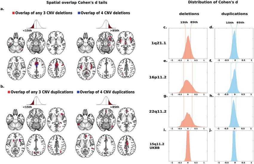
C. Modenato et al.
3
Table 1. Demographics.
Clinical ascertainment
CNV loci Copy number Age mean (SD) Male/Female TIV mean (SD) FSIQ mean (SD) ASD SCZ Other diagnosis
1q21.1 Deletions 29 (18) 11/18 1.22 (0.14) 90.85 (21.75) 1 – 7
N = 29 N = 26
Duplication 34 (17) 10/9 1.57 (0.11) 95.56 (23.19) 1 – 4
N = 19 N = 18
16p11.2 Deletions 17 (12) 47/36 1.54 (0.17) 82.17 (14.99 13 – 36
N = 83 N = 64
Duplication 31 (14.9) 41/32 1.33 (0.17) 85.47 (19.48) 10 1 19
N = 73 N = 63
22q11.2 Deletions 16 (8.6) 35/39 1.30 (0.15) 77.42 (13.51) 9 2 32
N = 74 N = 48
Duplication 20 (14.2) 15/7 1.47 (0.16) 97.83 (20.34) 2 – 8
N = 22 N = 12
Controls N = 331 26 (14.6) 189/142 1.46 (0.15) 106.73 (15.03) 1 – 23
N = 224
Non-clinical ascertainment
CNV loci Copy number Age mean (SD) Male/Female TIV mean (SD) UKB FI mean (SD) ASD SCZ Other diagnosis
1q21.1 Deletions 59.1 (6.7) 6/4 1.35(0.12) −0.8 (0.5) – 1* 3
N = 10 N=9
Duplication 60.6 (7) 2/7 1.55(0.14) 0.2 (1.3) – – –
N=9 N=9
15q11.2 Deletions 63.4 (7.6) 31/41 1.54(0.15) −0.3 (0.9) – – 2
N = 72 N = 63
Duplication 62.9 (7.3) 36/40 1.49(0.15) 0 (1.1) – – 6
N = 76 N = 71
16p11.2 Deletion 65.6 (3.2) 3/1 1.56(0.13) 0.8 (0.5) – – –
N=4 N=2
Duplication 69.3 (2.1) 1/3 1.29(0.11) −1.6 (0.2) – – –
N=4 N=4
22q11.2 Deletion 69.8 (–) 1/– 1.44(-) – – – –
N=1
Duplication 62 (9.5) 4/4 1.55(0.17) −0.2 (1.1) – – 1
N=8 N=8
Controls N = 965 62.1 (7.4) 358/607 1.51(0.14) 0 (1) – 2* 65
N = 866
CNV copy number variant, SD standard deviation, TIV total intracranial volume, FSIQ full scale IQ, UKB FI UK Biobank fluid intelligence, ASD autism spectrum
disorders, SCZ schizophrenia (including * ICD10 code F25.9 Schizoaffective disorder, unspecified).
CNV carriers and controls from the clinically ascertained group come from five different cohorts (Supplementary Table 1), while non-clinically ascertained
participants were identified in the UK Biobank. 16p11.2 and 22q11.2 from the UKBB were not included in the VBM and SBM due to small sample size. Other
diagnosis included: language disorder, major depressive disorder, posttraumatic stress disorder (PTSD), unspecified disruptive and impulse-control and
conduct disorder, social anxiety disorder, social phobia disorder, speech sound disorder, moderate intellectual disability, specific learning disorder, gambling
disorder, bipolar disorder, conduct disorder, attention deficit/hyperactivity disorder ADHD, Substance abuse disorder, global developmental delay, motor
disorder, obsessive compulsive disorder, sleep disorder, Tourette’s disorder, mood disorder, eating disorders, transient tic disorder, trichotillomania, pervasive
developmental disorder NOS, specific phobia, body dysmorphic disorder, mathematics disorder, dysthymic disorder.
Jointly modeling of gene-morphology dimensions using CCA of global effects differed across loci (Fig. 1a–c). Effects on GM and
We re-purposed CCA to simultaneously model the shared and distinct SA were less pronounced once adjusted for TIV (Supplementary
impact of the CNVs in causing distributed alterations in brain morpho- Fig. 1).
metry (130 grey matter regions) [29, 30]. This principled doubly-
multivariate approach, widely used in neuroimaging studies [29, 30], was
performed to identify modes of coherent co-variation that jointly Overlapping deletion effects on regional morphometry
characterize how CNVs and patterns of regional volumes systematically Whole-brain VBM analyses contrasting each deletion and duplica-
co-occur across subjects. Henceforth, we refer to the ensuing modes of co- tion group with controls showed mostly distinct brain patterns
variation as ‘CCA dimensions’ or ‘gene-morphology dimensions’. across CNVs (Fig. 2a, c, e, Supplementary Table 3). To investigate
potential overlap across the four genomic regions, we ranked
Cohen’s d maps and overlapped voxels with similar rankings.
RESULTS Using a threshold for voxels with Cohen’s d < 15th and >85th
CNV effects on global brain morphometry percentiles separately (Fig. 3c, e, g, i), we observed significant
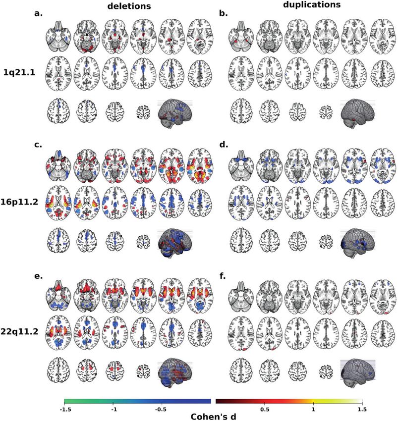
Deletions and duplications of each genomic loci showed opposing overlap between deletions (p valueSHUFFLE < 10e−4, Fig. 3a).
effects on one or more global metrics: TIV, total GM volume, total Volumes of the middle and anterior cingulate extending to the
SA, or mean CT (Fig. 1, Supplementary Table 2). The directionality supplementary motor cortex and of the cerebellum were
Translational Psychiatry (2021)11:399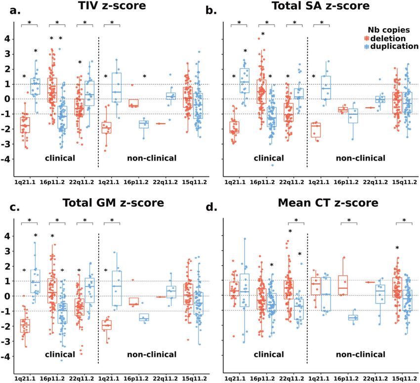
C. Modenato et al.
4

    Fig. 1 1q21.1, 16p11.2, 22q11.2, and 15q11.2 exert rich effects on global brain measures. Total intracranial volume (a), total surface area
    (b), total grey matter volume (c) and mean cortical thickness (d) for clinically and non-clinically ascertained CNVs. Z scores for clinically and
    non-clinically ascertained CNVs were calculated using 331 and 965 controls, respectively, adjusting for age, age2, sex and site as a random
    factor. Y axis values are z scores. X axis are CNV groups. Significant difference between CNV group and corresponding control group is
    indicated with a star. Horizontal bars with stars show significant differences between deletions and duplications within the same locus. TIV
    total intracranial volume, SA surface area, GM grey matter, CT cortical thickness.

    decreased in all deletions while volume was increased in the               Cohen’s d values 85th percentiles (Fig. 3d, f, h, j)
    thalamus (Fig. 3a).                                                        demonstrated spatial overlap across all four duplications (p
       Sensitivity analyses tested the effect of ascertainment and             valueSHUFFLE < 10e−4, Fig. 3b). The resulting pattern was mainly
    control groups: (1) We recomputed the deletion convergence map             distinct from the one observed in deletions and was characterized
    using 1q21.1 deletion carriers from UK Biobank instead of those            by smaller volumes in anterior insula and frontal operculum, and
    clinically ascertained (Table 1). The new deletion convergence             larger volumes in the middle cingulate gyrus and supplementary
    map was similar to the initial one presented above with a dice             motor cortex compared to controls.
    index of 39.4% (p valueSPIN < 10e−4); (2) We excluded all subjects            Sensitivity analysis testing the effect of clinical ascertainment,
    with autism, SZ, or other psychiatric diagnoses. Again, this did not       psychiatric diagnoses, control groups, and volume versus
    change the overlap (Supplementary Fig. 2); (3) We tested the               Freesurfer-derived measures demonstrated that results were
    effects of the control group by recomputing contrasts only using           robust (Supplementary Figs. 2–8).
    controls from the same site (instead of the initial ANOVA pooling             The deletion/duplication ratio of Cohen’s d distributions ranged
    all controls together and controlling for site). This again did not        from 1.24 to twofold across the four genomic loci (F-test, p <
    alter the convergence maps (Supplementary Fig. 3). Finally, we             10e−16, Fig. 3c–j, Supplementary Table 7). Similar effect-size ratios
    performed the same analysis using Freesurfer-derived SA and CT             were also observed for SA alterations (Supplementary Table 7),
    measures. We also identified spatial overlaps but regions                   except for the 15q11.2 locus.
    identified were different especially for CT (Supplementary Table               We tested opposing (mirror) effects on VBM contrast maps
    4 & Fig. 4). Overlap maps are provided in Supplementary Figs. 5–8          between deletion and duplications. The strongest anticorrelation
    and Tables 5, 6.                                                           of Cohen’s d values was observed for 16p11.2 (p valueSPIN < 10e
                                                                               −4) followed by 15q11.2 (p valueSPIN < 10e−4), 1q21.1 (p valueSPIN
    Overlapping duplication effects on regional morphometry                    < 0.033) and 22q11.2 (p valueSPIN < 0.038) (Supplementary Fig. 9
    Contrasts computed for duplications (Fig. 2b, d, f) showed smaller         and Tables 8–10). Mirror effects were observed in clinically and
    effect sizes compared to deletions. The same analysis using                non-clinically ascertained CNV carriers, as well as for SA at all four

                                                                                                    Translational Psychiatry (2021)11:399
C. Modenato et al.
5
Fig. 2 Cohen’s d maps of VBM regional brain differences in deletion and duplication carriers at the 1q21.1, 16p11.2, and 22q11.2 loci
compared to controls. Regional brain differences adjusted for total grey matter volume. Left and right columns show results for deletions (a,
c, e) and duplication (b, d, f) carriers, respectively. Color maps show the significant effects of each CNV, thresholded at q < 0.05 FWE. Color scale
represents positive and negative Cohen’s d effect sizes were estimated. The linear model was adjusted for sex, linear, and quadratic expansion
of age and total grey matter volume. 15q11.2 was not displayed because only a few voxels survived family-wise error (FWE) correction.
Corresponding maps for surface area and cortical thickness are reported in Supplementary Figs. 4 and 5.
genomic loci but not for CT (Supplementary Tables 8–10). Hence posterior insula, cerebellum, fusiform gyrus and thalamus were
mirror effects were observed in global metrics and, independently, also top regions altered across subsets of CNVs (Fig. 4a, b and
in regional alterations. Supplementary Table 12). The variance explained by both
components for each CNV’s Cohen’s d map ranged from 27% to
Quantifying distinct and shared effects on brain morphometry 82% (Fig. 4d). Finally, we performed the same analysis using
associated with eight CNVs Freesurfer-derived SA and CT measures which also provided latent
We performed a multivariate PCA based on Cohen’s d profiles dimensions with comparable variance explained, opposing load-
obtained from contrasts between the eight CNV groups and ings for deletions and duplications of each genomic loci
controls (using 130 neuromorphometric regional volumes, Sup- (Supplementary Fig. 10). However, CNV loadings differ across
plementary Table 11). The first two components explained 31.8 brain morphometry metrics.
and 28.7% of the variance of Cohen’s d maps, respectively. The
third component dropped to 13.8% and was therefore not Gene-morphology dimensions across eight CNVs
investigated further. As a next step, we performed a multi-view pattern-learning
Deletions and duplications at each genomic loci showed analysis, jointly analyzing the genetic and morphometry brain
opposite loading on PC1 or PC2 (Fig. 4c). Regions with the data. This doubly multivariate method allowed testing whether
highest loadings on PC1 and PC2 were also those identified in the shared dimensions could be identified in a data-driven approach,
convergence maps presented above: in particular the middle without performing any individual contrast. We interrogated 2
cingulate gyrus and the supplementary motor cortex. Anterior and hypotheses: (1) CNVs show levels of shared brain effects at the
Translational Psychiatry (2021)11:399C. Modenato et al.
6
Fig. 3 Spatial overlap across deletions and duplications at four genomic loci. Spatial overlap across clinically and non-clinically ascertained
deletions (a) and duplications (b) at four genomic loci shown separately for 85th percentile of Cohen’s d values. Overlap of all four
deletions (a) or all four duplications (b) is shown in blue. Overlaps of any combination of three deletions (a) or any combination of three
duplications (b) are shown in red. Top ranking Cohen’s d values used in (a, b) are presented on the density plots for all eight deletions and
duplications: 1q21.1 (c, d), 16p11.2 (e, f), 22q11.2 (g, h), 15q11.2 (i, j). The x axes values of the eight density plots are Cohen’s d. Corresponding
maps for surface area and cortical thickness are reported in Supplementary Figs. 6 and 7.
morphometry level and (2) deletions and duplications show variance. A second multivariate approach (CCA), jointly analyzing
opposing effects. We investigated the same 130 regional volumes genetic and morphometry data, confirmed the latent CNV-brain
in 484 carriers of CNVs at four genomic loci. To test hypothesis (2), dimensions identified by PCA. Genomic loci contributed to the
deletions and duplications were coded as opposing gene dosage. latent CCA dimensions in proportion to their effect sizes. Even for
CCA confirmed both hypotheses by identifying two significant small effect-size deletions at the 1q21.1 and 15q11.2 loci, the PCA
‘gene-morphometry dimensions’ (r = 0.84, 0.79, p value < 0.05, Fig. components explained between 43 and 65% of their Cohen’s d
4e, f). Regional brain contributions to canonical dimension 1 and 2 profile. All three approaches—spatial overlap, CCA, and PCA—
were well correlated with those of PC2 and 1, respectively (r = identified a similar set of regions altered by CNVs including the
0.83, r = −0.81). cingulate gyrus and supplementary motor cortex.
Top ranking brain regions contributing to either of the two CCA
dimensions of morphological variation included supplementary Distinct and shared effects of CNVs
motor cortex, posterior and anterior insula, middle cingulate Our results show that two-thirds of the average CNV effects on
gyrus, calcarine cortex, cuneus and accumbens (Supplementary brain morphometry are distinct. This is consistent with a recent
Fig. 11 and Supplementary Table 13). 16p11.2 and 22q11.2 study showing relative specificity of association between brain
preferentially contributed to dimension 1 and 2 respectively, and patterns of gene expression and patterns of cortical anatomy
1q21.1 loaded similarly on both dimensions. 15q11.2 CNVs changes across six CNVs and chromosomal aneuploidies [28]. One-
showed the smallest loadings on both dimensions (Fig. 4e). third of the effects on brain morphometry is shared as
Sensitivity analyses are detailed in supplementary material demonstrated by latent gene-morphology dimensions identified
(Supplementary Figs. 12–16 and Tables 14, 15). across subsets of CNVs. There is no single dimension explaining
CNV effects. Instead, subsets of CNVs load on either dimension,
which may suggest similar brain mechanisms within subgroups of
DISCUSSION CNV. Yet CNVs within subgroups were not characterized by the
Here, in the largest cross-CNV-neuroimaging study to date, we same risk for ASD or SCZ.
tested potentially shared effects of eight neuropsychiatric CNVs on These results have implications for our conceptualization of
brain morphometry. CNVs showed a combination of distinct and polygenic psychiatric conditions. Indeed, studies estimate that
shared profiles of brain alterations, as demonstrated by the spatial 70–100% of any 1-MB window in the human genome encom-
overlap of Cohen’s d maps across deletions and duplications. A passes variants (including CNVs) contributing to increased risk for
multivariate approach (PCA) quantified distinct and shared SZ and autism [4, 42]. Gene-morphology dimensions alone, can
alterations across subsets of CNVs and identified two latent not explain the fact that subgroups of CNVs are associated with a
dimensions explaining 31.8 and 28.7% of Cohen’s d map’s similar range of behavioral symptoms [43], and psychiatric
Translational Psychiatry (2021)11:399C. Modenato et al.
7
Fig. 4 Principal component analysis and canonical correlation analysis of brain alterations due to eight CNVs. a PCA dimension 1 and 2
regional relevances projected on axial brain slices. The darker the red or blue color, the stronger the positive or negative association with the
PCA dimensions. PCA was run on z-scored Cohen’s d values, with the eight CNVs as variables and 130 neuroanatomical GM regions as
observations. GM region volumes were adjusted for total grey matter, age, age2, sex, and site. The first two components explained respectively
31.77 and 28.66 % of the variance. b Loading of eight CNVs on the two PCA dimensions. Values are PC loading magnitudes and represent the
contribution of a CNV to the PC. c Variance explained (coefficient of determination, R-squared) of each CNV Cohen’s d profile by PC1 and PC2.
Values and color scale represent the “percent of variance”. d Loadings of the first and second CCA dimension on four CNV genomic loci. Shows
contribution of a CNV loci to the canonical dimension. e Loading of Neuromorphometrics Regions of Interests (ROIs) on the two PCA
dimensions. ROIs are averaged across the left and right hemisphere for visualization. The font size is correlated to the region’s contribution to
dimensions. ROI names are color coded as being part of the deletion (red), duplication (blue) and both deletion and duplication (magenta)
convergence patterns. f Scatterplot showing the participant/specific expressions of each of the 484 carriers of eight different CNVs along two
dominant gene-morphometry canonical correlation (CC) dimensions established using 130 neuroanatomical GM regions of CNV carriers. GM
region volumes were adjusted for total grey matter, age, age2, sex, and site. The empty and full symbols represent deletions and duplication,
respectively. The grey hexagonal bin plot represents the frequency of controls (n = 1296). Controls were not used to calculate the CCA and
were projected post hoc on the two dimensions using CCA prediction. CCA ROI loadings are reported in Supplementary Fig. 10. Results for
surface area and cortical thickness are reported in Supplementary Fig. 9 (PCA), 14–15 (CCA).
disorders [1, 2, 4, 44]. In fact, the large proportion of distinct CNV- effects with phenotypic traits. Alterations of the cingulate cortex
neuroimaging effects suggests that a broad diversity of brain have been associated with genetic and environmental risk for SZ
mechanisms increase the risk for autism and SZ. Extreme [48]. The supplementary motor cortex has been shown to play a
examples include CNVs associated with opposing loadings on critical role in 16p11.2, 22q11.2 CNVs as well as autism and SZ by
the same latent gene-morphology dimension while increasing risk functional connectivity studies, but not by cross-diagnostic
for the same psychiatric condition (ie. 16p11.2 deletions, duplica- neuroimaging structural studies [49, 50]. Several cerebellar regions
tions, and autism). The presence of such genomic variants in (vermis lobule VIII-X and cerebellar cortex) are highly sensitive to
studies of ASD and SZ may explain heterogeneity and small CNVs, which may be due to the cerebellum’s protracted
neuroimaging effect sizes [45, 46]. Why opposing effects on the development [51]. The cerebellum has either been excluded or
same latent brain dimension increase risk for the same psychiatric not reported by cross-disorder structural neuroimaging studies,
condition is an unsolved question. Further observations on a but volume alterations have been associated with autism and SZ
broad variety of genomic variants are required to address this separately [52, 53]. Multiple genetic mouse models of autism, as
question. well as Down Syndrome, also show abnormal cerebellar develop-
ment [54]. The same level of spatial overlap was observed for SA
Brain hubs vulnerable to altered gene dosage and CT but implicated mostly distinct sets of brain regions. This is
Insula, cingulate, fusiform gyrus, and hippocampus are regions in line with the distinct genetic contributions previously demon-
showing alterations across SZ, bipolar disorders, major depression, strated for these cortical metrics [55].
and obsessive-compulsive disorders [45, 47]. The cingulate, insula,
and fusiform gyrus were also among regions markedly altered Dissociation between global and regional effects
across eight CNVs. CNVs have either negative or positive effects on Results suggest that global and local effects may be mechan-
these brain regions, however, the number of CNVs included in this istically unrelated. 1q21.1 deletions and duplications highlight the
study did not allow us to associate the directionality of these contrast between very large effects on global measures, with small
Translational Psychiatry (2021)11:399C. Modenato et al.
8
regional effects once adjusted for total GM. Dissociation is also 10. Zufferey F, et al. A 600 kb deletion syndrome at 16p11.2 leads to energy
observed between the directionalities of global and regional imbalance and neuropsychiatric disorders. J Med Genet. 2012;49:660–8.
effects: all deletions are associated with a smaller cingulate and 11. D’Angelo D, et al. Defining the effect of the 16p11.2 duplication on cognition,
supplementary motor cortex volume irrespective of their effect on behavior, and medical comorbidities. JAMA Psychiatry. 2016;73:20–30.
12. Sun D, et al. Large-scale mapping of cortical alterations in 22q11.2 deletion
TIV and GM. Animal studies have proposed mechanisms for global
syndrome: Convergence with idiopathic psychosis and effects of deletion size.
[8, 56], but not regional effects of CNVs. Mol Psychiatry. 2018:1–13. https://doi.org/10.1038/s41380-018-0078-5.
13. Niarchou M, et al. Psychiatric disorders in children with 16p11.2 deletion and
Limitations duplication. Transl Psychiatry. 2019;9:1–8.
Multiple sites included in the study may have introduced noise, 14. Moberg PJ, et al. Neurocognitive functioning in patients with 22q11.2 deletion
but previous studies have shown that site effects do not influence syndrome: a meta-analytic review. Behav Genet. 2018;48:259–70.
the neuroanatomical patterns associated with CNVs at the 15. Gur RE, et al. Neurocognitive development in 22q11.2 deletion syndrome:
16p11.2, 22q11.2, and 15q11.2 loci [12, 19, 23]. While shared comparison with youth having developmental delay and medical comorbidities.
variation could have been influenced by clinical ascertainment or Mol Psychiatry. 2014;19:1205–11.
16. Lin A, et al. Mapping 22q11.2 gene dosage effects on brain morphometry. J
psychiatric diagnoses, our sensitivity analyses showed that this is
Neurosci. 2017;37:6183–99.
not the case. The effect of medication on CNVs brain alterations 17. Maillard AM, et al. The 16p11.2 locus modulates brain structures common to
could not be investigated in the current study as medication autism, schizophrenia and obesity. Mol Psychiatry. 2015;20:140–7.
information was not available for the whole dataset. We were 18. Qureshi AY, et al. Opposing brain differences in 16p11.2 deletion and duplication
underpowered to properly investigate potential sex-related effects carriers. J. Neurosci. 2014;34:11199–211.
of 1q21.1 and 15q11.2 on brain morphometry. Of note, previous 19. Martin-Brevet S, et al. Quantifying the effects of 16p11.2 copy number variants on
neuroimaging studies of large 22q11.2 and 16p11.2 samples were brain structure: a multisite genetic-first study. Biol Psychiatry. 2018;84:253–64.
unable to identify any sex-related effects [19, 25]. 20. Stefansson H, et al. CNVs conferring risk of autism or schizophrenia affect cog-
15q11.2 deletions and duplications have small effect sizes and nition in controls. Nature. 2014;505:361–6.
21. Silva AI, et al. Reciprocal White Matter Changes Associated With Copy Number
larger samples would improve the accuracy of the brain
Variation at 15q11.2 BP1-BP2: A Diffusion Tensor Imaging Study. Biological Psy-
morphometry signature. Systematic analysis through the two chiatry. 2019;85:563–72.
most widespread computational neuroanatomy frameworks 22. Ulfarsson MO, et al. 15q11.2 CNV affects cognitive, structural and functional
(voxel-based and surface-based) shows that effects could not be correlates of dyslexia and dyscalculia. Transl Psychiatry. 2017;7:e1109.
attributed to the processing pipeline. Extending our approach to 23. van der Meer D, et al. Association of copy number variation of the 15q11.2 BP1-
the rapidly expanding number of rare genomic variants associated BP2 Region With Cortical and Subcortical Morphology and Cognition. JAMA
with psychiatric disorders is required to draw a robust conclusion Psychiatry. 2019:1–11. https://doi.org/10.1001/jamapsychiatry.2019.3779.
on the distinct and shared effects of CNVs on brain structure. 24. Sønderby IE, et al. 1q21.1 distal copy number variants are associated with cer-
ebral and cognitive alterations in humans. Transl Psychiatry. 2021;11:1–16.
25. Sun D, et al. Large-scale mapping of cortical alterations in 22q11.2 deletion
syndrome: Convergence with idiopathic psychosis and effects of deletion size.
CONCLUSIONS Mol Psychiatry. 2018:1–13. https://doi.org/10.1038/s41380-018-0078-5.
The simultaneous analyses and comparisons of several genomic 26. Warland A, Kendall KM, Rees E, Kirov G, Caseras X. Schizophrenia-associated
variants demonstrate distinct CNV-associated alteration profiles as genomic copy number variants and subcortical brain volumes in the UK Biobank.
well as shared latent gene-morphology dimensions relevant to Mol Psychiatry. 2020;25:854–62.
subsets of CNVs. Large proportions of distinct effects may provide 27. Moreau CA, et al. Mutations associated with neuropsychiatric conditions
some answers to the small neuroimaging effect sizes reported in delineate functional brain connectivity dimensions contributing to autism and
idiopathic psychiatric conditions. The mechanisms underlying the schizophrenia. Nat Commun. 2020;11:5272.
identified latent dimensions remain unknown and pathway 28. Seidlitz J, et al. Transcriptomic and cellular decoding of regional brain vulner-
ability to neurogenetic disorders. Nat Commun. 2020;11:3358.
convergence may occur early on at the transcriptome and protein
29. Smith SM, et al. A positive-negative mode of population covariation links brain
level, or at later stages (i.e., brain architecture or behavior). The connectivity, demographics and behavior. Nat Neurosci. 2015;18:1565–7.
hotly debated omnigenic model postulates that convergence may 30. Wang H-T, et al. Finding the needle in high-dimensional haystack: a tutorial on
occur at early stages due to highly interconnected cell regulatory canonical correlation analysis. NeuroImage. 2020.
networks [57]. These approaches may help subgroup genomic 31. Miller KL, et al. Multimodal population brain imaging in the UK Biobank pro-
variants based on their morphometry signature and dissect the spective epidemiological study. Nat Neurosci. 2016;19:1523–36.
heterogeneity of psychiatric conditions. 32. Sudlow C, et al. UK biobank: an open access resource for identifying the causes of
a wide range of complex diseases of middle and old age. PLoS Med. 2015;12:
e1001779.
REFERENCES 33. Ashburner J, Friston KJ. Unified segmentation. NeuroImage. 2005;26:839–51.
34. Lorio S, et al. New tissue priors for improved automated classification of sub-
1. Sanders SJ, et al. Insights into autism spectrum disorder genomic architecture
cortical brain structures on MRI. Neuroimage. 2016;130:157–66.
and biology from 71 risk loci. Neuron. 2015;87:1215–33.
35. Ashburner J. A fast diffeomorphic image registration algorithm. NeuroImage.
2. Marshall CR, et al. Contribution of copy number variants to schizophrenia from a
2007;38:95–113.
genome-wide study of 41,321 subjects. Nat Genet. 2017;49:27–35.
36. Fischl B, Sereno MI, Dale AM. Cortical surface-based analysis: II: inflation,
3. Chawner SJRA, et al. A genetics-first approach to dissecting the heterogeneity of
flattening, and a surface-based coordinate system. NeuroImage.
autism: phenotypic comparison of autism risk copy number variants. AJP.
1999;9:195–207.
2021;178:77–86.
37. Desikan RS, et al. An automated labeling system for subdividing the human
4. Douard E, et al. Effect sizes of deletions and duplications on autism risk across the
cerebral cortex on MRI scans into gyral based regions of interest. NeuroImage.
genome. AJP. 2020;178:87–98.
2006;31:968–80.
5. Moreno-De-Luca D, et al. Using large clinical data sets to infer pathogenicity for
38. Cohen J. Statistical power analysis for the behavioral sciences. Psychology Press;
rare copy number variants in autism cohorts. Mol Psychiatry. 2013;18:1090–5.
1988.
6. Crawford K, et al. Medical consequences of pathogenic CNVs in adults: analysis of
39. Worsley KJ, et al. SurfStat: a matlab toolbox for the statistical analysis of univariate
the UK Biobank. J Med Genet. 2019;56:131–8.
and multivariate surface and volumetric data using linear mixed effects models
7. Mefford HC, et al. Recurrent rearrangements of chromosome 1q21.1 and variable
and random field theory. Neuroimage. 2009. https://doi.org/10.1016/S1053-8119
pediatric phenotypes. N Engl J Med. 2008;359:1685–99.
(09)70882-1.
8. Golzio C, et al. KCTD13 is a major driver of mirrored neuroanatomical phenotypes
40. Alexander-Bloch A, et al. On testing for spatial correspondence between maps of
associated with the 16p11.2 CNV. Nature. 2012;485:363–7.
human brain structure and function. Neuroimage. 2018;178:540–51.
9. Jonas RK, Montojo CA, Bearden CE. The 22q11.2 deletion syndrome as a window
41. Reardon PK, et al. Normative brain size variation and brain shape diversity in
into complex neuropsychiatric disorders over the lifespan. Biol Psychiatry.
humans. Science. 2018;360:1222–7.
2014;75:351–60.
Translational Psychiatry (2021)11:399C. Modenato et al.
9
42. Loh P-R, et al. Contrasting genetic architectures of schizophrenia and other Excellence Fund, Healthy Brains for Healthy Lives through the Canada First Research
complex diseases using fast variance-components analysis. Nat Genet. Excellence Fund. JS is a recipient of a Canada Research Chair in neurodevelopmental
2015;47:1385–92. disorders, and a chair from the Jeanne et Jean Louis Levesque Foundation. The
43. Huguet G, et al. Measuring and estimating the effect sizes of copy number Cardiff CNV cohort was supported by the Wellcome Trust Strategic Award “DEFINE”
variants on general intelligence in community-based samples. JAMA Psychiatry. and the National Centre for Mental Health with funds from Health and Care Research
2018;75:447–57. Wales (code 100202/Z/12/Z). The CHUV cohort was supported by the SNF (Maillard
44. Chawner SJRA, et al. Genotype–phenotype associations in children with copy Anne, Project, PMPDP3 171331). Data from the UCLA cohort provided by CEB
number variants associated with high neuropsychiatric risk in the UK (IMAGINE- (participants with 22q11.2 deletions or duplications and controls) was supported
ID): a case-control cohort study. The Lancet Psychiatry. 2019;6:493–505. through grants from the NIH (U54EB020403), NIMH (R01MH085953, R01MH100900,
45. Opel N, et al. Cross-disorder analysis of brain structural abnormalities in six major R03MH105808), and the Simons Foundation (SFARI Explorer Award). CMod was
psychiatric disorders: a secondary analysis of mega- and meta-analytical findings supported by the doc.mobility grant provided by the Swiss National Science
from the ENIGMA Consortium. Biol Psychiatry. 2020. https://doi.org/10.1016/j. Foundation (SNSF). KK was supported by The Institute of Data Valorization (IVADO)
biopsych.2020.04.027. Postdoctoral Fellowship program, through the Canada First Research Excellence
46. Bedford SA, et al. Large-scale analyses of the relationship between sex, age and Fund. DB was supported by the Healthy Brains Healthy Lives initiative (Canada First
intelligence quotient heterogeneity and cortical morphometry in autism spec- Research Excellence fund), and by the CIFAR Artificial Intelligence Chairs program
trum disorder. Mol Psychiatry. 2020;25:614–28. (Canada Institute for Advanced Research). BD is supported by the Swiss National
47. Goodkind M, et al. Identification of a common neurobiological substrate for Science Foundation (NCCR Synapsy, project grant numbers 32003B_135679,
mental illness. JAMA Psychiatry. 2015;72:305–15. 32003B_159780, 324730_192755 and CRSK-3_190185), the Roger De Spoelberch
48. Tost H, Champagne FA, Meyer-Lindenberg A. Environmental influence in the and the Leenaards Foundations. We thank all of the families participating at the
brain, human welfare and mental health. Nat Neurosci. 2015;18:1421–31. Simons Searchlight sites, as well as the Simons Searchlight Consortium. We
49. Moreau C, et al. Neuropsychiatric mutations delineate functional brain con- appreciate obtaining access to imaging and phenotypic data on SFARI Base.
nectivity dimensions contributing to autism and schizophrenia. 2019. https://doi. Approved researchers can obtain the Simons Searchlight population dataset
org/10.1101/862615. described in this study by applying at https://base.sfari.org. We are grateful to all
50. Kebets V, et al. Somatosensory-motor dysconnectivity spans multiple transdiag- families who participated in the 16p11.2 European Consortium.
nostic dimensions of psychopathology. Biol Psychiatry. 2019;86:779–91.
51. Sathyanesan A, et al. Emerging connections between cerebellar development,
behavior, and complex brain disorders. Nat Rev Neurosci. 2019;20:298–313. COMPETING INTERESTS
52. Moberget T, et al. Cerebellar volume and cerebellocerebral structural covariance MBMVdB reports grants from Takeda Pharmaceuticals, outside the submitted work.
in schizophrenia: a multisite mega-analysis of 983 patients and 1349 healthy All other authors reported no biomedical financial interests or potential conflicts of
controls. Mol Psychiatry. 2018;23:1512–20. interest.
53. Traut N, et al. Cerebellar volume in autism: literature meta-analysis and analysis of
the autism brain imaging data exchange cohort. Biol. Psychiatry. 2018;83:579–88.
54. Ellegood J, et al. Clustering autism: using neuroanatomical differences in 26
ADDITIONAL INFORMATION
mouse models to gain insight into the heterogeneity. Mol Psychiatry.
2015;20:118–25. Supplementary information The online version contains supplementary material
55. Grasby KL. The genetic architecture of the human cerebral cortex. Science. available at https://doi.org/10.1038/s41398-021-01490-9.
2020;367:6484.
56. Richter M, et al. Altered TAOK2 activity causes autism-related neurodevelop- Correspondence and requests for materials should be addressed to S.J.
mental and cognitive abnormalities through RhoA signaling. Mol Psychiatry.
2019;24:1329–50. Reprints and permission information is available at http://www.nature.com/
57. Boyle EA, Li YI, Pritchard JK. An expanded view of complex traits: from polygenic reprints
to omnigenic. Cell. 2017;169:1177–86.
Publisher’s note Springer Nature remains neutral with regard to jurisdictional claims
in published maps and institutional affiliations.
AUTHOR CONTRIBUTIONS
CMod, KK, BD, and SJ designed the study, analyzed imaging data, and drafted the
manuscript. CMod and KK did all the preprocessing and analysis of neuroimaging
data, DB provided scripts and mentored the CCA analysis. CMor, CEB, and DB Open Access This article is licensed under a Creative Commons
contributed in result interpretation and in the editing of the manuscript. CMod, AM, Attribution 4.0 International License, which permits use, sharing,
AP, SR, and SM-B recruited and scanned participants in the 16p11.2 European adaptation, distribution and reproduction in any medium or format, as long as you give
Consortium. SL, COM, NY, PT, ED, FT-D, VC, ARC, FD recruited and scanned appropriate credit to the original author(s) and the source, provide a link to the Creative
participants in the Brain Canada cohort. LK collected and provided the data for the Commons license, and indicate if changes were made. The images or other third party
UCLA cohort. DEJL, MJO, MBMVdB, JH, and AIS provided the data for the Cardiff material in this article are included in the article’s Creative Commons license, unless
cohort. All authors provided feedback on the manuscript. indicated otherwise in a credit line to the material. If material is not included in the
article’s Creative Commons license and your intended use is not permitted by statutory
regulation or exceeds the permitted use, you will need to obtain permission directly
FUNDING from the copyright holder. To view a copy of this license, visit http://creativecommons.
This research was supported by Calcul Quebec (http://www.calculquebec.ca) and org/licenses/by/4.0/.
Compute Canada (http://www.computecanada.ca), the Brain Canada Multi-
Investigator initiative, the Canadian Institutes of Health Research, CIHR_400528,
The Institute of Data Valorization (IVADO) through the Canada First Research © The Author(s) 2021
16P11.2 EUROPEAN CONSORTIUM
Marie-Claude Addor14, Joris Andrieux15, Benoît Arveiler16, Geneviève Baujat17, Frédérique Sloan-Béna18, Marco Belfiore19, Dominique
Bonneau20, Sonia Bouquillon21, Odile Boute22, Alfredo Brusco23, Tiffany Busa24, Jean- Hubert Caberg25, Dominique Campion26, Vanessa
Colombert27, Marie-Pierre Cordier28, Albert David29, François-Guillaume Debray30, Marie-Ange Delrue31, Martine Doco-Fenzy32, Ulrike
Dunkhase-Heinl33, Patrick Edery34, Christina Fagerberg35, Laurence Faivre36, Francesca Forzano37,38, David Genevieve39, Marion
Gérard40, Daniela Giachino41, Agnès Guichet42, Olivier Guillin43, Delphine Héron44, Bertrand Isidor45, Aurélia Jacquette46, Sylvie
Jaillard47, Hubert Journel48, Boris Keren49, Didier Lacombe50, Sébastien Lebon51, Cédric Le Caignec52, Marie-Pierre Lemaître53, James
Lespinasse54, Michèle Mathieu-Dramart55, Sandra Mercier56, Cyril Mignot57, Chantal Missirian58, Florence Petit59, Kristina Pilekær
Sørensen60, Lucile Pinson61, Ghislaine Plessis62, Fabienne Prieur63, Alexandre Raymond64, Caroline Rooryck-Thambo65, Massimiliano
Translational Psychiatry (2021)11:399C. Modenato et al.
10
Rossi66, Damien Sanlaville67, Britta Schlott Kristiansen68, Caroline Schluth-Bolard69, Marianne Till70, Mieke Van Haelst71 and Lionel Van
Maldergem72
14
Service de génétique médicale, Centre Hospitalier Universitaire Vaudois, Lausanne University, Lausanne, Switzerland. 15Institut de Génétique Médicale, CHRU de Lille, Hopital
Jeanne de Flandre, Lille, France. 16Service de génétique médicale, CHU de Bordeaux- GH Pellegrin, Bordeaux, France. 17Service de Génétique Médicale, CHU Paris - Hôpital
Necker-Enfants Malades, Paris, France. 18Service de médecine génétique, Hôpitaux Universitaires de Genève – HUG, Geneva, Switzerland. 19Service de génétique médicale, Centre
Hospitalier Universitaire Vaudois, Lausanne University, Lausanne, Switzerland. 20Service de génétique médicale, CHU d’Angers, Angers, France. 21Institut de Génétique Médicale,
Hopital Jeanne de Flandre, Lille, France. 22Hôpital Jeanne de Flandre, CHRU de Lille, Lille, France. 23Genetica Medica, Dipartimento di Scienze Mediche, Università di Torino,
Torino, Italy. 24Département de génétique médicale, CHU de Marseille, Hôpital de la Timone, Marseille, France. 25Centre de génétique humaine, CHU de Liège, Liège, Belgique.
26
Service de psychiatrie, Centre hospitalier de Rouvray, Sotteville lès Rouen, France. 27Service de génétique médicale, Centre Hospitalier Bretagne Atlantique CH Chubert, Vannes,
France. 28Service de génétique clinique, CHU de Lyon, Hospices Civils de Lyon, Lyon, France. 29Service de Génétique Médicale, CHU de Nantes, Hôtel Dieu, Paris, France. 30Service
de Génétique Humaine, CHU Sart Tilman, Liège, Belgique. 31Service de génétique médicale, CHU de Bordeaux, Hôpital Pellegrin, Bordeaux, France. 32Service de Génétique et
Biologie de la Reproduction, CHU de Reims, Hôpital Maison Blanche, Reims, France. 33Department of Pediatrics, Aabenraa Hospital, Sonderjylland, Denmark. 34Service de
génétique clinique, CHU de Lyon, Hospices Civils de Lyon, Lyon, France. 35Department of Clinical Genetics, Odense University hospital, Odense, Denmark. 36Centre de génétique,
Hôpital d’Enfants, CHU Dijon Bourgogne - Hôpital François Mitterrand, Dijon, France. 37Ambulatorio di Genetica Medica, Ospedali Galliera di Genova, Genoa, Italy. 38Clinical
Genetics Department, 7th Floor Borough Wing, Guy’s Hospital, Guy’s & St Thomas’ NHS Foundation Trust, Great Maze Pond, London SE1 9RT, UK. 39Département de Génétique
Médicale, Maladies Rares et Médecine Personnalisée, service de génétique clinique, Université Montpellier, Unité Inserm U1183, CHU Montpellier, Montpellier, France. 40Service
de Génétique, CHU de Caen, Hôpital Clémenceau, Caen, France. 41Genetica Medica, Dipartimento di Scienze Cliniche e Biologiche, Università di Torino, Torino, Italy. 42Service de
génétique, CHU d’Angers, Angers, France. 43Service de psychiatrie, Centre hospitalier du Rouvray, Sotteville lès Rouen, France. 44Service de Génétique clinique, CHU Paris-GH La
Pitié Salpêtrière-Charles Foix - Hôpital Pitié Salpêtrière, Paris, France. 45Service de Génétique Médicale, CHU de Nantes, Hôtel Dieu, Paris, France. 46Service de Génétique clinique,
CHU Paris-GH La Pitié Salpêtrière-Charles Foix - Hôpital Pitié-Salpêtrière, Foix, France. 47Service de Génétique Moléculaire et Génomique – Pôle biologie, CHU de Rennes, Hôpital
Pontchaillou, Rennes, France. 48Service de génétique médicale, Centre Hospitalier Bretagne Atlantique CH Chubert, Vannes, France. 49Centre de Génétique Moléculaire et
Chromosomique, CHU Paris-GH La Pitié Salpêtrière-Charles Foix - Hôpital Pitié-Salpêtrière, Paris, France. 50Service de génétique médicale, CHU de Bordeaux-GH Pellegrin,
Bordeaux, France. 51Pediatric Neurology Unit, Department of Pediatrics, Lausanne University Hospital, Lausanne, Switzerland. 52Service de Génétique Médicale - Institut de
Biologie, CHU de Nantes, Nantes, France. 53Service de Neuropédiatrie, Centre Hospitalier Régional Universitaire de Lille, Lille, France. 54Service génétique médicale et
oncogénétique, Hotel Dieu, Chambéry, France. 55Service de Génétique Clinique, CHU Amiens Picardie, Amiens, France. 56Service de Génétique Médicale, CHU de Nantes, Hôtel
Dieu, Paris, France. 57Service de Génétique clinique, CHU Paris-GH La Pitié Salpêtrière-Charles Foix - Hôpital Pitié-Salpêtrière, Paris, France. 58Département de génétique médicale,
CHU de Marseille, Hôpital de la Timone, Marseille, France. 59Service de génétique clinique Guy Fontaine, Hôpital Jeanne de Flandre, CHRU de Lille, Lille, France. 60Department of
Clinical Genetics, Odense University Hospital, Odense, Denmark. 61Département de Génétique Médicale, Maladies Rares et Médecine Personnalisée, service de génétique
clinique, Université Montpellier, Unité Inserm U1183, CHU Montpellier, Montpellier, France. 62Service de Génétique, CHU de Caen, Hôpital Clémenceau, Caen, France. 63Service de
génétique clinique, CHU de Saint-Etienne - Hôpital Nord, Saint-Priest-en-Jarez, France. 64Center for Integrative Genomics, Lausanne University, Lausanne, Switzerland.
65
Laboratoire de génétique moléculaire, CHU de Bordeaux-GH Pellegrin, Bordeaux, France. 66Service de génétique clinique, CHU de Lyon, Hospices Civils de Lyon, Lyon, France.
67
Laboratoire de Cytogénétique Constitutionnelle, CHU de Lyon, Hospices Civils de Lyon, Lyon, France. 68Department of Clinical Genetics, Odense University Hospital, Odense,
Denmark. 69Laboratoire de Cytogénétique Constitutionnelle, CHU de Lyon, Hospices Civils de Lyon, Lyon, France. 70Service de génétique clinique, CHU de Lyon, Hospices Civils de
Lyon, Lyon, France. 71Department of Genetics, University Medical Center Utrecht, Utrecht, Netherlands. 72Centre de Génétique humaine, CHRU de Besançon - Hôpital Saint-
Jacques, Besançon, France
SIMONS SEARCHLIGHT CONSORTIUM
Hanalore Alupay73, Benjamin Aaronson73, Sean Ackerman73, Katy Ankenman73, Ayesha Anwar73, Constance Atwell73, Alexandra Bowe73,
Arthur L. Beaudet73, Marta Benedetti73, Jessica Berg73, Jeffrey Berman73, Leandra N. Berry73, Audrey L. Bibb73, Lisa Blaskey73, Jonathan
Brennan73, Christie M. Brewton73, Randy Buckner73, Polina Bukshpun73, Jordan Burko73, Phil Cali73, Bettina Cerban73, Yishin Chang73,
Maxwell Cheong73, Vivian Chow73, Zili Chu73, Darina Chudnovskaya73, Lauren Cornew73, Corby Dale73, John Dell73, Allison G.
Dempsey73, Trent Deschamps73, Rachel Earl73, James Edgar73, Jenna Elgin73, Jennifer Endre Olson73, Yolanda L. Evans73, Anne Findlay73,
Gerald D. Fischbach73, Charlie Fisk73, Brieana Fregeau73, Bill Gaetz73, Leah Gaetz73, Silvia Garza73, Jennifer Gerdts73, Orit Glenn73, Sarah E.
Gobuty73, Rachel Golembski73, Marion Greenup73, Kory Heiken73, Katherine Hines73, Leighton Hinkley73, Frank I. Jackson73, Julian
JenkinsIII73, Rita J. Jeremy73, Kelly Johnson73, Stephen M. Kanne73, Sudha Kessler73, Sarah Y. Khan73, Matthew Ku73, Emily Kuschner73,
Anna L. Laakman73, Peter Lam73, Morgan W. Lasala73, Hana Lee73, Kevin LaGuerre73, Susan Levy73, Alyss Lian Cavanagh73, Ashlie V.
Llorens73, Katherine Loftus Campe73, Tracy L. Luks73, Elysa J. Marco73, Stephen Martin73, Alastair J. Martin73, Gabriela Marzano73,
Christina Masson73, Kathleen E. McGovern73, Rebecca McNally Keehn73, David T. Miller73, Fiona K. Miller73, Timothy J. Moss73, Rebecca
Murray73, Srikantan S. Nagarajan73, Kerri P. Nowell73, Julia Owen73, Andrea M. Paal73, Alan Packer73, Patricia Z. Page73, Brianna M. Paul73,
Alana Peters73, Danica Peterson73, Annapurna Poduri73, Nicholas J. Pojman73, Ken Porche73, Monica B. Proud73, Saba Qasmieh73, Melissa
B. Ramocki73, Beau Reilly73, Timothy P. L. Roberts73, Dennis Shaw73, Tuhin Sinha73, Bethanny Smith-Packard73, Anne Snow Gallagher73,
Vivek Swarnakar73, Tony Thieu73, Christina Triantafallou73, Roger Vaughan73, Mari Wakahiro73, Arianne Wallace73, Tracey Ward73, Julia
Wenegrat73 and Anne Wolken73
73
Simons Foundation, 160 Fifth Avenue, 7th Floor, New York, NY 10010, USA. 74These authors contributed equally: Claudia Modenato, Kuldeep Kumar. 75
These authors
contributed equally: Bogdan Draganski, Sébastien Jacquemont. ✉email: sebastien.jacquemont@umontreal.ca
Translational Psychiatry (2021)11:399You can also read