Chimpanzee histology and functional brain imaging show that the paracingulate sulcus is not human-specific - Nature
←
→
Page content transcription
If your browser does not render page correctly, please read the page content below
ARTICLE
                  https://doi.org/10.1038/s42003-020-01571-3                OPEN

                  Chimpanzee histology and functional brain
                  imaging show that the paracingulate sulcus
                  is not human-specific
1234567890():,;

                  Céline Amiez 1,8 ✉, Jérôme Sallet1,2,8, Jennifer Novek3, Fadila Hadj-Bouziane4, Camille Giacometti1,
                  Jesper Andersson5, William D. Hopkins6 & Michael Petrides7

                  The paracingulate sulcus -PCGS- has been considered for a long time to be specific to the
                  human brain. Its presence/absence has been discussed in relation to interindividual variability
                  of personality traits and cognitive abilities. Recently, a putative PCGS has been observed in
                  chimpanzee brains. To demonstrate that this newly discovered sulcus is the homologue of
                  the PCGS in the human brain, we analyzed cytoarchitectonic and resting-state functional
                  magnetic resonance imaging data in chimpanzee brains which did or did not display a PCGS.
                  The results show that the organization of the mid-cingulate cortex of the chimpanzee brain is
                  comparable to that of the human brain, both cytoarchitectonically and in terms of functional
                  connectivity with the lateral frontal cortex. These results demonstrate that the PCGS is not
                  human-specific but is a shared feature of the primate brain since at least the last common
                  ancestor to humans and great apes ~6 mya.

                  1 Univ Lyon, Université Lyon 1, Inserm, Stem Cell and Brain Research Institute U1208, 69500 Bron, France. 2 Wellcome Integrative Neuroimaging Centre,

                  Department of Experimental Psychology, University of Oxford, Oxford OX1 3SR, UK. 3 Montreal Neurological Institute, Department of Neurology and
                  Neurosurgery, McGill University, Montreal, Quebec, Canada. 4 Integrative Multisensory Perception Action & Cognition Team (ImpAct), INSERM U1028,
                  CNRS UMR5292, Lyon Neuroscience Research Center (CRNL), Lyon, France, University of Lyon 1, Lyon, France. 5 Wellcome Integrative Neuroimaging Centre,
                  fMRIB, University of Oxford, Headington, UK. 6 Department of Comparative Medicine, University of Texas MD Anderson Cancer Center, Bastrop, TX 78602,
                  USA. 7 Montreal Neurological Institute, Department of Neurology and Neurosurgery and Department of Psychology, McGill University, Montreal,
                  Quebec, Canada. 8These authors contributed equally: Céline Amiez, Jérôme Sallet. ✉email: celine.amiez@inserm.fr

                  COMMUNICATIONS BIOLOGY | (2021)4:54 | https://doi.org/10.1038/s42003-020-01571-3 | www.nature.com/commsbio                                         1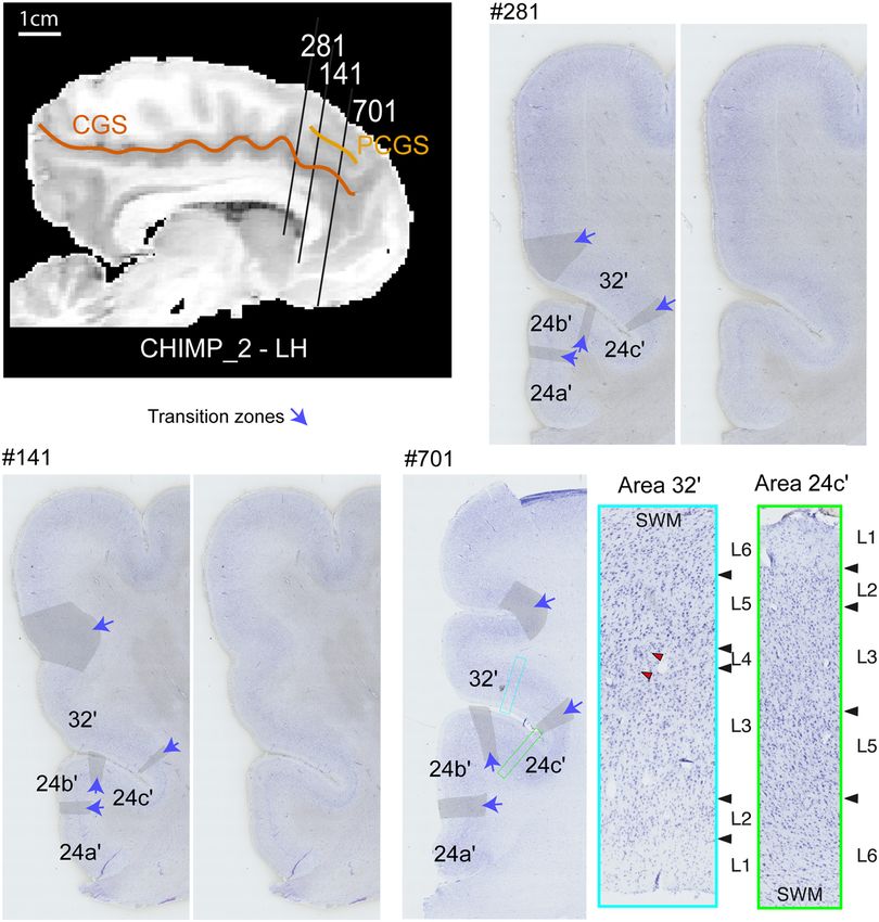
ARTICLE                                                                  COMMUNICATIONS BIOLOGY | https://doi.org/10.1038/s42003-020-01571-3

U
        nderstanding the mechanisms underlying brain evolution,                 often starts at the intersection with the sus-orbitalis and the
        and more specifically of the human brain, is still the topic             supra-rostral sulcus, in front and at the level of the anterior limit
        of intense debates1–4. Comparative neuroanatomical stu-                 of the genu of the corpus callosum, where the anterior cingulate
dies have demonstrated that ecological and social pressures are                 cortex (ACC) lies (Fig. 1a, b)13,14. We are referring here to the
key factors that have driven the expansion of the neocortex in                  cingulate subdivisions proposed by Vogt et al.15 (Fig. 1c). From
primates. But this expansion has differentially impacted brain                  the ACC, the PCGS runs caudally where the anterior mid-
circuits5. With the development of neuroimaging tools, one could                cingulate cortex (aMCC) lies, but it can also run as far posterior
address comparative neuroanatomy questions in vivo at different                 as the level of the anterior commissure (where the posterior mid-
levels of analysis, from gross morphology (e.g., sulcal pattern                 cingulate cortex (pMCC) lies) (Fig. 1). In the human brain, the
analysis) to brain connectivity (e.g., resting-state functional                 impact of the presence of a PCGS on the cytoarchitectonic
Magnetic Resonance Imaging analysis). Comparative neuroima-                     organization (i.e., the cellular organization of the cerebral cortex)
ging studies are principally relying on a comparison between                    of the aMCC is known10: when the PCGS is absent, areas 24c′ and
human brains and a limited number of non-human primate                          32′ occupy, respectively, the ventral and the dorsal banks of the
models, namely macaques and marmosets6,7, whose ancestors                       CGS; however, when a PCGS is present, area 24c′ occupies both
diverged from human ancestors 25 and 35 million years ago,                      banks of the CGS and area 32′ occupies the PCGS (Fig. 1).
respectively8. With a common evolutionary history until 7 million                  In the human brain, the morphological variability of the PCGS
years ago, the chimpanzee is a key model for better understanding               has been linked to personality traits16–18, and pathologies19–23,
the evolution of brain regions that have largely expanded in the                and has also been associated with higher-order cognitive pro-
human brain, such as the medial prefrontal cortex9. Among the                   cessing, i.e., several so-called human-specific processes12,24–27.
sulci that characterize the human medial frontal cortex, the                    These observations have led some investigators to suggest that
paracingulate sulcus (PCGS) is a secondary sulcus running dorsal                cortical area 32′ which occupies the PCGS when present and the
and parallel to the cingulate sulcus (CGS) in a rostro-caudal                   dorsal bank of the CGS when the PCGS is absent, might be
direction10,11 in the medial frontal cortex. The PCGS is observed               unique to the human brain28,29. However, a recent study has
in about 70% of subjects at least in one hemisphere10–13 and most               shown, based on morphological observations of the sulcal

Fig. 1 Morphological and cytoarchitectonic organization of the cingulate cortex in hemispheres without or with a PCGS in the human brain. a In
hemispheres displaying no PCGS, the CGS starts at the intersection with the supra-rostral sulcus (SUROS) and the sulcus sus-orbitalis (SOS) in front of the
genu of the corpus callosum. b In hemispheres with a PCGS, it is the PCGS that starts rostrally at the intersection with the SUROS and the SOS13,14. c The
4-regions model is represented in a hemisphere displaying a PCGS. This model identifies the limit between the ACC and the aMCC at the level of the
anterior limit of the genu of the corpus callosum, the limit between the aMCC and the pMCC as being the anterior commissure. In the aMCC, when a PCGS
is present, both banks of the CGS are occupied by area 24c′ whereas the ventral bank of the PCGS is occupied by area 32′. When a PCGS is absent, the
ventral and dorsal banks of the CGS are respectively occupied by area 24c′ and 32′. d Cytoarchitectonic organization of the aMCC in hemispheres with and
without a PCGS, as shown on coronal sections at the anteroposterior level displayed by the blue line in (c). a anterior, p posterior, d dorsal, v ventral, AC
anterior commissure, cc corpus callosum, ACC anterior cingulate cortex, CGS cingulate sulcus, MCC mid-cingulate cortex, PCC posterior cingulate cortex,
RSC retrosplenial cortex, PCGS paracingulate sulcus, SU-ROS supra-rostral sulcus, SOS sulcus sus-orbitalis. Figure 1c modified from Supplementary Fig. 3
in Amiez et al.13.

2                            COMMUNICATIONS BIOLOGY | (2021)4:54 | https://doi.org/10.1038/s42003-020-01571-3 | www.nature.com/commsbio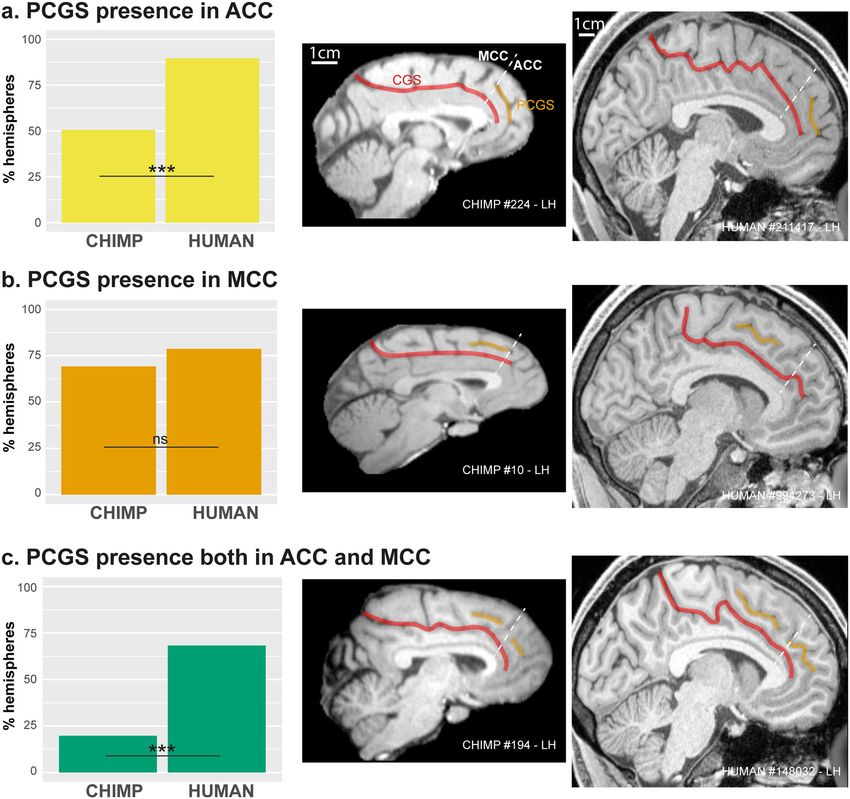
COMMUNICATIONS BIOLOGY | https://doi.org/10.1038/s42003-020-01571-3                                                                  ARTICLE

organization on structural MRI scans, the presence of a putative               (Fig. 2a, dependent variable: PCGS present (0/1), main effect species:
homologue of the human PCGS in 33.8% of chimpanzees, at least                  χ2 = 49.7, df = 1,p-value = 1.79e−12, logistic regression; source data
in one hemisphere13. Furthermore, opposing the view of a lack of               are provided as Supplementary data 1).
area 32′ in non-human primates, this transition area (i.e., area                  By contrast, the probability of observing a PCGS in the MCC is
32′) between the cingulate cortex and cortex of the medial frontal             comparable in human (78.7%) and chimpanzee (69.2%), i.e., it
gyrus has been shown in macaques30. However, the statement                     does not statistically differ between these two species (Fig. 2b)
that the PCGS is not human-specific and can be observed in                      (dependent variable: PCGS present in MCC (0/1), main effect
chimpanzee brains must be supported by cytoarchitectonic evi-                  species: χ2 = 2.9, df = 1, p-value = 0.09, logistic regression).
dence showing that the organization of this region in the chim-                Finally, the probability of observing a PCGS in both the ACC
panzee brain is comparable to that in the human brain.                         and the MCC is significantly higher in human (in 68.3% of
   In the chimpanzee brain, it is not known whether, as in the                 hemispheres displaying a PCGS) compared to chimpanzees (in
human brain, the PCGS starts in the ACC and runs caudally to                   19.8% of hemispheres displaying a PCGS, Fig. 2c) (dependent
the midcingulate cortex (MCC)13. In the present study, we                      variable: PCGS location (ACC/MCC), main effect species:
therefore first assessed the extent of the PCGS in both human and               χ2 = 60.2, df = 1, p-value = 8.48e−15, logistic regression).
chimpanzee brains and hypothesized that, if the sulcus that we
identified as a PCGS in the chimpanzee13 is homologous to the
human PCGS, the mapping of the cytoarchitectonic organization                  Cytoarchitectonic study. Note that our analysis is specifically
of the aMCC in the chimpanzee should be comparable to that in                  focused on the distribution of area 24c′ and area 32′ and the
the human brain.                                                               impact of the presence/absence of a PCGS on it. Previous studies
   It should be noted that, in the human brain, the functional                 had already investigated the dorsal-ventral or rostro-caudal
connectivity within the MCC of seeds located in the PCGS and in                organization of the cingulate cortex and adjacent areas of the
the CGS, when the PCGS is absent, is similar. Specifically, Loh                 medial frontal region7,33–36.
et al.31 have assessed the functional connectivity of the anterior                From the morphological inspection of the three chimpanzee
rostral cingulate zone (RCZa) which is located within the anterior             brains included in the following cytoarchitectonic analysis, we
part of the MCC. Within the RCZa, there are limb and face motor                selected four hemispheres. We analyzed the left hemisphere of
representations with the limb motor representations lying within               CHIMP_1 and the right hemisphere of CHIMP_3 in which the
the CGS even when a PCGS is present; the face motor repre-                     PCGS was absent. We also selected the right hemisphere of
sentations lie in the PCGS if present and in the CGS if the PCGS               CHIMP_1 and the left hemisphere of CHIMP_2 which displayed
is absent32. Loh et al.31 have shown that the functional con-                  a PCGS. In CHIMP_2, the PCGS was present in the anteriormost
nectivity of the face motor representation of RCZa with lateral                part of the aMCC, but absent in the posterior part of the aMCC.
prefrontal and lateral motor regions of interest is similarly                  Note that the remaining hemispheres (left hemisphere of
organized, regardless of whether it is located in the CGS in                   CHIMP_3 and right hemisphere of CHIMP_2) displayed
hemispheres without a PCGS or in the PCGS in hemispheres with                  no PCGS.
a PCGS. The functional connectivity is stronger with anterior
prefrontal regions and weaker with posterior motor regions31.                  Hemispheres without a PCGS. We examined first the MCC within
   In the present study, we examined (1) the extent of the PCGS                the left hemisphere of CHIMP_1, which did not display a PCGS.
in both chimpanzee and human brains, (2) the cytoarchitectonic                 As shown in Fig. 3a, proceeding from the corpus callosum dor-
organization of the aMCC with a specific emphasis on the dis-                   sally, we observed successively areas 24a′ and 24b′, respectively on
tribution of areas 24c′ and 32′ in three post-mortem chimpanzee                the ventral and dorsal part of the gyrus of the cingulate cortex,
brains which did or did not display a PCGS, and (3) in vivo                    and areas 24c′ and 32′, respectively in the ventral and dorsal bank
functional connectivity of this region in four anesthetized chim-              of the CGS (for the cytoarchitectonic characteristics, see “Meth-
panzees based on the availability of resting-state functional                  ods” section). Note that a transition zone was observed between
magnetic resonance imaging (rs-fMRI) data. The results                         each area (i.e. between areas 24a′ and 24b′, between areas 24b′
demonstrate that, in the chimpanzee brain, the impact the PCGS                 and 24c′, and between areas 24c′ and 32′). Proceeding dorsally
has on the cytoarchitectonic organization of the aMCC is com-                  along the cingulate gyrus, the cytoarchitecture does not change
parable to that observed in the human brain. The results also                  abruptly, but rather a smooth reorganization is observed. We also
show that the functional connectivity of the CGS and the PCGS is               examined the posterior part of the aMCC of CHIMP_2 (Fig. 4,
comparable to that observed in the human brain31. Altogether,                  slice 81) which does not display a PCGS (although the anterior
these results demonstrate that the PCGS in chimpanzee brains is                part of the aMCC does possess a PCGS). The results demon-
comparable in terms of cytoarchitecture and functional con-                    strated exactly the same cytoarchitectonic organization as in the
nectivity with the PCGS in human brains. These observations                    left MCC of CHIMP_1.
demonstrate that the PCGS is not human-specific and had
already emerged in the brains of the last common ancestor with                 Hemispheres with a PCGS. We examined the right hemisphere of
chimpanzees.                                                                   CHIMP_1 and CHIMP_3, both of which display a PCGS in the
                                                                               MCC. In both chimpanzees, we observed successively from the
                                                                               corpus callosum in a dorsal direction towards the lateral cortical
Results                                                                        surface, area 24a′ and area 24b′, respectively on the ventral and
Morphological study. We first re-analyzed data from Amiez et al.13              dorsal parts of the cingulate gyrus, area 24c′ in the ventral bank
to assess the occurrence of a PCGS in the ACC versus the MCC in                and in part of the dorsal bank of the CGS, and area 32′ which
197 human and 225 chimpanzee brains. Note that the ACC/MCC                     extends from a part of the dorsal bank of the CGS to the ventral
limit was identified using the probabilistic cytoarchitectonic map of           bank of the PCGS (for the cytoarchitectonic characteristics, see
the ACC from the JuBrain atlas (see “Methods”)33. This new analysis            the “Methods” section). As in hemispheres in which the PCGS is
demonstrates that, in hemispheres displaying a PCGS (i.e., n = 183             absent, we observed small transition zones between adjacent areas
human brain hemispheres, n = 91 chimpanzee brain hemispheres),                 (Fig. 3b). We also examined the anterior part of the aMCC in the
the probability of observing a PCGS in the ACC is higher (89.6%) in            left hemisphere of CHIMP_2 (Fig. 4, slices 141 and 701) which
human hemispheres than in chimpanzee (50.5%) hemispheres                       displays a PCGS. The results demonstrated exactly the same

COMMUNICATIONS BIOLOGY | (2021)4:54 | https://doi.org/10.1038/s42003-020-01571-3 | www.nature.com/commsbio                                         3
ARTICLE                                                                 COMMUNICATIONS BIOLOGY | https://doi.org/10.1038/s42003-020-01571-3

Fig. 2 Occurence of the PCGS in the ACC and MCC in the chimpanzee and the human brains. Probability of occurrence of a PCGS in the ACC (a), the
MCC (b), or in both ACC and MCC (c) in chimpanzee versus human brains. The putative limit between ACC and MCC is represented by the dashed line.
CGS and PCGS correspond to the red and yellow lines, respectively. Left diagrams show that, in hemispheres displaying a PCGS (i.e., in n = 183 human
brain hemispheres and n = 91 chimpanzee brain hemispheres), the probability of occurrence of a PCGS in the ACC as well as in both the ACC and the MCC
is higher in human than in chimpanzee brains (dependent variable: PCGS present (0/1), main effect species: χ2 = 49.7, df = 1, p-value = 1.79e−12, logistic
regression). By contrast, the probability of occurrence of a PCGS in the MCC is similar in human and chimpanzee (dependent variable: PCGS present in
MCC (0/1), main effect species: χ2 = 2.9, df = 1, p-value = 0.09, logistic regression). ACC anterior cingulate cortex, LH left hemisphere, MCC mid-
cingulate cortex, PCGS paracingulate sulcus, ***p < 0.001, logistic regression; ns non-significant logistic regression.

cytoarchitectonic organization as in the right MCC of CHIMP_1                  cortex (Fig. 5a). To be able to conduct a comparison of
and CHIMP_3.                                                                   connectivity fingerprints between species, ROIs were chosen for
                                                                               their known homologies between chimpanzee and human brains.
                                                                               The location of seeds and ROIs are displayed on the medial (left
Rs-fMRI study. From the morphological inspection of the four                   diagram) and the lateral cortical surface (right diagram) of a
chimpanzee brains included in the following rs-fMRI analysis, we               typical example (left hemisphere of CHIMP_C). The heat-maps
observed a PCGS in the left hemisphere of CHIMP_A, and in                      reflecting the average correlation strength between each pair of
both the left and right hemispheres of CHIMP_C. The right                      seed-ROI clusters in the three hemispheres displaying a PCGS
hemisphere of CHIMP_A, and both hemispheres of CHIMP_B                         (see “Methods”) are shown in Fig. 5a. The Boxplots further depict
and CHIMP_D did not display a PCGS.                                            the average Z values of correlations between the two seeds and
  We first assessed, in hemispheres displaying a PCGS, the intra-               ROIs across the three hemispheres displaying a PCGS (see
hemispheric functional connectivity profiles of areas 24c′ and 32′              “Methods”). The results demonstrate how the activity of each
with Regions Of Interest (ROIs) located in the lateral frontal                 seed is differentially correlated with the activity of lateral

4                            COMMUNICATIONS BIOLOGY | (2021)4:54 | https://doi.org/10.1038/s42003-020-01571-3 | www.nature.com/commsbio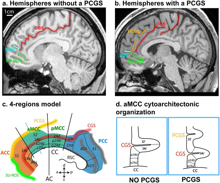
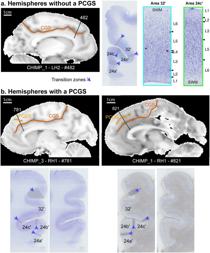
COMMUNICATIONS BIOLOGY | https://doi.org/10.1038/s42003-020-01571-3                                                                      ARTICLE

Fig. 3 Impact of the presentce of a PCGS on the cytoarchitectonic organization of the anterior MCC. Cytoarchitectonic organization of the anterior MCC
in hemispheres without (a) and with (b) a PCGS. a The MCC of the left hemisphere of CHIMP_1 is presented on a sagittal view of a post-mortem MRI scan
(left panel). The CGS is marked in red. The coronal section presented on the middle panel corresponds to the antero-posterior level defined by a black line
on the MRI image (slice 482). The right panels present the labeled and raw Nissl-stained slices. The black lines represent the limits between areas. The
gray zones identified by a blue arrow correspond to transition zones between two adjacent cytoarchitectonic areas. Area 24c′ occupies the ventral bank of
the CGS and area 32′ occupies the dorsal bank of the CGS. The photomicrographs of area 32′ (corresponding to the region identified by a blue box on the
coronal section) and area 24c′ (corresponding to the region identified by a green box on the coronal section) are displayed on the right panels. Results
show the presence of a dysgranular layer 4 (in red are displayed the granular patches) in area 32′ and the absence of this layer in area 24c′. b The MCC of
the right hemispheres of CHIMP_3 and CHIMP_1 are presented on sagittal views of post-mortem MRI scans. The CGS is marked in red, the PCGS in
orange. The coronal sections presented on each Nissl-stained slices correspond to the antero-posterior levels defined by a black line on the MRI images
(CHIMP_3: slice 781, CHIMP_1: slice 821). In both chimpanzees, area 24c′ occupies the ventral bank, the fundus, and the lateral-most part of the dorsal
bank of the CGS. Area 32′ occupies the dorsal bank of the CGS, the gyrus between the CGS and the PCGS and the ventral bank of the PCGS. CGS cingulate
sulcus, MRI magnetic resonance imaging, PCGS paracingulate sulcus, L1-6 cytoarchitectonic layers 1-6, SWM superficial white matter.

COMMUNICATIONS BIOLOGY | (2021)4:54 | https://doi.org/10.1038/s42003-020-01571-3 | www.nature.com/commsbio                                               5
ARTICLE                                                                 COMMUNICATIONS BIOLOGY | https://doi.org/10.1038/s42003-020-01571-3

Fig. 4 Cytoarchitectonic organization of the anterior MCC of a hemisphere displaying a PCGS in its anterior part and no PCGS in its posterior part. The
MCC of the left hemisphere of CHIMP_2 is presented on a sagittal view of a post-mortem MRI scan. The CGS is marked in red, the PCGS in orange. The
Nissl-stained coronal sections presented correspond to the antero-posterior levels defined by a black line on the MRI images (slice 281 where the PCGS is
absent, slices 141 and 701 where the PCG is present). On slice 281 where the PCGS is absent, (1) area 24c′ occupies the ventral bank, the fundus, and the
lateral-most part of the dorsal bank of the CGS, (2) area 32′ occupies the dorsal bank of the CGS, the gyrus between the CGS and the PCGS and the ventral
bank of the PCGS. On slices 141 and 701 where the PCGS is present, (1) area 24c′ occupies the ventral bank, the fundus, and the lateral-most part of the
dorsal bank of the CGS, (2) area 32′ occupies the dorsal bank of the CGS, the gyrus between the CGS and the PCGS and the ventral bank of the PCGS. The
gray zones identified by a blue arrow correspond to transition zones between two adjacent cytoarchitectonic areas. The photomicrographs of area 32′
(corresponding to the region identified by a blue box on the coronal section of slice #701) and area 24c′ (corresponding to the region identified by a green
box on the coronal section of slice #701) are displayed on the right panels. Results show the presence of a dysgranular layer 4 (in red are displayed the
granular patches) in area 32′ and the absence of this layer in area 24c′. CGS cingulate sulcus, PCGS paracingulate sulcus, LH left hemisphere, L1-6
cytoarchitectonic layers 1-6, SWM superficial white matter.

prefrontal/motor ROIs (see “Methods”). For each seed, we tested                value (8) corresponds to the M1-Hand region (the most posterior
these differences in connectivity z values with a generalized linear           ROI).
model with ROI zones (prefrontal zones: Area 10, DLPFC                            We performed the same analysis in hemispheres without a
(dorsolateral prefrontal cortex), Area 45, Area 44, and Fo (Frontal            PCGS. The location of seeds and ROIs are displayed on the
operculum), and motor zones: FEF (Frontal Eye Field), M1Face                   medial (left diagram) and the lateral cortical surface (right
and M1Hand) as a fixed effect. To account for the variability                   diagram) of a typical example (left hemisphere of CHIMP_B).
observed across individuals, the chimpanzee ID was used as a                   The results indicated that, as in hemispheres displaying a PCGS,
random effect. The results indicated that, as in the human brain,              the correlation strength between Area 24c′ and Area 32′ with the
the correlation strength between Area 24c′ and Area 32′ with the               prefrontal areas is significantly higher than with the motor zones
prefrontal cortex is significantly higher than with the motor zones             as demonstrated by heat-maps and boxplots (Fig. 5b, Area 24c′:
(Area 24c′: df = 7, F = 154.8, p < 2.2e−16; Area 32’: df = 7, F =              df = 7, F = 123.5, p < 2.2e−16; Area 32′: df = 7, F = 110.1, p <
157, p < 2.2e−16, ANOVA). We then assessed the linearity of                    2.2e−16, ANOVA; source data are provided as Supplementary
correlation trends with lateral frontal areas along the rostro-                data 2).
caudal axis (Fig. 5c). To test and quantify these linear trends from              We then performed multiple linear regressions on the
anterior prefrontal to motor areas, we recoded the various lateral             correlation values with seed identity, seed location, and ROIline
frontal ROIs into a numeric axis variable (ROIline) that                       as predictors. A significant negative linear trend (slope) was
corresponded to their relative posterior-to-anterior positions                 observed for both seeds (stronger correlation with rostral
(see “Methods”). Based on this coding, the lowest value (1)                    prefrontal areas) and in both morphology types (presence or
corresponds to Area 10 (the most anterior ROI) and the highest                 absence of a PCGS) (Fig. 5Cc. These negative slopes were

6                            COMMUNICATIONS BIOLOGY | (2021)4:54 | https://doi.org/10.1038/s42003-020-01571-3 | www.nature.com/commsbio
COMMUNICATIONS BIOLOGY | https://doi.org/10.1038/s42003-020-01571-3                                                                          ARTICLE

Fig. 5 Intra-hemispheric rostro-caudal functional organization between areas 24c′ and 32′ with the lateral frontal cortex in hemispheres displaying a
PCGS (a, N = 3) and no PCGS (b, N = 5). a The location of each seed is shown in a typical example of a hemisphere displaying a PCGS (CHIMP_C – LH).
The location of each region of interest (ROI) is shown on the cortical surface of the same hemisphere. The heat-map represents the averaged seed-ROI Z
values in hemispheres displaying a PCGS. Boxplots displaying the mean ± SD Z-transformed connectivity between each seed (areas 24c′ and 32′) with the
various ROIs in hemispheres displaying a PCGS. Results show that the correlation strength between Area 24c′ and Area 32′ with the prefrontal cortex is
significantly higher than with the motor zones (Area 24c′: df = 7, F = 154.8, p < 2.2e−16; Area 32′: df = 7, F = 157, p < 2.2e−16, ANOVA). b The location of
each seed is shown in a typical example of a hemisphere displaying no PCGS (CHIMP_B – LH). The heat-map represents the averaged seed-ROI Z values in
hemispheres displaying a PCGS. Boxplots displaying the mean ± SD Z-transformed connectivity between each seed (areas 24c′ and 32′) with the various
ROIs in hemispheres displaying no PCGS. Results show that the correlation strength between Area 24c’ and Area 32′ with the prefrontal areas is
significantly higher than with the motor zones (Area 24c′: df = 7, F = 123.5, p < 2.2e−16; Area 32’: df = 7, F = 110.1, p < 2.2e−16, ANOVA). c Significant
negative linear trend of connectivity (slope) of each seed with the rostral-caudal position of lateral frontal ROIs (ROIlines) in hemispheres displaying or not
a PCGS. The ROIline was obtained by recoding the ROIs in terms of their relative rostro-caudal rank: 1, Area 10; 2, DLPFC; 3, Area 45; 4, Area 44; 5, Fo; 6,
FEF; 7, M1Face; 8, M1Hand. Results show that these negative slopes were statistically similar for both seeds (Area 24c′ and Area 32′) and for both
morphologies (presence or absence of a PCGS) (interaction between seed identity, seed location, and ROIline, F = 3.03, p > 0.05, ns, 3-ways ANOVA). LH
left hemisphere; *** statistically significant at p < 0.001.

COMMUNICATIONS BIOLOGY | (2021)4:54 | https://doi.org/10.1038/s42003-020-01571-3 | www.nature.com/commsbio                                                   7
ARTICLE                                                              COMMUNICATIONS BIOLOGY | https://doi.org/10.1038/s42003-020-01571-3

statistically similar for both seeds (Area 24c′ and Area 32′) and           of areas 24c′ and 32′ that occupy the banks of the CGS. In a rare
for both morphologies (presence or absence of a PCGS)                       study investigating the functional properties of the dorsal and
(interaction between seed identity, seed location, and ROIline,             ventral banks of the CGS in macaques, major differences were
F = 3.03, p = 0.08, ns, 3-ways ANOVA).                                      reported56. Only neurons in the dorsal bank were modulated by
   Thus, the connectivity profiles of areas 24c′ and 32′ with the            the oculomotor saccade direction and, in addition, neurons in the
lateral frontal cortex regions follow the same pattern in hemi-             dorsal bank were most active prior to the choice, while neurons in
spheres with a PCGS and those hemispheres that do not display a             the ventral bank were most active at the outcome phase.
PCGS: both areas 24c′ and 32′ display equally stronger functional               Sensorimotor properties of the human cingulate cortex have
coupling with the lateral prefrontal cortex and weaker with the             been well characterized57. The aMCC contains a cingulate motor
motor cortex.                                                               area (the anterior Rostral Cingulate Zone, RCZa) that is soma-
                                                                            totopically organized. Whereas the face motor representations
                                                                            (mouth and eye) are located in the PCGS when present and in the
Discussion                                                                  CGS when the PCGS is absent (and, therefore, putatively in area
The present study demonstrates that the PCGS in the common                  32′), the limb motor representations (hand and foot) are located
chimpanzee (Pan troglodytes), as previously identified by mor-               in the CGS regardless of the presence or absence of the PCGS
phological examination13, does correspond to the PCGS observed              (and thus putatively in area 24c′)31,32,37,50. In exploratory situa-
in the human (Homo sapiens sapiens)10,11, both cytoarchitecto-              tions in which the learning is driven by behavioral feedback, the
nically and in term of functional connectivity.                             analysis of visual, gustatory, and auditory feedback recruits a
   When a PCGS is absent, the ventral and dorsal banks of the               region located in the PCGS when present and in the CGS when
CGS are, respectively, occupied by areas 24c′ and 32′; when a               the PCGS is absent37,38,40, a region that is co-localized with the
PCGS is present, we observed an expansion of area 32′ on the                face motor area of RCZa37,50. Altogether, these data led us to
medial wall above the CGS up to the fundus of the PCGS.                     hypothesize that the role of the aMCC may be to perform an
Importantly, the functional connectivity of both areas 24c′ and             embodied analysis of feedback in exploratory situations, i.e. juice/
32′ with the lateral frontal cortex is similarly organized to that in       visual/voice feedback recruit the face motor area of RCZa,
the human brain31: both areas display equally stronger con-                 whereas somatosensory feedback on the hand recruits the hand
nectivity with rostral prefrontal areas than with caudal motor              motor area of RCZa37,50. Within this framework, areas 32′ and
areas. Importantly, this gradient of functional connectivity can be         24c′ might support effector specific comparable feedback-related
observed despite the positioning of each ROI on the basis of the            functional processes in exploratory situations. In rhesus maca-
sulcal morphological organization in each chimpanzee brain (see             ques, recordings from the ventral and dorsal banks of the CGS
“Methods” and Supplementary Fig. 1). Specifically, the position-             have highlighted the role of both structures in reward processing
ing of each ROI was based on (1) the fMRI literature on the                 and behavioral adaptation54–56,58. Given that both the cytoarch-
human brain concerning the precise relationship between the                 itectonic and the functional connectivity organization of the
local sulcal organization and functional activity in the regions of         aMCC is comparable in macaque, chimpanzee and human brains,
interest37–41, and (2) studies revealing that the sulcal organization       it is reasonable to hypothesize that the role of the aMCC in higher
between human and chimpanzee is well preserved13,37,42,43. We               cognitive processing may also be preserved. However, in hemi-
chose this methodology because, for both ethical and methodo-               spheres displaying a PCGS, area 24c′ occupies about half of the
logical considerations, there is no study assessing the direct              dorsal bank of the CGS in human brains10,44, but it occupies only
relationships between local sulcal morphology and functional                the fundus of the CGS and the most lateral part of the dorsal bank
activity in behaving chimpanzees. The present study strongly                of the CGS in chimpanzee. One should be cautious in interpreting
suggests that, in addition to preservation of the sulcal and                this difference as the boundaries between cingulate cortical areas
cytoarchitectonic organization from the chimpanzee to the                   24c′ and 32′ in human brains are not consistent in the
human brain, the sulcal-functional organization is also preserved.          literature59.
This is of importance because it indicates that the understanding               Unlike the human brain11,12,24,60, we did not observe at the
of the sulcal organization in great apes may allow us to infer the          population level a left/right PCGS asymmetry in the chimpanzee.
functional organization of the brain in chimpanzees.                        Although brain asymmetry is not a specifically human trait, its
   Based on the morphological sulcal organization of the medial             origins in a population have yet to be determined61. One hypothesis
prefrontal cortex13 and on the present study, three differences can         is that behavioral and brain asymmetry observed in primates might
be identified between the human and the chimpanzee cingulate                 be related to the gradual evolution of language62–64. At the indi-
organization: (1) the PCGS is present in fewer hemispheres in the           vidual subject level, future studies should aim at investigating a
chimpanzee (33.8% of chimpanzees display a PCGS at least in one             putative link between PCGS morphology and cognitive abilities in
hemisphere, compared to about 70% of humans); (2) the PCGS is               chimpanzees, as has been done in humans12,65. Finally, another
more frequently observed in the left hemisphere than in the right           difference between human and chimpanzee brains is the prevalence
hemisphere in the human but not in the chimpanzee brains; (3)               of the PCGS in the ACC. While the PCGS is present in MCC in
the PCGS has a more caudal distribution in chimpanzee than in               both chimpanzees and humans, the PCGS is more often observed in
human brains, implying that the PCGS is more commonly found                 ACC in human brains. This finding suggests that differential evo-
in the MCC compared to the ACC in chimpanzees.                              lutionary pressures impacted the ACC. This is a surprising result as
   The anatomo-functional organization of the cingulate cortex              some studies showed that the hotspot of cortical expansion in the
has received considerable attention. Along the rostral-caudal axis,         primate medial frontal cortex may be located in the MCC rather
several anatomical and functional subdivisions have been                    than in the ACC66,67. Other studies, however, showed that the ACC
identified15,44–50. Similarly, differences between the cingulate             presents high structural variability across subjects in several primate
gyrus and the CGS have been demonstrated44,51–53. One could                 species, contrasting with the MCC which presents less
identify similar functional properties of neurons in the ventral            variability13,14,68. The latter studies suggest that the ACC under-
and dorsal banks of the CGS. For instance, neurons sensitive to             went greater expansion than the MCC during primate evolution.
distance to reward in a reward-guided sequential task have been             This expansion is however associated with a preserved cytoarchi-
recorded in both banks of the CGS54,55. However, very few stu-              tectonic organization of the areas composing the ACC and adjacent
dies have directly investigated what might be the respective roles          ventromedial prefrontal cortex (vmPFC)30,35,47.

8                         COMMUNICATIONS BIOLOGY | (2021)4:54 | https://doi.org/10.1038/s42003-020-01571-3 | www.nature.com/commsbio
COMMUNICATIONS BIOLOGY | https://doi.org/10.1038/s42003-020-01571-3 ARTICLE
Within the ACC, resting-state analyses have shown that area colony of apes housed at the Yerkes National Primate Research Center of Emory
24c has stronger coupling with the anterior insula, the striatum University.
The chimpanzees were all born in captivity and had all lived in social groups
and the ventrolateral prefrontal cortex, while perigenual area 32 ranging from 2 to 13 individuals at the Yerkes National Primate Research Center
has stronger coupling with the dorsolateral prefrontal cortex, the and were housed according to institutional guidelines. Chimpanzee data collection
amygdala, and the hippocampus33. The ACC in the human brain was approved by the Institutional Animal Care and Use Committees at YNPRC
has been associated with value-based computations in economic and UTMDACC and followed the guidelines of the Institute of Medicine in the use
and social domains69–74. Similar properties have been identified of chimpanzees in research. We note here that all in vivo MRI scans were obtained
prior to changes in NIH policy on the acquisition of neuroimaging data from
in the non-human primate brain52,75,76. Implicit mentalizing chimpanzees (Nov 2015).
abilities have been described in chimpanzees and macaques77,78,
and are affected by reversible lesions of the ACC in macaques78.
Recursive thinking and counterfactual manipulation of informa- MRI data acquisition
tion to guide behavior have also been observed in macaques75,79. Structural MRI of post-mortem chimpanzee brains. Each post-mortem chimpanzee
brain was scanned overnight in a 3 T Siemens Prisma MRI scanner to obtain
Counterfactual reasoning was also impacted by reversible lesions structural T1 volumetric images (repetition time = 23 ms, echo time = 5.65 ms,
of the ACC in macaques75. However, human subjects have been voxel resolution = 0.4 × 0.4 × 0.4 mm). Each brain was scanned in a container filled
shown to understand more complex relationships between social with 10% formalin and supported with padding to prevent scanning artifacts from
agents and their intentions than non-human primates70,80. The occurring near the edges of the container. 24, 4, and 36 repetitions of T1 scans of
respectively CHIMP_1, CHIMP_2, and CHIMP_3 were obtained and averaged.
more complex the information about intentionality of social
agents a human subject can comprehend, the larger the gray In vivo rs-fMRI acquisition in chimpanzee. In vivo resting-state functional magnetic
matter volume in the ACC and vmPFC81. Altogether, these resonance imaging (rs-fMRI) data came from Dr Hopkins’ laboratory. These data
results suggest that the building blocks of a human ACC have were acquired in early 2015 at the Yerkes National Primate Research Center
been present since the last common ancestor to human and (YNPRC) on the four adult chimpanzees at the time they were being surveyed for
macaques, but its evolution might reflect the development of their annual physical examinations. Subjects were first immobilized by ketamine
(10 mg/kg) or telazol (3–5 mg/kg) and subsequently anaesthetized with propofol
recursive thinking in hominids82. (40–60 mg/[kg/h]) following standard procedures at the YNPRC. The subjects
To conclude, using multimodal data, the present study remained anaesthetized for the duration of the scans as well as the time needed to
demonstrates that chimpanzee brains do possess a PCGS in the transport them between their home cage and the imaging facility (between 5 and
MCC that is comparable to that in the human brain in terms of 10 min). Chimpanzees were placed in the scanner chamber in a supine position
with their head fitted inside the human-head coil.
cytoarchitecture and functional connectivity. The similarities They were scanned using a 3.0-T scanner (Siemens Trio; Siemens Medical Solutions
between ACC and MCC in primates and rodents are still a matter USA, Inc., Malvern, PA, USA). T1-weighted images were collected using a three-
of debate7,83. dimensional gradient-echo sequence (repetition time = 2300 ms, echo time = 4.4 ms,
number of signals averaged = 2, voxel resolution = 0.625 × 0.625 × 0.60 mm. In
addition, 2 runs of 350 measurements each (16 min/session) of rs-fMRI scans were
collected using a three-dimensional gradient-echo sequence (repetition time = 2683 ms,
Methods echo time = 25 ms, voxel resolution = 1.9 × 1.9 × 1.9 mm, right-left phase-encoding
Subjects/specimens
direction). An additional short run was performed with the same characteristics except
Human subjects. The first step in this investigation was a reanalysis of the mor-
that the phase-encoding was in the opposite direction (left-right). Scan duration was
phological organization of the PCGS in 197 human brains13 to refine our previous
about 90 min.
analysis by assessing the location and extent of the PCGS in the ACC and/or the
After completing MRI procedures, the subjects were temporarily housed in a
MCC. High-resolution anatomical scans of these brains were obtained from the
single enclosure for 6–12 h to allow the effects of the anesthesia to wear off before
Human Connectome Project (HCP) database [http://www.humanconnectome.org/
being returned to their social group.
]. Only data from subjects with no family relationships were analyzed. The parti-
cipants in the HCP study were recruited from the Missouri Family and Twin
Registry that includes individuals born in Missouri84. Acquisition parameters
of T1 anatomical scans are the following: whole head, 0.7 mm3 isotropic Sulcal morphology of the medial frontal cortex. On the basis of structural T1
resolution, TR = 2.4 s, TE = 2.14 ms, flip angle = 8° (more details can be found at MRI scans of each hemisphere of all chimpanzee and human brains, we established
[https://humanconnectome.org/storage/app/media/documentation/s1200/ the presence or absence of a PCGS in each cerebral hemisphere. A PCGS was
HCP_S1200_Release_Appendix_I.pdf]). The full set of inclusion and exclusion marked as present if running parallel and dorsal to the CGS12,13. We then
criteria is detailed elsewhere. Briefly, the HCP subjects are healthy individuals free examined whether this PCGS was located within the ACC, within the MCC, or
from major psychiatric or neurological illnesses. They are drawn from ongoing extending along both regions. Note that the ACC-MCC limit was based on Vogt’s
longitudinal studies84, in which they had received extensive assessments, including four-region model (Fig. 1)15,45,85,86.
the history of drug use, and emotional and behavioral problems. The experiments
were performed in accordance with relevant guidelines and regulations and all Morphological analysis: occurrence and location of PCGS in human and chim-
experimental protocols were approved by the Institutional Review Board (IRB) panzee. To establish the probability of occurrence of a PCGS in the MCC and the
(IRB #201204036; Title: ‘Mapping the Human Connectome: Structure, Function, ACC in human and chimpanzee brains, we first reanalyzed the neuroimaging T1
and Heritability’). All subjects provided written informed consent on forms anatomical data of 197 human brains and 225 chimpanzee brains from our pre-
approved by the Institutional Review Board of Washington University in St Louis. vious study13. From this inspection, we identified 76 chimpanzees displaying a
In addition, the present study received approval (n°15-213) from the Ethics PCGS at least in one hemisphere (15, 29, and 32 displaying a PCGS in both
Committee of Inserm (IORG0003254, FWA00005831) and from the Institutional hemispheres, only in the left hemisphere, and only in the right hemispheres,
Review Board (IRB00003888) of the French Institute of Medical Research and respectively), for a total of 91 hemispheres displaying a PCGS. We also identified
Health. 139 human brains displaying a PCGS at least in one hemisphere (45, 69, and 25
displaying a PCGS in both hemispheres, only in the left hemisphere, and only in
Chimpanzee (Pan troglodytes). Three chimpanzee groups were examined in (1) the the right hemispheres, respectively), for a total of 184 hemispheres displaying a
morphological sulcal organization of the medial frontal cortex (Group 1, N = 225), PCGS. In these hemispheres, we then identified whether the PCGS was present in
(2) in the cytoarchitectonic analysis of the MCC (Group 2, N = 3), and (3) in the the ACC, the MCC, or in both the ACC and MCC. The limit between the ACC and
functional connectivity analysis of the MCC (Group 3, N = 4, note that these four the MCC was identified using the probabilistic cytoarchitectonic map of the ACC
chimpanzees were also part of group 1). from the JuBrain atlas (https://jubrain.fz-juelich.de/apps/cytoviewer2/cytoviewer-
Specifically, the morphological analysis aimed at refining our previous analysis main.php#)33.
on 225 chimpanzees13 by assessing the location and extent of the PCGS in the ACC
and/or MCC. In the cytoarchitectonic analysis, we examined three post-mortem Cytoarchitectonic analysis: occurrence and location of PCGS in chimpanzee. From
male chimpanzee brains that died from natural causes (CHIMP_1, CHIMP_2, and the morphological inspection of the three chimpanzee brains included in this
CHIMP_3; ages at death 38, 33, and 37 years, respectively). Within 14 h of each analysis, we selected four hemispheres for the cytoarchitectonic analysis:
subject’s death, the brain was removed and immersed in 10% formalin at necropsy. The left hemisphere of CHIMP_1, in which the PCGS is absent.
In the in vivo resting-state fMRI study, we analyzed the data obtained in four The right hemisphere of CHIMP_1, which displays a PCGS.
chimpanzees: three females (CHIMP_A, CHIMP_B, CHIMP_C, respectively 16, The left hemisphere of CHIMP_2, where a PCGS is present in the anterior-most
17, and 27 years of age at the time of the experiment) and one male (CHIMP_D, 15 part of the aMCC, but absent in the posterior part of the aMCC.
years of age). All four chimpanzees were captive born and were members of the The right hemisphere of CHIMP_3, in which the PCGS is absent.
COMMUNICATIONS BIOLOGY | (2021)4:54 | https://doi.org/10.1038/s42003-020-01571-3 | www.nature.com/commsbio 9ARTICLE COMMUNICATIONS BIOLOGY | https://doi.org/10.1038/s42003-020-01571-3
Note that the remaining hemispheres (left hemisphere of CHIMP_3 and right Seed selection in the MCC. The seeds consisted of 2.5-mm radius spheres and were
hemisphere of CHIMP_2) displayed no PCGS. positioned in the aMCC at the same antero-posterior level as in the cytoarchi-
tectonic study as follows:
Rs-fMRI analysis: occurrence and location of PCGS in chimpanzee. We identified In hemispheres displaying no PCGS (N = 5): a seed assigned to area 24c′ was
three hemispheres with a PCGS in the four chimpanzees included in this analysis: positioned in the ventral bank of the CGS and a seed assigned to area 32′ was
Both the left and right hemispheres of CHIMP_C, and the left hemisphere of positioned in the dorsal bank of the CGS.
CHIMP_A, displayed a PCGS. In hemispheres displaying a PCGS (N = 3): a seed assigned to area 24c′ was
The right hemisphere of CHIMP_A, and both hemispheres of CHIMP_D and positioned in the CGS and a seed for area 32′ was positioned in the ventral bank of
CHIMP_B did not display a PCGS. the PCGS.
In both cases, the two seeds were positioned on an imaginary line going through
the posterior limit of the genu of the corpus callosum and perpendicular to the axis
Blocking and histological processing. Based on the averaged structural MRI data on which the CGS and PCGS are running (see Supplementary Fig. 1 for positioning
of the chimpanzee brains and using a neuronavigation system (Brainsight), each of seeds in all chimpanzees and hemispheres). In hemispheres displaying no PCGS,
selected brain was cut in several blocks to obtain histological sections optimal for the two seeds (area 24c′ and area 32′) displayed a small overlap. This overlap was
the study of the architecture of the regions of interest (see Novek et al.87 for the removed before performing the ROI-based resting-state data analysis.
method used). The blocks including the MCC were optimized to allow for histo-
logical sections that were perpendicular to the orientation of both the cingulate Selection of regions of interest (ROIs). For a stricter comparison of the present
and/or paracingulate sulci. Note that the remaining blocks were processed for results with results obtained in our previous study which assessed the functional
ongoing studies aiming to assess cytoarchitectonic areas of various regions of the connectivity of the CGS and PCGS in the anterior part of the human MCC31, we
chimpanzee cortex. All blocks were cryoprotected by immersion in buffered used the same ROIs (see below and Supplementary Fig. 1 for positioning of ROIs in
sucrose solutions from 10 to 30% until they sank, frozen to −60 ˚C in a bath of 2- all chimpanzees and hemispheres). Each ROI consisted of a sphere with a 5-mm
methylbutane chilled by a surrounding mixture of dry ice and ETOH, then stored radius.
at −80 ˚C until use, where the block was kept surrounded by dry ice on a frozen
microtome stage during sectioning. The histological sections were cut at a thickness
of 30 μm, three out of every ten sections were kept, and a photograph was taken ROIs selection in motor cortical areas. For each subject, 3 ROIs within the motor
before each set of the kept sections throughout the entire blocks to aid with 3D cortex of both hemispheres were identified based on sulcal morphology. These
reconstructions. included the hand motor region (the precentral knob) within the central sulcus
–M1Hand–43 and the primary face motor region within the ventral part of the
posterior part of the precentral gyrus –M1Face–92. We also included the frontal eye
Cytoarchitectonic analysis. The detailed cytoarchitectonic analysis of the mid- field –FEF–. In the human brain, this region is located within the ventral branch of
cingulate cortex of the selected blocks was carried out from the sections that were the superior precentral sulcus39. As the chimpanzee presents the same sulcal
cut in a coronal orientation. Every tenth section was mounted on 2” × 3” coated pattern in this region, including a ventral branch of the superior precentral sulcus,
slides and stained with cresyl violet, a Nissl cell body stain, for cytoarchitectonic we tentatively included this ROI in our analysis.
analysis; the remaining sections are being used in ongoing studies.
The architectonic organization of the MCC and, more specifically, the
boundaries between the cytoarchitectonic areas composing the MCC, were
identified on high-resolution tilted images of the entire region of interest from the Selection of ROIs in the prefrontal cortex. For each subject, 6 ROI locations
cresyl violet sections, obtained under bright field with a Zeiss Axio Scan.Z1 at ×10 within the left prefrontal cortex were identified based on the local anatomy. On a
magnification. rostro-caudal axis:
The assessment of the architectonic organization of the MCC was based on The frontopolar cortex –Area 10–. In the human brain, this region is located at
prior studies of this region in both the human10 and in the macaque monkey30,88 the intersection between the vertical segment of the intermediate frontal sulcus, the
brains. Proceeding from the corpus callosum in a dorsal direction towards the lateral and the medial frontomarginal sulcus, see41. Because chimpanzee brains
lateral surface of the frontal lobe, the following areas were identified in the display a similar sulcal pattern, we hypothesized that Area 10 most likely lies at the
cingulate region: area 24a′, area 24b′, area 24c′, and area 32′. Note that, dorsal to same location.
these cingulate cortical areas, the medial frontal areas 6. 8, and 9 are located. Area The dorsolateral prefrontal cortex -DLPFC-. In the human brain, area 9/46 of
24a′ is an agranular area located on the gyrus just dorsal to the corpus callosum. It the DLPFC lies at the rostral level of the genu of the corpus callosum, above the
is characterized by a clear layer II, a thick layer III, a dense layer V, and a poorly inferior frontal sulcus. Because the chimpanzee displays also an inferior frontal
defined layer VI. Area 24b’ is an agranular area located dorsal to area 24a′ and ends sulcus that is also present at the level of the rostral level of the genu of the corpus
where the CGS starts. It is characterized by a broad layer III containing distinct callosum, we hypothesized that area 9/46 lies at the same location.
layers IIIa, b, and c, as well as a highly prominent layer V composed of large Broca’s region: The two cytoarchitectonic areas that comprise Broca’s region,
pyramidal neurons. Area 24c′ is an agranular area located in the ventral bank of the namely area 44 and area 45, have been shown to be located in the chimpanzee
CGS and is characterized by thin layers II and III, as well as the presence of more brain between the inferior precentral sulcus and the fronto-orbital sulcus, and
densely packed large pyramidal neurons in layer V in comparison with area 24b’. anterior to the fronto-orbital sulcus42,93.
Finally, area 32′ is a dysgranular cingulo-frontal transitional area located dorsal to The frontal operculum –Fo– (intersection between the frontal operculum and
area 24c′ in the dorsal bank of the CGS when there is no PCGS in the human brain the circular sulcus, see ref. 38.
and located within the PCGS if present10. In the macaque, area 32′ (labeled as 32/6
or 32/8 in macaque) is located in the dorsal bank of the CGS 30,88. It displays a wide Statistics and reproducibility. The mean signal from Seeds and ROI regions was
layer IIIc containing large pyramidal neurons, a dysgranular layer IV, and a thinner then extracted using AFNI software. For each chimpanzee brain, correlation
and less dense layer V than in area 24c′. coefficients between the two seeds with the various ROIs in the prefrontal cortex
and the motor cortex were computed and normalized using the Fisher’s r-to-z
transform formula. The significant threshold at the individual subject level was
Rs-fMRI data analysis. Data were collected using 2 phase-encoding directions (2 Z = 0.1 (p < 0.05). These normalized correlation coefficients, which corresponded
full runs in Right-Left, and a shorter 3d run in Left-Right directions). It resulted in to the functional connectivity strength between each seed and each ROI in indi-
two pairs of images with distortions going in opposite directions (pair 1: 1st run in vidual chimpanzee brains, were subsequently processed with R statistical software
right-left and 3d run in left-right direction; pair 2: 2d run in right-left and 3d run in (https://www.r-project.org/) for all the following analyses.
left-right direction). Distortions were corrected using TOPUP’s FSL tool. First, To compare the connectivity profile of each seed with the various lateral frontal
TOPUP estimated from these pairs the susceptibility-induced off-resonance field ROIs, we constructed boxplots corresponding to the correlation strength of each
using a method similar to that described by Andersson et al.89 as implemented in seed location with each of the ROIs. Based on these boxplots, it can be discerned
FSL90. Once estimated, the images were then combined into corrected ones. Two that both Area 24c′ and Area 32′ seeds have stronger connectivity with prefrontal
runs, corrected for distortions, resulted from this analysis and were further regions and weaker connectivity with premotor and motor areas (see “Results”).
preprocessed. We then characterized this rostro-caudal functional axis based on the correlation
The preprocessing of resting-state scans was then performed with SPM 12. The profiles of Area 24c′ and Area 32′, when a PCGS is present and also when it is not
first 5 volumes of each run were removed to allow for T1 equilibrium effects. The present, with the lateral frontal cortex by estimating linear trends in the correlation
head motion correction was applied using rigid body realignment and we then strength for each seed with the rostro-caudal lateral frontal ROIs (for details, see
applied a slice timing correction using the time center of the volume as reference. “Methods“ in Loh et al.31). The 8 ROIs were first ranked along a rostro-caudal axis
Then, using the AFNI software91, the segmentation of each brain was performed on based on their average Y coordinate values across chimpanzee brains and recoded
skull-stripped brains. A temporal filtering was then applied to extract the into a numeric axis variable (ROIline): Area 10 (most anterior)-1, DLPFC-2, Area
spontaneous slowly fluctuating brain activity (0.01–0.1 Hz). Finally, linear 45-3, Area 44-4, Fo-5, FEF-6, M1Face-7, M1Hand (most posterior)-8. We then
regression was used to remove nuisance variables (the cerebrospinal fluid and white performed multiple linear regressions on the correlation z values with seed identity
matter signals from the segmentation) and spatial smoothing with a 4-mm FWHM (area 24c′ and area 32′), sulcal morphology (PCGS absent or present), and the
Gaussian kernel was applied to the output of the regression. linear axis variable (ROIline) as predictors. We assessed whether the linear trends
10 COMMUNICATIONS BIOLOGY | (2021)4:54 | https://doi.org/10.1038/s42003-020-01571-3 | www.nature.com/commsbioCOMMUNICATIONS BIOLOGY | https://doi.org/10.1038/s42003-020-01571-3 ARTICLE
(slopes) observed for each seed were identical or not in each sulcal morphology 26. Borst, G. et al. Folding of the anterior cingulate cortex partially explains
using a 3-ways ANOVA. inhibitory control during childhood: a longitudinal study. Dev. Cogn.
Neurosci. 9, 126–135 (2014).
Reporting summary. Further information on research design is available in the Nature 27. Cachia, A. et al. Longitudinal stability of the folding pattern of the anterior
Research Reporting Summary linked to this article. cingulate cortex during development. Dev. Cogn. Neurosci. 19, 122–127 (2016).
28. Cole, M. W., Yeung, N., Freiwald, W. A. & Botvinick, M. Cingulate cortex:
Data availability diverging data from humans and monkeys. Trends Neurosci. 32, 566–574 (2009).
The source data underlying Figs. 1 and 5 are provided as Source Data file (respectively 29. Vogt, B. A. Architecture, Neurocytology and Comparative Organization of
Supplementary Data 1 and Supplementary Data 2). Human anatomical MRI scans are Monkey and Human Cingulate Cortices. In Cingulate Neurobiology and
available from the Human Connectome Project database [http://www. Disease. (ed. Vogt, B. A.) 65–93 (Oxford University Press, 2009).
humanconnectome.org/]94, and Chimpanzee anatomical MRI and rs-fMRI scans are 30. Petrides, M. & Pandya, D. N. Comparative architectonic analysis of the
available from Dr. W. Hopkins [http://www.chimpanzeebrain.org/]95. A reporting human and the macaque frontal cortex. In Handbook of Neuropsychology,
summary for this Article is available as a Supplementary Information file. (eds. Boller, F. & Grafman, J.), 9, 17–58 (Amsterdam, Elsevier, 1994).
31. Loh, K. K., Hadj-Bouziane, F., Petrides, M., Procyk, E. & Amiez, C. Rostro-
caudal organization of motor areas and lateral frontal regions. Front. Neurosci.
Received: 1 September 2020; Accepted: 25 November 2020; 11, 753 (2018).
32. Amiez, C. & Petrides, M. Neuroimaging evidence of the anatomo-functional
organization of the human cingulate motor areas. Cereb. Cortex 24, 563–578
(2014).
33. Palomero-Gallagher, N. et al. Human pregenual anterior cingulate cortex:
structural, functional, and connectional heterogeneity. Cereb. Cortex 29,
References 2552–2574 (2019).
1. Dunbar, R. I. M. & Shultz, S. Why are there so many explanations for primate 34. Mackey, S. & Petrides, M. Architecture and morphology of the human
brain evolution? Phil. Trans. R. Soc. B 372, 20160244 (2017). ventromedial prefrontal cortex. Eur. J. Neurosci. 40, 2777–2796 (2014).
2. Barton, R. A. & Montgomery, S. H. Proportional versus relative size as metrics 35. Mackey, S. & Petrides, M. Quantitative demonstration of comparable
in human brain evolution. Proc. Natl Acad. Sci. USA 116, 3–4 (2019). architectonic areas within the ventromedial and lateral orbital frontal cortex in
3. DeCasien, A. R., Williams, S. A. & Higham, J. P. Primate brain size is the human and the macaque monkey brains. Eur. J. Neurosci. 32, 1940–1950
predicted by diet but not sociality. Nat. Ecol. Evol. 1, 112 (2017). (2010).
4. Mars, R. B. et al. Primate comparative neuroscience using magnetic resonance 36. Vogt, B. A. Regions and Subregions of the Cingulate Cortex. In Cingulate
imaging: promises and challenges. Front. Neurosci. 8, 298 (2014). Neurobiology and Disease. (ed. Vogt, B. A.) 3–30 (Oxford University Press,
5. DeCasien, A. R. & Higham, J. P. Primate mosaic brain evolution reflects selection 2009).
on sensory and cognitive specialization. Nat. Ecol. Evol. 3, 1483–1493 (2019). 37. Loh, K. K. et al. Cognitive control of orofacial motor and vocal responses in
6. Sallet, J. et al. The organization of dorsal frontal cortex in humans and the ventrolateral and dorsomedial human frontal cortex. Proc. Natl Acad. Sci.
macaques. J. Neurosci. 33, 12255–12274 (2013). USA 117, 4994–5005 (2020).
7. Schaeffer, D. J. et al. Divergence of rodent and primate medial frontal cortex 38. Amiez, C. et al. The location of feedback-related activity in the
functional connectivity. Proc. Natl Acad. Sci. USA 117, 21681–21689 (2020). midcingulate cortex is predicted by local morphology. J. Neurosci. 33,
8. Schrago, C. G. & Russo, C. A. M. Timing the origin of New World monkeys. 2217–2228 (2013).
Mol. Biol. Evol. 20, 1620–1625 (2003). 39. Amiez, C., Kostopoulos, P., Champod, A.-S. & Petrides, M. Local morphology
9. Passingham, R. How good is the macaque monkey model of the human brain? predicts functional organization of the dorsal premotor region in the human
Curr. Opin. Neurobiol. 19, 6–11 (2009). brain. J. Neurosci. 26, 2724–2731 (2006).
10. Vogt, B. A., Nimchinsky, E. A., Vogt, L. J. & Hof, P. R. Human cingulate 40. Amiez, C. et al. Single subject analyses reveal consistent recruitment of frontal
cortex: surface features, flat maps, and cytoarchitecture. J. Comp. Neurol. 359, operculum in performance monitoring. Neuroimage 133, 266–278 (2016).
490–506 (1995). 41. Petrides, M. The Human Cerebral Cortex. An MRI Atlas of the Sulci and Gyri
11. Paus, T. et al. Human cingulate and paracingulate sulci: pattern, variability, in MNI Stereotaxic Space. (Academic Press, New York, 2012).
asymmetry, and probabilistic map. Cereb. Cortex 6, 207–214 (1996). 42. Schenker, N. M. et al. Broca’s area homologue in chimpanzees (Pan
12. Amiez, C., Wilson, C. R. E. & Procyk, E. Variations of cingulate troglodytes): probabilistic mapping, asymmetry, and comparison to humans.
sulcal organization and link with cognitive performance. Sci. Rep. 8, 13988 (2018). Cereb. Cortex 20, 730–742 (2010).
13. Amiez, C. et al. Sulcal organization in the medial frontal cortex provides 43. Hopkins, W. D. et al. Evolution of the central sulcus morphology in primates.
insights into primate brain evolution. Nat. Commun. 10, 3437 (2019). Brain Behav. Evol. 84, 19–30 (2014).
14. Lopez-Persem, A., Verhagen, L., Amiez, C., Petrides, M. & Sallet, J. The 44. Palomero-Gallagher, N., Mohlberg, H., Zilles, K. & Vogt, B. Cytology and
human ventromedial prefrontal cortex: sulcal morphology and its influence on receptor architecture of human anterior cingulate cortex. J. Comp. Neurol.
functional organization. J. Neurosci. 39, 3627–3639 (2019). 508, 906–926 (2008).
15. Vogt, B. A., Berger, G. R. & Derbyshire, S. W. G. Structural and functional 45. Palomero-Gallagher, N., Vogt, B. A., Schleicher, A., Mayberg, H. S. & Zilles, K.
dichotomy of human midcingulate cortex. Eur. J. Neurosci. 18, 3134–3144 (2003). Receptor architecture of human cingulate cortex: evaluation of the four-region
16. Mériau, K. et al. A neural network reflecting individual differences in cognitive neurobiological model. Hum. Brain Mapp. 30, 2336–2355 (2009).
processing of emotions during perceptual decision making. Neuroimage 33, 46. Alexander, W. H. & Brown, J. W. Medial prefrontal cortex as an action-
1016–1027 (2006). outcome predictor. Nat. Neurosci. 14, 1338–1344 (2011).
17. Whittle, S. et al. Variations in cortical folding patterns are related to individual 47. Neubert, F.-X., Mars, R. B., Sallet, J. & Rushworth, M. F. S. Connectivity
differences in temperament. Psychiatry Res. 172, 68–74 (2009). reveals relationship of brain areas for reward-guided learning and decision
18. Kanai, R. & Rees, G. The structural basis of inter-individual differences in making in human and monkey frontal cortex. Proc. Natl Acad. Sci. USA 112,
human behaviour and cognition. Nat. Rev. Neurosci. 12, 231–242 (2011). E2695–E2704 (2015).
19. Yücel, M. et al. Paracingulate morphologic differences in males with 48. Heilbronner, S. R. & Hayden, B. Y. Dorsal anterior cingulate cortex: a bottom-
established schizophrenia: a magnetic resonance imaging morphometric up view. Annu. Rev. Neurosci. 39, 149–170 (2016).
study. Biol. Psychiatry 52, 15–23 (2002). 49. Kolling, N. et al. Value, search, persistence and model updating in anterior
20. Yücel, M. et al. Anterior cingulate dysfunction: implications for psychiatric cingulate cortex. Nat. Neurosci. 19, 1280–1285 (2016).
disorders? J. Psychiatry Neurosci. 28, 350–354 (2003). 50. Procyk, E. et al. Midcingulate motor map and feedback detection: converging
21. Le Provost, J.-B. et al. Paracingulate sulcus morphology in men with early- data from humans and monkeys. Cereb. Cortex 26, 467–476 (2016).
onset schizophrenia. Br. J. Psychiatry 182, 228–232 (2003). 51. Rudebeck, P. H., Buckley, M. J., Walton, M. E. & Rushworth, M. F. S. A role
22. Shim, G. et al. Reduced cortical folding of the anterior cingulate cortex in for the macaque anterior cingulate gyrus in social valuation. Science 313,
obsessive-compulsive disorder. J. Psychiatry Neurosci. 34, 443–449 (2009). 1310–1312 (2006).
23. Meredith, S. M. et al. Anterior cingulate morphology in people at genetic high- 52. Chang, S. W. C., Gariépy, J.-F. & Platt, M. L. Neuronal reference frames for
risk of schizophrenia. Eur. Psychiatry 27, 377–385 (2012). social decisions in primate frontal cortex. Nat. Neurosci. 16, 243–250 (2013).
24. Fornito, A. et al. Individual differences in anterior cingulate/paracingulate 53. Apps, M. A. J., Rushworth, M. F. S. & Chang, S. W. C. The anterior cingulate
morphology are related to executive functions in healthy males. Cereb. Cortex gyrus and social cognition: tracking the motivation of others. Neuron 90,
14, 424–431 (2004). 692–707 (2016).
25. Buda, M., Fornito, A., Bergström, Z. M. & Simons, J. S. A specific brain 54. Procyk, E., Tanaka, Y. L. & Joseph, J. P. Anterior cingulate activity during
structural basis for individual differences in reality monitoring. J. Neurosci. 31, routine and non-routine sequential behaviors in macaques. Nat. Neurosci. 3,
14308–14313 (2011). 502–508 (2000).
COMMUNICATIONS BIOLOGY | (2021)4:54 | https://doi.org/10.1038/s42003-020-01571-3 | www.nature.com/commsbio 11You can also read