Out-of-step: brain-heart desynchronization in anxiety disorders - Georg ...
←
→
Page content transcription
If your browser does not render page correctly, please read the page content below
Molecular Psychiatry
                                    https://doi.org/10.1038/s41380-021-01029-w

                                        REVIEW ARTICLE

                                    Out-of-step: brain-heart desynchronization in anxiety disorders
                                                           1,2                          3                        1
                                    Shankar Tumati               ●
                                                                     Martin P. Paulus       ●
                                                                                                Georg Northoff

                                    Received: 19 June 2020 / Revised: 30 December 2020 / Accepted: 12 January 2021
                                    © The Author(s), under exclusive licence to Springer Nature Limited 2021

                                    Abstract
                                    Imaging studies in anxiety disorders (AD) show abnormal functional connectivity primarily in the salience network (SN),
                                    somatomotor network (SMN), and default mode network (DMN). However, it is not clear how precisely these network
                                    changes occur including their relation to psychopathological symptoms. Here, we show that the functional networks affected
                                    in AD overlap with cortical regions that receive visceral inputs (the so-called central/visceral autonomic network). Focusing
                                    on cardiac afferents, we suggest that network changes in AD may be due to reduced phase synchronization between ongoing
                                    neural and cardiac activity. This neuro-cardiac desynchronization occurs due to the abnormal phase resetting of neural
                                    activity at the onset of each heartbeat, as measured by a lower intertrial coherence and heartbeat-evoked potential.
1234567890();,:
                  1234567890();,:

                                    Biochemically, cardiac afferents reach subcortical serotonergic raphe nuclei and noradrenergic locus coeruleus (among
                                    others) which, in turn, are known to reciprocally modulate the DMN and SMN/SN on the cortical level. Consistent with the
                                    network changes in AD, decreases in serotonergic and noradrenergic activity are known to increase connectivity in both
                                    SMN and SN while, at the same time, they decrease DMN connectivity. SMN and SN increases, in turn, lead to increased
                                    emotional arousal/anxiety and bodily awareness whereas decreased DMN connectivity leads to an unstable sense-of-self in
                                    AD. Finally, we integrate our proposal with interoceptive predictive processing models suggesting neuro-cardiac
                                    desynchronization as a mechanism for “noisy” bottom-up information leading to a persistently uncertain bodily state in top-
                                    down models. In sum, integrating theories on active interference and hyperarousal, we propose a precise neuro-cardiac and
                                    biochemically -driven mechanisms for key psychopathological symptoms of AD.

                                    Introduction                                                             changes occur and how they may be related to the various
                                                                                                             symptoms in these disorders.
                                    Anxiety disorders (AD) include panic disorder (PD), gen-                    Here, we suggest that functional network changes in
                                    eralized anxiety disorder (GAD), social anxiety disorder                 AD may be due to an abnormal biochemically -driven
                                    (SAD), and specific phobias, which share symptoms of                      interaction between neural activity and cardiac inputs to
                                    excessive worry or fearful responses to benign stimuli [1].              the brain. Specifically, we show that (1) regions in the
                                    In addition to uncontrollable worry, somato-cardiac symp-                affected networks receive cardiac and other visceral
                                    toms such as heart palpitations and lower heart rate varia-              inputs, (2) cardiac activity resets the phase of neural
                                    bility (HRV) are also common across these disorders [2].                 activity in these regions, and may be abnormal in AD, (3)
                                    Imaging studies in these disorders show changes in large                 the impaired phase-resetting process affects the flow of
                                    scale functional networks/regions during rest [3] and var-               information from the body to the brain, which leads
                                    ious tasks [4]. However, it remains unclear how these neural             to increased uncertainty of the bodily state, which (4)
                                                                                                             induces top-down corrective signals to enhance ‘noisy’
                                                                                                             bodily signals, (5) resulting in altered phase coherence
                                                                                                             (functional connectivity) within functional networks, as
                                    * Shankar Tumati
                                      shankar.tumati@gmail.com
                                                                                                             well as causing anxious apprehension and somatic
                                                                                                             symptoms of anxious arousal such as palpitations. (6)
                                    1
                                         Mind, Brain imaging, and Neuroethics, Institute of Mental Health    Lastly, we suggest that the subcortical monoaminergic
                                         Research, University of Ottawa, Ottawa, ON, Canada                  neurotransmitter system including serotonin and nora-
                                    2
                                         Neuropsychopharmacology research group, Sunnybrook Research         drenaline may modulate the reciprocal balance in the
                                         Institute, University of Toronto, Toronto, ON, Canada               activity on the cortical level leading to the specific net-
                                    3
                                         Laureate Institute for Brain Research, Tulsa, OK, USA               work findings in AD.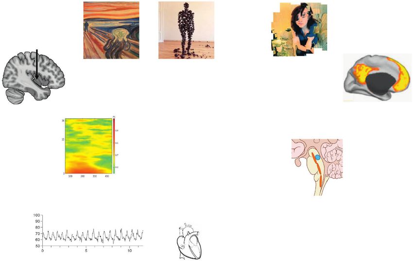
S. Tumati et al.

   We base our suggestion on evidence that converges           chronic and unrelenting anxiety, are preferentially suscep-
upon the role of abnormal spatiotemporal mechanisms of         tible to “top-down constructed dysfunctions”, i.e., they are
cardiac interoception in anxiety. We focus more on AD          the consequences of a persistent mismatch between top-
rather than primarily on anxiety as a trans-diagnostic         down predicted body states and bottom-up afferent signals
symptom because of the relative strength of evidence in        from the body. It has been proposed that sustained and
these disorders for our proposal. We also focused on car-      exaggerated mismatches dysregulate the ability to accu-
diac function rather than on interoceptive abnormalities in    rately sense what is happening in the body, resulting in a
general because recent advances show how cardiac activity      turbulent reference state (i.e., a “noisy baseline”) [21],
influences neural activity at a high temporal resolution.       attentional bias toward threats [22], increased worry and
More generally, our proposal attributes psychopathologi-       self-related cognitions, dysfunctional learning about bodily
cal symptoms of AD to spatiotemporal mechanisms, such          states over time [23], and increased allostatic load leading to
as brain-heart synchronization, rather than to affective or    increased stress and mental illness [24, 25].
cognitive abnormalities [5–11].                                   In an adaptive individual, corrective action in the pre-
                                                               sence of somatic error can be achieved by adjusting the
                                                               expectations (priors) to match the current physiological
Anxiety—an asymmetric response to benign                       state, or by engaging in regulatory actions which change
stimuli                                                        the afferent signal, leading the current physiological state
                                                               to conform more closely with the expectations. In either
Anxiety may be defined as a state of worry due to the           case, successful corrective action reduces somatic error,
anticipation of uncertain or undesirable outcomes [12]. This   which results in homeostatic balance within the nervous
can be useful in healthy individuals as uncertainty may        system. This can break down in two ways. Firstly,
prompt increased attention and arousal in order to avoid       hyperprecise priors, i.e., having very strong beliefs that a
negative events. However, persistent worry where innoc-        certain model is correct driven by prolonged periods of
uous stimuli are considered threatening constitutes a mental   worry and rumination, may not get appropriately updated
disorder and limits day-to-day functioning. Anxiety may be     by the evidence, which can create persistent somatic
subdivided into anxious apprehension (enduring worry of        errors or a “noisy” baseline state [21]. Secondly, context
negative events) and anxious arousal (hyperarousal and         rigidity, i.e., the lack of one’s ability to adjust expectation
exaggerated response to benign stimuli) [13]. These con-       as a function of context, may contribute to the persistent
structs are regarded as traits, suggesting that individuals    experience of somatic error because the individual does
have a propensity for anxiety.                                 not adjust her belief about different models in a new
   The above view considers psychological processes of         environment. In its most severe form, the somatic error
prospecting and uncertainty reduction to underlie anxiety;     becomes so pervasive that the only corrective action that
a broader view suggests abnormal interoceptive proces-         seems to quell the error is avoidance of all perceived
sing as a basis for anxiety [14–16]. In this view, beha-       triggers (i.e., agoraphobia).
vioral and autonomic symptoms are attributed to top-              The symptoms of AD are considered as responses either
down modulation from brain regions that monitor inter-         to reduce uncertainty or to avoid consequent symptoms
oceptive signals [14, 15, 17, 18]. It has been hypothesized    [12, 26]. However, each of the disorders show a con-
that the central nervous system implements active              stellation of overlapping and distinct symptoms (see Box 1).
inference perceptual processing hierarchically and bi-         GAD maybe described by the presence of internally focused
directionally [19], i.e., models at a lower level of the       apprehension without prominent external symptoms.
hierarchy serve as evidence for models at a higher level,      Patients with PD, on the other hand, suffer from sudden
i.e., bottom-up modulation. In turn, higher-level models       unpredictable bouts of extreme anxiety, a sense of
modify lower-level models, i.e., top-down modulation, to       impending doom, and overwhelming somatic symptoms. In
match their prediction. Minimization of the energetic cost,    the context of interoceptive basis for anxiety, the prominent
i.e., the free energy principle proposed by Friston, aims to   cardiac symptoms and heightened bodily awareness are
provide a computational explanation for how the brain          especially noteworthy. In contrast to GAD and PD, feelings
optimizes (selects) perceptions in the presence of multiple    of anxiety in SAD and specific phobias are cue-specific.
expectations and models [20]. Via the active inference         Overt anxiety in SAD is limited to specific circumstances
process, the brain ultimately settles on a perception that     such as social settings or public-speaking, which may lead
results in the least divergence between the available evi-     patients to avoid these situations or endure them with dis-
dence and the predicted model.                                 tress. In both PD and SAD, the somatic or “external”
   A consequence of active inference models is that certain    symptoms are more prominent than in GAD where the
psychiatric disorders, especially those characterized by       symptoms are more internally oriented. This heterogeneity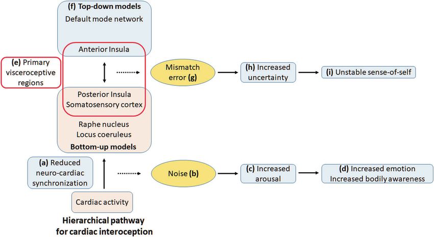
Out-of-step: brain-heart desynchronization in anxiety disorders

                                                                              In GAD, RSFC is reduced between the midline regions of
 Box 1. Anxiety disorders: specific-symptom constellations
                                                                           the DMN—the perigenual and posterior cingulate cortex
 GAD is characterized by presence of excessive worry over                  (pgACC, PCC) [38]. However, another study found both
 multiple issues and anxious apprehension of untoward outcomes             increased and decreased RSFC within regions of the DMN
 [1]. Despite the absence of specific sources, feelings of anxiety are
 difficult to control. Somatic symptoms are less prominent than             [39]. In a resting state and task-based intervention study in
 other AD and include restlessness, excessive fatigue, irritability,       GAD, resting state scans were obtained before and after a
 difficulty concentrating, muscle tension, sleep disturbance, head-         perseverative cognition task asking subjects to recall worrying
 aches, and gastrointestinal symptoms. These symptoms must be              episodes [40]. A greater increase in worry was associated with
 present for more than 6 months. The internally oriented rumination
 over varied concerns with mild physical symptoms differentiate            a greater decline in amygdala—VMPFC connectivity.
 GAD from other AD that are triggered by external cues.                       In SAD, midline regions of the DMN showed reduced
 SAD is characterized by fear or worry limited to social situations        RSFC [41, 42]. Specifically, RSFC is consistently reduced
 in which either the individual may be scrutinized or are afraid of        between the amygdala and the medial prefrontal cortex and
 acting in a way that elicits negative responses from others [1]. To
 avoid the symptoms, social situations are avoided or endured under        posterior cingulate cortex [41, 43–46]. Whole-brain RSFC
 distress. Somatic symptoms are typically limited to social                studies also report similar results [42, 47]. However, albeit
 situations, and may include palpitations, excessive sweating,             in a small sample, increased RSFC in the amygdala has
 tremors, blushing, or muscle tension.                                     been reported [36]. In PD, both increased and reduced
 PD is characterized by sudden and unexpected attacks of extreme
 anxiety accompanied by strong somatic symptoms including                  RSFC within the DMN were reported [45, 48, 49]. In the
 palpitations, tightness of chest, feeling short of breath, sweating,      SMN, changes in RSFC appear to be specific to PD. Using a
 and trembling [1]. Patients feel a fear of losing control and a fear of   whole-brain approach, Cui et al. [50] observed increased
 death. An attack is followed by a persistent fear of a recurrence.        RSFC between the postcentral cortex (i.e., sensory cortex)
 The abrupt nature of the attack along with dominant somatic
 (especially cardiac) symptoms distinguish PD from GAD                     and the thalamus, which also correlated with the degree of
 and SAD.                                                                  anxiety (see also [37]).
                                                                              Thus, studies in AD show a shared reduction in RSFC
                                                                           in the midline regions of DMN and SN. In addition, RSFC
in symptom clusters of AD along with a commonality in                      between networks (DMN to executive network, SN to
anxiety symptoms is also reflected on the neural level where                SMN) is also reduced, whereas RSFC between the
these disorders show common as well as specific brain                       amygdala and anterior DMN appears to be increased
changes [27].                                                              [3, 29]. In individual disorders, changes in the SN and
                                                                           SMN appear to be relatively more specific—increased
                                                                           RSFC in SN in SAD, and increased RSFC from SMN to
Decoupled networks: brain changes in                                       thalamus in PD (Fig. 1).
anxiety disorders
                                                                           Task-evoked abnormalities in SN, DMN, and SMN
Resting state abnormalities in DMN, SN, and SMN—                           during interoception of heartbeats
functional connectivity
                                                                           The anterior insula, cingulate cortex, and somatomotor
The large number of functional imaging studies on AD                       cortex, which show abnormal RSFC in AD, are ‘activated’
implicate a variety of regions (see [3, 28–32] for reviews                 when healthy subjects are asked to focus on their heart-
and meta-analyses). Broadly speaking, the aggregate chan-                  beat [51–55]. Awareness of heartbeats is also exaggerated
ges in functional connectivity across these disorders appear               in anxiety and AD [15, 56], which may be linked to the
to be reduced [3]. However, like their symptoms, these                     neural changes in these disorders. Indeed, interoceptive
disorders show common and distinct patterns of con-                        tasks in AD show increased activation, which is correlated
nectivity changes [29]. These regions include the amygdala,                to anxiety symptoms [57–59]. Furthermore, this increased
insula, somatosensory cortex, ventromedial prefrontal cor-                 activation maybe linked to RSFC in GAD [60]. In drug-
tex (VMPFC), and the precuneus and posterior cingulate                     naïve patients, the insula showed increased activity during
cortex. Together, these regions are part of three canonical                a cardiac interoception task as well as reduced RSFC to
functional networks—the salience network (SN), the                         the VMPFC. In addition, the task-evoked activity was
somatomotor network (SMN), and the default mode net-                       correlated with ‘psychic anxiety’ and reduced RSFC was
work (DMN) [33, 34]. Broadly, the DMN shows reduced                        negatively correlated with ‘somatic anxiety’ (on the
RSFC in GAD, and SAD, whereas in PD, studies show both                     Hamilton Anxiety Rating Scale). Although more studies
increased and decreased RSFC. The SN shows increased                       are needed, this result shows that interoception in AD may
RSFC specifically in SAD; and the SMN shows increased                       be related to abnormalities in the same regions affected in
RSFC specifically in PD [35–37].                                            the resting state.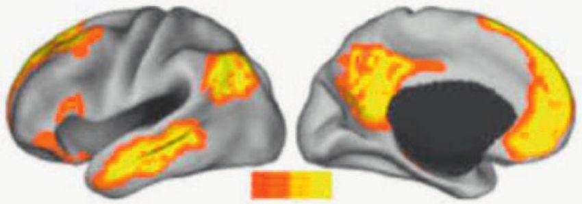
S. Tumati et al.

      Default-mode network: Reduced RSFC in GAD, SAD, and PD
                                                                          Mechanisms of neuro-cardiac coupling in the
                                                                          healthy brain

                                                                          Regions showing neuro-cardiac coupling are part of
                                                                          the DMN, SN, and SMN

                                                                          Regions in the DMN, SMN, and SN receive visceral inputs
                                                                          such as those from the heart, lungs, and gastric activity (see
                                                                          [69, 70] for recent reviews). Together, these regions are termed
             Somatomotor network: Increased RSFC in PD
                                                                          as the ‘visceral/central autonomic network’, which monitor the
                                                                          internal state of the body [71]. As described below, studies
                                                                          show that cardiac activity modulates neural activity in these
                                                                          regions through a phase-based mechanism [72].
                                                                             In healthy subjects, HRV is correlated with variability in
                                                                          neural activity in the insula, amygdala, and anterior cingulate
                                                                          cortex [73–75]. The high-frequency component of HRV in
                                                                          particular is correlated with the variability of RSFC in the
                                                                          regions of the ‘central autonomic network’—the VMPFC
                                                                          (and adjacent perigenual anterior cingulate cortex/PACC),
              Salience network: Increased RSFC in SAD                     somatosensory and somatomotor cortex, and subcortical
                                                                          regions (like periaquaeductal gray, thalamus, and lentiform
                                                                          nucleus) [74–77]. Considering that cardiac interoceptive tasks
                                                                          also evoke activity in the same regions [51–53, 78–81], these
                                                                          findings further suggest that cardiac and neural activity share
                                                                          a temporal inter-dependence in these regions.
                                                                             In the context of AD, the overlap between regions involved
                                                                          in processing cardiac activity and neural activity in affected
Fig. 1 Altered resting state functional connectivity (RSFC) in dif-
ferent neural networks in anxiety disorders. Systematic and meta-         functional networks suggests that the ongoing integration of
analytic reviews show that anxiety disorders are associated with          cardiac inputs in these regions may underlie the changes in
reduced functional connectivity within and between the salience net-      RSFC (Fig. 2). Furthermore, the mechanism for neuro-cardiac
work, the somatomotor network, and the default mode network.
                                                                          synchronization in these regions may account for the abnor-
However, a closer look shows disorder-specific network changes such
as a higher RSFC in the salience network in social anxiety disorder and   mal interoceptive processing in these disorders.
in the somatomotor network in panic disorder.
                                                                          Temporal synchronization of heart and brain—
                                                                          heartbeat-evoked potential and intertrial coherence
   The SN and specifically the insula are implicated in AD
by other task paradigms, such as emotion regulation,                      Magnetoencephalography          and    intracranial     electro-
emotion recognition, and fear regulation [28–30, 61, 62]                  encephalography studies show that cardiac activity mod-
(GAD-specific studies [63–65], and SAD-specific studies                     ulates neural activity as well as functions ranging from
[66, 67]). Abnormal activation in the amygdala is found in                perception to the sense-of-self (see [70, 82, 83] for reviews).
GAD, SAD, and PD whereas the medial prefrontal cortex                     These studies suggest that neural activity in these regions
and posterior insula changes are specifically associated                   are phase-locked to cardiac inputs [72, 84]. Similar to a task
with PD [27, 68].                                                         stimulus, each heartbeat induces an event-related potential
   Taken together, neural changes in AD can be localized to               in the brain, termed as ‘heartbeat-evoked potential’ (HEP)
affective regions such as the amygdala, regions in the SN—                [70, 85, 86]. The HEP is the increased amplitude of neural
particularly the insula, the anterior and posterior DMN                   activity that is time-locked to the R-wave of cardiac activity.
regions, and regions in the primary somatosensory cortex.                    Park et al. [72, 87–89] observed that the HEP induces a
RSFC changes within these networks are mostly reduced,                    reset in the phase of ongoing neural activity resulting in the
though disorder-specific increase in RSFC also occurs.                     phase of ongoing neural fluctuations being locked to the
Importantly, the neural correlates of interoception in healthy            timing of heartbeats. This phase-locking (between 4 and 7
and anxious individuals also localize to these regions.                   Hz frequencies) can be measured by intertrial coherence
However, it is not clear how abnormal interoceptive pro-                  (ITC), and is high after each HEP [72]. Moreover, higher
cessing in AD is related to its network changes.                          values of ITC were associated with a higher HEP. The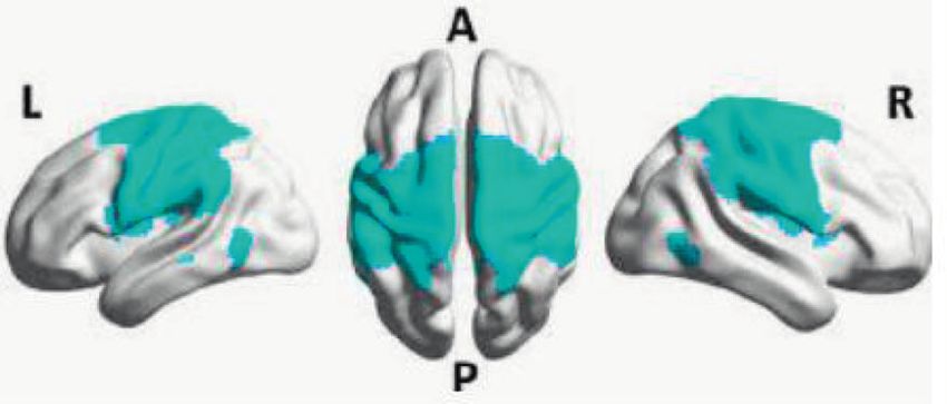
Out-of-step: brain-heart desynchronization in anxiety disorders

                                                                          Is neuro-cardiac phase synchronization related to
                                                                          functional connectivity?

                                                                          The relation between neuro-cardiac synchronization and
                                                                          brain-wide functional networks is less clear. We suggest a
                                                                          theoretical-computational and biochemical basis relating
                                                                          visceral inputs such as from the heart to the brain’s
                                                                          functional connectivity. The alignment between rhythmic
                                                                          neural and cardiac activity feeds information about the
                                                                          visceral state to the brain. As per active inference-based
                                                                          theories, top-down predictive models are matched with
                                                                          this bottom-up information. Visceral inputs ascend
                                                                          to the brainstem nuclei and are relayed forward to the
                                                                          thalamus and the cortex, where the posterior insula and
                                                                          primary somatosensory cortex are its primary targets. This
                                                                          information is fed to the anterior insula, which is regarded
                                                                          as a primary visceromotor cortex, i.e., the higher-level
                                                                          region generating predictions of the bodily state and
                                                                          modulating interoceptive inputs to match predicted states
                                                                          [14, 21, 55].
                                                                             Changes in the bodily state increase the mismatch error
Fig. 2 Overlap in regions processing cardiac activity and regions         leading to increased interoceptive awareness. This change
affected in anxiety disorders. The regions underlying the functional
networks altered in anxiety disorders show a significant overlap with      is accompanied by activation of monoaminergic brainstem
regions that are associated with low and high-frequency heart rate        nuclei that receive this input—namely the serotonergic
variability. These regions are also suggested to receive visceroceptive   raphe nucleus and noradrenergic locus coeruleus, which in
inputs, and are proposed to form a visceral or central autonomic net-     turn increase activity in the amygdala, insula, and orbi-
work. Regions in this network modulate lower and higher cognitive
functions such as perception and sense of self.                           tofrontal cortex (the so-called ‘fear circuit’). Tracer stu-
                                                                          dies show strong reciprocal connections between these
                                                                          regions—ascending inputs from brainstem nuclei are
authors therefore conclude that the amplitude of HEP can, at              relayed to the thalamus and then to the posterior insula
least partly, be linked to the phase of neural activity [72].             and primary somatosensory cortex. The posterior insula
Both HEP and ITC can therefore be regarded as markers of                  modulates connectivity of the anterior insula, which
neuro-cardiac synchronization                                             shows bi-directional connections to dorsal anterior cin-
   Neuro-cardiac phase coupling may also be related to                    gulate cortex, orbitofrontal cortex, and the amygdala
RSFC, which is typically derived from fMRI data in the ~0.1               [55, 69]. Taken together, the visceral (including cardiac)
Hz frequency range. Using fMRI, Pfurtscheller et al. [90, 91]             input to the brain, and its prediction and modulation by
demonstrated that the vascular component of the BOLD                      both top-down processes and neurotransmitters provide a
signal could be separated from neural oscillations based on               basis to explain how abnormal interoception may lead to
their timing, with the former preceding the latter. Moreover,             altered functional networks.
in healthy subjects with high anxiety levels in the fMRI
scanner, neuro-cardiac phase coupling in the insula and pre-
central gyrus as well as between the amygdala and medial                  Neurotransmitter systems—targets of
prefrontal cortex was increased. These findings allow us to                visceral inputs and opposite modulators of
extend phase-resetting, a M/EEG measure of neuronal com-                  SMN/SN and DMN
munication and information transfer [92, 93] to fMRI data for
abnormal neuro-cardiac synchronization in AD.                             Monoaminergic neurotransmitter systems originate in brain-
   More generally, neural activity comprises of continuous                stem nuclei and are targets of visceral afferents, which ascend
fluctuations and oscillations [94, 95]. The phase of this                  through the spinal cord to reach them (Fig. 3, see [55] for a
rhythmic activity shifts in response to external stimuli such             detailed description of interoceptive pathways). Projections
as when listening to music—described as entrainment                       from these nuclei carry the afferents forward to the thalamus
[96, 97] or alignment [98]. A similar response to continuous              and then to primary visceroceptive cortical regions. These
internal stimuli like heartbeats appear to entrain neural                 structures include the serotonergic raphe nucleus and the
activity in regions supporting allostasis.                                noradrenergic locus coeruleus, which are implicated in AD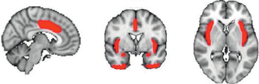
S. Tumati et al.

                                                                                Increased                         Increased                                               Unstable
                                                                 (g)
                                                                             emotion & anxiety                 bodily awareness                                          sense of self

                       Posterior Insula

                                                                 (c)        Salience network               Somatomotor network                                       Default mode network

                                                                                               Increased                                                                   Decreased        (f)
                                                                  (d) Lower phase
                                                                  synchronization
                                                                                                                            Functional Connectivity
                                               Freq. (log Hz)

                                                                                                           Opposite reciprocal modulation of cortical networks (e)

                                                                                                                  (b) Raphe nucleus (serotonin, in orange)
                                                                                                                  Locus coeruleus (noradrenaline, in blue)
                                                                       Time (ms)

                                                                                                                                         Visceral afferent
                                                                (h) Reduced variability
                            Heart rate (bpm)

                                                                                                                                 Cardiac activity (a)

                                                                        Time (min)

Fig. 3 Neuro-cardiac desynchronization, functional networks, and                                                                      evoked potential. This desynchronization alters serotonergic and nor-
symptoms of anxiety disorders. Cardiac afferents (a) first reach the                                                                   adrenergic activity and leads to opposite reciprocal modulation of
serotonergic raphe nucleus and noradrenergic locus coeruleus (b), and                                                                 cortical networks (e) with increased RSFC in the salience network and
are then relayed onwards to the primary visceral cortex (posterior                                                                    in the somatomotor network whereas RSFC decreases in the DMN (f).
insula and primary somatosensory cortex) (c), which are abnormal in                                                                   These network changes manifest as heightened emotions and anxiety,
anxiety disorders. We suggest that synchronization between the car-                                                                   increased bodily awareness, and an unstable sense-of-self (g). Notably,
diac and neural regions is abnormal in these disorders and can be                                                                     anxiety disorders are also associated with lower cardiac variability (h)
measured by lower intertrial coherence (ITC) (d), and heartbeat-                                                                      potentially indicating visceromotor modulation.

[99]. These nuclei modulate functional networks and may                                                                               Moreover, this process shapes mental features such as
help explain the network changes seen in AD.                                                                                          visual perception [87, 89], emotion, bodily awareness
   Conio et al. [100] demonstrate that the serotonergic raphe                                                                         [72], and sense-of-self [88, 104, 105]. The degree of
nucleus modulate SMN/SN and DMN connectivity in an                                                                                    neuro-cardiac synchronization in the visual cortex and
opposite reciprocal manner. They suggest that increased                                                                               insula was associated with visual perception [87, 89].
serotonergic signaling is associated with decreased SMN                                                                               Similarly, the amplitude of HEP in the insula and the
activity and increased DMN activity. Although the locus                                                                               somatosensory cortex were related to bodily awareness
coeruleus was not directly associated with cortical network                                                                           [88]. Higher-order functions such as subjects’ experience
modulation by Conio et al., it is worth noting that it receives                                                                       of their self as either “I” (‘subjective self’ referring to
visceral afferents, is activated by stress and in AD, and                                                                             thoughts with the self as the agent) or “me” (‘objective
increases connectivity in multiple networks including the                                                                             self’ referring to thoughts about themselves) was related
SN, SMN, and amygdala [101–103]. Together, the findings                                                                                to the amplitude of the HEP specifically in the VMPFC
show opposite modulation of cortical SMN/SN and DMN                                                                                   (‘Me’), and in the PCC and right anterior insula (‘I’)
by subcortical serotoninergic raphe nucleus with potential                                                                            [88, 105].
involvement of noradrenergic locus coeruleus. This is                                                                                    The DMN is known to be involved in our sense-of-self
consistent with AD where opposite changes in these net-                                                                               [106–108], the SMN is related to bodily awareness [109],
works as well as abnormalities in serotonergic and nora-                                                                              and the SN is central to mediating the sense-of-self
drenergic systems occur.                                                                                                              [110, 111] and emotions [112]. Given the findings by
                                                                                                                                      Tallon-Baudry et al., neuro-cardiac synchronization can
Brain-heart synchronization shapes mental                                                                                             be said to modulate mental features like emotions, bodily
functions—emotion, bodily awareness, and the                                                                                          awareness, and sense-of-self (see [83] who make this
mental self                                                                                                                           point; also see [69, 113]). In the context of interoceptive
                                                                                                                                      deficits in AD, reduced neuro-cardiac synchronization
The aforementioned studies show that phase-resetting of                                                                               may reduce the efficacy of functions localized to these
neural activity may underlie neuro-cardiac coupling.                                                                                  regions/networks.
Out-of-step: brain-heart desynchronization in anxiety disorders
Brain-heart desynchronization and trigger altered activity (decrease in the serotonergic sys-
abnormal cortical networks in AD tem, and increase in the noradrenergic system), which
leads to an imbalance between the SMN or SN and the
It was recently suggested that neuro-cardiac synchroniza- DMN (Fig. 3).
tion underlies the RSFC changes and symptoms of AD
[114]. Here, we expand this proposal by describing the Abnormal neuro-cardiac synchronization affects
process that may underlie the observed dysfunctions in each emotional processing and interoceptive awareness
network.
Symptoms of AD are congruent with the functions attrib-
Reduced heart rate variability in anxiety disorders uted to the affected networks. Increased emotional sensi-
tivity, bodily awareness, and unstable sense-of-self can be
HRV, a marker of health [115, 116], is consistently attributed to the SN, SMN, and DMN, respectively [114]
reduced in AD [2]. HRV is also reduced in major (Fig. 3). Hence, symptoms and RSFC in AD can be linked
depressive disorder, where anxiety is common [117]. It is to neuro-cardiac synchronization in these regions. Indeed,
measured either by the root mean square of the successive features of AD such as fear processing and interoception are
differences (RMSSD) or by high (HF) and low frequency linked to cardiac function [84, 122]. Garfinkel et al. found
(LF) variability. Studies in GAD, PD, and SAD that fear processing was more sensitive and intense when
show lower RMSSD and HF HRV (medium effect size) the cardiac cycle was in systole (corresponding to the HEP),
but normal LF HRV, which suggests a stronger para- showing that neuro-cardiac coupling alters emotional pro-
sympathetic input to the heart [2]. cessing. Pollatos et al. showed that time perception was
Reduced HRV in AD contributed to an influential model positively correlated to phase-locking with cardiac activity
of neurovisceral integration [71, 118, 119], which describes and interoceptive sensitivity [84]. These studies suggest that
how cardiac and other visceral activity is regulated across ITC/HEP may be related to emotional and interoceptive
the neural hierarchy. In this model, the relation between sensitivity in AD.
cardiac (visceral) activity, and emotional and cognitive Notably, subjective (sensibility) and objective (sensitiv-
functions is explained using concepts from dynamical sys- ity/accuracy) interoceptive processing may not be correlated
tems theory and predictive coding. Deviations (or predictive in AD. Subjects with autism, who also have anxiety, were
errors) in cognitive or sensory inputs are proposed to reduce found to perceive bodily signals more strongly (higher
cortical inhibitory input (parasympathetic/vagal tone) to the sensibility) but were worse in interoceptive accuracy
heart, leading to reduced HRV. (objective measures). The discrepancy between their sub-
jective sense of accuracy (confidence) and the difference
Serotonergic and noradrenergic systems—opposite between objective and subjective measures of interoception
reciprocal modulators of abnormal functional predicted their anxiety symptoms [79, 123].
networks in AD A few studies have recently investigated neuro-cardiac
synchronization with HEP/ITC in AD. In GAD subjects,
The monoaminergic transmitters play a key role in the HEP was abnormally high in the eyes-open condition, but
understanding and treating AD through their innervation the change in HEP from eyes-open to eyes-closed con-
of the so-called “fear circuit” (amygdala, insula, and dition was lower than in controls [124]. Moreover, HEP in
orbitofrontal cortex) [99]. They may also explain how the prefrontal cortex was correlated to anxiety symptoms.
cardiac inputs from the periphery affect cortical networks. In another study, false cues of increased heart rate induced
As described above, the serotonergic raphe nuclei and larger HEP in healthy subjects with high social anxi-
noradrenergic locus coeruleus receive cardiac inputs, and ety [125]. These studies provide the first evidence for
relay it forward to the thalamus and then to the primary neuro-cardiac mechanisms in AD. Further studies are
visceral cortex (Fig. 3). In AD, lower serotonergic activity needed to spatially localize these changes and relate ITC
is consistently reported [99] (though this has been ques- to symptoms.
tioned recently [120, 121]). This lower serotonergic Finally, we suggest that inter-individual variations in
activity, as per Conio et al. [100], suggests increased neuro-cardiac synchronization may explain the propensity
SMN RSFC and lower DMN RSFC. Similarly, increased for anxiety and AD in some individuals. This suggestion is
noradrenergic activity suggests increased SN RSFC and consistent with the view that visceral inputs shape cognitive
decreased DMN RSFC. These predictions are consistent functions [17, 69]; rather than driven by external inputs,
with the observed network abnormalities in AD. Thus, internal (visceral) inputs predispose neural responses to
taken together, abnormal neuro-cardiac synchronization in external stimuli. A mis-alignment between the brain and the
the subcortical targets of cardiac afferents is suggested to body may predispose individuals to anxiety.S. Tumati et al.
Fig. 4 Abnormal neuro-cardiac synchronization in an inter- emotion and somatic symptoms (d). The noisy bottom-up models in
oceptive predictive coding framework. Neuro-cardiac synchroniza- the primary visceroceptive regions (e) do not match predictive models
tion provides a mechanism for the interoceptive predictive processing generated in the higher-order cortical regions (f), generating persistent
framework and neurovisceral intergration model. Lower phase syn- somatic errors (g) that lead to increased uncertainty (h) and at the
chronization (a) leads to bottom-up models being perceived as ‘noisy’ mental level, to an unstable sense-of-self and worry (i).
(b) leading to increased arousal (c), which manifests in increased
Predictive interoceptive processing—noise, higher subjective interoceptive sensibility (own belief that
desynchronization of functional networks, they are accurate) despite lower objective accuracy [123].
and symptoms of AD The mismatch error triggered corrective signals may reflect
functional connectivity, which would be consistent with the
The insula, along with the somatosensory cortex, is con- increased RSFC in AD in the SN and SMN—networks
sidered to be the primary visceroceptive cortical region. We containing the primary visceral cortex. The uncertainty in
suggest that an abnormal synchronization in the posterior top-down models could be related to the decreased RSFC
insula may result in a higher degree of neuronal ‘noise’ in the DMN, which does not receive direct visceroceptive
(Fig. 4). That is, a lower degree of synchronization reduces inputs.
certainty about information regarding the bodily state, Finally, the isolated decrease in DMN RSFC raises the
thereby leading to a mismatch between higher and lower- possibility that the degree of neuro-cardiac desynchroniza-
level models in the neural computational hierarchy. The tion influences the manifested symptoms. A small error may
Embodied Predictive Interoception Coding hypothesis increase neural and psychological uncertainty but not trig-
suggests that higher-level predictions are represented in the ger strong top-down corrective signaling. This would
anterior insula [14]. A mismatch between higher and lower- manifest as lower RSFC in the DMN without changes in the
level models are suggested to trigger corrective viscer- SN or SMN, and correspondingly lead to persistent anxiety
omotor signals from the anterior insula to the posterior but without overwhelming somatic symptoms, which is
insula to either correct the mismatch or to adapt signal characteristic of GAD. Abnormal DMN connectivity across
sampling so that subsequent errors are minimized (Fig. 4). the disorders also indicates a basic deficit in self-related
The neuronal noise may also result in non-neuronal or processing [126]. As discussed above, cardiac inputs affect
perceptual noise (Fig. 4). Noisy afferent inputs may reduce all levels of the neural hierarchy from perception to the
certainty of the bodily state in higher-level models. This meta-cognitive objective self. That is, visceral inputs are
uncertainty may result in a persistent somatic error and part of automatic self-related processing and hence, deficits
hence, a strong belief of an abnormal bodily state (hyper- in this processing would lead to increased self-specificity or
precise prior). The persistent error would also lead to con- self-prioritization. This persistent deficit may contribute to a
text rigidity (i.e., inability to change beliefs despite ‘fearful self’ that continuously anticipates threats [127].
changing contexts) as bottom-up information would con- Together, the abnormal neuro-cardiac phase synchroni-
tinue to remain noisy in varying contexts, preventing zation in the primary visceroceptive regions leads to neu-
updating of top-down models. Consistent with this sug- ronal and perceptual noise or uncertainty of the bodily
gestion, anxiety symptoms in autism are correlated with state. Higher-order regions trigger corrective visceromotorOut-of-step: brain-heart desynchronization in anxiety disorders
signaling to reduce this noise. The desynchronization and Publisher’s note Springer Nature remains neutral with regard to
jurisdictional claims in published maps and institutional affiliations.
top-down signaling may lead to activation of subcortical
and cortical regions reflected in increased functional con-
nectivity in the SMN/SN and corresponding decrease in the
DMN. This in turn, may manifest in the particular symptom References
constellation of AD with changes in body awareness,
1. American Psychiatric Association. Diagnostic and statistical
emotion, and sense-of-self. manual of mental disorders. 5th ed. American Psychiatric
Association; 2013.
2. Chalmers JA, Quintana DS, Abbott MJ-A, Kemp AH. Anxiety
disorders are associated with reduced heart rate variability: a
Conclusion meta-analysis. Front Psychiatry. 2014;5:80.
3. Xu J, Van Dam NT, Feng C, Luo Y, Ai H, Gu R, et al. Anxious
In summary, we identified abnormal phase-resetting, which brain networks: a coordinate-based activation likelihood estimation
can be experimentally measured by ITC and HEP, as a meta-analysis of resting-state functional connectivity studies in
anxiety. Neurosci Biobehav Rev. 2019;96:21–30.
mechanism for neuro-cardiac desynchronization in AD.
4. Etkin A, Wager TD. Functional neuroimaging of anxiety: a
This neuro-cardiac desynchronization underlies changes in meta-analysis of emotional processing in PTSD, social anxiety
serotonergic and noradrenergic neurotransmission that lead disorder, and specific phobia. Am J Psychiatry. 2007;164:
to opposite modulation of cortical networks like SMN/SN 1476–88.
5. Fingelkurts AA, Fingelkurts AA. Brain space and time in mental
and DMN and the typical AD symptom constellation with
disorders: paradigm shift in biological psychiatry. Int J Psychiatry
increases in bodily awareness and anxiety accompanied by Med. 2019;54:53–63.
an unstable sense-of-self. We also integrate neuro-cardiac 6. Northoff G. Spatiotemporal psychopathology I: no rest for the
desynchronization in interoceptive predictive processing brain’s resting state activity in depression? Spatiotemporal psy-
chopathology of depressive symptoms. J Affect Disord. 2016;
models as a source of noise and mismatch between bottom-
190:854–66.
up and top-down models, leading to somatic errors and 7. Northoff G. Spatiotemporal psychopathology II: how does a
further to the particular symptom constellation of AD. psychopathology of the brain’s resting state look like? Spatio-
Together, we propose neuro-cardiac desynchronization temporal approach and the history of psychopathology. J Affect
Disord. 2016;190:867–79.
as a primarily spatiotemporal basis for the particular
8. Northoff G. The brain’s spontaneous activity and its psycho-
symptom constellation of AD, in line with “Spatiotemporal pathological symptoms - ‘Spatiotemporal binding and integra-
Psychopathology” [6, 7]. tion’. Progress in Neuro-Psychopharmacology and Biological
Our suggestions also raise further questions. These Psychiatry. 2017. https://doi.org/10.1016/j.pnpbp.2017.03.019.
9. Northoff G, Magioncalda P, Martino M, Lee H-C, Tseng Y-C,
include—(1) is HRV related to neural variability in visceral
Lane T. Too fast or too slow? Time and neuronal variability in
regions of the brain? (2) Are these region-specific correla- bipolar disorder—a combined theoretical and empirical investi-
tions related to specific symptoms? (3) Subcortical regions gation. Schizophr Bull. 2018;44:54–64.
also receive visceral inputs [69, 71] and are affected in AD 10. Northoff G, Duncan NW. How do abnormalities in the brain’s
spontaneous activity translate into symptoms in schizophrenia?
[3]. Are ITC/HEP in these regions abnormal in these dis-
From an overview of resting state activity findings to a proposed
orders, and if so, what is their relation to the implicated spatiotemporal psychopathology. Prog Neurobiol. 2016. https://
cortical regions. (4) We did not consider other visceral doi.org/10.1016/j.pneurobio.2016.08.003.
inputs—respiration and gastric rhythm, which also affect 11. Northoff G, Stanghellini G. How to link brain and experience?
Spatiotemporal psychopathology of the lived body. Front Hum
interoception [128, 129] due to a paucity of studies in AD.
Neurosci. 2016;10:76.
The phase coupling of respiratory and gastric activity to 12. Grupe DW, Nitschke JB. Uncertainty and anticipation in anxiety:
neural activity in AD would be of interest as these subjects an integrated neurobiological and psychological perspective. Nat
often show abnormal perception of respiratory and/or gas- Rev Neurosci. 2013;14:488–501.
13. Sharp PB, Miller GA, Heller W. Transdiagnostic dimensions of
tric activity. (5) Do other oscillatory measures, such as
anxiety: neural mechanisms, executive functions, and new
phase amplitude coupling [97] or those from dynamical directions. Int J Psychophysiol J Int Organ Psychophysiol. 2015;
systems [71] provide additional insight into these disorders? 98:365–77.
Finally, (6) can interventions be targeted towards ser- 14. Barrett LF, Simmons WK. Interoceptive predictions in the brain.
Nat Rev Neurosci. 2015;16:419–29.
otonergic- and/or noradrenergic-driven neuro-cardiac
15. Paulus MP, Stein MB. Interoception in anxiety and depression.
desynchronization to yield novel therapies for these Brain Struct Funct. 2010;214:451–63.
disorders? 16. Stephan KE, Manjaly ZM, Mathys CD, Weber LAE, Paliwal S,
Gard T, et al. Allostatic self-efficacy: a metacognitive theory of
dyshomeostasis-induced fatigue and depression. Front Hum
Compliance with ethical standards Neurosci. 2016;10:550.
17. Paulus MP, Feinstein JS, Khalsa SS. An active inference approach
Conflict of interest The authors declare that they have no conflict of to interoceptive psychopathology. Annu Rev Clin Psychol. 2019;
interest. 15:97–122.S. Tumati et al.
18. Seth AK, Friston KJ. Active interoceptive inference and the 38. Andreescu C, Sheu LK, Tudorascu D, Walker S, Aizenstein H.
emotional brain. Philos Trans R Soc Lond B Biol Sci. 2016;371: The ages of anxiety—differences across the lifespan in the
1708.20160007. default mode network functional connectivity in generalized
19. Friston K. Hierarchical models in the brain. PLoS Comput Biol. anxiety disorder. Int J Geriatr Psychiatry. 2014;29:704–12.
2008;4:e1000211. 39. Wang W, Hou J, Qian S, Liu K, Li B, Li M, et al. Aberrant
20. Friston K, Schwartenbeck P, FitzGerald T, Moutoussis M, regional neural fluctuations and functional connectivity in gen-
Behrens T, Dolan RJ The anatomy of choice: dopamine and eralized anxiety disorder revealed by resting-state functional
decision-making. Philos Trans R Soc Lond B Biol Sci. 2014;369. magnetic resonance imaging. Neurosci Lett. 2016;624:78–84.
21. Paulus MP, Stein MB. An insular view of anxiety. Biol Psy- 40. Makovac E, Watson DR, Meeten F, Garfinkel SN, Cercignani M,
chiatry. 2006;60:383–7. Critchley HD, et al. Amygdala functional connectivity as a
22. Hakamata Y, Lissek S, Bar-Haim Y, Britton JC, Fox NA, Leibenluft longitudinal biomarker of symptom changes in generalized
E, et al. Attention bias modification treatment: a meta-analysis anxiety. Soc Cogn Affect Neurosci. 2016;11:1719–28.
toward the establishment of novel treatment for anxiety. Biol 41. Hahn A, Stein P, Windischberger C, Weissenbacher A, Spin-
Psychiatry. 2010;68:982–90. delegger C, Moser E, et al. Reduced resting-state functional
23. Van den Bergh O, Witthöft M, Petersen S, Brown RJ. Symptoms connectivity between amygdala and orbitofrontal cortex in social
and the body: taking the inferential leap. Neurosci Biobehav anxiety disorder. NeuroImage. 2011;56:881–9.
Rev. 2017;74:185–203. 42. Liu F, Zhu C, Wang Y, Guo W, Li M, Wang W, et al. Disrupted
24. Peters A, McEwen BS, Friston K. Uncertainty and stress: why it cortical hubs in functional brain networks in social anxiety dis-
causes diseases and how it is mastered by the brain. Prog Neu- order. Clin Neurophysiol J Int Fed Clin Neurophysiol.
robiol. 2017;156:164–88. 2015;126:1711–6.
25. Sterling P. Homeostasis vs allostasis: implications for brain func- 43. Dodhia S, Hosanagar A, Fitzgerald DA, Labuschagne I,
tion and mental disorders. JAMA Psychiatry. 2014;71:1192–3. Wood AG, Nathan PJ, et al. Modulation of resting-state amyg-
26. LeDoux JE, Moscarello J, Sears R, Campese V. The birth, death dala-frontal functional connectivity by oxytocin in generalized
and resurrection of avoidance: a reconceptualization of a trou- social anxiety disorder. Neuropsychopharmacol Publ Am Coll
bled paradigm. Mol Psychiatry. 2017;22:24–36. Neuropsychopharmacol. 2014;39:2061–9.
27. Fonzo GA, Ramsawh HJ, Flagan TM, Sullivan SG, Letamendi A, 44. Liao W, Qiu C, Gentili C, Walter M, Pan Z, Ding J, et al. Altered
Simmons AN, et al. Common and disorder-specific neural effective connectivity network of the amygdala in social anxiety
responses to emotional faces in generalised anxiety, social anxiety disorder: a resting-state FMRI study. PlOS ONE. 2010;5:e15238.
and panic disorders. Br J Psychiatry J Ment Sci. 2015;206:206–15. 45. Prater KE, Hosanagar A, Klumpp H, Angstadt M, Phan KL.
28. Fonzo GA, Etkin A. Affective neuroimaging in generalized Aberrant amygdala-frontal cortex connectivity during perception
anxiety disorder: an integrated review. Dialogues Clin Neurosci. of fearful faces and at rest in generalized social anxiety disorder.
2017;19:169–79. Depress Anxiety. 2013;30:234–41.
29. Kim Y-K, Yoon H-K. Common and distinct brain networks 46. Yuan H, Ding L, Zhu M, Zotev V, Phillips R, Bodurka J.
underlying panic and social anxiety disorders. Prog Neu- Reconstructing large-scale brain resting-state networks from
ropsychopharmacol Biol Psychiatry. 2018;80:115–22. high-resolution EEG: spatial and temporal comparisons with
30. MacNamara A, DiGangi J, Phan KL. Aberrant spontaneous and fMRI. Brain Connect. 2016;6:122–35.
task-dependent functional connections in the anxious brain. Biol 47. Liu F, Guo W, Fouche J-P, Wang Y, Wang W, Ding J, et al.
Psychiatry Cogn Neurosci Neuroimaging. 2016;1:278–87. Multivariate classification of social anxiety disorder using whole
31. Peterson A, Thome J, Frewen P, Lanius RA. Resting-state brain functional connectivity. Brain Struct Funct. 2015;220:101–15.
neuroimaging studies: a new way of identifying differences and 48. Shin Y-W, Dzemidzic M, Jo HJ, Long Z, Medlock C, Dydak U,
similarities among the anxiety disorders? Can J Psychiatry Rev et al. Increased resting-state functional connectivity between the
Can Psychiatr. 2014;59:294–300. anterior cingulate cortex and the precuneus in panic disorder:
32. Sylvester CM, Corbetta M, Raichle ME, Rodebaugh TL, Schlaggar resting-state connectivity in panic disorder. J Affect Disord.
BL, Sheline YI, et al. Functional network dysfunction in anxiety 2013;150:1091–5.
and anxiety disorders. Trends Neurosci. 2012;35:527–35. 49. Lai C-H, Wu Y-T. The alterations in inter-hemispheric functional
33. Greicius MD, Krasnow B, Reiss AL, Menon V. Functional coordination of patients with panic disorder: the findings in the
connectivity in the resting brain: a network analysis of the default posterior sub-network of default mode network. J Affect Disord.
mode hypothesis. Proc Natl Acad Sci USA. 2003;100:253–8. 2014;166:279–84.
34. Seeley WW, Menon V, Schatzberg AF, Keller J, Glover GH, 50. Cui H, Zhang J, Liu Y, Li Q, Li H, Zhang L, et al. Differential
Kenna H, et al. Dissociable intrinsic connectivity networks for alterations of resting-state functional connectivity in generalized
salience processing and executive control. J Neurosci J Soc anxiety disorder and panic disorder. Hum Brain Mapp. 2016;37:
Neurosci. 2007;27:2349–56. 1459–73.
35. Pannekoek JN, Veer IM, van Tol M-J, van der Werff SJA, 51. Critchley HD, Wiens S, Rotshtein P, Ohman A, Dolan RJ.
Demenescu LR, Aleman A, et al. Aberrant limbic and salience Neural systems supporting interoceptive awareness. Nat Neu-
network resting-state functional connectivity in panic disorder rosci. 2004;7:189–95.
without comorbidity. J Affect Disord. 2013;145:29–35. 52. Wiebking C, Duncan NW, Qin P, Hayes DJ, Lyttelton O, Gravel
36. Pannekoek JN, Veer IM, van Tol M-J, van der Werff SJA, P, et al. External awareness and GABA−a multimodal imaging
Demenescu LR, Aleman A, et al. Resting-state functional con- study combining fMRI and [18F]flumazenil-PET. Hum Brain
nectivity abnormalities in limbic and salience networks in social Mapp. 2014;35:173–84.
anxiety disorder without comorbidity. Eur Neuropsychopharmacol. 53. Wiebking C, Duncan NW, Tiret B, Hayes DJ, Marjaǹska M,
2013;23:186–95. Doyon J, et al. GABA in the insula - a predictor of the neural
37. Lai C-H, Wu Y-T. The changes in the low-frequency fluctuations response to interoceptive awareness. NeuroImage 2014;86:10–18.
of cingulate cortex and postcentral gyrus in the treatment of 54. Kleint NI, Wittchen H-U, Lueken U. Probing the interoceptive
panic disorder: The MRI study. World J Biol Psychiatry J World network by listening to heartbeats: an fMRI study. PloS ONE.
Fed Soc Biol Psychiatry. 2016;17:58–65. 2015;10:e0133164.Out-of-step: brain-heart desynchronization in anxiety disorders
55. Quadt L, Critchley HD, Garfinkel SN. The neurobiology of 73. Chang C, Metzger CD, Glover GH, Duyn JH, Heinze H-J, Walter
interoception in health and disease. Ann N. Y. Acad Sci. 2018; M. Association between heart rate variability and fluctuations in
1428:112–28. resting-state functional connectivity. NeuroImage 2013;68:93–104.
56. Domschke K, Stevens S, Pfleiderer B, Gerlach AL. Interoceptive 74. Rabellino D, D’Andrea W, Siegle G, Frewen PA, Minshew R,
sensitivity in anxiety and anxiety disorders: an overview and Densmore M, et al. Neural correlates of heart rate variability in
integration of neurobiological findings. Clin Psychol Rev. PTSD during sub- and supraliminal processing of trauma-related
2010;30:1–11. cues. Hum Brain Mapp. 2017;38:4898–907.
57. Caseras X, Murphy K, Mataix-Cols D, López-Solà M, Soriano- 75. Thome J, Densmore M, Frewen PA, McKinnon MC, Théberge J,
Mas C, Ortriz H, et al. Anatomical and functional overlap within Nicholson AA, et al. Desynchronization of autonomic response
the insula and anterior cingulate cortex during interoception and and central autonomic network connectivity in posttraumatic
phobic symptom provocation. Hum Brain Mapp. 2013;34:1220–9. stress disorder. Hum Brain Mapp. 2017;38:27–40.
58. Grossi D, Longarzo M, Quarantelli M, Salvatore E, Cavaliere C, 76. Benarroch EE. The central autonomic network: functional
De Luca P, et al. Altered functional connectivity of interoception organization, dysfunction, and perspective. Mayo Clin Proc.
in illness anxiety disorder. Cortex J Devoted Study Nerv Syst 1993;68:988–1001.
Behav. 2017;86:22–32. 77. Jennings JR, Sheu LK, Kuan DC-H, Manuck SB, Gianaros PJ.
59. Tan Y, Wei D, Zhang M, Yang J, Jelinčić V, Qiu J. The role of Resting state connectivity of the medial prefrontal cortex cov-
mid-insula in the relationship between cardiac interoceptive aries with individual differences in high-frequency heart rate
attention and anxiety: evidence from an fMRI study. Sci Rep. variability. Psychophysiology. 2016;53:444–54.
2018;8:17280. 78. Forkmann T, Scherer A, Meessen J, Michal M, Schächinger H,
60. Cui H, Zhang B, Li W, Li H, Pang J, Hu Q, et al. Insula shows Vögele C, et al. Making sense of what you sense: disentangling
abnormal task-evoked and resting-state activity in first-episode interoceptive awareness, sensibility and accuracy. Int J Psycho-
drug-naïve generalized anxiety disorder. Depress anxiety. 2020. physiol J Int Organ Psychophysiol. 2016;109:71–80.
https://doi.org/10.1002/da.23009. 79. Garfinkel SN, Seth AK, Barrett AB, Suzuki K, Critchley HD.
61. Hilbert K, Lueken U, Beesdo-Baum K. Neural structures, func- Knowing your own heart: distinguishing interoceptive accuracy
tioning and connectivity in Generalized Anxiety Disorder and from interoceptive awareness. Biol Psychol. 2015;104:65–74.
interaction with neuroendocrine systems: a systematic review. J 80. Kuehn E, Mueller K, Lohmann G, Schuetz-Bosbach S. Inter-
Affect Disord. 2014;158:114–26. oceptive awareness changes the posterior insula functional con-
62. Mochcovitch MD, da Rocha Freire RC, Garcia RF, Nardi AE. A nectivity profile. Brain Struct Funct. 2016;221:1555–71.
systematic review of fMRI studies in generalized anxiety dis- 81. Wiebking C, de Greck M, Duncan NW, Heinzel A, Tempelmann
order: evaluating its neural and cognitive basis. J Affect Disord. C, Northoff G. Are emotions associated with activity during rest
2014;167:336–42. or interoception? An exploratory fMRI study in healthy subjects.
63. Etkin A, Prater KE, Schatzberg AF, Menon V, Greicius MD. Neurosci Lett. 2011;491:87–92.
Disrupted amygdalar subregion functional connectivity and 82. Garfinkel SN, Critchley HD. Threat and the body: how the heart
evidence of a compensatory network in generalized anxiety supports fear processing. Trends Cogn Sci. 2016;20:34–46.
disorder. Arch Gen Psychiatry. 2009;66:1361–72. 83. Tallon-Baudry C, Campana F, Park H-D, Babo-Rebelo M. The
64. Fonzo GA, Ramsawh HJ, Flagan TM, Sullivan SG, Simmons neural monitoring of visceral inputs, rather than attention,
AN, Paulus MP, et al. Cognitive-behavioral therapy for gen- accounts for first-person perspective in conscious vision. Cortex
eralized anxiety disorder is associated with attenuation of limbic J Devoted Study Nerv Syst Behav. 2018;102:139–49.
activation to threat-related facial emotions. J Affect Disord. 84. Pollatos O, Yeldesbay A, Pikovsky A, Rosenblum M. How much
2014;169:76–85. time has passed? Ask your heart. Front Neurorobotics. 2014;8:15.
65. Palm ME, Elliott R, McKie S, Deakin JFW, Anderson IM. 85. Park H-D, Blanke O. Coupling inner and outer body for self-
Attenuated responses to emotional expressions in women with consciousness. Trends Cogn Sci. 2019;23:377–88.
generalized anxiety disorder. Psychol Med. 2011;41:1009–18. 86. Pollatos O, Herbert BM, Mai S, Kammer T. Changes in inter-
66. Gorka SM, Fitzgerald DA, Labuschagne I, Hosanagar A, Wood oceptive processes following brain stimulation. Philos Trans R
AG, Nathan PJ, et al. Oxytocin modulation of amygdala func- Soc Lond B Biol Sci. 2016;371:1708.20160016.
tional connectivity to fearful faces in generalized social anxiety 87. Park H-D, Correia S, Ducorps A, Tallon-Baudry C. Spontaneous
disorder. Neuropsychopharmacology. 2015;40:278–86. fluctuations in neural responses to heartbeats predict visual
67. Klumpp H, Angstadt M, Phan KL. Insula reactivity and con- detection. Nat Neurosci. 2014;17:612–8.
nectivity to anterior cingulate cortex when processing threat in 88. Park H-D, Bernasconi F, Bello-Ruiz J, Pfeiffer C, Salomon R,
generalized social anxiety disorder. Biol Psychol. 2012;89:273–6. Blanke O. Transient modulations of neural responses to heart-
68. Neufang S, Geiger MJ, Homola GA, Mahr M, Schiele MA, beats covary with bodily self-consciousness. J Neurosci J Soc
Gehrmann A, et al. Cognitive-behavioral therapy effects on Neurosci. 2016;36:8453–60.
alerting network activity and effective connectivity in panic 89. Park H-D, Tallon-Baudry C. The neural subjective frame: from
disorder. Eur Arch Psychiatry Clin Neurosci. 2019;269:587–98. bodily signals to perceptual consciousness. Philos Trans R Soc
69. Azzalini D, Rebollo I, Tallon-Baudry C. Visceral signals shape Lond B Biol Sci. 2014;369:20130208.
brain dynamics and cognition. Trends Cogn Sci. 2019;23:488–509. 90. Pfurtscheller G, Schwerdtfeger A, Seither-Preisler A, Brunner C,
70. Park H-D, Blanke O. Heartbeat-evoked cortical responses: Aigner CS, Calisto J, et al. Synchronization of intrinsic 0.1-Hz
underlying mechanisms, functional roles, and methodological blood-oxygen-level-dependent oscillations in amygdala and
considerations. NeuroImage. 2019;197:502–11. prefrontal cortex in subjects with increased state anxiety. Eur J
71. Smith R, Thayer JF, Khalsa SS, Lane RD. The hierarchical basis of Neurosci. 2018;47:417–26.
neurovisceral integration. Neurosci Biobehav Rev. 2017;75:274–96. 91. Pfurtscheller G, Schwerdtfeger A, Fink D, Brunner C, Aigner
72. Park H-D, Bernasconi F, Salomon R, Tallon-Baudry C, Spinelli L, CS, Brito J, et al. MRI-related anxiety in healthy individuals,
Seeck M, et al. Neural sources and underlying mechanisms of neural intrinsic BOLD oscillations at 0.1 Hz in precentral gyrus and
responses to heartbeats, and their role in bodily self-consciousness: insula, and heart rate variability in low frequency bands. PlOS
an intracranial EEG study. Cereb Cortex. 2018;28:2351–64. ONE. 2018;13:e0206675.You can also read