Activation of transmembrane receptor tyrosine kinase DDR1-STAT3 cascade by extracellular matrix remodeling promotes liver metastatic colonization ...
←
→
Page content transcription
If your browser does not render page correctly, please read the page content below
Signal Transduction and Targeted Therapy                                                                                                   www.nature.com/sigtrans

                  ARTICLE                 OPEN

                  Activation of transmembrane receptor tyrosine kinase
                  DDR1-STAT3 cascade by extracellular matrix remodeling
                  promotes liver metastatic colonization in uveal melanoma
                  Wei Dai1,2, Shenglan Liu3, Shubo Wang1, Li Zhao1, Xiao Yang1, Jingfeng Zhou3, Yun Wang1, Jing Zhang3, Ping Zhang3, Ke Ding4,
                  Yangqiu Li2 and Jingxuan Pan3

                     Colonization is believed a rate-limiting step of metastasis cascade. However, its underlying mechanism is not well understood.
                     Uveal melanoma (UM), which is featured with single organ liver metastasis, may provide a simplified model for realizing the
                     complicated colonization process. Because DDR1 was identified to be overexpressed in UM cell lines and specimens, and abundant
                     pathological deposition of extracellular matrix collagen, a type of DDR1 ligand, was noted in the microenvironment of liver in
                     metastatic patients with UM, we postulated the hypothesis that DDR1 and its ligand might ignite the interaction between UM cells
                     and their surrounding niche of liver thereby conferring strengthened survival, proliferation, stemness and eventually promoting
                     metastatic colonization in liver. We tested this hypothesis and found that DDR1 promoted these malignant cellular phenotypes and
                     facilitated metastatic colonization of UM in liver. Mechanistically, UM cells secreted TGF-β1 which induced quiescent hepatic stellate
1234567890();,:

                     cells (qHSCs) into activated HSCs (aHSCs) which secreted collagen type I. Such a remodeling of extracellular matrix, in turn, activated
                     DDR1, strengthening survival through upregulating STAT3-dependent Mcl-1 expression, enhancing stemness via upregulating
                     STAT3-dependent SOX2, and promoting clonogenicity in cancer cells. Targeting DDR1 by using 7rh, a specific inhibitor, repressed
                     proliferation and survival in vitro and in vivo outgrowth. More importantly, targeting cancer cells by pharmacological inactivation of
                     DDR1 or targeting microenvironmental TGF-β1-collagen I loop exhibited a prominent anti-metastasis effect in mice. In conclusion,
                     targeting DDR1 signaling and TGF-β signaling may be a novel approach to diminish hepatic metastasis in UM.

                     Signal Transduction and Targeted Therapy (2021)6:176                                    ; https://doi.org/10.1038/s41392-021-00563-x

                  INTRODUCTION                                                                           eventually allow cancer cells to gain strengthened survival
                  Distant organ metastasis (in particular to life-maintaining organs                     capacity, strengthened proliferative capacity, strengthened stem-
                  such as liver, brain, and lung) is one of the important causes of                      ness, and construction of supportive niche.4 Drivers of coloniza-
                  therapeutic failure and mortality in patients with cancers at the                      tion are not well understood. However, during such a colonization
                  terminal stage. While patients with breast cancer, lung cancer,                        process, ECM is postulated to not only serve a scaffold of
                  cutaneous melanoma manifest metastasis to different organ                              outgrowth, but also provide an amplifier of oncogenic signaling in
                  sites,1 patients with uveal melanoma (UM) are featured with                            tumor cells because of its unique features of three-dimensional
                  single organ liver metastasis.2 The single organ-specific metastasis                    super molecular structures with distinct biochemical and biome-
                  may provide a simplified model for realizing the complicated                            chanical properties that regulate cellular survival, proliferation and
                  metastasis process.                                                                    stemness by ligating specific receptors.5 Collagen deposition,
                     Metastasis can be principally dissected into intravasation,                         elevated expression of matrix remodeling genes such as MMPs
                  circulation, extravasation, and colonization. Colonization is a                        and collagen cross-linkers are observed to be predictive of a poor
                  rate-limiting step of metastasis cascade. It was reported that in a                    prognosis in patients with breast cancer and pancreatic cancer.6
                  melanoma experimental metastasis model, the majority (>80%) of                         Patients with fibrosis and cirrhosis of organs such as liver and lung
                  injected tumor cells can survive the circulation and successfully                      in which abnormal accumulation of collagen have an increased
                  extravasate into liver. Despite so, only 1 out of 40 cells formed                      risk of metastasis in pancreatic ductal adenocarcinoma (PDAC).7
                  micrometastases by day 3, and only 1 out of 100 micrometastases                           The increased collagen deposition can ligate their correspond-
                  grew into macroscopic metastases by day 10.3                                           ing surface receptors [e.g., the discoidin domain receptor (DDR)
                     Colonization is believed an outcome of the interaction between                      family of receptor tyrosine kinases and certain types of integrin
                  “seed and soil” through cell–cell and cell–extracellular matrix                        (e.g., α1β1, α2β1, α10β1, and α11β1)] of cancer cells.8 DDR,
                  (ECM) adhesion and the release of soluble factors, which                               composed of two members, DDR1 and DDR2, regulates

                  1
                   Jinan University Institute of Tumor Pharmacology, College of Pharmacy, Jinan University, Guangzhou, China; 2Integrated Chinese and Western Medicine Postdoctoral Research
                  Station, Jinan University, Guangzhou, China; 3State Key Laboratory of Ophthalmology, Zhongshan Ophthalmic Center, Sun Yat-sen University, Guangzhou, China and
                  4
                   International Cooperative Laboratory of Traditional Chinese Medicine Modernization and Innovative Drug Development of Chinese Ministry of Education (MOE), Guangzhou City
                  Key Laboratory of Precision Chemical Drug Development, College of Pharmacy, Jinan University, Guangzhou, China
                  Correspondence: Jingxuan Pan (panjx2@mail.sysu.edu.cn)

                  Received: 8 March 2020 Revised: 14 February 2021 Accepted: 15 March 2021

                  © The Author(s) 2021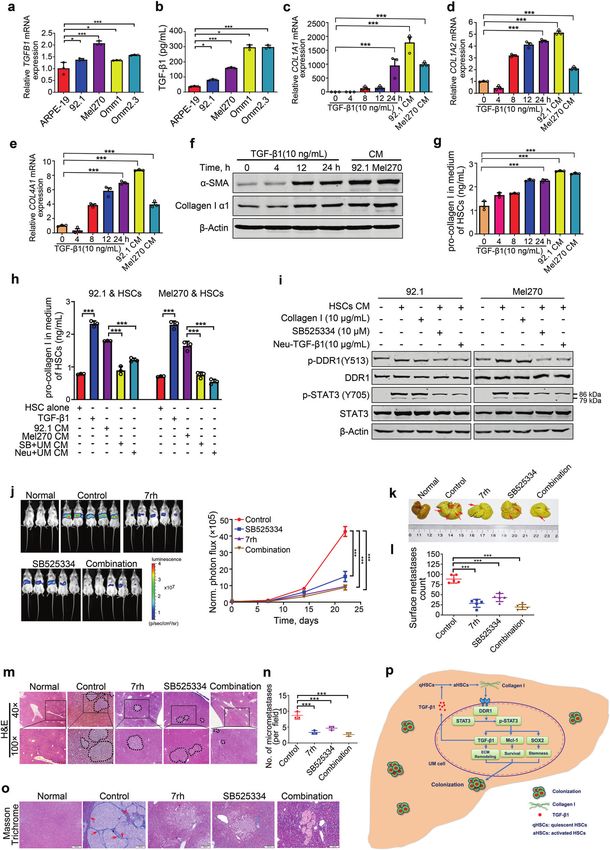
Activation of transmembrane receptor tyrosine kinase DDR1-STAT3 cascade. . .
                                                                     Dai et al.
2
        downstream molecules (e.g., STAT3, STAT5),9 which eventually               DDR1 enhances cellular survival via pro-survival Mcl-1 gene
        coordinates cell adhesion, migration, proliferation, and matrix            Because cellular survival is a critical prerequisite for coloniza-
        remodeling.9                                                               tion when cancer cells are disseminated into host organs, the
           Given that the microenvironment of liver in metastatic patients         pro-survival role of DDR1 in UM cells was examined. The UM
        with UM contains pathological collagen deposition,10 we hypothe-           cells were treated with various concentrations of 7rh for 48 h or
        sized that DDR and its ligand might ignite the interaction between         a fixed concentration (15 µM) for various durations, apoptosis
        metastasis-initiating cells and their surrounding niche thereby            was assessed by flow cytometry after dually stained with
        conferring colonization. In the present study, we tested this              Annexin V-FITC and propidium iodide (Fig. 1m). The results
        hypothesis by dissecting the expression of DDR1 and its signaling          showed that percentages of dead cells were increased in a
        in UM cells. We found that DDR1-overexpressing UM cells                    concentration- and time-dependent manner after 7rh treat-
        educated hepatic stellate cells (HSCs) to secret collagen I, which         ment (Fig. 1n and Supplementary Fig. S2a). As well, 7rh
        in turn ligated DDR1 to activate UM cells to release HSCs-                 induced a concentration- (Fig. 1o) and time-dependent
        activating soluble cytokine TGF-β1, forming a positive loop to             (Supplementary Fig. S2b) PARP cleavage and caspase-3
        enhance survival, proliferation and stemness, which eventually             activation. 7rh treatment elicited an increase in cell population
        promotes liver metastasis. These findings may shed light on the             with loss of mitochondrial potential (ΔΨm), as measured by
        understanding of modus operandi by which tumor cells seduce                flow cytometry after chloromethyl-X-rosamine and MTGreen
        the host liver microenvironment, providing rationale targets for           double staining in UM cells (Supplementary Fig. S2c, d).
        liver metastatic colonization in UM.                                       Collectively, these data together suggest that 7rh may induce
                                                                                   mitochondrial damage and apoptosis in UM cells.

        RESULTS                                                                    7rh downregulates Mcl-1 and induces apoptosis in UM cells
        DDR1 is overexpressed in cell lines and primary specimens of UM            To investigate the mechanism of 7rh-induced apoptosis in UM
        Western blotting analysis showed that the protein levels of DDR1           cells, we evaluated the expression of apoptosis-related proteins.
        rather than DDR2 were considerably higher in the four lines of             Western blotting analysis showed that the protein levels of pro-
        human UM cells than those in ARPE-19 cells (Fig. 1a). In parallel,         survival Mcl-1 were obviously decreased without considerably
        qRT-PCR analysis revealed that the mRNA levels of DDR1 rather              change in the levels of other apoptosis-related family members
        than DDR2 were significantly increased in the UM cells relative to          (XIAP, Bcl-2, Bcl-XL, Bax, and Survivin) (Supplementary Fig. S3a). We
        ARPE-19 cells (Fig. 1b, c). The significantly increased mRNA levels         next determined the role of Mcl-1 in 7rh-induced apoptotic cell
        of DDR1 were also found in the primary UM tissues versus normal            death in UM cells. Forced overexpression of Mcl-1 attenuated the
        choroid tissues (Fig. 1d). These results suggest that the over-            7rh-induced apoptosis in Mel270 cells as reflected by PARP
        expression of DDR1 in UM cells occurs at the transcriptional layer.        cleavage (Supplementary Fig. S3b, left) and trypan blue staining
        After the specificity of the anti-DDR1 antibody was verified                 cells (Supplementary Fig. S3c). Conversely, knockdown of Mcl-1 by
        (Supplementary Fig. S1a), we measured the expression of DDR1 in            siRNA duplexes significantly accelerated the lethal effect of 7rh in
        the primary ocular tumor specimens from patients with UM by                UM cells (Supplementary Fig. S3b, right and Supplementary Fig.
        using immunohistochemistry (IHC) staining with anti-DDR1. In               S3d). These results reveal that DDR1 may exert its pro-survival role
        contrast to the undetectable levels of DDR1 in the adjacent                via Mcl-1.
        normal tissues (Supplementary Fig. S1b), the positive staining
        (ranging from low to high by IHC score) of DDR1 was observed in            7rh downregulates Mcl-1 transcription through STAT3
        57 out of 62 (92%) of the tested UM cases (Fig. 1e, f). Moreover,          We further explored the underlying mechanism that Mcl-1 was
        the expression of DDR1 was positively correlated with the largest          downregulated by DDR1 inhibition. qRT-PCR analysis showed
        basal diameter (P = 0.00006) and thickness (P = 0.0386) of primary         that DDR1 deletion inhibited the transcription of Mcl-1 in UM
        tumors, which are two important independent predictors of UM               cells (Supplementary Fig. S3e). It has been reported that Mcl-1
        metastatic death.11 It was also positively correlated with the TNM         is the target gene of transcription factor STAT3 implicated in
        stage (P = 0.0019) in cohort (Supplementary Table S2). Taken               tumor survival.13 In addition, STAT3 is a critical downstream of
        together, these results suggest that the overexpressed DDR1                DDR1 signaling,14 we, therefore, hypothesized that STAT3 may
        predicts a poor prognosis in UM.                                           be a mediator between DDR1 and Mcl-1 in UM. As expected,
                                                                                   ChIP assay indicated that knockdown of DDR1 obviously
        DDR1 promotes cellular proliferation in UM cells                           decreased the STAT3 binding to the promoter of Mcl-1 gene,
        The overexpression of DDR1 in UM cells and tissues prompted us             which was reversed by forced overexpressing STAT3 in Mel270
        to ask whether DDR1 promoted the growth of UM cells. 92.1 and              cells. There was no significant enrichment at a downstream
        Omm1 cells with DDR1 stably silenced by lentiviral shRNA                   intronic region on Mcl-1 lacking STAT3 binding sequence
        manifested a significantly decreased clonogenicity as evaluated             (Supplementary Fig. S3f, g).
        in soft agar-containing culture (Fig. 1g, h). We next employed 7rh
        (Fig. 1i), a specific small-molecule inhibitor of DDR1 (15-fold             Pharmacological inactivation of DDR1 tyrosine kinase impedes
        selectivity relative to DDR2) as described in our previous report,12       outgrowth of xenografted Omm1 cells and PDX in NOD-SCID mice
        to confirm the role of DDR1 in UM cells. UM cells were treated with         To evaluate the in vivo function of DDR1 activity, the NOD-SCID
        increasing concentrations of 7rh for 48 h, western blotting analysis       mice (4–6-week-old) were subcutaneously injected with
        showed that 7rh decreased the phosphorylation of DDR1 without              Omm1 cells. When the tumor xenografts were palpable
        alternating the protein levels of DDR1, suggesting that 7rh can            (~100 mm3), the mice were randomly divided into two groups
        effectively inhibit the cellular DDR1 kinase activity in UM cells (Fig.    administered with vehicle or 7rh for 2 weeks. The results showed
        1j). Cell viability assay showed that 7rh concentration-dependently        that the increase in tumor volume was impeded in 7rh-treated
        dampened the growth of UM cells with IC50 values ranged from 4             group when compared to vehicle-treated mice (Fig. 2a). Con-
        to 10 μM (Fig. 1k). Separately, the UM cells were treated with             sistently, tumor weight of the 7rh-treated mice was significantly
        increasing concentrations of 7rh for 24 h, and then seeded in soft         lower than that of the vehicle-treated mice (Fig. 2b, c).
        agar culture in the absence of 7rh. The colony formation was               Furthermore, IHC staining assessment of Ki67 validated the
        concentration-dependently reduced by 7rh (Fig. 1l). Taken                  remarkable decrease of cell proliferation upon 7rh treatment
        together, these data suggest that DDR1 promotes the proliferation          (Fig. 2d, e). Western blotting analysis of cell lysates from four
        of UM cells.                                                               tumors of each group indicated that the phosphorylation of DDR1

                                                                                     Signal Transduction and Targeted Therapy (2021)6:176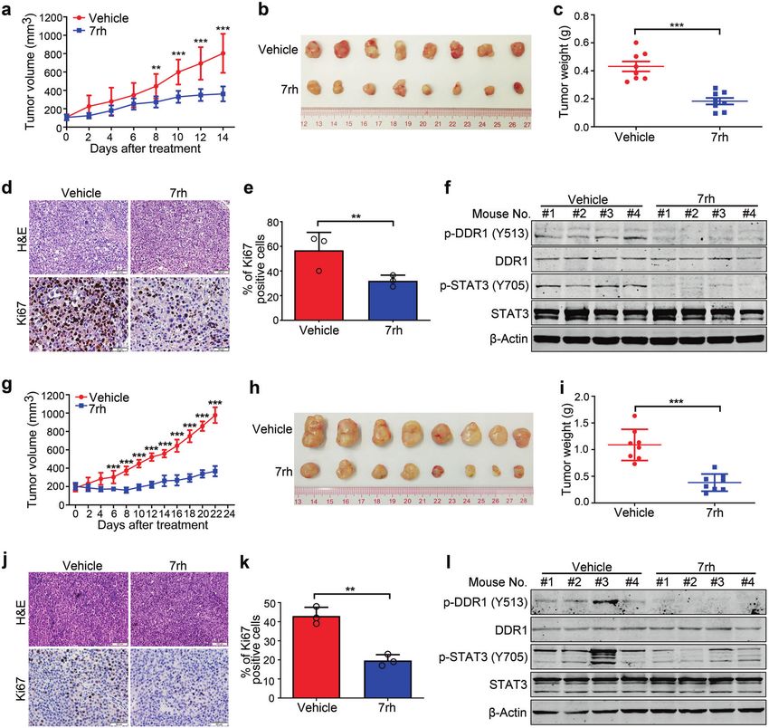
Activation of transmembrane receptor tyrosine kinase DDR1-STAT3 cascade. . .
                                                                    Dai et al.
                                                                                                                                                   3

and its downstream signaling STAT3 were blocked after 7rh           DDR1 confers maintenance of cancer stem-like cell traits via SOX2
administering (Fig. 2f). Similar results were obtained in a human   in UM
UM MP41 PDX model (Fig. 2g–l). These data demonstrate that          Because cancer stem-like cells (CSCs) disseminated into the
pharmacological inactivation of DDR1 kinase by 7rh impedes the      host organs likely function as ‘seeds’ of colonization,15 we
outgrowth of UM cells in vivo.                                      asked whether DDR1 maintained the CSC traits. Exposure of

Signal Transduction and Targeted Therapy (2021)6:176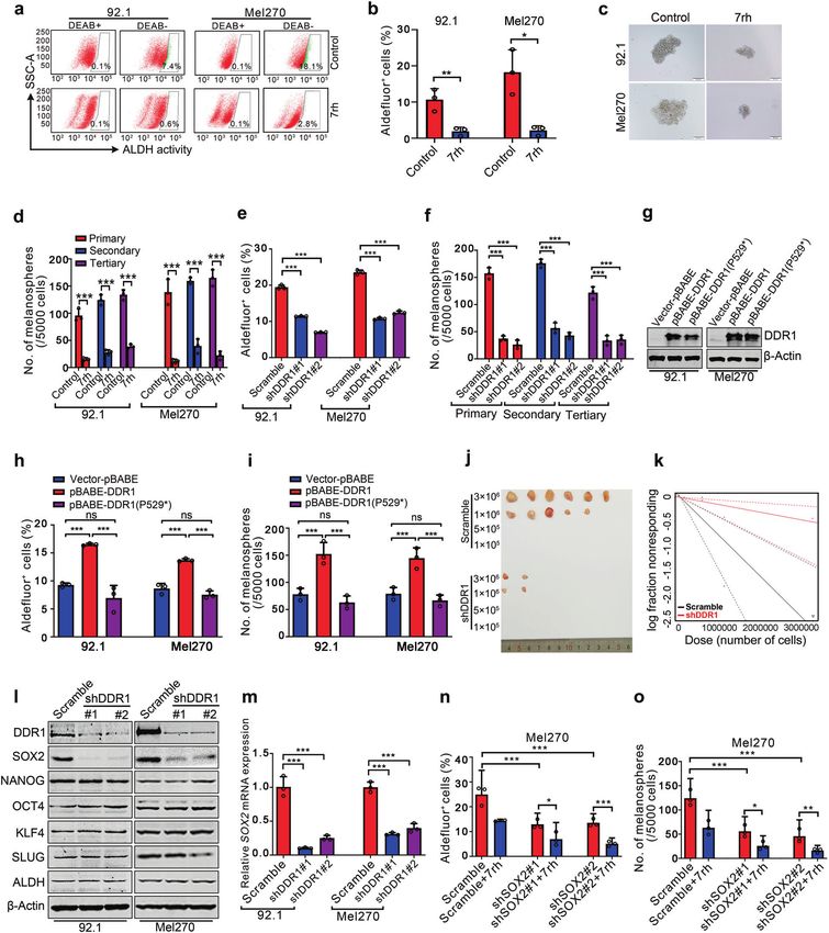
Activation of transmembrane receptor tyrosine kinase DDR1-STAT3 cascade. . .
Dai et al.
4
Fig. 1 DDR1 is overexpressed in uveal melanoma and inhibition of DDR1 reduces colony formation capacity and induces apoptosis in human
UM cells. a–c Protein and mRNA levels of DDR1 and DDR2 in human uveal melanoma (UM) cells (Mel270, 92.1, Omm1, and Omm2.3) and
retinal pigment epithelial (ARPE-19) cells were determined by western blotting and qRT-PCR analysis, respectively. ns, no significant. Data are
shown as the mean ± SD (n = 3). d The mRNA levels of DDR1 and DDR2 genes in normal choroid tissues and UM tissues were assessed by qRT-
PCR. Data are shown as the mean ± SD (n = 3). e, f Statistical analysis of DDR1 expression in paraffin-embedded tissues from the patients with
UM (n = 62) is shown. The staining intensity was scored on four levels (negative, low, medium, and high). Data are shown as the mean ± SD. g
92.1 and Omm1 cells stably transduced with control shRNA (Scramble) or two DDR1 shRNAs lentivirus underwent western blotting analysis or
h colony-formation assay in soft agar-containing culture, Data are shown as the mean ± SD (n = 3). i The chemical structure of DDR1 inhibitor
7rh is shown. j The effect of 7rh on the phosphorylation of DDR1 was ascertained by western blotting analysis after 92.1 and Omm1 cells
exposure to 7rh for 24 h. k UM cells were treated with escalating concentrations of 7rh for 72 h, cell viability was determined by MTS assay.
Data are shown as the mean ± SD (n = 3). l After treatment with 7rh at the indicated concentrations for 48 h, the UM cells were harvested and
seeded in drug-free soft agar-containing culture for 14 days, colonies were counted and analyzed. Data are shown as the mean ± SD (n = 3). m,
n UM cells were exposed to increasing concentrations of 7rh for 48 h, or 15 μM 7rh for the different durations, flow cytometry was employed
to detect the apoptosis after dual-staining with Annexin V-FITC and PI. Representative flow cytometry dot plots for 92.1 cells (m) and
quantitative analysis of results from three independent experiments (n) are shown. The Y-axis is the sum of the top left, top right, and bottom
right quadrants. Data are shown as the mean ± SD (n = 3). o Cleavage of PARP and activation of caspase-3 were determined by western
blotting after UM cells were treated with escalating concentrations of 7rh. *P < 0.05; ***P < 0.001, one-way ANOVA, post hoc comparisons,
Tukey’s test for result in b, c, h and n; *P < 0.05; ***P < 0.001, Student’s t test for results in (d, e)
UM cells to 7rh to block DDR1 kinase activity resulted in a STAT3-dependent SOX2 upregulation by DDR1 facilitates traits of
significant decrease of the percentage of ALDH+ cells (Fig. 3a, CSCs and metastasis in UM
b and Supplementary Fig. S4a). 7rh also abrogated the self- Given that STAT3 can directly bind to the promoter of SOX2 gene
renewal capacity as reflected by the serially-replating melano- and that DDR1 can activate STAT3,14,16 we detected whether
sphere ability (Fig. 3c, d and Supplementary Fig. S4b). DDR1 mediated SOX2 transcription via STAT3 in UM. Mel270 cells
Consistently, silencing DDR1 by lentiviral shRNA against transduced with lentiviral STAT3-encoding construct were
DDR1 significantly eliminated CSC properties as displayed exposed to 7rh for 24 h, western blotting analysis and CSCs traits
repressed the percentage of ALDH+ cells (Fig. 3e and were evaluated. Western blotting analysis unveiled that the forced
Supplementary Fig. S4c) and serial melanosphere formation expression of STAT3 reversed the 7rh-mediated decrease in SOX2
(Fig. 3f and Supplementary Fig. S4d). While forced expression (Fig. 4h). The results of ChIP assay revealed that depletion of DDR1
of wild-type DDR1 but not the loss-of-function mutant DDR1 attenuated the abundance of STAT3 at the promoter region of
(Fig. 3g and Supplementary Fig. S4e) significantly elevated the SOX2 compared with Scramble control cells, which was at least
percentage of ALDH+ cells (Fig. 3h and Supplementary Fig. S4f) partially reversed by forced expression of STAT3 (Fig. 4i, j). There
as well as serial melanosphere formation (Fig. 3i and was no significant enrichment at a downstream intronic region on
Supplementary Fig. S4g). Furthermore, in vivo limiting dilution SOX2 lacking STAT3 binding sequence. These results indicate that
assay revealed that silencing DDR1 by shRNA reduced UM CSCs STAT3 is fundamental for DDR1-mediated SOX2 transcription.
frequency 5.3-fold (Scramble: 1.09 × 10⁻⁶; shDDR1: 5.82 × 10⁻⁶) Moreover, forced expression of STAT3 elevated the proportion
(Fig. 3j, k and Supplementary Table S3). All these data indicate of ALDH+ cells (Fig. 4k) as well as serial melanosphere formation
that abolishing DDR1 eliminates CSCs in UM. (Fig. 4l). Ectopic expression of STAT3 attenuated the 7rh-mediated
To explore the molecular mechanism that DDR1 maintains the decrease in the percentage of ALDH+ cells (Fig. 4k) and
phenotypes of CSCs, we detected the expression of stemness- melanoshpere formation and replating ability (Fig. 4l). We next
associated proteins. Western blotting assay revealed an appreci- carried out experiments to validate the effect of STAT3 on
able decrease in SOX2 in DDR1-silenced UM cells without metastatic colonization. As expected, administration of 7rh
obvious alteration in SLUG, KLF4, NANOG, ALDH, and OCT4 (Fig. significantly decreased the bioluminescence signal intensity of
3l and Supplementary Fig. S4h). qRT-PCR analysis showed that liver tumor burdens (Fig. 4m, n), as well as the number of
the mRNA levels of SOX2 were also inhibited in DDR1-silenced metastatic nodules on liver surface (Fig. 4o–q), which was at least
cells (Fig. 3m). Notably, disrupting SOX2 by shRNA diminished partially reversed by forced expression of STAT3. Taken together,
the percentage of ALDH+ cells and serially-replating capacity of these data suggest that STAT3 is required for DDR1 to promote
melanosphere formation (Fig. 3n, o). Silencing SOX2 potentiated CSCs, and metastatic colonization in UM.
the 7rh-mediated decrease in ALDH+ cells and melanosphere
formation in UM cells (Fig. 3n, o). Collectively, these results UM cells educate hepatic stellate cells to secret type Ia collagen
indicate that SOX2 positively regulates CSCs properties, and that Studies have demonstrated that ECM remodeling in the host
DDR1 inactivation by 7rh decreases SOX2 and eradicates CSCs in organ can create a favorable microenvironment (niche) which is
UM cells. critical for colonization of tumor cells.17 HSCs, one of the most
important stromal cell types in liver, are an important source of
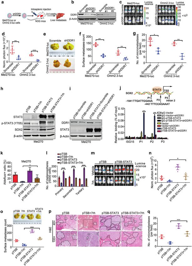
DDR1 knockdown diminishes liver metastatic colonization in UM ECM remodeling impetus in hepatic fibrosis and carcinoma.18 We
To assess the effect of DDR1 on metastatic colonization, Mel270- thus wondered whether UM cells were capable of activating HSCs
luc or Omm 2.3-luc cells stably transduced with lentiviral to favor colonization in liver. Exposure of HSCs to the conditioned
Scramble or shDDR1 were intrasplenically injected into NOG medium (CM) prepared from co-cultured UM cells with HSCs
mice (Fig. 4a). We found that DDR1 knockdown (Fig. 4b) (direct or indirect as illustrated in Fig. 5a) elicited activation of
effectively retarded bioluminescence signals in livers on day 21 HSCs evidenced by increased α-SMA as examined with immuno-
(Fig. 4c, d), as well as restrained the counts of liver surface fluoresence staining and western blotting analysis (Fig. 5b, c). By
metastatic nodules (Fig. 4e, f). In accordance, H&E staining contrast, the CM derived from mono-cultured HSCs using either
showed a remarkable reduction in density and size of the HSC-specific medium or RPMI1640 medium did not induce such
metastatic nodules in the livers of shDDR1 group relative to an activating effect on HSCs (Fig. 5b, c). These results suggest that
those in the Scramble group (Fig. 4g and Supplementary Fig. the UM cells may secret soluble factor(s) to activate HSCs.
S5a, b). These results demonstrate that DDR1 inhibition We next collected the CM from the activated HSCs to the
suppresses metastatic colonization in UM. culture of melanosphere of UM cells transduced with either
Signal Transduction and Targeted Therapy (2021)6:176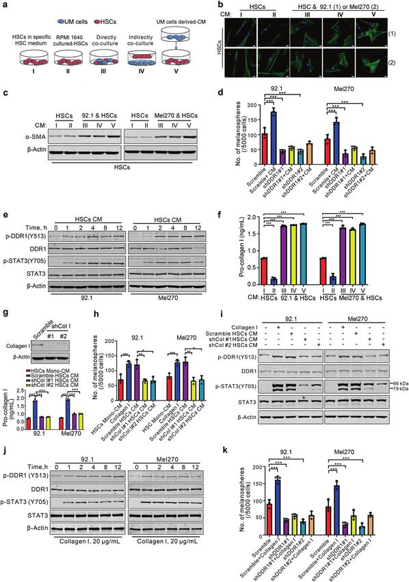
Activation of transmembrane receptor tyrosine kinase DDR1-STAT3 cascade. . .
                                                                         Dai et al.
                                                                                                                                                        5

Fig. 2 7rh impedes outgrowth of xenografted Omm1 cells and MP41 cells patient-derived xenograft model in NOD-SCID mice. a–c Male NOD-
SCID mice (4–6 weeks) bearing palpable Omm1 xenografted tumors were randomly administered with either vehicle (ddH2O:DMSO:EtOH:
Cremophor EL = 90:2:4:4) or 7rh (25 mg/kg in vehicle, orally) every day for 14 days (n = 8 per group). a The tumor volumes estimated every
other day measurement by caliper versus time were plotted. Data are shown as the mean ± SD (n = 8). b Representative tumors resected from
mice treatment with vehicle or 7rh for 14 days are shown. c Weights of tumor from mice of each group are shown. Data are shown as the
mean ± SD (n = 8). d The xenograft tissues from the vehicle- or 7rh-treated mice were subject to H&E staining and IHC analysis with anti-Ki67.
Scale bar: 100 µm (H&E staining), 50 µm (IHC staining). e The total number of Ki67-positive cells (brown-stained nuclei, regardless of staining
intensity were counted as positive) in three random microscopic fields was counted by Image-Pro Plus 6.0. Data are shown as the mean ± SD
(n = 3). f Western blotting detection of DDR1, phospho-DDR1 and its downstream signal phospho-STAT3 in tumor tissues from each group of
mice is shown. g–l The effect of 7rh on UM PDX model. Male NOD-SCID mice (4–6 weeks) bearing palpable MP41 cells xenografted tumors
were randomly administered with either vehicle (ddH2O:DMSO:EtOH:Cremophor EL = 90:2:4:4) or 7rh (25 mg/kg in vehicle, orally) every day
for 22 days. g The tumor volumes estimated every other day measurement by caliper versus time were plotted. Data are shown as the mean ±
SD (n = 8). h, i The mice were sacrificed and tumors were collected, then tumors were photographed and weights were measured and
analyzed. Data are shown as the mean ± SD (n = 8). j, k Tumor sections from two groups were subject to H&E staining and IHC analysis with
anti-Ki67. The total number of Ki67-positive cells was counted. Data are shown as the mean ± SD (n = 3). Scale bar: 100 µm (H&E staining),
50 µm (IHC staining). l Western blotting analysis of cell lysates of tumors was performed to detect DDR1, phospho-DDR1 and its downstream
signal phospho-STAT3 after vehicle or 7rh treatment. **P < 0.01; ***P < 0.001, Student’s t test for results in (a, c, e, g, i and k)

Scramble or shDDR1. The results showed that the CM from the              blotting analysis of whole-cell lysates of these UM cells showed
activated HSCs induced by the CM from UM cells significantly              that HSCs-derived CM pretreated with UM cells-derived CM led to
elevated ability of melanosphere formation in UM cells transduced        increased phosphorylation in DDR1 and STAT3 in UM cells (Fig.
with lentiviral vector (Fig. 5d). In contrast, the CM from the           5e), suggesting that HSCs may be activated to release soluble
activated HSCs did not elevate ability of melanosphere formation         factors to ligate membrane receptor DDR1 on UM cells in response
of UM cells transduced with DDR1 shRNA (Fig. 5d). Western                to soluble factors derived from UM cells.

Signal Transduction and Targeted Therapy (2021)6:176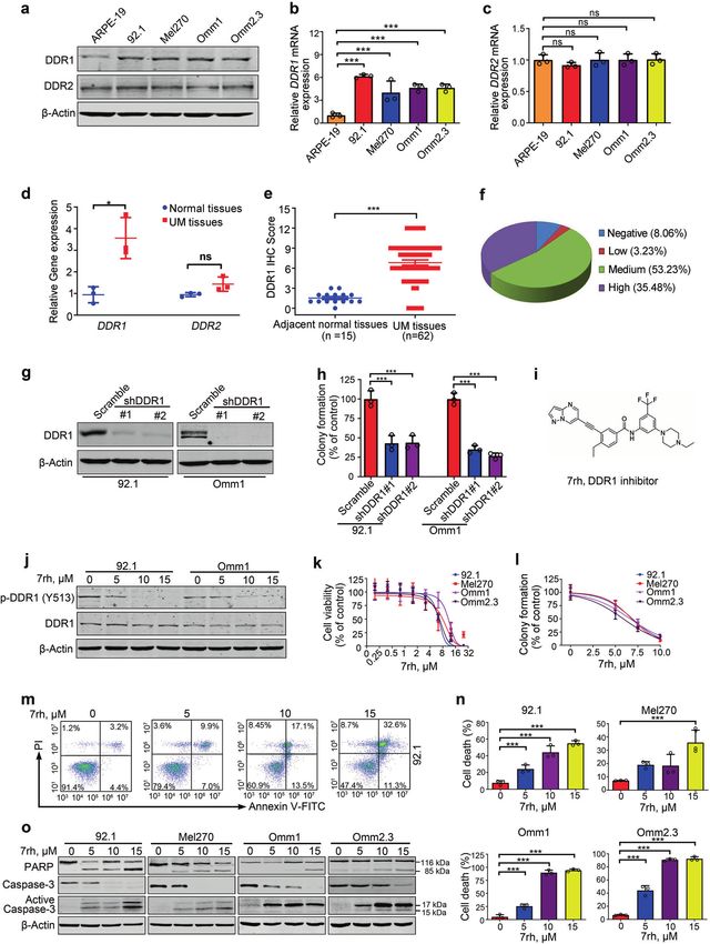
Activation of transmembrane receptor tyrosine kinase DDR1-STAT3 cascade. . .
                                                                     Dai et al.
6

        Collagen I is an active factor in the CM from the activated HSC            I-depleted HSCs caused less phosphorylation in DDR1 and STAT3
        Linking that HSCs can be activated into contractile myofibroblasts          as well as reduced the self-renewal capacity of CSCs in UM cells
        to secret collagens into ECM,19 we, therefore, decide to determine         compared with the CM prepared from Scramble HSCs (Fig. 5g–i).
        the pro-collagen I content in the HSCs CM. ELISA assay results                We next examined whether the in vitro artificial collagen I
        showed an increase of pro-collagen I content in the CM from HSCs           recapitulated the effect of HSCs CM. The results indicated that
        that only pre-co-cultured with UM cells or pretreated with UM              addition of recombinant human collagen I to the culture obviously
        cells-derived CM when compared with that in the CM from mono-              led to phosphorylation in DDR1 and STAT3 in UM cells (Fig. 5j).
        cultured HSCs (Fig. 5f). These results support that our hypothesis         Furthermore, treatment with recombinant human collagen I
        that UM cells generate certain soluble factor(s) to activate HSCs          significantly elevated the capacity of melanosphere formation in
        secretion, which may trigger ECM remodeling.                               UM cells stably transduced with Scramble lentivirus, but not in UM
           To further verify the requirement of collagen I, CM from the            cells stably transduced with shDDR1 lentivirus (Fig. 5k). These
        HSCs depleted collagen I by shRNA was exposed to UM cells.                 results reveal that the promoting effect of collagen I on self-
        Western blotting analysis indicated that CM derived from collagen          renewal of CSCs was exerted via DDR1.

                                                                                     Signal Transduction and Targeted Therapy (2021)6:176
Activation of transmembrane receptor tyrosine kinase DDR1-STAT3 cascade. . .
Dai et al.
7
Fig. 3 DDR1 inhibition suppresses the properties of cancer stem-like cells through lowering SOX2 in UM cells. a, b 92.1 and Mel270 cells were
treated with 10 μM 7rh for 48 h, the ALDH activity was detected by flow cytometry. Representative flow cytometry (a) and quantitative analysis
of ALDH+ cells (b) from three independent experiments are shown. Data are shown as the mean ± SD (n = 3). c, d 92.1 and Mel270 cells were
treated with 10 μM 7rh for 48 h, and then drug-freely cultured for three rounds of melanosphere assay for 14 days. Data are shown as the
mean ± SD (n = 3). Scale bar: 200 µm. e 92.1 and Mel270 cells stably transduced with lentiviral Scramble or shDDR1 were assayed by flow
cytometry for the proportion of ALDH+ cells. Quantitative analysis of ALDH+ cells from three independent experiments is shown. Data are
shown as the mean ± SD (n = 3). f 92.1 cells stably transduced with lentiviral Scramble or shDDR1 were plated in the stem cell culture medium.
Melanospheres were counted on day 14. The cells were harvested and replated for the secondary and tertiary rounds of evaluation,
respectively. Data are shown as the mean ± SD (n = 3). ***P < 0.001, one-way ANOVA, post hoc comparisons, Tukey’s test. g Ectopic expression
of wild-type DDR1 or mutant DDR1 (DDR1 P529*) in 92.1 and Mel270 cells were determined by western blotting analysis. h Overexpression of
wild-type DDR1 but not mutant DDR1 (DDR1 P529*) increased the percentage of ALDH+ cells as detected by flow cytometry in 92.1 and
Mel270 cells. ns no significant. Data are shown as the mean ± SD (n = 3). i Ectopic expression of wild-type DDR1 rather than mutant DDR1
(DDR1 P529*) potentiated self-renewal capacity as evaluated by melanosphere growth and serially-replating assay in 92.1 and Mel270 cells. ns
no significant. Data are shown as the mean ± SD (n = 3). j, k Silencing DDR1 decreased in vivo frequency of CSCs in UM cells. Omm1 cells
stably transduced with lentiviral Scramble or shDDR1 were subjected to limiting dilution assay in NOD-SCID mice. Representative image of
tumors removed from the mice of each group (n = 6) are shown. The frequency of CSCs was calculated by L-Cal software. l DDR1 deletion
decreased the expression of SOX2. The protein levels of stemness-related proteins were detected by western blotting in 92.1 and Mel270 cells
stably transduced with Scramble or shDDR1 lentivirus. m DDR1 deletion downregulated the mRNA levels of SOX2. The mRNA levels of SOX2 in
92.1 and Mel270 cells stably transduced with Scramble or shDDR lentivirus were determined by qRT-PCR. Data are shown as the mean ± SD (n
= 3). n, o The percentage of ALDH+ cells and melanosphere assay were performed in Mel270 cells stably transduced with Scramble or shSOX2
lentivirus with or without 7rh. Data are shown as the mean ± SD (n = 3). *P < 0.05; **P < 0.01; ***P < 0.001, Student’s t test for results in (b, d). *P
< 0.05; **P < 0.01; ***P < 0.001, one-way ANOVA, post hoc comparisons, Tukey’s test for results in (e, f, h, i and m–o)
TGF-β1 secreted from UM cells activates HSCs to release collagen I approximately liver area (Fig. 6j and Supplementary Fig. S8a, b)
To define the specific factor(s) in the UM-derived CM activated and increased numbers and size of metastatic nodules on liver
HSCs, the expression of the cytokines (e.g., PDGFA, PDGFC, IGF, (Fig. 6k, l and Supplementary Fig. S8c). In a sharp contrast, the
and Endothelin) that are critical for activation of HSCs was mice administrated with either 7rh or SB525334 manifested much
detected by qRT-PCR. The results indicated that only TGF-β1 was less bioluminescence signal intensity in the liver area (Fig. 6j and
higher in UM cells than that in ARPE-19 cells (Fig. 6a and Supplementary Fig. S8a, b) and numbers of metastatic nodules on
Supplementary Fig. S6a–d). Considering that TGF-β1 is the most liver surface (Fig. 6k, l and Supplementary Fig. S8c). In accordance,
important cytokine which can activate quiescent HSCs into H&E staining showed an obvious reduction in density and size of
myofibroblast,19 and the transcription of TGF-β1 gene can be the metastatic nodules in the livers of treated group relative to
regulated by DDR1-STAT3 axis (Supplementary Fig. S7), we paid those in non-treated group (Fig. 6m, n and Supplementary Fig.
our attention to TGF-β1. Similarly, the increased content of TGF-β1 S8d, e). Of note, the efficacy that 7rh or SB525334 inhibited the
was detected in the CM of UM cells compared with that in ARPE- liver metastasis of UM was similar to the combinational treatment
19 cells using ELISA assay (Fig. 6b). with 7rh and SB525334, suggesting that they did not produce
Exposure of HSCs to recombinant human TGF-β1 as well as the synergy or additive anti-metastasis effect. The results of Masson
CM from UM cells dramatically elicited 1000-fold increase in the Trichrome staining revealed large thick bundles of collagen
expression of collagen Iα1 with limited increase in the expression surrounding tumor cell nests to form a niche in liver with
of collagen Iα2 and collagen IV (Fig. 6c–e). Given that collagen I is metastasis compared with the normal liver, and collagen was
the major type of collagen produced by the activated HSCs in effectively decreased in the liver by 7rh, SB525334 or combination
response to TGF-β1, and that collagen I is an important ligand of treatment (Fig. 6o and Supplementary Fig. S8f). These findings
DDR1. The significant elevation of collagen Iα1 was further indicate that UM cells in liver may interact with HSCs, and activate
confirmed in HSCs after exposure to recombinant human TGF- HSCs to secret collagen I, forming a beneficial niche for
β1 or the CM from UM cells with western blotting assay (Fig. 6f). colonization of UM cells (Fig. 6p).
ELISA assay analysis of the CM from HSCs stimulated by
recombinant human TGF-β1 as well as the CM from UM cells
further validated a significant upregulation in pro-collagen I, DISCUSSION
which was abrogated in the presence of TGF-β1 inhibitor, Little is known about the mechanism of metastatic colonization. In
SB525334 or anti-TGF-β1 neutralizing antibody (Fig. 6g, h). the present study, we found that DDR1 is overexpressed in the
Consistently, western blotting results showed that the DDR1 cancerous cells and primary tissues of UM and promotes cellular
activation was attenuated in UM cells treated with HSC CM from proliferation and survival. Pharmacological inactivation of DDR1
TGF-β1 inhibitor, SB525334 or anti-TGF-β1 neutralizing antibody tyrosine kinase impedes outgrowth of xenografted Omm1 cells
group when compared with the non-treated HSC CM or and PDX in NOD-SCID mice. Mechanistically, DDR1 confers
recombinant collagen I stimulation (Fig. 6i). These results support maintenance of CSCs traits via STAT3-dependent SOX2 upregula-
a tumor cells-microenvironment interaction loop that TGF- tion in UM cells. Further, co-culture experiments show that UM
β1 secreted from UM cells activates stromal HSCs to release cells secrete TGF-β1 which in turn induces HSCs to produce
collagen I which in turn ligate cell surface DDR1 on UM cells. collagen type Iα1, remodeling ECM. DDR1 responds to collagen I,
resulting in strengthened survival, proliferation, and stemness in
Interrupting the TGF-β1-collagen I-DDR1-STAT3 loop abrogates cancer cells, and promoting metastatic colonization in liver.
liver metastatic colonization in UM
The tumor cells-microenvironment interaction loop predicted that DDR1 is highly expressed in UM
blockade of either TGF-β1 signaling in microenvironmental HSCs DDRs, unique tyrosine kinase receptors that function as a sensor
or DDR1 signaling in UM cells might inhibit in vivo metastatic ECM, are implicated in a wide range of cellular processes.20 Our
colonization. To test this hypothesis, the NOG mice received results show that DDR1 rather than DDR2 is overexpressed in UM
intrasplenic injection of Mel270-luc or Omm2.3-luc cells were cells as well as specimens from patients with UM, which is in line
administrated with 7rh, SB525334 or combination. The mice with the previous studies that DDR1 expression is restricted to
treated with vehicle manifested strong bioluminescence signal in epithelial cells in solid tissues.21 DDR1 is reported to be also
Signal Transduction and Targeted Therapy (2021)6:176Activation of transmembrane receptor tyrosine kinase DDR1-STAT3 cascade. . .
Dai et al.
8
overexpressed in melanoma arising from cutaneous melano- stemness of UM cells. Pharmacological inactivation of DDR1
cytes.22 In addition, DDR1 is overexpressed in prostate cancer, tyrosine kinase impedes outgrowth of xenografted Omm1 cells
glioma, and human hepatocellular carcinoma.20 and PDX in NOD-SCID mice. Given that DDR1 is actually not
In the present study, we demonstrated that targeting DDR1 by essential because DDR1-null mice does not impair embryonic
shRNA or inhibitor 7rh, restrained proliferation, survival and development,23 it is likely that tumor cells may become addicted
Signal Transduction and Targeted Therapy (2021)6:176Activation of transmembrane receptor tyrosine kinase DDR1-STAT3 cascade. . .
Dai et al.
9
Fig. 4 Silencing DDR1 suppresses the metastatic colonization of UM cells in liver. a Experimental schematic of liver metastasis model. b
DDR1 knockdown in Mel270-luc and Omm2.3-luc cells stably transduced with Scramble or shDDR1 lentivirus was confirmed by Western
blotting. c, d Mel270-luc and Omm2.3-luc cells stably transduced with Scramble or shDDR1 were intrasplenically inoculated in NOG mice for
3–4 weeks (n = 5 per group). Liver metastasis was analyzed by luciferase-based bioluminescence imaging system. Representative images (c)
and quantitative analysis (d) of photon flux on day 21 are shown. Data are shown as the mean ± SD (n = 5). e, f The mice were sacrificed to
count metastatic nodules on liver surface. Representative images (e) and quantitative analysis of liver surface nodules (f) are shown. Data are
shown as the mean ± SD (n = 5). g Quantitative analysis of micrometastases in H&E staining liver sections from Scramble and shDDR1 mice.
Data are shown as the mean ± SD (n = 3). **P < 0.01, Student’s t test for results in (d, f, g). h Mel270 cells transduced with lentiviral vector
(pTSB) or construct encoding human STAT3 cDNA (pTSB-STAT3) were exposed to 10 μM 7rh for 24 h, and subjected to western blotting
analysis with the indicated antibodies. i DDR1 knockdown and STAT3 overexpression was confirmed by western blotting in Mel270 cells stably
expressed shDDR1 with or without STAT3 overexpression. j Experimental schematic diagram showing the location of STAT3-binding sites of
SOX2 regulatory region. P1, P2 represent STAT3-binding sites at the SOX2 gene promoter. P3 was used as negative control and located in
intronic region. ISG15 as a known non-target gene of STAT3 served as a negative control (top). ChIP-PCR analysis for STAT3 occupancy at the
SOX2 gene promoter in Mel270 cells stably expressed shDDR1 with or without STAT3 overexpression (bottom). Data are shown as the mean ±
SD (n = 3). k Enforced expression of STAT3 attenuated the 7rh-mediated decrease in percentage of ALDH+ cells in UM cells. Mel270 cells stably
expressed with STAT3 were treated with or without 10 μM 7rh for 24 h, and then subjected to ALDH+ cells analysis by flow cytometry.
Quantitive analysis of ALDH+ cells from three independent experiments is shown. Data are shown as the mean ± SD (n = 3). l Overexpression
of STAT3 reversed the 7rh-mediated decrease in melanosphere growth and serially-replating capacity in UM. Mel270 cells stably expressed
with STAT3 were treated with or without 10 μM 7rh for 24 h, and then subjected to melanosphere-formation assay. Data are shown as the
mean ± SD (n = 3). m–q Mel270-luc cells stably transduced with lentiviral vector (pTSB) or constructs encoding human STAT3 cDNA (pTSB-
STAT3) underwent intrasplenic injection in NOG mice, the mice were then administrated with vehicle (ddH2O:DMSO:EtOH:Cremophor EL =
90:2:4:4) or 7rh (25 mg/kg in vehicle, orally) every day for 21 days (n = 5 per group). Liver metastasis was analyzed by luciferase-based
bioluminescence imaging and liver surface nodules were counted. Representative images were taken on day 21 post injection of cells (m) and
quantitative analysis of bioluminescence intensity (n) are shown. Data are shown as the mean ± SD (n = 5). o Representative bright field
images of liver surface and quantitative analysis of surface metastatic nodules on liver are shown. Data are shown as the mean ± SD (n = 5).
p, q Microscopic observation of H&E staining in liver paraffin section from each group verified metastastic nodules. Data are shown as the
mean ± SD (n = 3). Scale bar: 500 µm (40×), 200 µm (100×). *P < 0.05; ***P < 0.001, one-way ANOVA, post hoc comparisons, Tukey’s test for
results in (j, k, l, n, o and q)
to the increased levels of oncogenic DDR1. The ECM remodeling DDR1 facilitates the traits of CSCs in UM
such as collagens accumulation in the microenvironment can In our study, we found that targeting DDR1 by 7rh or shRNA led to
easily lead to activation of DDR1. Of note, unlike the pattern of decrease in ALDH+ cells, ability of melanosphere formation and
instantaneous activation and fast fading in soluble cytokine serially-replating, and in vivo CSCs frequency. Further study
binding receptor tyrosine kinases, collagen binding to DDR1 showed that among the stemness-related proteins, SOX2 was
eliciting a slow and sustainable activation pattern lasting even up reduced in the DDR1-depleted UM cells. With ChIP assay, STAT3
to 18 h.24 At the present time, it’s not clear about the underlying was involved in the DDR1 knockdown-enabled SOX2 transcription
mechanism that DDR1 is overexpressed in UM. TCGA analysis downregulation. Therefore, it’s reasonable to postulate DDR1
showed that amplification is the dominant alternation in DDR1, facilitates the traits of CSCs involved in STAT3 mediated-SOX2
accounting for only ~10% cases of UM.25 Other potential transcription in UM. SOX2, a protein initially is known as a
mechanisms (e.g., miRNA regulation, protein stability alteration) regulator of self-renewal in mouse and human embryonic stem
for deregulated DDR1 need to be further clarified. cells, as well function as a key transcription factor to induce
pluripotent stem cells from fibroblast cells.29 Mounting evidence
DDR1 promotes cellular survival of cancer cells suggests that SOX2 is a critical biomarker of self-renewal and
Enhanced survival capability, an important hallmark of cancer, is maintains the stemness of CSCs in types of cancer, including
critical for outgrowth of cancer metastasis. One of the principal breast cancer, osteosarcomas and glioma.30
mechanisms of pro-survivial or resistance to apoptosis in cancer is
the deregulation of members of the Bcl-2 protein family which ECM remodeling and metastasis
controls the integrity of the outer mitochondrial membrane.26 Almost every cell in the body is exposed to ECM, a complex and
Mcl-1, a member of the Bcl-2 protein family, has been shown to be dynamic network of macromolecules with different physical and
up-regulated in numerous hematological and solid tumor biochemical properties.31 ECM is not just an inert supportive
malignancies.27 Its overexpression positively correlates with tumor scaffold, but regulates diverse cellular behaviors.31 Of importance,
grade and resistance to standard chemotherapy.27 Loss of cell ECM is a dynamic compartment because of alternation in
viability was observed in response to siRNA-induced reduction of production, degradation, and remodeling of its components.31
Mcl-1 or small-molecule inhibitor targeting transcription of Mcl-1 Alteration in a specific ECM component may impact on the
in cancer cells.28 Additionally, some kinase inhibitors are believed biochemical, biomechanical, and physical properties of the ECM,
to exert their antiproliferative effects by indirectly downregulation leading to disorganized network and eventually remodeling.32
of Mcl-1 mRNA via inhibition of RNA polymerase II-dependent Collagens accounting for 30% of body proteins are a major type of
transcription inhibition.28 component of ECM31. In liver disease, HSCs are particularly
Consistent with previous studies, our results showed that vigorous compared with the rest types of residue cells (e.g.,
targeting DDR1 with 7rh induced apoptosis with decline in Mcl-1 hepatocytes and liver sinusoidal endothelial cells). In the
transcription. Mcl-1 has been reported a target gene of STAT3.13 pathogenesis of virus-associated hepatic fibrosis or cirrhosis, a
Plausibly, the presence of STAT3 binding sites in the promoter of critical step is the activation of HSCs from the quiescent state
Mcl-1 was confirmed by using ChIP assay in this study; the results leading to collagen excessive accumulation.33 In this study,
reveal that the repressed Mcl-1 transcription during the 7rh- progressive and excessive accumulation in collagens was
induced apoptosis may be via DDR1-STAT3 axis. Therefore, our observed in the tumor cell-infiltrated liver of NOG mice
results support that inhibition of Mcl-1 may offer a therapeutic (Supplementary Fig. S9), which is consistent with the previous
target in metastatic UM. finding that increased number of activated HSCs and accumulated
Signal Transduction and Targeted Therapy (2021)6:176Activation of transmembrane receptor tyrosine kinase DDR1-STAT3 cascade. . .
Dai et al.
10
collagen existed in the hepatectomy samples of patients with exposed to CM of UM cells. In accord with this, HSCs secretes
UM.10 Experiments of liver metastasis also showed that co- collagen I in the presence of hepatoma cells.34 Because collagen
inoculation of HSCs increased the number of UM hepatic Iα1 is a potent ligand of DDR1 receptor, it’s plausible to postulate
metastases.10 Our further analysis suggested that collagen Iα1 is that ECM remodeling featured with excessive collagen Iα1 elicits
the major type of collagens that HSCs secreted when they are sustaining activation of DDR1 and metastatic colonization.
Signal Transduction and Targeted Therapy (2021)6:176Activation of transmembrane receptor tyrosine kinase DDR1-STAT3 cascade. . .
Dai et al.
11
Fig. 5 UM cells educate hepatic stellate cells (HSCs) to secret collagen I. a Schema of mono-cultured HSCs or co-cultured HSCs with UM cells. I,
mono-cultured HSCs with HSC-specific culture medium; II, mono-cultured HSCs with RPMI 1640 culture medium; III, directly co-culture HSCs
mixed 1:1 with UM cells for 24 h; IV, HSCs were co-cultured with UM cells in Transwell systems for 24 h; V, UM cells-derived conditioned
medium (CM) were added to culture starved HSCs for 6 h and replaced with HSCs specific medium for another 24 h. b, c CM were isolated
from HSCs grown in mono-culture or co-cultured with 92.1 or Mel270 cells as indicated approaches (a), and then added into the culture of
HSCs for 24 h, the marker of activated-HSCs α-SMA was determined by immunofluorescence assay (b) and western blotting (c). Scale bar:
10 µm. d DDR1 is required for melanosphere-formation in UM cells stimulated by HSCs-derived CM. 92.1 and Mel270 cells stably expressing
Scramble or shDDR1 were cultured with HSCs-derived CM and then subjected to melanosphere assay. The medium of UM cells was added to
culture starved HSCs for 6 h and replaced with HSCs specific medium for another 24 h, which collected as HSCs-derived CM. Data are shown as
the mean ± SD (n = 3). e The CM from HSCs that were educated by UM cells activated DDR1 kinase in UM cells. The HSCs-derived CM was
added into the culture of 92.1 and Mel270 cells for different times, and then phosphorylation of DDR1 and its downstream signal
phosphorylation of STAT3 were analyzed by western blotting. f ELISA evaluation of the collagen type Iα was performed in various CM collected
from different culture systems as illustrated in (a). Data are shown as the mean ± SD (n = 3). g HSCs were infected with lentiviral Scramble or
specific shRNA targeting collagen type I, and verified by western blotting (top). The level of collagen type Iα in the CM harvested from HSCs
that were stably transduced with lentiviral Scramble or shCol I were measured by ELISA assay (bottom). Data are shown as the mean ± SD (n =
3). h The medium of 92.1 or Mel270 cells was added to culture starved HSCs expressing Scramble or two shRNAs against Collagen I for 6 h and
replaced with HSCs specific medium for another 24 h, which collected as Scramble HSC CM, shCol I #1 HSC CM, and shCol I #2 HSC CM,
respectively. The indicated HSC-derived CM and recombinant collagen type I (10 μg/mL) were used for melanosphere assay in 92.1 or Mel270
cells. Data are shown as the mean ± SD (n = 3). i Western blotting was conducted after 92.1 or Mel270 cells incubation with recombinant
collagen type I (20 μg/mL) or Scramble HSC CM, shCol I #1 HSC CM, and shCol I #2 HSC CM as illustrated in (h) for 24 h. j After UM cells treated
with 20 μg/mL collagen I for different durations, the phosphorylation of DDR1 and STAT3 were examined by western blotting. k 92.1 and
Mel270 cells stably transduced lentiviral Scramble or shDDR1 were cultured in the absence or presence of recombinant human collagen type I
(10 μg/mL) to evaluate melanospheres formation. Data are shown as the mean ± SD (n = 3). *P < 0.05; **P < 0.01; ***P < 0.001, one-way ANOVA,
post hoc comparisons, Tukey’s test for results in (d, f, g, h and k)
The possibility of involvement of integrin receptors in the TGF- results showed that DDR1 deletion did not affect the expression of
β1-collagen I loop cannot be excluded at the present time. It has BAP1 (Supplementary Fig. S10c). On the other hand, knockdown
been reported that integrins such as collagen receptor α1β1 are of BAP1 did not change the protein levels of DDR1 and its
expressed in UM cells.35 ECM collagen I can bind cell surface downstream signal pathway (Supplementary Fig. S10d). These
integrin α1β1, likely either activating STAT3 in a Src-dependent results reveal that the expression of DDR1 and BAP1 may be
manner or cross-talking with DDR1 signalling pathways,36 and independent in UM cells.
thereby being involved the TGF-β1-collagen I loop.
TGF-β inhibitors and DDR inhibitors
Tumor cells-derived TGF-β1 activates HSCs In our study, the results that using SB525334 to block TGF-
In the hepatectomy tissues from patients with UM, activated HSCs β1 signaling interrupted the TGF-β1-collagen I-DDR1 loop and
and their pathological matrix were observed surrounding the inhibited liver metastasis in UM. Because the anti-TGF-β1-based
malignant foci.37 It’s well known that TGF-β1 is a major potent therapy is an attractive therapeutic target of microenvironment,
profibrogenic cytokine to activate HSCs. TGF-β1 employs SMAD novel drugs blocking the TGF-β pathway, including blocking
pathway that induces expression of the genes needed for resting production of TGF-β ligands with antisense molecules, small-
HSCs activation to activated HSCs,38 TGF-β1 plays a critical role in molecule inhibitors of the kinase activity of TGFβRI and TGFβRII,
promoting collagen secretion and upregulation of MMPs in monoclonal antibodies that block TGF-β signaling, and soluble
activated-HSCs, which can exacerbate the malignant phenotype forms of TGFβRII and TGFβRIII that function as ligand traps, have
and lead to increased invasion and subsequent metastasis.39 been developed and have shown efficacy in preclinical and clinical
Targeting TGF-β1 may be a promising strategy for management of studies.45 More than 100 clinical trials (https://clinicaltrials.gov) in
metastasis by restricting ECM activation and attenuating collagen which the effect of TGF-β has been studied in patients with
secretion. different diseases are undergoing. The first clinical human dose-
Overexpression of TGF-β is commonly observed in solid escalation study in solid cancer (e.g., melanoma, breast cancer,
tumors.40 Because transformed cells (e.g., esophageal cancer, hepatocellular carcinoma, and prostate cancer) patients
gastric cancer, hepatocellular carcinoma) can secret cytokines,41 (NCT02160106) targeting TGFβRI with TEW-7197 causing down-
we measured the expression of cytokines (e.g., TGF-β1, PDGFA, regulation of TGF-β signaling is currently completed with
PDGFC, IGF, and Endothelin) of UM cells and found that the encouraging results. This line of progress may help clinical trials
expression of TGF-β1 was increased. ELISA assay also showed that of TGF-β inhibitors in prevention and treatment in patients with
TGF-β1 was increased in the conditioned medium of UM cells. metastatic UM in future.
However, the mechanism of the high expression of TGF-β in tumor The concentration of 7rh required for a pharmacodynamic or
cells remains unclear. Some studies showed that abnormal DNA antiproliferative effect sounds higher in UM cells in the present
methylation on its promoter may lead to its anomalous study than other types of carcinoma (e.g., nasopharyngeal
transcription of TGF-β.42 The mechanism of overexpression of carcinoma, pancreatic cancer).46,47 The differential sensitivity
TGF-β in tumor cells requires further study. may due to discrepancy in cell type context and culture system.
Apart from 7rh, other promising small-molecule inhibitors of
DDR1 is a druggable target independent of BAP1 in UM DDR1 have been developed.12 Treatment with 7rh or SB525334
Recently, mutations of several genes such as GNAQ, GNA11, BAP1, exhibited negligible toxicity to the mice as reflected by body
SF3B1, EIF1AX, PLCB4, and CYSLTR2 have been identified in UM.43 weight changes (Supplementary Fig. S11). Targeting DDR1 in
In particular, loss-of-function mutations of BAP1 gene are found to combination with targeting microenvironment TGF-β may a new
be associated with a poor outcome in 84% of metastatic UM attractive approach for treatment of UM liver metastasis.
patients, hinting a tumor suppressor role of BAP1 in metastasis. In conclusion, our results reveal that ECM remodeling activates
However, the context of BAP1 signaling pathway in UM metastasis the overexpressed extracellular matrix collagen receptor DDR1
remains unclear.44 UM cells used for metastatic experiments in this promoting colonization phenotypes (e.g., survival, outgrowth and
study harbor wild-type BAP1 (Supplementary Fig. S10a, b), our stemness of cancer cells in the metastatic site). The UM cells secret
Signal Transduction and Targeted Therapy (2021)6:176Activation of transmembrane receptor tyrosine kinase DDR1-STAT3 cascade. . .
Dai et al.
12
cytokine TGF-β1 activating HSCs to secret collagen I, remodeling MATERIALS AND METHODS
ECM and forming a positive feedback loop. We, therefore, propose Chemicals and antibodies
a working model (Fig. 6p). Targeting DDR1 signaling and TGF- DDR1 inhibitor 7rh (chemical structure, Fig. 1i) was synthesized in
β1 signaling may be a novel approach to diminish hepatic our laboratory.12 TGFβRI inhibitor SB525334 was obtained from
metastasis in UM. Selleck (Shanghai, China). Annexin V-FITC and collagen I were from
Signal Transduction and Targeted Therapy (2021)6:176Activation of transmembrane receptor tyrosine kinase DDR1-STAT3 cascade. . .
Dai et al.
13
Fig. 6 TGF-β1 secreted by UM cells activates microenvironmental HSCs to release collagen type Iα which in turn ligates cell surface DDR1 on
UM cells. a Expression of TGF-β1 was analyzed by qRT-PCR assay in UM cells and ARPE-19 cells. Data are shown as the mean ± SD (n = 3). b
ELISA analysis of TGF-β1 in the CM of UM cells and ARPE-19 cells is shown. Data are shown as the mean ± SD (n = 3). c–e qRT-PCR analysis of
collagen Iα1 (c), collagen Iα2 (d) and collagen IV (e) in HSCs after treated with recombinant TGF-β1 (10 ng/mL) for indicated time periods or CM
isolated from 92.1 and Mel270 cells for 6 h, then replaced with fresh HSC medium for 24 h. Data are shown as the mean ± SD (n = 3). f, g HSCs
were cultured in the presence of 10 ng/mL recombinant TGF-β1 at different time points or the CM derived from 92.1 and Mel270 cells for 6 h,
then replaced with fresh HSC medium for 24 h, The cells were pelleted by centrifugation to extract whole-cell lysates for western blotting
analysis (f) and the corresponding supernatants were analyzed levels of pro-collagen I using ELISA assay (g). Data are shown as the mean ± SD
(n = 3). h, i HSCs were treated with recombinant human TGF-β1 (10 ng/mL), or UM-derived CM in the presence or absence of neutralizing anti-
TGF-β1 antibody or SB525334 (TGFβRI inhibitor) for 6 h, then replaced with fresh HSC medium for another 24 h. The collagen type Iα in the
resultant medium was determined by ELISA assay (h). The resultant medium or HSC-derived CM was added into the culture of 92.1 and
Mel270 cells for 12 h, and then subjected to western blotting analysis of DDR1 and its signaling molecules (i). Data are shown as the mean ±
SD (n = 3). j–n After 5 × 10⁵ Mel270-luc cells were intrasplenically inoculated, the NOG mice were administered with vehicle (ddH2O:DMSO:
EtOH:Cremophor EL = 90:2:4:4), 7rh (25 mg/kg, orally), SB525334 (30 mg/kg/day, i.p.) alone or combination 7rh (25 mg/kg, orally) with
SB525334 (30 mg/kg/day, i.p.) every day for 21 days (n = 5 per group). j Representative images of luciferase signals on day 21 after treatment
with vehicle, 7rh, SB525334 alone or combination 7rh with SB525334 (left). Quantitative analysis of photon flux for hepatic metastases in NOG
mice was performed every week (right). Data are shown as the mean ± SD (n = 5). The mice were sacrificed to count metastatic nodules on
liver surface. Representative images (k) and quantitative analysis (l) of liver nodules are shown. Data are shown as the mean ± SD (n = 5). m, n
Metastasis nodules in paraffin sections of liver tissue from each group were identified by H&E staining. Scale bar: 500 µm (40×), 200 µm (100×).
Data are shown as the mean ± SD (n = 3). o Collagens were detected by Masson trichrome staining in paraffin sections of liver tissue. Scale bar:
200 µm (100×). p A proposed working model is shown. UM cells secrete TGF-β1 which induces quiescent hepatic stellate cells (qHSCs) into
activated HSCs (aHSCs) which secretes collagen type I in turn activates DDR1, strengthening survival through upregulating STAT3-dependent
Mcl-1, strengthening stemness via upregulating STAT3-dependent SOX2, and clonogenicity in cancer cells and ultimately promotes liver
colonization and metastasis in UM. *P < 0.05; **P < 0.01; ***P < 0.001, one-way ANOVA, post hoc comparisons, Tukey’s test for results in (a–e, g,
h, j, l and n)
Sigma-Aldrich (Shanghai, China). Recombinant TGF-β1 was cells were pretreated with 7rh (0, 5, 7.5, 10 μM) for 24 h and plated
purchased from Invitrogen (Shanghai, China). Antibodies against at 5000 cells/well resuspended in complete RPMI1640 medium
DDR1, Survivin, and Bcl-XL were from Santa Cruz Biotech (Santa containing 0.5% agar, and seeded over a layer of 1% agar in 24-
Cruz, CA). Antibodies against PARP (clone 4C10-5), caspase-3, XIAP, well plates for 2 weeks at 37 °C. Colonies composed of >50 cells
and Bcl-2 were purchased from BD Biosciences (San Jose, CA). were counted using an inverted phase-contrast microscope.
Antibodies against DDR2, STAT3, p-STAT3 (Y705), p-DDR1 (Y513),
active Caspase-3, Bax, α-SMA, SOX2, NANOG, OCT4, KLF4, SLUG Real-time quantitative RT-PCR
and ALDH were from Cell Signaling Technology (Beverly, MA). Total mRNA from cells or tissues was prepared using Trizol reagent
Anti-β-actin was from Sigma-Aldrich (Shanghai, China). Secondary (Invitrogen). The first-strand complementary DNA (cDNA) was
antibodies used were fluorescent-conjugated anti-mouse IgG and synthesized with maxima first-strand cDNA synthesis kit (Thermo
anti-rabbit IgG (LI-COR Biotechnology, Nebraska, USA). Fisher). According to the manufacturer’s instructions, the qRT-PCR
reaction was carried on a BIO-RAD CFX96 Real-Time Thermocycler
Cell culture (CFX96, Bio-Rad Laboratories, Hercules, CA) with SYBR Premix Ex
The UM cell lines (92.1, Mel270, Omm1, and Omm2.3) were Taq (Perfect Real-time; Takara Bio). Relative expression of each
generously provided by Dr. M.J Jager (Leiden University Medical gene was normalized to GAPDH by utilizing the 2−ΔΔCT method,
Center, Leiden, the Netherlands) and cultured in where ΔΔCT = (CT, Target gene − CT, GAPDH) treated cells − (CT, Target gene
RPMI1640 supplemented with 10% FBS (Biological Industries, − CT, GAPDH) control cells. PCR primers were listed in Supplementary
Kibbutz Beit Haemek, Israel). Human adult retinal pigmented Table S4.
epithelium (ARPE-19) cells and MP41 cells were purchased from
American Type Culture Collection (ATCC, Manassas, VA) cultured in Western blotting analysis
RPMI1640 supplemented with 10% FBS and 20% FBS, respec- Whole-cell lysates were prepared by sonication in cold RIPA buffer
tively.48 Hepatic stellate cells LX-2 obtained from ATCC were containing 1× protease inhibitor cocktail (Roche), 10 mM
cultured in stellate cell medium (Sciencell, San Diego, CA) β-glycerophosphate, 1 mM sodium orthovanadate, 10 mM sodium
supplemented with 2% FBS and specific cytokines. For packaging fluoride, and 1 mM phenylmetnylsulfonyl fluoride. Equal amounts
virus, human embryonic kidney 293 T cells were obtained from of protein samples were separated by SDS-PAGE gel electrophor-
ATCC grown in DMEM medium with 10% FBS. All cells were kept esis and transferred to nitrocellulose membranes, then incubated
in a 37 °C humidified incubator in the presence of 5% CO2. with the primary antibodies overnight. The β-actin was used as the
internal control of protein loading, and the immunoblots were
Cell viability assay recorded with the Odyssey infrared imaging system (LI-COR) after
MTS assay was used to measure cell viability as previously exposure to appropriate IRDye 680CW or IRDye 800CW secondary
described.49 Briefly, UM cells were seeded at a density of 5000 antibodies (LI-COR, Lincoln, Nebraska) for 1 h at room
cells per well into 96-well plates and treated with escalating temperature.49
concentrations of 7rh from 0 to 20 μM. After 68 h, 20 μl MTS
(CellTiter 96 Aqueous One Solution reagent; Promega) was added Apoptosis assay by flow cytometry
into each well and incubated for another 4 h. Absorbance was Apoptosis were conducted using Annexin V-FITC (fluoroisothio-
recorded at 490 nm with a Synergy HT Microplate Reader (Bio Tek) cyanate) kit (Sigma-Aldrich, Shanghai) as previously described.49
and IC50 value was calculated via curve fitting of the sigmoidal Briefly, after UM cells exposed to elevating concentration of 7rh (0,
dose–response curve (GraphPad Prism 5, La Jolla, CA). 5, 10, 15 μM) for 48 h or 15 μM 7rh for different durations (0, 24, 36,
48 h), 2 × 10⁵ cells were collected and resuspended in 100 μl
Colony-formation assays binding buffer (10 mM HEPES/NaOH [pH7.4], 1 M CaCl2, 4 M NaCl)
Double layer soft agar system was utilized to evaluate the colony- mixed with 0.3 μL Annexin V-FITC. After incubation for 15 min at
forming capacity of UM cells as previously described.49 Briefly, UM room temperature in dark, the cell pellets were washed and
Signal Transduction and Targeted Therapy (2021)6:176You can also read