Highly accurate protein structure prediction with AlphaFold
←
→
Page content transcription
If your browser does not render page correctly, please read the page content below
Article

Highly accurate protein structure prediction
with AlphaFold

https://doi.org/10.1038/s41586-021-03819-2                        John Jumper1,4 ✉, Richard Evans1,4, Alexander Pritzel1,4, Tim Green1,4, Michael Figurnov1,4,
                                                                  Olaf Ronneberger1,4, Kathryn Tunyasuvunakool1,4, Russ Bates1,4, Augustin Žídek1,4,
Received: 11 May 2021
                                                                  Anna Potapenko1,4, Alex Bridgland1,4, Clemens Meyer1,4, Simon A. A. Kohl1,4,
Accepted: 12 July 2021                                            Andrew J. Ballard1,4, Andrew Cowie1,4, Bernardino Romera-Paredes1,4, Stanislav Nikolov1,4,
                                                                  Rishub Jain1,4, Jonas Adler1, Trevor Back1, Stig Petersen1, David Reiman1, Ellen Clancy1,
Published online: 15 July 2021
                                                                  Michal Zielinski1, Martin Steinegger2,3, Michalina Pacholska1, Tamas Berghammer1,
Open access                                                       Sebastian Bodenstein1, David Silver1, Oriol Vinyals1, Andrew W. Senior1, Koray Kavukcuoglu1,
                                                                  Pushmeet Kohli1 & Demis Hassabis1,4 ✉
    Check for updates

                                                                  Proteins are essential to life, and understanding their structure can facilitate a
                                                                  mechanistic understanding of their function. Through an enormous experimental
                                                                  effort1–4, the structures of around 100,000 unique proteins have been determined5, but
                                                                  this represents a small fraction of the billions of known protein sequences6,7. Structural
                                                                  coverage is bottlenecked by the months to years of painstaking effort required to
                                                                  determine a single protein structure. Accurate computational approaches are needed
                                                                  to address this gap and to enable large-scale structural bioinformatics. Predicting the
                                                                  three-dimensional structure that a protein will adopt based solely on its amino acid
                                                                  sequence—the structure prediction component of the ‘protein folding problem’8—has
                                                                  been an important open research problem for more than 50 years9. Despite recent
                                                                  progress10–14, existing methods fall far short of atomic accuracy, especially when no
                                                                  homologous structure is available. Here we provide the first computational method
                                                                  that can regularly predict protein structures with atomic accuracy even in cases in which
                                                                  no similar structure is known. We validated an entirely redesigned version of our neural
                                                                  network-based model, AlphaFold, in the challenging 14th Critical Assessment of protein
                                                                  Structure Prediction (CASP14)15, demonstrating accuracy competitive with
                                                                  experimental structures in a majority of cases and greatly outperforming other
                                                                  methods. Underpinning the latest version of AlphaFold is a novel machine learning
                                                                  approach that incorporates physical and biological knowledge about protein structure,
                                                                  leveraging multi-sequence alignments, into the design of the deep learning algorithm.

The development of computational methods to predict                                                the steady growth of experimental protein structures deposited in
three-dimensional (3D) protein structures from the protein sequence                                the Protein Data Bank (PDB)5, the explosion of genomic sequencing
has proceeded along two complementary paths that focus on either the                               and the rapid development of deep learning techniques to interpret
physical interactions or the evolutionary history. The physical interac-                           these correlations. Despite these advances, contemporary physical
tion programme heavily integrates our understanding of molecular                                   and evolutionary-history-based approaches produce predictions that
driving forces into either thermodynamic or kinetic simulation of pro-                             are far short of experimental accuracy in the majority of cases in which
tein physics16 or statistical approximations thereof17. Although theoreti-                         a close homologue has not been solved experimentally and this has
cally very appealing, this approach has proved highly challenging for                              limited their utility for many biological applications.
even moderate-sized proteins due to the computational intractability                                  In this study, we develop the first, to our knowledge, computational
of molecular simulation, the context dependence of protein stability                               approach capable of predicting protein structures to near experimental
and the difficulty of producing sufficiently accurate models of protein                            accuracy in a majority of cases. The neural network AlphaFold that we
physics. The evolutionary programme has provided an alternative in                                 developed was entered into the CASP14 assessment (May–July 2020;
recent years, in which the constraints on protein structure are derived                            entered under the team name ‘AlphaFold2’ and a completely different
from bioinformatics analysis of the evolutionary history of proteins,                              model from our CASP13 AlphaFold system10). The CASP assessment is
homology to solved structures18,19 and pairwise evolutionary correla-                              carried out biennially using recently solved structures that have not
tions20–24. This bioinformatics approach has benefited greatly from                                been deposited in the PDB or publicly disclosed so that it is a blind test
1
 DeepMind, London, UK. 2School of Biological Sciences, Seoul National University, Seoul, South Korea. 3Artificial Intelligence Institute, Seoul National University, Seoul, South Korea. 4These
authors contributed equally: John Jumper, Richard Evans, Alexander Pritzel, Tim Green, Michael Figurnov, Olaf Ronneberger, Kathryn Tunyasuvunakool, Russ Bates, Augustin Žídek, Anna
Potapenko, Alex Bridgland, Clemens Meyer, Simon A. A. Kohl, Andrew J. Ballard, Andrew Cowie, Bernardino Romera-Paredes, Stanislav Nikolov, Rishub Jain, Demis Hassabis.
✉e-mail: jumper@deepmind.com; dhcontact@deepmind.com

                                                                                                                                          Nature | Vol 596 | 26 August 2021 | 583
Article
         a                                      b                                             c                                                        d
                                                                  N terminus
                           4

                                                        C terminus
Median Cα r.m.s.d.95 (Å)

                           3

                           2

                           1

                           0                                 AlphaFold Experiment                                                                                AlphaFold Experiment
                                                                                                                AlphaFold Experiment
                                   G427
                               AlphaFold
                                   G009
                                   G473
                                   G129
                                   G403
                                   G032
                                   G420
                                   G480
                                   G498
                                   G488
                                   G368
                                   G324
                                   G362
                                   G253
                                   G216

                                                       r.m.s.d.95 = 0.8 Å; TM-score = 0.93                 r.m.s.d. = 0.59 Å within 8 Å of Zn              r.m.s.d.95 = 2.2 Å; TM-score = 0.96

         e                                                                                                                                                                      High
                                                                                                                                  Single repr. (r,c)                         confidence
                                                                                         MSA
                                     Genetic                          +             representation                                                                                       Low
                                    database                                            (s,r,c)                                                                                       confidence
                                     search
                                                      MSA

                                                                                                                                                           Structure
                                                                                                            Evoformer
                                                                                                                                                            module
                                                                                                           (48 blocks)
        Input sequence                                                                                                                                     (8 blocks)

                                     Pairing
                                                                                        Pair                                             Pair
                                                                                                                                                                                3D structure
                                                                          +        representation                                  representation
                                                                                       (r,r,c)                                          (r,r,c)

                                    Structure
                                    database
                                     search
                                                    Templates

                                                                                                                           ← Recycling (three times)

Fig. 1 | AlphaFold produces highly accurate structures. a, The performance                           An example of a well-predicted zinc-binding site (AlphaFold has accurate side
of AlphaFold on the CASP14 dataset (n = 87 protein domains) relative to the top-                     chains even though it does not explicitly predict the zinc ion). d, CASP target
15 entries (out of 146 entries), group numbers correspond to the numbers                             T1044 (PDB 6VR4)—a 2,180-residue single chain—was predicted with correct
assigned to entrants by CASP. Data are median and the 95% confidence interval                        domain packing (the prediction was made after CASP using AlphaFold without
of the median, estimated from 10,000 bootstrap samples. b, Our prediction of                         intervention). e, Model architecture. Arrows show the information flow among
CASP14 target T1049 (PDB 6Y4F, blue) compared with the true (experimental)                           the various components described in this paper. Array shapes are shown in
structure (green). Four residues in the C terminus of the crystal structure are                      parentheses with s, number of sequences (Nseq in the main text); r, number of
B-factor outliers and are not depicted. c, CASP14 target T1056 (PDB 6YJ1).                           residues (Nres in the main text); c, number of channels.

for the participating methods, and has long served as the gold-standard                              structures; in this dataset, all structures were deposited in the PDB after
assessment for the accuracy of structure prediction25,26.                                            our training data cut-off and are analysed as full chains (see Methods,
   In CASP14, AlphaFold structures were vastly more accurate than                                    Supplementary Fig. 15 and Supplementary Table 6 for more details).
competing methods. AlphaFold structures had a median backbone                                        Furthermore, we observe high side-chain accuracy when the back-
accuracy of 0.96 Å r.m.s.d.95 (Cα root-mean-square deviation at 95%                                  bone prediction is accurate (Fig. 2b) and we show that our confidence
residue coverage) (95% confidence interval = 0.85–1.16 Å) whereas                                    measure, the predicted local-distance difference test (pLDDT), reliably
the next best performing method had a median backbone accuracy                                       predicts the Cα local-distance difference test (lDDT-Cα) accuracy of the
of 2.8 Å r.m.s.d.95 (95% confidence interval = 2.7–4.0 Å) (measured on                               corresponding prediction (Fig. 2c). We also find that the global super-
CASP domains; see Fig. 1a for backbone accuracy and Supplementary                                    position metric template modelling score (TM-score)27 can be accu-
Fig. 14 for all-atom accuracy). As a comparison point for this accuracy,                             rately estimated (Fig. 2d). Overall, these analyses validate that the high
the width of a carbon atom is approximately 1.4 Å. In addition to very                               accuracy and reliability of AlphaFold on CASP14 proteins also transfers
accurate domain structures (Fig. 1b), AlphaFold is able to produce                                   to an uncurated collection of recent PDB submissions, as would be
highly accurate side chains (Fig. 1c) when the backbone is highly accu-                              expected (see Supplementary Methods 1.15 and Supplementary Fig. 11
rate and considerably improves over template-based methods even                                      for confirmation that this high accuracy extends to new folds).
when strong templates are available. The all-atom accuracy of Alpha-
Fold was 1.5 Å r.m.s.d.95 (95% confidence interval = 1.2–1.6 Å) compared
with the 3.5 Å r.m.s.d.95 (95% confidence interval = 3.1–4.2 Å) of the best                          The AlphaFold network
alternative method. Our methods are scalable to very long proteins with                              AlphaFold greatly improves the accuracy of structure prediction by
accurate domains and domain-packing (see Fig. 1d for the prediction                                  incorporating novel neural network architectures and training proce-
of a 2,180-residue protein with no structural homologues). Finally, the                              dures based on the evolutionary, physical and geometric constraints
model is able to provide precise, per-residue estimates of its reliability                           of protein structures. In particular, we demonstrate a new architecture
that should enable the confident use of these predictions.                                           to jointly embed multiple sequence alignments (MSAs) and pairwise
   We demonstrate in Fig. 2a that the high accuracy that AlphaFold dem-                              features, a new output representation and associated loss that enable
onstrated in CASP14 extends to a large sample of recently released PDB                               accurate end-to-end structure prediction, a new equivariant attention

584 | Nature | Vol 596 | 26 August 2021
a 0.30 b 1.0 architecture, use of intermediate losses to achieve iterative refinement
of predictions, masked MSA loss to jointly train with the structure,
Fraction of correct F1 rotamers
0.25 0.9
learning from unlabelled protein sequences using self-distillation and
Fraction of proteins
0.20
0.8
self-estimates of accuracy.
The AlphaFold network directly predicts the 3D coordinates of all
0.15
0.7 heavy atoms for a given protein using the primary amino acid sequence
0.10 and aligned sequences of homologues as inputs (Fig. 1e; see Methods
0.05
0.6 for details of inputs including databases, MSA construction and use of
templates). A description of the most important ideas and components
0 0.5
is provided below. The full network architecture and training procedure
0–0.5
0.5–1
1–2
2–4
4–8
>8
20 40 60 80 100
are provided in the Supplementary Methods.
lDDT-Cα of a residue
Full chain Cα r.m.s.d.95 (Å) The network comprises two main stages. First, the trunk of the net-
c 100
work processes the inputs through repeated layers of a novel neural
network block that we term Evoformer to produce an Nseq × Nres array
100
80 (Nseq, number of sequences; Nres, number of residues) that represents
a processed MSA and an Nres × Nres array that represents residue pairs.
60 The MSA representation is initialized with the raw MSA (although
lDDT-Cα
see Supplementary Methods 1.2.7 for details of handling very deep
90
40 MSAs). The Evoformer blocks contain a number of attention-based
and non-attention-based components. We show evidence in ‘Interpret-
ing the neural network’ that a concrete structural hypothesis arises
20
early within the Evoformer blocks and is continuously refined. The key
80
innovations in the Evoformer block are new mechanisms to exchange
80 90 100
0
information within the MSA and pair representations that enable direct
0 20 40 60 80 100
Average pLDDT on the resolved region reasoning about the spatial and evolutionary relationships.
The trunk of the network is followed by the structure module that
d 1.0
introduces an explicit 3D structure in the form of a rotation and transla-
1.0 tion for each residue of the protein (global rigid body frames). These
0.8
representations are initialized in a trivial state with all rotations set to
the identity and all positions set to the origin, but rapidly develop and
0.6 refine a highly accurate protein structure with precise atomic details.
TM-score
0.9
Key innovations in this section of the network include breaking the
0.4 chain structure to allow simultaneous local refinement of all parts of
the structure, a novel equivariant transformer to allow the network to
0.2 implicitly reason about the unrepresented side-chain atoms and a loss
term that places substantial weight on the orientational correctness
0.8
0.8 0.9 1.0 of the residues. Both within the structure module and throughout
0
0 0.2 0.4 0.6 0.8 1.0
the whole network, we reinforce the notion of iterative refinement
pTM on the resolved region by repeatedly applying the final loss to outputs and then feeding the
outputs recursively into the same modules. The iterative refinement
Fig. 2 | Accuracy of AlphaFold on recent PDB structures. The analysed
using the whole network (which we term ‘recycling’ and is related to
structures are newer than any structure in the training set. Further filtering is
approaches in computer vision28,29) contributes markedly to accuracy
applied to reduce redundancy (see Methods). a, Histogram of backbone
with minor extra training time (see Supplementary Methods 1.8 for
r.m.s.d. for full chains (Cα r.m.s.d. at 95% coverage). Error bars are 95%
details).
confidence intervals (Poisson). This dataset excludes proteins with a template
(identified by hmmsearch) from the training set with more than 40% sequence
identity covering more than 1% of the chain (n = 3,144 protein chains). The
overall median is 1.46 Å (95% confidence interval = 1.40–1.56 Å). Note that this
Evoformer
measure will be highly sensitive to domain packing and domain accuracy; a The key principle of the building block of the network—named Evo-
high r.m.s.d. is expected for some chains with uncertain packing or packing former (Figs. 1e, 3a)—is to view the prediction of protein structures
errors. b, Correlation between backbone accuracy and side-chain accuracy. as a graph inference problem in 3D space in which the edges of the
Filtered to structures with any observed side chains and resolution better than graph are defined by residues in proximity. The elements of the pair
2.5 Å (n = 5,317 protein chains); side chains were further filtered to representation encode information about the relation between the
B-factor
Article
a 48 blocks (no shared weights)
Row-wise
Column-
gated
MSA wise MSA
self- Tran-
representation
attention
+ gated + sition
+ representation
(s,r,c) self- (s,r,c)
with pair
attention
bias
Outer
product
mean
Triangle Triangle
Triangle Triangle
self- self-
Pair update update Pair
attention attention Tran-
representation + using + using + around
+ around
+ sition
+ representation
(r,r,c) outgoing incoming (r,r,c)
starting ending
edges edges
node node
b Pair representation Corresponding edges c Triangle multiplicative update Triangle multiplicative update Triangle self-attention around Triangle self-attention around
(r,r,c) in a graph using ‘outgoing’ edges using ‘incoming’ edges starting node ending node
i j k
ji i ij j i ij j i ij j i ij j
i ij ik i ij j
ik jk ik jk
ki kj ki kj
j ji jk jk
ki
ik kj
k ki kj k k k k
k
Pair
representation
(r,r,c)
d 8 blocks (shared weights) e f
Predict F angles
and compute all
atom positions (R ,
k t )
k
~
IPA (R , ~
Single repr. (r,c)
module + Single repr. (r,c) k t )
k
~ xi
xi
Predict relative
rotations and
translations
Backbone frames
(r, 3×3) and (r,3) Backbone frames
(initially all at the origin) (r, 3×3) and (r,3)
Fig. 3 | Architectural details. a, Evoformer block. Arrows show the information module. The single representation is a copy of the first row of the MSA
flow. The shape of the arrays is shown in parentheses. b, The pair representation representation. e, Residue gas: a representation of each residue as one
interpreted as directed edges in a graph. c, Triangle multiplicative update and free-floating rigid body for the backbone (blue triangles) and χ angles for the
triangle self-attention. The circles represent residues. Entries in the pair side chains (green circles). The corresponding atomic structure is shown below.
representation are illustrated as directed edges and in each diagram, the edge f, Frame aligned point error (FAPE). Green, predicted structure; grey, true
being updated is ij. d, Structure module including Invariant point attention (IPA) structure; (R k, t k), frames; xi, atom positions.
representation—for a pairwise description of amino acids to be represent-
able as a single 3D structure, many constraints must be satisfied including End-to-end structure prediction
the triangle inequality on distances. On the basis of this intuition, we The structure module (Fig. 3d) operates on a concrete 3D backbone
arrange the update operations on the pair representation in terms of structure using the pair representation and the original sequence row
triangles of edges involving three different nodes (Fig. 3c). In particular, (single representation) of the MSA representation from the trunk. The
we add an extra logit bias to axial attention31 to include the ‘missing edge’ 3D backbone structure is represented as Nres independent rotations
of the triangle and we define a non-attention update operation ‘triangle and translations, each with respect to the global frame (residue gas)
multiplicative update’ that uses two edges to update the missing third (Fig. 3e). These rotations and translations—representing the geometry
edge (see Supplementary Methods 1.6.5 for details). The triangle multipli- of the N-Cα-C atoms—prioritize the orientation of the protein back-
cative update was developed originally as a more symmetric and cheaper bone so that the location of the side chain of each residue is highly
replacement for the attention, and networks that use only the attention or constrained within that frame. Conversely, the peptide bond geometry
multiplicative update are both able to produce high-accuracy structures. is completely unconstrained and the network is observed to frequently
However, the combination of the two updates is more accurate. violate the chain constraint during the application of the structure mod-
We also use a variant of axial attention within the MSA representation. ule as breaking this constraint enables the local refinement of all parts
During the per-sequence attention in the MSA, we project additional of the chain without solving complex loop closure problems. Satisfac-
logits from the pair stack to bias the MSA attention. This closes the loop tion of the peptide bond geometry is encouraged during fine-tuning
by providing information flow from the pair representation back into by a violation loss term. Exact enforcement of peptide bond geometry
the MSA representation, ensuring that the overall Evoformer block is is only achieved in the post-prediction relaxation of the structure by
able to fully mix information between the pair and MSA representations gradient descent in the Amber32 force field. Empirically, this final relaxa-
and prepare for structure generation within the structure module. tion does not improve the accuracy of the model as measured by the
586 | Nature | Vol 596 | 26 August 2021
global distance test (GDT)33 or lDDT-Cα34 but does remove distracting           a                                                  Test set of CASP14 domains        Test set of PDB chains

stereochemical violations without the loss of accuracy.                                         With self-distillation training
   The residue gas representation is updated iteratively in two stages
(Fig. 3d). First, a geometry-aware attention operation that we term                                                 Baseline

‘invariant point attention’ (IPA) is used to update an Nres set of neural                                      No templates
activations (single representation) without changing the 3D positions,
                                                                                               No auxiliary distogram head
then an equivariant update operation is performed on the residue gas
using the updated activations. The IPA augments each of the usual                                            No raw MSA
                                                                                            (use MSA pairwise frequencies)
attention queries, keys and values with 3D points that are produced
                                                                                              No IPA (use direct projection)
in the local frame of each residue such that the final value is invariant
to global rotations and translations (see Methods ‘IPA’ for details). The                   No auxiliary masked MSA head

3D queries and keys also impose a strong spatial/locality bias on the                                           No recycling
attention, which is well-suited to the iterative refinement of the protein
                                                                                             No triangles, biasing or gating
structure. After each attention operation and element-wise transition                                   (use axial attention)

block, the module computes an update to the rotation and translation            No end-to-end structure gradients
                                                                                           (keep auxiliary heads)
of each backbone frame. The application of these updates within the
                                                                                                   No IPA and no recycling
local frame of each residue makes the overall attention and update
block an equivariant operation on the residue gas.                                                                                –20      –10         0        –4       –2       0       2
   Predictions of side-chain χ angles as well as the final, per-residue                                                             GDT difference compared            lDDT-Cα difference
accuracy of the structure (pLDDT) are computed with small per-residue                                                                    with baseline               compared with baseline

networks on the final activations at the end of the network. The estimate       b           100
of the TM-score (pTM) is obtained from a pairwise error prediction that
is computed as a linear projection from the final pair representation. The                    80

final loss (which we term the frame-aligned point error (FAPE) (Fig. 3f))
                                                                               Domain GDT
                                                                                              60
compares the predicted atom positions to the true positions under
many different alignments. For each alignment, defined by aligning                            40
the predicted frame (Rk, tk) to the corresponding true frame, we com-
                                                                                                                                                                                  T1024 D1
pute the distance of all predicted atom positions xi from the true atom                       20                                                                                  T1024 D2
positions. The resulting Nframes × Natoms distances are penalized with a                                                                                                          T1064 D1
                                                                                               0
clamped L1 loss. This creates a strong bias for atoms to be correct relative                       0                       48                    96                   144                 192
to the local frame of each residue and hence correct with respect to its
                                                                                                                                          Evoformer block
side-chain interactions, as well as providing the main source of chirality
for AlphaFold (Supplementary Methods 1.9.3 and Supplementary Fig. 9).          Fig. 4 | Interpreting the neural network. a, Ablation results on two target sets:
                                                                               the CASP14 set of domains (n = 87 protein domains) and the PDB test set of
                                                                               chains with template coverage of ≤30% at 30% identity (n = 2,261 protein
Training with labelled and unlabelled data                                     chains). Domains are scored with GDT and chains are scored with lDDT-Cα. The
                                                                               ablations are reported as a difference compared with the average of the three
The AlphaFold architecture is able to train to high accuracy using only
                                                                               baseline seeds. Means (points) and 95% bootstrap percentile intervals (error
supervised learning on PDB data, but we are able to enhance accuracy
                                                                               bars) are computed using bootstrap estimates of 10,000 samples. b, Domain
(Fig. 4a) using an approach similar to noisy student self-distillation35.      GDT trajectory over 4 recycling iterations and 48 Evoformer blocks on CASP14
In this procedure, we use a trained network to predict the structure of        targets LmrP (T1024) and Orf8 (T1064) where D1 and D2 refer to the individual
around 350,000 diverse sequences from Uniclust3036 and make a new              domains as defined by the CASP assessment. Both T1024 domains obtain the
dataset of predicted structures filtered to a high-confidence subset. We       correct structure early in the network, whereas the structure of T1064 changes
then train the same architecture again from scratch using a mixture of         multiple times and requires nearly the full depth of the network to reach the
PDB data and this new dataset of predicted structures as the training          final structure. Note, 48 Evoformer blocks comprise one recycling iteration.
data, in which the various training data augmentations such as crop-
ping and MSA subsampling make it challenging for the network to
recapitulate the previously predicted structures. This self-distillation       Evoformer block—in which each intermediate represents the belief of
procedure makes effective use of the unlabelled sequence data and              the network of the most likely structure at that block. The resulting
considerably improves the accuracy of the resulting network.                   trajectories are surprisingly smooth after the first few blocks, show-
   Additionally, we randomly mask out or mutate individual residues            ing that AlphaFold makes constant incremental improvements to the
within the MSA and have a Bidirectional Encoder Representations from           structure until it can no longer improve (see Fig. 4b for a trajectory of
Transformers (BERT)-style37 objective to predict the masked elements of        accuracy). These trajectories also illustrate the role of network depth.
the MSA sequences. This objective encourages the network to learn to           For very challenging proteins such as ORF8 of SARS-CoV-2 (T1064),
interpret phylogenetic and covariation relationships without hardcoding        the network searches and rearranges secondary structure elements
a particular correlation statistic into the features. The BERT objective is    for many layers before settling on a good structure. For other proteins
trained jointly with the normal PDB structure loss on the same training        such as LmrP (T1024), the network finds the final structure within the
examples and is not pre-trained, in contrast to recent independent work38.     first few layers. Structure trajectories of CASP14 targets T1024, T1044,
                                                                               T1064 and T1091 that demonstrate a clear iterative building process
                                                                               for a range of protein sizes and difficulties are shown in Supplementary
Interpreting the neural network                                                Videos 1–4. In Supplementary Methods 1.16 and Supplementary Figs. 12,
To understand how AlphaFold predicts protein structure, we trained             13, we interpret the attention maps produced by AlphaFold layers.
a separate structure module for each of the 48 Evoformer blocks in                Figure 4a contains detailed ablations of the components of AlphaFold
the network while keeping all parameters of the main network fro-              that demonstrate that a variety of different mechanisms contribute
zen (Supplementary Methods 1.14). Including our recycling stages,              to AlphaFold accuracy. Detailed descriptions of each ablation model,
this provides a trajectory of 192 intermediate structures—one per full         their training details, extended discussion of ablation results and the

                                                                                                                                        Nature | Vol 596 | 26 August 2021 | 587
Article
          a                                                                 b
                 100

                 80

                 60
       lDDT-Cα

                 40

                 20
                                                          Coverage < 0.3
                                                          Coverage > 0.6                                      AlphaFold Experiment
                  0
                       100      101           102          103        104
                             Median per-residue Neff for the chain

Fig. 5 | Effect of MSA depth and cross-chain contacts. a, Backbone accuracy        number of non-gap residues for each position in the MSA (using the Neff
(lDDT-Cα) for the redundancy-reduced set of the PDB after our training data        weighting scheme; see Methods for details) and taking the median across
cut-off, restricting to proteins in which at most 25% of the long-range contacts   residues. The curves are obtained through Gaussian kernel average smoothing
are between different heteromer chains. We further consider two groups of          (window size is 0.2 units in log10(Neff )); the shaded area is the 95% confidence
proteins based on template coverage at 30% sequence identity: covering more        interval estimated using bootstrap of 10,000 samples. b, An intertwined
than 60% of the chain (n = 6,743 protein chains) and covering less than 30% of     homotrimer (PDB 6SK0) is correctly predicted without input stoichiometry
the chain (n = 1,596 protein chains). MSA depth is computed by counting the        and only a weak template (blue is predicted and green is experimental).

effect of MSA depth on each ablation are provided in Supplementary                 match traditional, hand-crafted structure prediction pipelines51. In paral-
Methods 1.13 and Supplementary Fig. 10.                                            lel, the success of attention-based networks for language processing52
                                                                                   and, more recently, computer vision31,53 has inspired the exploration of
                                                                                   attention-based methods for interpreting protein sequences54–56.
MSA depth and cross-chain contacts
Although AlphaFold has a high accuracy across the vast majority of
deposited PDB structures, we note that there are still factors that affect         Discussion
accuracy or limit the applicability of the model. The model uses MSAs              The methodology that we have taken in designing AlphaFold is a combi-
and the accuracy decreases substantially when the median alignment                 nation of the bioinformatics and physical approaches: we use a physical
depth is less than around 30 sequences (see Fig. 5a for details). We               and geometric inductive bias to build components that learn from PDB
observe a threshold effect where improvements in MSA depth over                    data with minimal imposition of handcrafted features (for example,
around 100 sequences lead to small gains. We hypothesize that the MSA              AlphaFold builds hydrogen bonds effectively without a hydrogen bond
information is needed to coarsely find the correct structure within the            score function). This results in a network that learns far more efficiently
early stages of the network, but refinement of that prediction into a              from the limited data in the PDB but is able to cope with the complexity
high-accuracy model does not depend crucially on the MSA information.              and variety of structural data.
The other substantial limitation that we have observed is that AlphaFold              In particular, AlphaFold is able to handle missing the physical context
is much weaker for proteins that have few intra-chain or homotypic con-            and produce accurate models in challenging cases such as intertwined
tacts compared to the number of heterotypic contacts (further details              homomers or proteins that only fold in the presence of an unknown
are provided in a companion paper39). This typically occurs for bridging           haem group. The ability to handle underspecified structural conditions
domains within larger complexes in which the shape of the protein is               is essential to learning from PDB structures as the PDB represents the
created almost entirely by interactions with other chains in the complex.          full range of conditions in which structures have been solved. In gen-
Conversely, AlphaFold is often able to give high-accuracy predictions for          eral, AlphaFold is trained to produce the protein structure most likely
homomers, even when the chains are substantially intertwined (Fig. 5b).            to appear as part of a PDB structure. For example, in cases in which a
We expect that the ideas of AlphaFold are readily applicable to predicting         particular stochiometry, ligand or ion is predictable from the sequence
full hetero-complexes in a future system and that this will remove the dif-        alone, AlphaFold is likely to produce a structure that respects those
ficulty with protein chains that have a large number of hetero-contacts.           constraints implicitly.
                                                                                      AlphaFold has already demonstrated its utility to the experimental
                                                                                   community, both for molecular replacement57 and for interpreting
Related work                                                                       cryogenic electron microscopy maps58. Moreover, because AlphaFold
The prediction of protein structures has had a long and varied develop-            outputs protein coordinates directly, AlphaFold produces predictions
ment, which is extensively covered in a number of reviews14,40–43. Despite         in graphics processing unit (GPU) minutes to GPU hours depending on
the long history of applying neural networks to structure prediction14,42,43,      the length of the protein sequence (for example, around one GPU min-
they have only recently come to improve structure prediction10,11,44,45.           ute per model for 384 residues; see Methods for details). This opens up
These approaches effectively leverage the rapid improvement in com-                the exciting possibility of predicting structures at the proteome-scale
puter vision systems46 by treating the problem of protein structure                and beyond—in a companion paper39, we demonstrate the application
prediction as converting an ‘image’ of evolutionary couplings22–24 to an           of AlphaFold to the entire human proteome39.
‘image’ of the protein distance matrix and then integrating the distance              The explosion in available genomic sequencing techniques and data
predictions into a heuristic system that produces the final 3D coordinate          has revolutionized bioinformatics but the intrinsic challenge of experi-
prediction. A few recent studies have been developed to predict the 3D             mental structure determination has prevented a similar expansion in
coordinates directly47–50, but the accuracy of these approaches does not           our structural knowledge. By developing an accurate protein structure

588 | Nature | Vol 596 | 26 August 2021
prediction algorithm, coupled with existing large and well-curated                                      30. Mirabello, C. & Wallner, B. rawMSA: end-to-end deep learning using raw multiple
                                                                                                            sequence alignments. PLoS ONE 14, e0220182 (2019).
structure and sequence databases assembled by the experimental                                          31. Huang, Z. et al. CCNet: criss-cross attention for semantic segmentation. In Proc. IEEE/CVF
community, we hope to accelerate the advancement of structural                                              International Conference on Computer Vision 603–612 (2019).
bioinformatics that can keep pace with the genomics revolution. We                                      32. Hornak, V. et al. Comparison of multiple Amber force fields and development of
                                                                                                            improved protein backbone parameters. Proteins 65, 712–725 (2006).
hope that AlphaFold—and computational approaches that apply its                                         33. Zemla, A. LGA: a method for finding 3D similarities in protein structures. Nucleic Acids
techniques for other biophysical problems—will become essential                                             Res. 31, 3370–3374 (2003).
tools of modern biology.                                                                                34. Mariani, V., Biasini, M., Barbato, A. & Schwede, T. lDDT: a local superposition-free score for
                                                                                                            comparing protein structures and models using distance difference tests. Bioinformatics
                                                                                                            29, 2722–2728 (2013).
                                                                                                        35. Xie, Q., Luong, M.-T., Hovy, E. & Le, Q. V. Self-training with noisy student improves
Online content                                                                                              imagenet classification. In Proc. IEEE/CVF Conference on Computer Vision and Pattern
                                                                                                            Recognition 10687–10698 (2020).
Any methods, additional references, Nature Research reporting sum-                                      36. Mirdita, M. et al. Uniclust databases of clustered and deeply annotated protein
maries, source data, extended data, supplementary information,                                              sequences and alignments. Nucleic Acids Res. 45, D170–D176 (2017).
acknowledgements, peer review information; details of author con-                                       37. Devlin, J., Chang, M.-W., Lee, K. & Toutanova, K. BERT: pre-training of deep bidirectional
                                                                                                            transformers for language understanding. In Proc. 2019 Conference of the North
tributions and competing interests; and statements of data and code                                         American Chapter of the Association for Computational Linguistics: Human Language
availability are available at https://doi.org/10.1038/s41586-021-03819-2.                                   Technologies 1, 4171–4186 (2019).
                                                                                                        38. Rao, R. et al. MSA transformer. In Proc. 38th International Conference on Machine
                                                                                                            Learning PMLR 139, 8844–8856 (2021).
1.    Thompson, M. C., Yeates, T. O. & Rodriguez, J. A. Advances in methods for atomic                  39. Tunyasuvunakool, K. et al. Highly accurate protein structure prediction for the human
      resolution macromolecular structure determination. F1000Res. 9, 667 (2020).                           proteome. Nature https://doi.org/10.1038/s41586-021-03828-1 (2021).
2.    Bai, X.-C., McMullan, G. & Scheres, S. H. W. How cryo-EM is revolutionizing structural            40. Kuhlman, B. & Bradley, P. Advances in protein structure prediction and design. Nat. Rev.
      biology. Trends Biochem. Sci. 40, 49–57 (2015).                                                       Mol. Cell Biol. 20, 681–697 (2019).
3.    Jaskolski, M., Dauter, Z. & Wlodawer, A. A brief history of macromolecular crystallography,       41. Marks, D. S., Hopf, T. A. & Sander, C. Protein structure prediction from sequence variation.
      illustrated by a family tree and its Nobel fruits. FEBS J. 281, 3985–4009 (2014).                     Nat. Biotechnol. 30, 1072–1080 (2012).
4.    Wüthrich, K. The way to NMR structures of proteins. Nat. Struct. Biol. 8, 923–925 (2001).         42. Qian, N. & Sejnowski, T. J. Predicting the secondary structure of globular proteins using
5.    wwPDB Consortium. Protein Data Bank: the single global archive for 3D macromolecular                  neural network models. J. Mol. Biol. 202, 865–884 (1988).
      structure data. Nucleic Acids Res. 47, D520–D528 (2018).                                          43. Fariselli, P., Olmea, O., Valencia, A. & Casadio, R. Prediction of contact maps with neural
6.    Mitchell, A. L. et al. MGnify: the microbiome analysis resource in 2020. Nucleic Acids Res.           networks and correlated mutations. Protein Eng. 14, 835–843 (2001).
      48, D570–D578 (2020).                                                                             44. Yang, J. et al. Improved protein structure prediction using predicted interresidue
7.    Steinegger, M., Mirdita, M. & Söding, J. Protein-level assembly increases protein sequence            orientations. Proc. Natl Acad. Sci. USA 117, 1496–1503 (2020).
      recovery from metagenomic samples manyfold. Nat. Methods 16, 603–606 (2019).                      45. Li, Y. et al. Deducing high-accuracy protein contact-maps from a triplet of coevolutionary
8.    Dill, K. A., Ozkan, S. B., Shell, M. S. & Weikl, T. R. The protein folding problem. Annu. Rev.        matrices through deep residual convolutional networks. PLOS Comput. Biol. 17,
      Biophys. 37, 289–316 (2008).                                                                          e1008865 (2021).
9.    Anfinsen, C. B. Principles that govern the folding of protein chains. Science 181, 223–230        46. He, K., Zhang, X., Ren, S. & Sun, J. Deep residual learning for image recognition. In
      (1973).                                                                                               Proc. IEEE Conference on Computer Vision and Pattern Recognition 770–778 (2016).
10.   Senior, A. W. et al. Improved protein structure prediction using potentials from deep             47. AlQuraishi, M. End-to-end differentiable learning of protein structure. Cell Syst. 8,
      learning. Nature 577, 706–710 (2020).                                                                 292–301 (2019).
11.   Wang, S., Sun, S., Li, Z., Zhang, R. & Xu, J. Accurate de novo prediction of protein contact      48. Senior, A. W. et al. Protein structure prediction using multiple deep neural networks in the
      map by ultra-deep learning model. PLOS Comput. Biol. 13, e1005324 (2017).                             13th Critical Assessment of Protein Structure Prediction (CASP13). Proteins 87, 1141–1148
12.   Zheng, W. et al. Deep-learning contact-map guided protein structure prediction in                     (2019).
      CASP13. Proteins 87, 1149–1164 (2019).                                                            49. Ingraham, J., Riesselman, A. J., Sander, C. & Marks, D. S. Learning protein structure with a
13.   Abriata, L. A., Tamò, G. E. & Dal Peraro, M. A further leap of improvement in tertiary                differentiable simulator. in Proc. International Conference on Learning Representations
      structure prediction in CASP13 prompts new routes for future assessments. Proteins 87,                (2019).
      1100–1112 (2019).                                                                                 50. Li, J. Universal transforming geometric network. Preprint at https://arxiv.org/
14.   Pearce, R. & Zhang, Y. Deep learning techniques have significantly impacted protein                   abs/1908.00723 (2019).
      structure prediction and protein design. Curr. Opin. Struct. Biol. 68, 194–207 (2021).            51. Xu, J., McPartlon, M. & Li, J. Improved protein structure prediction by deep learning
15.   Moult, J., Fidelis, K., Kryshtafovych, A., Schwede, T. & Topf, M. Critical assessment of              irrespective of co-evolution information. Nat. Mach. Intell. 3, 601–609 (2021).
      techniques for protein structure prediction, fourteenth round. CASP 14 Abstract Book              52. Vaswani, A. et al. Attention is all you need. In Advances in Neural Information Processing
      https://www.predictioncenter.org/casp14/doc/CASP14_Abstracts.pdf (2020).                              Systems 5998–6008 (2017).
16.   Brini, E., Simmerling, C. & Dill, K. Protein storytelling through physics. Science 370,           53. Wang, H. et al. Axial-deeplab: stand-alone axial-attention for panoptic segmentation. in
      eaaz3041 (2020).                                                                                      European Conference on Computer Vision 108–126 (Springer, 2020).
17.   Sippl, M. J. Calculation of conformational ensembles from potentials of mean force.               54. Alley, E. C., Khimulya, G., Biswas, S., AlQuraishi, M. & Church, G. M. Unified rational
      An approach to the knowledge-based prediction of local structures in globular proteins.               protein engineering with sequence-based deep representation learning. Nat. Methods 16,
      J. Mol. Biol. 213, 859–883 (1990).                                                                    1315–1322 (2019).
18.   Šali, A. & Blundell, T. L. Comparative protein modelling by satisfaction of spatial               55. Heinzinger, M. et al. Modeling aspects of the language of life through transfer-learning
      restraints. J. Mol. Biol. 234, 779–815 (1993).                                                        protein sequences. BMC Bioinformatics 20, 723 (2019).
19.   Roy, A., Kucukural, A. & Zhang, Y. I-TASSER: a unified platform for automated protein             56. Rives, A. et al. Biological structure and function emerge from scaling unsupervised
      structure and function prediction. Nat. Protocols 5, 725–738 (2010).                                  learning to 250 million protein sequences. Proc. Natl Acad. Sci. USA 118, e2016239118
20.   Altschuh, D., Lesk, A. M., Bloomer, A. C. & Klug, A. Correlation of co-ordinated amino acid           (2021).
      substitutions with function in viruses related to tobacco mosaic virus. J. Mol. Biol. 193,        57. Pereira, J. et al. High-accuracy protein structure prediction in CASP14. Proteins https://doi.
      693–707 (1987).                                                                                       org/10.1002/prot.26171 (2021).
21.   Shindyalov, I. N., Kolchanov, N. A. & Sander, C. Can three-dimensional contacts in protein        58. Gupta, M. et al. CryoEM and AI reveal a structure of SARS-CoV-2 Nsp2, a multifunctional
      structures be predicted by analysis of correlated mutations? Protein Eng. 7, 349–358 (1994).          protein involved in key host processes. Preprint at https://doi.org/10.1101/2021.05.10.
22.   Weigt, M., White, R. A., Szurmant, H., Hoch, J. A. & Hwa, T. Identification of direct residue         443524 (2021).
      contacts in protein–protein interaction by message passing. Proc. Natl Acad. Sci. USA
      106, 67–72 (2009).
23.   Marks, D. S. et al. Protein 3D structure computed from evolutionary sequence variation.           Publisher’s note Springer Nature remains neutral with regard to jurisdictional claims in
      PLoS ONE 6, e28766 (2011).                                                                        published maps and institutional affiliations.
24.   Jones, D. T., Buchan, D. W. A., Cozzetto, D. & Pontil, M. PSICOV: precise structural contact
      prediction using sparse inverse covariance estimation on large multiple sequence
                                                                                                                            Open Access This article is licensed under a Creative Commons Attribution
      alignments. Bioinformatics 28, 184–190 (2012).
                                                                                                                            4.0 International License, which permits use, sharing, adaptation, distribution
25.   Moult, J., Pedersen, J. T., Judson, R. & Fidelis, K. A large-scale experiment to assess protein
                                                                                                                            and reproduction in any medium or format, as long as you give appropriate
      structure prediction methods. Proteins 23, ii–iv (1995).
                                                                                                        credit to the original author(s) and the source, provide a link to the Creative Commons license,
26.   Kryshtafovych, A., Schwede, T., Topf, M., Fidelis, K. & Moult, J. Critical assessment of
                                                                                                        and indicate if changes were made. The images or other third party material in this article are
      methods of protein structure prediction (CASP)-round XIII. Proteins 87, 1011–1020 (2019).
                                                                                                        included in the article’s Creative Commons license, unless indicated otherwise in a credit line
27.   Zhang, Y. & Skolnick, J. Scoring function for automated assessment of protein structure
                                                                                                        to the material. If material is not included in the article’s Creative Commons license and your
      template quality. Proteins 57, 702–710 (2004).
                                                                                                        intended use is not permitted by statutory regulation or exceeds the permitted use, you will
28.   Tu, Z. & Bai, X. Auto-context and its application to high-level vision tasks and 3D brain
                                                                                                        need to obtain permission directly from the copyright holder. To view a copy of this license,
      image segmentation. IEEE Trans. Pattern Anal. Mach. Intell. 32, 1744–1757 (2010).
                                                                                                        visit http://creativecommons.org/licenses/by/4.0/.
29.   Carreira, J., Agrawal, P., Fragkiadaki, K. & Malik, J. Human pose estimation with iterative
      error feedback. In Proc. IEEE Conference on Computer Vision and Pattern Recognition
      4733–4742 (2016).                                                                                 © The Author(s) 2021

                                                                                                                                                Nature | Vol 596 | 26 August 2021 | 589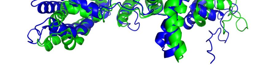
Article
Methods                                                                      FAMSA65 and computed the HMMs following the Uniclust HH-suite
                                                                             database protocol36.
Full algorithm details                                                          The following versions of public datasets were used in this study. Our
Extensive explanations of the components and their motivations are           models were trained on a copy of the PDB5 downloaded on 28 August
available in Supplementary Methods 1.1–1.10, in addition, pseudocode         2019. For finding template structures at prediction time, we used a copy
is available in Supplementary Information Algorithms 1–32, network           of the PDB downloaded on 14 May 2020, and the PDB7066 clustering
diagrams in Supplementary Figs. 1–8, input features in Supplementary         database downloaded on 13 May 2020. For MSA lookup at both training
Table 1 and additional details are provided in Supplementary Tables 2, 3.    and prediction time, we used Uniref9067 v.2020_01, BFD, Uniclust3036
Training and inference details are provided in Supplementary Methods         v.2018_08 and MGnify6 v.2018_12. For sequence distillation, we used
1.11–1.12 and Supplementary Tables 4, 5.                                     Uniclust3036 v.2018_08 to construct a distillation structure dataset.
                                                                             Full details are provided in Supplementary Methods 1.2.
IPA                                                                             For MSA search on BFD + Uniclust30, and template search against
The IPA module combines the pair representation, the single repre-           PDB70, we used HHBlits61 and HHSearch66 from hh-suite v.3.0-beta.3
sentation and the geometric representation to update the single rep-         (version 14/07/2017). For MSA search on Uniref90 and clustered MGnify,
resentation (Supplementary Fig. 8). Each of these representations            we used jackhmmer from HMMER368. For constrained relaxation of
contributes affinities to the shared attention weights and then uses         structures, we used OpenMM v.7.3.169 with the Amber99sb force field32.
these weights to map its values to the output. The IPA operates in 3D        For neural network construction, running and other analyses, we used
space. Each residue produces query points, key points and value points       TensorFlow70, Sonnet71, NumPy72, Python73 and Colab74.
in its local frame. These points are projected into the global frame using      To quantify the effect of the different sequence data sources, we
the backbone frame of the residue in which they interact with each           re-ran the CASP14 proteins using the same models but varying how the
other. The resulting points are then projected back into the local frame.    MSA was constructed. Removing BFD reduced the mean accuracy by
The affinity computation in the 3D space uses squared distances and          0.4 GDT, removing Mgnify reduced the mean accuracy by 0.7 GDT, and
the coordinate transformations ensure the invariance of this module          removing both reduced the mean accuracy by 6.1 GDT. In each case, we
with respect to the global frame (see Supplementary Methods 1.8.2            found that most targets had very small changes in accuracy but a few
‘Invariant point attention (IPA)’ for the algorithm, proof of invariance     outliers had very large (20+ GDT) differences. This is consistent with the
and a description of the full multi-head version). A related construc-       results in Fig. 5a in which the depth of the MSA is relatively unimportant
tion that uses classic geometric invariants to construct pairwise fea-       until it approaches a threshold value of around 30 sequences when the
tures in place of the learned 3D points has been applied to protein          MSA size effects become quite large. We observe mostly overlapping
design59.                                                                    effects between inclusion of BFD and Mgnify, but having at least one
   In addition to the IPA, standard dot product attention is computed        of these metagenomics databases is very important for target classes
on the abstract single representation and a special attention on the pair    that are poorly represented in UniRef, and having both was necessary
representation. The pair representation augments both the logits and         to achieve full CASP accuracy.
the values of the attention process, which is the primary way in which
the pair representation controls the structure generation.                   Training regimen
                                                                             To train, we use structures from the PDB with a maximum release date
Inputs and data sources                                                      of 30 April 2018. Chains are sampled in inverse proportion to cluster
Inputs to the network are the primary sequence, sequences from evo-          size of a 40% sequence identity clustering. We then randomly crop
lutionarily related proteins in the form of a MSA created by standard        them to 256 residues and assemble into batches of size 128. We train the
tools including jackhmmer60 and HHBlits61, and 3D atom coordinates           model on Tensor Processing Unit (TPU) v3 with a batch size of 1 per TPU
of a small number of homologous structures (templates) where avail-          core, hence the model uses 128 TPU v3 cores. The model is trained until
able. For both the MSA and templates, the search processes are tuned         convergence (around 10 million samples) and further fine-tuned using
for high recall; spurious matches will probably appear in the raw MSA        longer crops of 384 residues, larger MSA stack and reduced learning
but this matches the training condition of the network.                      rate (see Supplementary Methods 1.11 for the exact configuration). The
  One of the sequence databases used, Big Fantastic Database (BFD),          initial training stage takes approximately 1 week, and the fine-tuning
was custom-made and released publicly (see ‘Data availability’) and          stage takes approximately 4 additional days.
was used by several CASP teams. BFD is one of the largest publicly avail-       The network is supervised by the FAPE loss and a number of auxil-
able collections of protein families. It consists of 65,983,866 families     iary losses. First, the final pair representation is linearly projected to
represented as MSAs and hidden Markov models (HMMs) covering                 a binned distance distribution (distogram) prediction, scored with
2,204,359,010 protein sequences from reference databases, metage-            a cross-entropy loss. Second, we use random masking on the input
nomes and metatranscriptomes.                                                MSAs and require the network to reconstruct the masked regions
  BFD was built in three steps. First, 2,423,213,294 protein sequences       from the output MSA representation using a BERT-like loss37. Third,
were collected from UniProt (Swiss-Prot&TrEMBL, 2017-11)62, a soil refer-    the output single representations of the structure module are used to
ence protein catalogue and the marine eukaryotic reference catalogue7,       predict binned per-residue lDDT-Cα values. Finally, we use an auxiliary
and clustered to 30% sequence identity, while enforcing a 90% align-         side-chain loss during training, and an auxiliary structure violation loss
ment coverage of the shorter sequences using MMseqs2/Linclust63.             during fine-tuning. Detailed descriptions and weighting are provided
This resulted in 345,159,030 clusters. For computational efficiency,         in the Supplementary Information.
we removed all clusters with less than three members, resulting in              An initial model trained with the above objectives was used to make
61,083,719 clusters. Second, we added 166,510,624 representative pro-        structure predictions for a Uniclust dataset of 355,993 sequences with
tein sequences from Metaclust NR (2017-05; discarding all sequences          the full MSAs. These predictions were then used to train a final model
shorter than 150 residues)63 by aligning them against the cluster rep-       with identical hyperparameters, except for sampling examples 75% of
resentatives using MMseqs264. Sequences that fulfilled the sequence          the time from the Uniclust prediction set, with sub-sampled MSAs, and
identity and coverage criteria were assigned to the best scoring cluster.    25% of the time from the clustered PDB set.
The remaining 25,347,429 sequences that could not be assigned were              We train five different models using different random seeds, some
clustered separately and added as new clusters, resulting in the final       with templates and some without, to encourage diversity in the predic-
clustering. Third, for each of the clusters, we computed an MSA using        tions (see Supplementary Table 5 and Supplementary Methods 1.12.1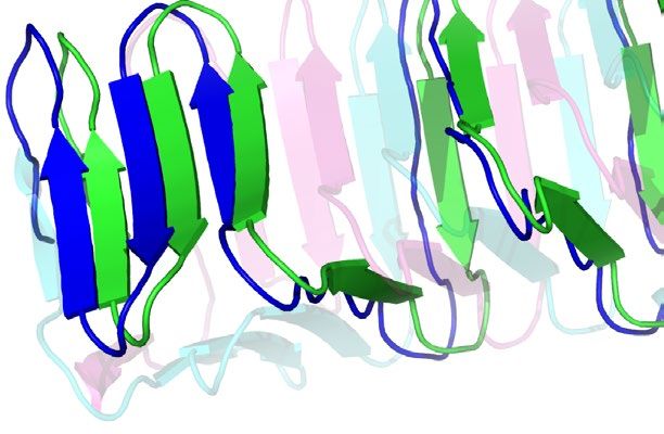
for details). We also fine-tuned these models after CASP14 to add a             structures with less than 16 resolved residues, with unknown residues
pTM prediction objective (Supplementary Methods 1.9.7) and use the              or solved by NMR methods were removed. As the PDB contains many
obtained models for Fig. 2d.                                                    near-duplicate sequences, the chain with the highest resolution was
                                                                                selected from each cluster in the PDB 40% sequence clustering of the
Inference regimen                                                               data. Furthermore, we removed all sequences for which fewer than
We inference the five trained models and use the predicted confidence           80 amino acids had the alpha carbon resolved and removed chains with
score to select the best model per target.                                      more than 1,400 residues. The final dataset contained 10,795 protein
   Using our CASP14 configuration for AlphaFold, the trunk of the net-          sequences.
work is run multiple times with different random choices for the MSA               The procedure for filtering the recent PDB dataset based on prior
cluster centres (see Supplementary Methods 1.11.2 for details of the            template identity was as follows. Hmmsearch was run with default
ensembling procedure). The full time to make a structure prediction             parameters against a copy of the PDB SEQRES fasta downloaded
varies considerably depending on the length of the protein. Repre-              15 February 2021. Template hits were accepted if the associated struc-
sentative timings for the neural network using a single model on V100           ture had a release date earlier than 30 April 2018. Each residue position
GPU are 4.8 min with 256 residues, 9.2 min with 384 residues and 18 h           in a query sequence was assigned the maximum identity of any template
at 2,500 residues. These timings are measured using our open-source             hit covering that position. Filtering then proceeded as described in
code, and the open-source code is notably faster than the version we            the individual figure legends, based on a combination of maximum
ran in CASP14 as we now use the XLA compiler75.                                 identity and sequence coverage.
   Since CASP14, we have found that the accuracy of the network with-              The MSA depth analysis was based on computing the normalized
out ensembling is very close or equal to the accuracy with ensembling           number of effective sequences (Neff ) for each position of a query
and we turn off ensembling for most inference. Without ensembling,              sequence. Per-residue Neff values were obtained by counting the num-
the network is 8× faster and the representative timings for a single            ber of non-gap residues in the MSA for this position and weighting the
model are 0.6 min with 256 residues, 1.1 min with 384 residues and              sequences using the Neff scheme76 with a threshold of 80% sequence
2.1 h with 2,500 residues.                                                      identity measured on the region that is non-gap in either sequence.
   Inferencing large proteins can easily exceed the memory of a single
GPU. For a V100 with 16 GB of memory, we can predict the structure              Reporting summary
of proteins up to around 1,300 residues without ensembling and the              Further information on research design is available in the Nature
256- and 384-residue inference times are using the memory of a single           Research Reporting Summary linked to this paper.
GPU. The memory usage is approximately quadratic in the number of
residues, so a 2,500-residue protein involves using unified memory so
that we can greatly exceed the memory of a single V100. In our cloud            Data availability
setup, a single V100 is used for computation on a 2,500-residue protein         All input data are freely available from public sources.
but we requested four GPUs to have sufficient memory.                              Structures from the PDB were used for training and as templates
   Searching genetic sequence databases to prepare inputs and final             (https://www.wwpdb.org/ftp/pdb-ftp-sites; for the associated
relaxation of the structures take additional central processing unit            sequence data and 40% sequence clustering see also https://ftp.wwpdb.
(CPU) time but do not require a GPU or TPU.                                     org/pub/pdb/derived_data/ and https://cdn.rcsb.org/resources/
                                                                                sequence/clusters/bc-40.out). Training used a version of the PDB
Metrics                                                                         downloaded 28 August 2019, while the CASP14 template search used
The predicted structure is compared to the true structure from the              a version downloaded 14 May 2020. The template search also used the
PDB in terms of lDDT metric34, as this metric reports the domain accu-          PDB70 database, downloaded 13 May 2020 (https://wwwuser.gwdg.
racy without requiring a domain segmentation of chain structures.               de/~compbiol/data/hhsuite/databases/hhsuite_dbs/).
The distances are either computed between all heavy atoms (lDDT)                   We show experimental structures from the PDB with accession num-
or only the Cα atoms to measure the backbone accuracy (lDDT-Cα).                bers 6Y4F77, 6YJ178, 6VR479, 6SK080, 6FES81, 6W6W82, 6T1Z83 and 7JTL84.
As lDDT-Cα only focuses on the Cα atoms, it does not include the pen-              For MSA lookup at both the training and prediction time, we used
alty for structural violations and clashes. Domain accuracies in CASP           UniRef90 v.2020_01 (https://ftp.ebi.ac.uk/pub/databases/uniprot/
are reported as GDT33 and the TM-score27 is used as a full chain global         previous_releases/release-2020_01/uniref/), BFD (https://bfd.mmseqs.
superposition metric.                                                           com), Uniclust30 v.2018_08 (https://wwwuser.gwdg.de/~compbiol/
   We also report accuracies using the r.m.s.d.95 (Cα r.m.s.d. at 95% cov-      uniclust/2018_08/) and MGnify clusters v.2018_12 (https://ftp.ebi.ac.uk/
erage). We perform five iterations of (1) a least-squares alignment of the      pub/databases/metagenomics/peptide_database/2018_12/). Uniclust30
predicted structure and the PDB structure on the currently chosen Cα            v.2018_08 was also used as input for constructing a distillation structure
atoms (using all Cα atoms in the first iteration); (2) selecting the 95%        dataset.
of Cα atoms with the lowest alignment error. The r.m.s.d. of the atoms
chosen for the final iterations is the r.m.s.d.95. This metric is more robust
to apparent errors that can originate from crystal structure artefacts,         Code availability
although in some cases the removed 5% of residues will contain genuine          Source code for the AlphaFold model, trained weights and inference
modelling errors.                                                               script are available under an open-source license at https://github.
                                                                                com/deepmind/alphafold.
Test set of recent PDB sequences                                                  Neural networks were developed with TensorFlow v.1 (https://github.
For evaluation on recent PDB sequences (Figs. 2a–d, 4a, 5a), we used            com/tensorflow/tensorflow), Sonnet v.1 (https://github.com/deep-
a copy of the PDB downloaded 15 February 2021. Structures were fil-             mind/sonnet), JAX v.0.1.69 (https://github.com/google/jax/) and Haiku
tered to those with a release date after 30 April 2018 (the date limit for      v.0.0.4 (https://github.com/deepmind/dm-haiku). The XLA compiler is
inclusion in the training set for AlphaFold). Chains were further filtered      bundled with JAX and does not have a separate version number.
to remove sequences that consisted of a single amino acid as well as              For MSA search on BFD+Uniclust30, and for template search against
sequences with an ambiguous chemical component at any residue                   PDB70, we used HHBlits and HHSearch from hh-suite v.3.0-beta.3
position. Exact duplicates were removed, with the chain with the most           release 14/07/2017 (https://github.com/soedinglab/hh-suite). For MSA
resolved Cα atoms used as the representative sequence. Subsequently,            search on UniRef90 and clustered MGnify, we used jackhmmer from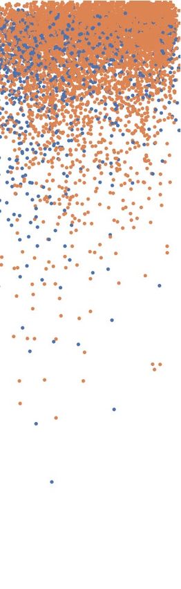
You can also read