Low dietary protein content alleviates motor symptoms in mice with mutant dynactin/dynein-mediated neurodegeneration
←
→
Page content transcription
If your browser does not render page correctly, please read the page content below
Human Molecular Genetics, 2015, Vol. 24, No. 8              2228–2240

                                                                                doi: 10.1093/hmg/ddu741
                                                                                Advance Access Publication Date: 30 December 2014
                                                                                Original Article

ORIGINAL ARTICLE

Low dietary protein content alleviates motor symptoms

                                                                                                                                                        Downloaded from https://academic.oup.com/hmg/article/24/8/2228/652057 by guest on 18 September 2021
in mice with mutant dynactin/dynein-mediated
neurodegeneration
Diana Wiesner1,†, Jérome Sinniger3,4,†, Alexandre Henriques3,4,
Stéphane Dieterlé3,4, Hans-Peter Müller1, Volker Rasche1, Boris Ferger5,
Sylvie Dirrig-Grosch3,4, Rana Soylu-Kucharz6, Asa Petersén6, Paul Walther2,
Birgit Linkus1, Jan Kassubek1, Philip C. Wong7, Albert C. Ludolph1
and Luc Dupuis3,4, *
1
  Department of Neurology, 2Central Facility for Electron Microscopy, Ulm University, 89081 Ulm, Germany,
3
  Inserm U1118, Mécanismes Centraux et Périphériques de la Neurodégénérescence, Strasbourg F-67085, France,
4
  Université de Strasbourg, Fédération de Médecine Translationnelle (FMTS), UMRS1118, Strasbourg F-67085,
France, 5CNS Diseases Research, Boehringer Ingelheim Pharma GmbH & Co. KG, 88397 Biberach an der Riss,
Germany, 6Translational Neuroendocrine Research Unit, Department of Experimental Medical Sciences, Lund
University, 22184 Lund, Sweden and 7Department of Pathology and Neuroscience and Division of
Neuropathology, The Johns Hopkins University School of Medicine, Baltimore, USA
*To whom correspondence should be addressed at: INSERM U1118, Faculté de médecine, 11 rue Humann, 67085, Strasbourg, France.
Tel: +33 368853082; Fax: +33 368853065; Email: ldupuis@unistra.fr

Abstract
Mutations in components of the molecular motor dynein/dynactin lead to neurodegenerative diseases of the motor system or
atypical parkinsonism. These mutations are associated with prominent accumulation of vesicles involved in autophagy and
lysosomal pathways, and with protein inclusions. Whether alleviating these defects would affect motor symptoms remain
unknown. Here, we show that a mouse model expressing low levels of disease linked-G59S mutant dynactin p150Glued develops
motor dysfunction >8 months before loss of motor neurons or dopaminergic degeneration is observed. Abnormal accumulation
of autophagosomes and protein inclusions were efficiently corrected by lowering dietary protein content, and this was
associated with transcriptional upregulations of key players in autophagy. Most importantly this dietary modification partially
rescued overall neurological symptoms in these mice after onset. Similar observations were made in another mouse strain
carrying a point mutation in the dynein heavy chain gene. Collectively, our data suggest that stimulating the autophagy/
lysosomal system through appropriate nutritional intervention has significant beneficial effects on motor symptoms of dynein/
dynactin diseases even after symptom onset.

†
 The authors wish it to be known that, in their opinion, the first two authors should be regarded as joint First Authors.
Received: October 11, 2014. Revised: December 8, 2014. Accepted: December 22, 2014
© The Author 2014. Published by Oxford University Press. All rights reserved. For Permissions, please email: journals.permissions@oup.com

                                                                                                                                               2228
Human Molecular Genetics, 2015, Vol. 24, No. 8 | 2229
Introduction age, and disease end-stage was reached at ∼18 months of age.
Consistent with the initial study, we observed denervation like
Neurodegenerative diseases are characterized by multiple altera-
spontaneous muscle electrical activity using electromyography
tions observed in brain tissue of patients. Loss of neuronal cell
(EMG) in end-stage animals (Supplementary Material, Fig. S1).
bodies as well as loss of synapses is generally called neurodegen-
However, these EMG abnormalities were never observed in mice
eration, and is accompanied by other neuronal alterations, in
younger than 9 months of age (Supplementary Material, Fig. S1)
particular protein inclusions and abnormal protein recycling path-
and neuromuscular junctions (NMJs) of 10-month-old G59S mice
ways such as autophagy/lysosomal pathway. In many cases, the
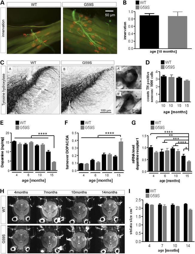
were normally innervated (Fig. 2A and B). Thus, the obvious
physician considers that a period for ‘neuronal dysfunction’
motor deficit observed at 10 months of age is not due to motor
occurs before degenerative changes, especially in slowly progres-
neuron degeneration or NMJ dysfunction. Mutations in DCTN1
sive neurodegenerative diseases, such as spastic paraplegia or
gene have also been linked with atypical Parkinson’s disease
juvenile motor neuron diseases. The use of post-mortem tissue
(10). However, the number of dopaminergic neurons in the sub-
precludes, however, to determine which pathological alterations
stantia nigra in 10- and 15-month-old G59S mice was similar to
occur first in patients and which one(s) actually cause the neuro-
wild-type littermate mice (Fig. 2C and D). Dopaminergic neurons
Downloaded from https://academic.oup.com/hmg/article/24/8/2228/652057 by guest on 18 September 2021
logical symptoms. Getting access to pre-symptomatic events
were, however, affected late in the disease, since dopamine (DA)
should in principle be easier in animal models, and their careful
levels dropped in 15 month-old G59S mice (Fig. 2E), while DA turn-
analysis might help us to determine the sequence of pathological
over increased (Fig. 2F) and D1 receptor mRNA decreased (Fig. 2G).
alterations and their relationships with symptoms. However, most
Thus, G59S mutation also affects dopaminergic neurons, although
commonly used mouse models develop very rapid progression
in late stages and in the absence of neuronal loss. To more broadly
and severe disease, which is not always the case in patients.
detect a potential degenerative phenotype in G59S, we performed
Thus, slowly progressive mouse models of disease could help to
longitudinal magnetic resonance imaging (MRI) analysis. As
understand the sequence of events upon longitudinal follow-up.
shown in Figure 2H and I, there was no obvious degeneration of
Dysfunction of vesicle trafficking is thought to be an early
specific brain region nor was there global brain atrophy in these
event in many neurodegenerative diseases. Recently, genetic
animals. In all, G59S mice neuronal loss appears restricted to
mutations in two components of dynein, the molecular motor
motor neurons, and to some extent to nigrostriatal dopaminergic
carrying cargoes in the retrograde direction in axons were linked
neurons, and occurs several months after motor symptoms.
to familial neurodegenerative disease (1,2). Mutations in dynein
heavy chain or its associated protein dynactin, cause diseases
affecting either motor neurons (3–6), sensory neurons (7,8), Low protein diet partially rescues motor deficits
medium spiny neurons (9), nigrostriatal neurons (10) and cortical in G59S and Cramping mice
development (5,11,12). Germline mutations in the murine dynein
If motor symptoms occur months before actual neuronal loss,
heavy chain lead to proprioceptive neuropathy and striatal dys-
then they are not the consequence of neuronal loss. Previous
function, but do not trigger neurodegeneration. On the contrary,
studies had indicated that mutant dynactin overexpression
overexpression of disease-linked G59S mutation in dynactin
leads to proliferation of membrane bound vesicles including
p150Glued (G59S mice) leads to motor neuron degeneration in
lysosomes (14) and autophagosomes (13) in motor neurons. Low-
end-stage mice (13,14) while knock-in of this same mutation
ering protein content of the diet increases lysosomal and autop-
did not lead to observable motor phenotypes (15). Interestingly,
hagic turnover in the periphery (16), and we hypothesized that
G59S mice also develop protein inclusions, and abnormal autop-
such dietary treatment could also ameliorate symptoms of
hagy/lysosomal pathways (13,14). The slow progression of their
G59S mice. Two groups of 6-month-old G59S mice (n = 16–18)
disease makes these mice a suitable model to study the sequence
and wild-type littermates (n = 11–12) were trained to rotarod dur-
of events leading to neurodegeneration and symptoms.
ing 1 week, assessed for motor coordination for the next 2 weeks
Here, we performed a longitudinal analysis of behavioural
and then fed with either chow or low protein diet [LPD, compos-
symptoms and neurodegenerative features in the M2 line of
ition in Supplementary Material, Table S1 identical to that of 16).
Laird et al. (13). This M2 line, later referred as G59S mice, is the
From Week 3 of LPD, we observed that LPD increased rotarod per-
mouse strain with the slowest disease progression that these
formance of G59S mice as compared both with baseline and their
authors generated. We observed that loss of neurons and synapses
G59S littermates under chow diet (Fig. 3A). The amelioration plat-
occurs extremely late, months after onset of motor symptoms.
eaued after 6 weeks of LPD, and as a whole, LPD rescued about
Lowering dietary protein content concomitantly mitigated symp-
half of the motor defect. Grip strength also tended to increase
toms, reverted autophagosome defects and decreased protein
upon LPD in G59S animals (Fig. 3B). As expected from previous
inclusions. Our results suggest that nutritional therapies stimulat-
studies, LPD also decreased the final body weight of the animals
ing autophagy/lysosomal pathway might be useful for symptom-
of 3–5 g. To provide further evidence that LPD might correct es-
atic relief and not only as neuroprotective strategies.
tablished motor symptoms caused by defective axonal transport,
we performed similar experiments in Cramping mice. These mice
carry a mutant allele of Dync1h1 (17), the gene encoding the heavy
Results chain of dynein, and this mutation lies in the same domain than
Late and selective neurodegeneration in transgenic human mutations associated with motor and sensory neuropa-
thies (4,5,8), and triggers early sensory-motor defects without
G59S mice
neurodegeneration (7,9,18). This mouse strain is maintained on
We sought to correlate motor symptoms and neuronal loss in G59S a pure C3H background, different from the Bl6 SJL background
mice, as these mice have only been characterized at end-stage in of G59S mice. In this mouse line, motor defects are established
the initial study (13). Reduced muscle grip strength of G59S mice within the first weeks after birth and remain essentially stable
(13) occurred after 6 months of age (Fig. 1A), similar to decreased through life (9,17). In this completely independent mouse
rotarod performance (Fig. 1B). Body weight loss (Fig. 1C) as well model, we also observed an increased motor performance of
as cramping and tremors (Fig. 1D) occurred after 8 months of Cramping mice upon LPD either in rotarod performance or in2230 | Human Molecular Genetics, 2015, Vol. 24, No. 8
Downloaded from https://academic.oup.com/hmg/article/24/8/2228/652057 by guest on 18 September 2021
Figure 1. Early motor dysfunction in G59S mice: (A) grip muscle strength of all limbs in wild-type (black) and G59S-mice (grey) from 1 to 18 months of age, normalized to
body weight. ***P < 0.001 versus corresponding wild type (n = 15 mice per group). (B) latency to fall in an accelerating rotarod test in wild-type (black) and G59S-mice (grey)
from 1 to 18 months of age. ****P < 0.0001 versus corresponding wild type (n = 15 mice per group). (C) Body weight of wild-type (black) and G59S-mice (grey) from 5 to 70
weeks of age. ****P < 0.0001 versus corresponding wild type (n = 15 mice per group). (D) Kaplan–Meier plot showing incidence of tremor and cramping in G59S-mice, tremor
(black line) and cramping (grey line) (n = 15 mice per group). The shaded grey part of each graph indicates time points at which muscle denervation is observed (see Fig. 2).
The dashed line indicates the time point at which LPD was initiated in Figure 3.
grip strength (Fig. 3C and D). Thus, decreasing protein content of autophagosome marker LC3-II. Consistent with a correction of
the diet was sufficient to mitigate established motor symptoms defective autophagy by LPD, increased LC3-II levels observed in
in these two mouse models, at a time point of disease when neur- chow-fed G59S mice were reverted by LPD (Fig. 6A). To directly
onal loss is not established. demonstrate effects on autophagic flux in vivo, we used electron
microscopy to unambiguously identify autophagosomes and
blocked lysosomal proteolysis using chloroquine in a subset of
Low protein diet normalizes autophagosomal
mice (22). Consistent with increased LC3-II, G59S mice displayed
defects in G59S mice
many more autophagosomes in neurons than wild-type litter-
LPD might be protective towards motor symptoms through mul- mates (Fig. 6B–D). Chloroquine administration led to significantly
tiple mechanisms, including improved axonal transport, direct more autophagosomes in G59S as compared with wild types,
effects on autophagic machinery or indirect peripheral effects. showing that G59S mice displayed increased autophagosome for-
To control that LPD could directly activate relevant long-term mation in basal conditions (Fig. 6B–D). LPD completely abolished
responses in the central nervous system, we monitored mRNA increased autophagosome counts in G59S mice. Importantly,
levels of Snat2, the primary amino acid transporter that is blocking autophagosomal clearance with chloroquine preven-
induced at the level of transcription when mammalian cells are ted the decrease in number of autophagosomes elicited by LPD
deprived of amino acids (19,20). Twelve weeks of LPD increased showing that autophagosome clearance was increased upon
Snat2 mRNA levels in the cerebral cortex (Fig. 4A) and spinal LPD. However, autophagosome counts remained higher in the
cord (Fig. 4B) of G59S mice as well as in spinal cord of Cramping presence of chloroquine in chow fed G59S mice as compared
mice (Fig. 4C). Similar trends were observed in wild-type litter- with LPD fed G59S mice, demonstrating that LPD also decreased
mates. Since LPD had effects on the CNS, we hypothesized that autophagosome formation. Thus, LPD was able to correct auto-
it could improve axonal transport, thereby alleviating symptoms phagosome defects through both decreased autophagosome
(21). To test this hypothesis, we injected fluorogold in hind limb generation and increased autophagosome clearance. In the
muscles of G59S mice either chow diet or LPD fed, and sacrificed course of the analysis of electron microscopy (EM) images, we
mice 24 h later to quantify retrograde transport of the dye in noticed the occurrence of prominent protein inclusions in G59S
spinal motor neurons. We observed that G59S mutation led to neurons (Fig. 6C–E). LPD decreased the frequency of these
drastic loss of fluorogold labelling in motor neurons (Fig. 5). LPD inclusions in G59S neurons, an effect prevented by chloroquine.
did not modify this axonal transport defect. Thus, autophagic flux is impaired in G59S neurons, and LPD
LPD could also alleviate motor symptoms through effects restores normal autophagosome numbers and resolves protein
on autophagy, and we thus studied protein levels of the aggregates.Human Molecular Genetics, 2015, Vol. 24, No. 8 | 2231
Downloaded from https://academic.oup.com/hmg/article/24/8/2228/652057 by guest on 18 September 2021
Figure 2. Late neurodegeneration in G59S mice. (A) Representative immunohistochemistry of neuromuscular junctions (NMJs) in the tibialis anterior muscle of wild-type
and G59S-mice at the age of 10 months of age (n = 5 mice per group). The post-synaptic part of the NMJ was labelled with bungarotoxin (red), and innervating axons were
labelled with anti-neurofilament antibodies (green). Note that all post-synaptic structures are properly innervated at that age. (B) Quantification of previous experiment.
The percentage of innervated NMJs is shown. (C) Immunohistological staining of tyrosine hydroxylase (TH)-positive neurons in wild-type and G59S-mice at the age of
15 months. (D) Stereological estimation of the number of TH-positive neurons in the substantia nigra in wild-type and G59S-mice at the age of 10 and 15 months (n = 5
mice per group). (E and F) Dopamine levels (E) and DA turnover (F) as judged on DOPAC/DA ratio from 4 to 15 months of age in wild-type (black) and G59S-mice (grey) (n = 12
mice per group). (G) mRNA levels of DA receptor1 in 4–15-month-old wild-type (black) and G59S-mice (grey) (n = 12 mice per group). (H) Representative horizontal
T2-weighted MRI slices of wild-type and G59S-mice at 4, 7, 10 and 14 months of age. (I) Striatal volume of wild-type (black) and G59S-mice (grey) at 4, 7, 10 and 14
months of age. *P < 0.05; **P < 0.01; ***P < 0.001; ****P < 0.0001 versus corresponding wild type.
Low protein diet differentially reprogrammes gene
hormesis (23). We hypothesized that this would also stand true
expression in different genetic backgrounds for LPD, and that LPD would stimulate expression of the cellular
Long-term dietary changes, such as calorie restriction, induce systems to recycle amino acids in particular macro-autophagy,
activation of cellular adaptative responses in a process called chaperone mediated autophagy (CMA) and/or lysosomal2232 | Human Molecular Genetics, 2015, Vol. 24, No. 8
Downloaded from https://academic.oup.com/hmg/article/24/8/2228/652057 by guest on 18 September 2021
Figure 3. Low protein diet partially rescues motor deficits in G59S and Cramping mice. (A and B) latency to fall in an accelerating rotarod test (A) or muscle grip strength
normalized to body weight (B) in wild-type (black) and G59S-mice (grey) fed with chow diet (filled symbols) or low protein diet (LPD, open symbols). (C and D) latency to fall
in an accelerating rotarod test (C) or muscle grip strength normalized to body weight (D) in wild-type (black) and Dync1h1Cra/+ mice (grey). Arrows show the time point mice
were switched from chow to LPD. *P < 0.05; ***P < 0.001 versus corresponding wild type. In (B) and (D) grip strength of wild-type mice under chow or LPD were omitted for
clarity.
biogenesis. Consistent with chronic autophagy impairment (24), Discussion
LC3 expression was slightly increased in chow fed G59S mice as
Here, we show that motor symptoms of dynein/dynactin diseases
compared with wild-type chow fed mice. Consistent with EM
occur long before actual neuronal or axonal loss and are alle-
quantifications, LC3 gene expression was decreased in G59S
viated by low dietary protein content. This is likely mediated
mice fed with LPD. mRNA levels of autophagy key genes, such
through correction of dysfunctional autophagy/lysosomal
as p62 and FoxO3 were unchanged in chow diet, but slightly in-
systems.
creased upon LPD selectively in G59S mice (Fig. 7A). mRNA levels
A first major result is that motor symptoms in G59S mice are
of key CMA player Lamp2-A were increased (Fig. 7B) and this was
not caused by neuronal death or synaptic loss. We chose to study
mirrored in protein levels (Fig. 7C). However, there was no coordi-
the correlation between features of neurodegeneration, such as
nated upregulation of genes involved in lysosomal biogenesis muscle denervation, and motor symptoms in the slowly progres-
(Fig. 7D). sive M2 line (13). Indeed, this mouse strain develops very slow
The lysosomal/autophagy pathway was also altered in Cramp- motor symptoms, which are accompanied by detectable motor
ing mice upon LPD, although with different features. LPD did not neuron degeneration, as shown by EMG, molecular and histo-
change Lamp2-A protein levels, but led to decreased LC3-I levels logical endpoints (13 and this study). Thus, these mice recapitu-
in C3H control littermates but not in Cramping mice (Fig. 8A and B). late as closely as possible the symptoms observed in patients
Gene expression was dramatically altered in Cramping mice upon carrying the G59S mutation. In our G59S mice, we observed
LPD, with increased expression of LC3 and p62 but also of key strong motor phenotypes in the complete absence of NMJ
lysosomal proteins such as Lamp1, Cathepsins B and D, and denervation or EMG abnormalities, thus demonstrating that
Atp6v1h and key CMA player Lamp2-A (Fig. 8C and D). All of degeneration of motor neurons and NMJs is uncoupled from
these regulations did not occur in C3H control littermates. Sum- motor symptoms. Similarly, dopaminergic involvement occurs
marizing, LPD activates a transcriptional programme in diseased late, and neither DA levels, nor tyrosine hydroxylase (TH)-posi-
mice with different features as a function of the genetic back- tive cell counts are changed at 10 months of age despite obvious
ground. In Bl6SJL mice, key players of macro-autophagy and of motor symptoms. MRI imaging did neither show specific atrophy
CMA, but not lysosomal biogenesis are upregulated, while in of the brain nor obvious lesions, and we consider thus unlikely
C3H mice, transcriptional upregulation is broader. that degeneration of other neurons than motor neurons orHuman Molecular Genetics, 2015, Vol. 24, No. 8 | 2233
Downloaded from https://academic.oup.com/hmg/article/24/8/2228/652057 by guest on 18 September 2021
Figure 4. LPD increases expression of Snat2 in the CNS. mRNA levels of the amino acid transporter Snat2 (Slc38a2) in the cortex (A) and spinal cord (B) of wild-type (black)
and G59S-mice (grey) and spinal cord of Cramping mice and their controls (C), fed with chow diet (−) or LPD (LPD, +).
Figure 5. Low protein diet does not modify axonal transport defect in G59S mice. (A) representative microphotographs of fluorogold fluorescence in lumbar spinal cord of
wild-type (upper row) and G59S-mice (lower row) fed with chow diet (−LPD) or low protein diet (+LPD). Motor neurons of wild type mice are filled with fluorogold-positive
vesicles (arrows), while motor neurons of G59S mice show less distinct staining, indicative of a decreased rate of fluorogold transport to the spinal cord. The left images
show lower magnifications of images in the middle column (box). (B) Number of fluorogold-positive lumbar motor neurons (L3–L5) per ventral horn in wild-type (black
columns) and G59S-mice (grey columns) fed with chow diet (−LPD) or low protein diet (+LPD). **P < 0.01 versus wild type, chow diet.
dopaminergic neurons cause disease. Summarizing, we observe Legs at odd angles (Loa) mice develop motor and behavioural symp-
a complete dissociation between motor symptoms and neuronal toms while neurodegeneration is absent except for developmen-
loss in this model. Interestingly, other mouse models of dynein/ tal death of proprioceptive neurons (7,9,25). Thus, the motor
dynactin inhibition also show dissociation between degener- behaviour caused by inhibition of dynein/dynactin is dissociated
ation of neurons and symptoms. For instance, Cramping and from neurodegeneration, and cannot be caused by it. In patients,2234 | Human Molecular Genetics, 2015, Vol. 24, No. 8
Downloaded from https://academic.oup.com/hmg/article/24/8/2228/652057 by guest on 18 September 2021
Figure 6. Low protein diets corrected autophagosome defects in G59S-mice. (A) Left: representative western blot of LC3-I (upper band) and LC3-II (lower band) in the
cerebral cortex of wild type or G59S mice, fed with chow or low protein diet. Actin western blot is provided as a standard. Right: LC3 II/LC3 I ratio, as measured using
western blotting in the cortex and spinal cord of wild-type (black columns) and G59S-mice (grey columns) fed with chow diet (−LPD) or low protein diet (+LPD).
**P < 0.01; ****P < 0.0001 versus corresponding wild type. (B and C) electron micrographs of neuronal cells in spinal cord from age-matched wild-type (WT) and G59S-
mice (G59S) under chow or LPD, and either injected with saline (NaCl) or chloroquine. Arrows indicate autophagosomes including degradative organelles (B) or protein
inclusions (C); scale bar is 1000 nm. (D) quantitative analysis of autophagosomes (D), and protein inclusions (E) per spinal neuronal cells in G59S-mice and wild-type age-
matched controls under the different treatment groups, *P < 0.05; **P < 0.01; ***P < 0.001; ****P < 0.0001 versus corresponding wild type.
EMG abnormalities were reported in individuals with dynein several patients with H306R DYNC1H1 mutation in their third
(5,26) and dynactin (27) mutations, but these patients were exam- decade or during childhood (6,8) suggesting that, consistent
ined in their fifth decade while some displayed symptoms al- with observations in mice, symptoms might precede neurode-
ready during childhood (26). Indeed, EMG was found normal in generation for a long time.Human Molecular Genetics, 2015, Vol. 24, No. 8 | 2235
Downloaded from https://academic.oup.com/hmg/article/24/8/2228/652057 by guest on 18 September 2021
Figure 7. Gene expression in response to LPD in G59S mice. (A) mRNA levels of genes involved in macro-autophagy (LC3, p62, FoxO3 and Beclin1) in the spinal cord of wild-
type (black) and G59S-mice (grey) fed with chow diet (−) or low protein diet (LPD, +). (B and C) mRNA (B) and protein (C) levels of Lamp2-A in the spinal cord of wild-type
(black) and G59S-mice (grey) fed with chow diet (−) or low protein diet (LPD, +). (D) mRNA levels of genes involved in lysosomal biogenesis (Lamp1, Cathepsin B, Cathepsin
D and ATP6v1h) in the spinal cord of wild-type (black) and G59S-mice (grey) fed with chow diet (−) or low protein diet (LPD, +).
Our study provides insights into underlying pathome- underlie increased accumulation of lysosomal/autophagic vesi-
chanisms. A major pathological finding in various mouse strains cles. First, increased autophagosome numbers could be a conse-
expressing mutant dynactin was that lysosomes and/or autopha- quence of increased biogenesis. Indeed, a recent study showed
gosomes were in abnormal numbers. Chevalier-Larsen et al. (14) that motor neurons with Loa mutation display aberrantly
showed lysosomal proliferation in their G59S line, while we pre- increased signalling response to serum starvation, a well-known
viously observed a progressive and abnormal accumulation of autophagy inducer (28). Such a response could in principle alter
vesicles, and in particular of autophagosomes in another G59S autophagosome generation. A second, non-mutually exclusive,
line (line M1) (13). Lai et al. (15) observed increased synaptic vesi- hypothesis is that G59S mutation impairs autophagic clearance
cles proteins at NMJs. This was corroborated by increased LC3-II through defective vesicle trafficking. Indeed, the G59S mutation
levels, and by increased LC3 gene expression in the mouse line decreases axonal transport rate in vitro (28–30), and we provide
studied here. Many mechanisms involving dynactin could here similar evidence in vivo using retrograde fluorogold labelling2236 | Human Molecular Genetics, 2015, Vol. 24, No. 8
Downloaded from https://academic.oup.com/hmg/article/24/8/2228/652057 by guest on 18 September 2021
Figure 8. Low protein diet effects in Cramping mice. (A) Left: representative western blot of LC3-I (upper band), LC3-II (lower band), Lamp2-A and actin in the spinal cord of
wild type or Cramping mice, fed with chow or low protein diet. (B) Quantification of the western blots shown in A. *P < 0.05 versus wild-type chow fed mice. N = 3–4 per group.
(C) mRNA levels of genes involved in macro-autophagy (LC3, p62, FoxO3) and in chaperone-mediated autophagy (Lamp2-A) and Beclin1) in the spinal cord of wild-type
(black) and Cramping mice (grey) fed with chow diet (−) or low protein diet (LPD, +). (D) mRNA levels of genes involved in lysosomal biogenesis (Lamp1, Cathepsin B,
Cathepsin D and ATP6v1h) in the spinal cord of wild-type (black) and Cramping mice (grey) fed with chow diet (−) or low protein diet (LPD, +).
in motor neurons. Since maturation of autophagosomes to previous work showing that dynein mutations impair autophagic
autophagolysosomes occurs during their transport in the axons clearance of aggregate prone proteins (32) and autophagic
(21), the accumulation of autophagosomes we observe could perinuclear clustering of defective mitochondria (18). The down-
be the consequence of decreased transport rate. Dynactin is stream mechanisms linking impaired autophagic flux to actual
required for transport initiation at distal neurites, and G59S motor symptoms remain to be identified. Among many
mutation also affects this function (29,30). Last, dynactin is also other possibilities, we would like to speculate that impaired
required for the stability of microtubule cytoskeleton (31). autophagy/lysosomal pathways perturbs axonal homeostasis,
In our study, we provide direct evidence for both increased autop- for example, by modifying retrograde signalling pathways or im-
hagosome generation and decreased clearance in G59S mice. pairing mitochondrial bioenergetics.
Indeed, chloroquine administration increased much more dra- Our results demonstrate that lowering dietary protein content
matically autophagosomes in wild types than in G59S neurons, mitigates motor symptoms after their establishment, as well as
showing that at least part of the increased autophagosome levels restores autophagosome defects. About 50% of rotarod defects
were due to impaired clearance. However, the difference between were compensated by LPD, while grip strength, less affected at
these two groups remained significant, consistent with a higher that age, was also increased. Similar observations were done
activity of autophagosome generation in G59S mice. That G59S in Cramping mice, showing that low dietary content generally
mutation impairs autophagic flux in neurons is consistent with protected animal models of dynein/dynactin diseases. It mightHuman Molecular Genetics, 2015, Vol. 24, No. 8 | 2237
seem surprising a priori that a dietary modification leads to such they developed eye infections. For histological analysis, animals
drastic alterations in the CNS. However, calorie restriction also were deeply anaesthetized with 1 mg/kg body weight ketamine
leads to significant adaptative changes in the CNS (33), and, chlorhydrate and 0.5 mg/kg body weight xylazine, and transcar-
LPD is long known to trigger effects in the CNS on various neuro- dially perfused with 4% paraformaldehyde in 0.1 pH 7.4 phos-
chemical parameters such as tyrosine levels (34) or on transduc- phate buffer. Tissues were then quickly dissected, post-fixed for
tion pathways of amino acid sensing (35). 24 h in 4% paraformaldehyde and cryoprotected for 48 h. For the
How could LPD correct autophagosome defects? The effect of biochemical analysis, animals were sacrificed and tissues were
LPD on autophagy were complex, and depending on the genetic quickly dissected, frozen in liquid nitrogen. All animal experi-
background. Measurement of autophagic flux in G59S mice ments in this study were approved by the Ethical Committee of
showed that LPD both decreased autophagosome generation, Baden-Württemberg under number 1077.
while increasing autophagosome clearance. G59S mice also
displayed several transcriptional upregulations, such as p62 or Dietary treatments
Lamp2-A that might underlie the resolving of protein aggregates.
In Cramping mice, LPD maintained the pool of LC3-I, likely Mice were fed either a LPD (no S0052-E712 EF M by Ssniff ) or nor-
Downloaded from https://academic.oup.com/hmg/article/24/8/2228/652057 by guest on 18 September 2021
through a transcriptional mechanism, and led to a generalized mal standard food (CHOW) as indicated (Supplementary Mater-
transcriptional upregulation of genes involved in macro- ial, Table S1 and (16)). Mice were randomly distributed per diet,
autophagy, CMA and lysosomal biogenesis. with at least one mouse per genotype per litter allocated to
We propose that LPD, as a mild, chronic, nutritional stress, each experimental condition. For practical reasons in the animal
leads to an adaptative hormetic response, leading to a coordi- facility, experimentators could not be blinded to the diet.
nated upregulation of genes helping in resolving the initial
stimulus that is decreased amino acid availability. This is consist- Behaviour and motor performance
ent with the simultaneous upregulation of amino acid transpor-
One week before starting experiments, male mice were brought
ters, such as Snat2, as well as of various actors of protein recycling
to the behaviour facility and handled every day. Once per week,
in both G59S and Cramping mice fed with LPD. A potential candi-
starting at the age of 4 weeks, body weight and fore and all limb
date transcription factor stimulated by LPD would be TFEB, that is
grip strength were measured for a cohort of 15 each of transgenic
critically involved in coordinating autophagy and lysosomal gene
and wild-type mice. Grip strength measurements were taken
expression (36), and was found inhibited by mutant androgen
using a grip strength meter (Bioseb gripmeter, Vitrolles, France).
receptor in SBMA, another motor neuron disease and causing
To take into account that the maximal performance of the animal
decreased autophagic flux (37). In Cramping mice, the overall pic-
is strictly dependent upon its body mass, we divided the measure
ture observed would be reminiscent of TFEB activation. However,
obtained by the body weight of the animal, according to the pro-
only one target of TFEB ( p62), was overexpressed in G59S mice.
vider’s recommendations. Each measurement was performed in
Whether the different genetic backgrounds of the mice display
triplicates and data were averaged. To test the motor coordin-
different sensitivities to TFEB stimulation is a possibility.
ation and balance, rotarod test was performed twice per month
It should also be mentioned that Cramping mice display a number
(Rotarod Version 1.2.0. MED Associates Inc. St. Albans, VT). Mice
of neuronal and non-neuronal phenotypes (7,9,18,38) that could
were placed onto a rotating rod with auto acceleration from 4 to
each contribute to the effects of LPD. In general, the transcrip-
40 rpm in 300 s. The length of time the mouse stayed on the rotat-
tional mechanisms accounting for the adaptative response
ing rod was recorded. Every mouse had to perform three trials
elicited by LPD, as well as the potential involvement of TFEB,
separated by 15 min each other, the three trials were averaged.
deserve further investigation.
Identical protocols were performed for G59S and Cramping
Our study shows that lowering dietary content is protective
experiments.
against symptoms of dynein/dynactin diseases, even after the
onset of motor symptoms. This expands the clinical spectrum of
diseases in which such diet could be beneficial (16), and other neu- Electromyography
rodegenerative diseases that display strong lysosomal/autophagy Mice were anesthetized with ketamine/xylazine and electrical
dysfunction such as Huntington’s disease or Parkinson’s diseases, activity was recorded using a monopolar needle electrode (diam-
could represent other conditions in which to test this easy nutri- eter 0.3 mm; 9013R0312; Medtronic, Minneapolis, MN, USA)
tional intervention (39–41). Consistently, LPD is currently sug- inserted into the tail of the mouse. Recordings were made with
gested in advanced PD, as it is postulated to improve L-DOPA a concentric needle electrode (diameter 0.3 mm; 9013S0011; Med-
bioavailability. Our results suggest further that increased autopha- tronic). Electrical activity was monitored in both gastrocnemius
gic flux could indeed also contribute to the benefits of this dietary and tibialis anterior on both legs for at least 2 min. Spontaneous
intervention in advanced PD patients (42,43). activity was differentiated from voluntary activity by visual and
auditory inspection.
Materials and Methods
Immunohistochemistry
Animals
Brain and spinal cord 10 µm cryosections were prepared on
Heterozygous G59S mice in B6 SJL background and Cramping mice gelatin coated slides and processed according to previously pub-
in C3H background were maintained and genotyped as described lished protocols (7). Neuromuscular junction histology was per-
(7,13). Mice were maintained at 22°C with a 12 h light/dark cycle formed on muscle bundles as previously described (7). Primary
and had food and water ad libitum. Unless otherwise mentioned, antibodies were incubated overnight at room temperature. Sec-
mice were fed with chow diet (Supplementary Material, Table S1). ondary antibodies (donkey anti-goat antibody coupled with
Mice were regularly monitored to assess onset and progression of Alexa 555, A21432; Invitrogen, Carlsbad CA, USA; donkey anti-
symptoms and were considered at end stage when they could not mouse coupled with Alexa 488, A21204; Invitrogen, Carlsbad
right themselves within 20 s when placed on their back, or when CA, USA) were incubated 90 min at room temperature. Slides2238 | Human Molecular Genetics, 2015, Vol. 24, No. 8
were mounted using Aqua-Poly/Mount (Polysciences, Warring- antibodies were used: LC3 (Sigma; 1 : 2500 buffered in 1% BSA;
ton PA, USA). Images were acquired with a laser scanning micro- 0.02% NaN3 in PBS containing 0.05% Tween 20); Lamp2-A
scope (LSM 510; Carl Zeiss, Thornwood, NY, USA) equipped (abcam; 1 : 1000 buffered in 1% BSA; 0.02% NaN3 in PBS containing
with a Plan-Apochromat 63× oil DIC immersion lens (numerical 0.05% Tween 20); overnight at 4°C. After washing in PBS/0.05%
aperture 1.4). Tween 20, membranes were incubated at room temperature for
1 h with the second antibody (Bio-Rad; 1 : 10 000 in 2.5% Milk
Stereological analyses of TH-positive cells Goat Anti-Rabbit IgG-HRPconjugated) and washed again. The
blot was visualized (ECL-immunodetection) using Image Quant
Stereological assessment of the number of A9 TH-positive cells in
LAS4000. Samples were corrected for background and quantified
the substantia nigra was performed on a Nikon 80i microscope
using Image Quant LAS 4000. All values were normalized to
equipped with an XY motorized stage (Märzhauser, Wetzlar,
housekeeping protein (β-actin).
Germany), a Z-axis motor and a high precision linear encoder
(Heidenhain, Traunreut, Germany). The delineation of the areas
of interest was done under the 4× objective and the counting Autophagic flux measurements in vivo
Downloaded from https://academic.oup.com/hmg/article/24/8/2228/652057 by guest on 18 September 2021
was performed at high magnification with a 60× Plan-Apo oil
For investigation of autophagy, only female mice were taken.
immersive objective (NA = 1.4). The unbiased stereological quan-
At the age of 30 days, half of the mice get a LPD instead (Supple-
tification principles were applied to estimate the numbers of
mentary Material, Table S1) of regular chow. At 50 days of age,
immunopositive cells by using the optical dissector method (44).
mice were daily injected (intra peritoneal) with 50 mg/kg chloro-
The assessment was performed in a blinded fashion and the
quine or 0.9% NaCl. After 10 days of injection, mice were taken for
procedure was carried out with a random start systematic sam-
biochemical or histological analysis as previously described.
pling routine (New Cast Module in VIS software; Visiopharm A/S,
Horsholm, Denmark). To minimize the coefficient of error, the
sampling interval was adjusted to count at least 100 cells for Electron microscopy
each animal.
Mice were perfused with 4% paraformaldehyde, spinal cord was
dissected and fixed in 2.5% glutaraldehyde for 24 h. Samples
Dopamine measurements were post-fixed in 2% osmiumtetroxide for 1 h and dehydrated
To measure striatal DA levels 4-, 6-, 10-month-old mice and end- in graded series of ethanol. Fully dehydrated samples were
stage mice were sacrificed and striata were quickly dissected, fro- then embedded into epon. The embedded samples were cut, col-
zen in liquid nitrogen and stored at −80°C. Tissues were then lected on copper grids, stained with uranyl acetate and analyzed
homogenized by ultrasonification for 10 s in 0.4 perchloric acid in a TEM (Zeiss EM 10) equipped with digital camera. Three ani-
and centrifuged at 14 000g for 15 min at 4°C. Afterwards, the super- mals per group and at least 10 fields per animal were analysed.
natants were passed through a 0.2 μm filter (MinisartRC4, Sartorius The numbers of lysosomes, inclusions and autophagosomes
AG, Göttingen, Germany). Measurements of DA and its metabolites were counted using Axiovision Rel.4.8. and correlated to the
3,4-dihydroxyphenylacetic acid (DOPAC), 3-methoxytyramine numbers of neuronal cells. Image analysis was performed
(3-MT) and homovanillic acid were performed using high-perform- blinded to the genotype and treatment groups.
ance liquid chromatography (HPLC) combined with electrochem-
ical detection under isocratic conditions as previously described
Retrograde transport evaluations
(45). For data acquisition and calculation, Chromeleon™ version
7.1 HPLC software (Thermo Fisher Scientific Inc., USA) was used. Fluorogold (7 µl of a 10 mg/ml solution in PBS, 10% DMSO) was
slowly injected in 1 min in the gastrocnemius muscle using a
Striatal volume by MRI 10 µl Hamilton syringe (Hamilton, Reno, NV, USA). The syringe
was gently withdrawn and any leakage tracer was removed
Ten G59S mice and 10 age- and gender-matched wild types under- from the surface of the muscle. After 24 h, mice were euthanized
went MRI at an 11.7T small bore animal scanner (Biospec 117/16, and L3–L5 spinal cord was processed for histology and cut in
Bruker, Ettlingen, Germany) with a 4-element receive-only surface 40 µm sections using a vibratome. Fluorogold fluorescence was
brain array coil at four time points with a time interval of 3 months then acquired using regular epifluorescence microscopy. The
(T2-weighted protocol, TE/TR 41.6/2000 ms, matrix 512 × 512, number of motor neurons with fluorogold was quantified by an
in-plane resolution 68 µm × 68 µm, 30 coronar slices of 250 µm). observer blinded to the genotype and treatment in at least 10 sec-
Analysis procedures followed (9). Image analysis was performed tions per animal (n = 5–6 per group).
blinded to the genotype.
Statistical analysis
RT-qPCR
For the experimental data all statistical analysis was done using
Total RNA was extracted using RNeasy Lipid Tissue Mini Kit (Qia-
Prism, version 5.0 (GraphPad Software). For comparison of groups
gen). RT-qPCR was performed using the iScript cDNA Synthesis
without repeated measurements, ANOVA was used. Differences
Kit (Bio-Rad) and the iQ SYBR Green SupermixR (Bio-Rad) using
between means were determined by post hoc comparisons using
a CFX96 thermocycler (Bio-Rad). Primer sequences are provided
the Fisher’s LSD Test. For longitudinal analysis, we used two-way
in Supplementary Material, Table S2. Data were analysed using
ANOVA, with genotype and age as co-variates. Differences were
the Cycler software and normalized to the normalization factor
considered statistically significant if P < 0.05. Values are pre-
calculated from the reference genes (Pol2, β-actin and HPRT).
sented as means ± SEM.
Western blotting
For western Blot analysis mouse tissue were homogenized in
Supplementary Material
RIPA-Buffer containing protease inhibitor. The following primary Supplementary Material is available at HMG online.Human Molecular Genetics, 2015, Vol. 24, No. 8 | 2239
Acknowledgements B., Chapon, F. et al. (2009) DCTN1 mutations in Perry syn-
drome. Nat. Genet., 41, 163–165.
We thank Renate Kunz and Reinhard Weih from the Central Facil-
11. Poirier, K., Lebrun, N., Broix, L., Tian, G., Saillour, Y., Boscher-
ity for Electron Microscopy, Ulm University, Aram Kobalyan, and
on, C., Parrini, E., Valence, S., Pierre, B.S., Oger, M. et al. (2013)
the imaging platform (Neuropole de Strasbourg) for their help.
Mutations in TUBG1, DYNC1H1, KIF5C and KIF2A cause mal-
formations of cortical development and microcephaly. Nat.
Conflict of Interest statement. None declared.
Genet., 45, 639–647.
12. Willemsen, M.H., Vissers, L.E., Willemsen, M.A., van Bon, B.
W., Kroes, T., de Ligt, J., de Vries, B.B., Schoots, J., Lugtenberg,
Funding D., Hamel, B.C. et al. (2012) Mutations in DYNC1H1 cause se-
This work was supported by the Agence Nationale de la Recher- vere intellectual disability with neuronal migration defects.
che (Dynemit to L.D.); Association pour la recherche sur la SLA et J. Med. Genet., 49, 179–183.
les autres maladies du motoneurone (to L.D.); Thierry Latran 13. Laird, F.M., Farah, M.H., Ackerley, S., Hoke, A., Maragakis, N.,
Foundation (SpastALS to L.D.); Helmholtz Institute “RNA dysme- Rothstein, J.D., Griffin, J., Price, D.L., Martin, L.J. and Wong,
Downloaded from https://academic.oup.com/hmg/article/24/8/2228/652057 by guest on 18 September 2021
tabolism in ALS and FTD”(to L.D. and A.C.L.); ALS association P.C. (2008) Motor neuron disease occurring in a mutant dy-
(Grant #2235 and 3209 to L.D.); the Frick foundation for ALS nactin mouse model is characterized by defects in vesicular
research (to L.D.); National Institute of Neurological Disorder trafficking. J. Neurosci., 28, 1997–2005.
and Stroke (Grant R01 NS40014 to P.C.W.); Department of Defense 14. Chevalier-Larsen, E.S., Wallace, K.E., Pennise, C.R. and Holz-
(Grant AL100078 to P.C.W.) and the Robert Packard Center for ALS baur, E.L. (2008) Lysosomal proliferation and distal degener-
Research (to P.C.W.). ation in motor neurons expressing the G59S mutation in
the p150Glued subunit of dynactin. Hum. Mol. Genet., 17,
1946–1955.
15. Lai, C., Lin, X., Chandran, J., Shim, H., Yang, W.J. and Cai, H.
References (2007) The G59S mutation in p150(glued) causes dysfunction
1. Eschbach, J. and Dupuis, L. (2011) Cytoplasmic dynein in neu- of dynactin in mice. J. Neurosci., 27, 13982–13990.
rodegeneration. Pharmacol. Ther., 130, 348–363. 16. Grumati, P., Coletto, L., Sabatelli, P., Cescon, M., Angelin, A.,
2. Schiavo, G., Greensmith, L., Hafezparast, M. and Fisher, E.M. Bertaggia, E., Blaauw, B., Urciuolo, A., Tiepolo, T., Merlini, L.
(2013) Cytoplasmic dynein heavy chain: the servant of et al. (2010) Autophagy is defective in collagen VI muscular
many masters. Trends Neurosci., 36, 641–651. dystrophies, and its reactivation rescues myofiber degener-
3. Puls, I., Jonnakuty, C., LaMonte, B.H., Holzbaur, E.L., Tokito, ation. Nat. Med., 16, 1313–1320.
M., Mann, E., Floeter, M.K., Bidus, K., Drayna, D., Oh, S.J. 17. Hafezparast, M., Klocke, R., Ruhrberg, C., Marquardt, A.,
et al. (2003) Mutant dynactin in motor neuron disease. Nat. Ahmad-Annuar, A., Bowen, S., Lalli, G., Witherden, A.S.,
Genet., 33, 455–456. Hummerich, H., Nicholson, S. et al. (2003) Mutations in dynein
4. Harms, M.B., Ori-McKenney, K.M., Scoto, M., Tuck, E.P., Bell, S., link motor neuron degeneration to defects in retrograde
Ma, D., Masi, S., Allred, P., Al-Lozi, M., Reilly, M.M. et al. (2012) transport. Science, 300, 808–812.
Mutations in the tail domain of DYNC1H1 cause dominant 18. Eschbach, J., Sinniger, J., Bouitbir, J., Fergani, A., Schlagowski,
spinal muscular atrophy. Neurology, 78, 1714–1720. A.I., Zoll, J., Geny, B., Rene, F., Larmet, Y., Marion, V. et al. (2013)
5. Fiorillo, C., Moro, F., Yi, J., Weil, S., Brisca, G., Astrea, G., Sever- Dynein mutations associated with hereditary motor neuro-
ino, M., Romano, A., Battini, R., Rossi, A. et al. (2014) Novel pathies impair mitochondrial morphology and function
dynein DYNC1H1 neck and motor domain mutations link with age. Neurobiol. Dis., 58C, 220–230.
distal spinal muscular atrophy and abnormal cortical devel- 19. Palii, S.S., Kays, C.E., Deval, C., Bruhat, A., Fafournoux, P. and
opment. Hum. Mutat., 35, 298–302. Kilberg, M.S. (2009) Specificity of amino acid regulated
6. Tsurusaki, Y., Saitoh, S., Tomizawa, K., Sudo, A., Asahina, N., gene expression: analysis of genes subjected to either
Shiraishi, H., Ito, J., Tanaka, H., Doi, H., Saitsu, H. et al. (2012) complete or single amino acid deprivation. Amino Acids, 37,
A DYNC1H1 mutation causes a dominant spinal muscular 79–88.
atrophy with lower extremity predominance. Neurogenetics, 20. Gazzola, R.F., Sala, R., Bussolati, O., Visigalli, R., Dall’Asta, V.,
13, 327–332. Ganapathy, V. and Gazzola, G.C. (2001) The adaptive regula-
7. Dupuis, L., Fergani, A., Braunstein, K.E., Eschbach, J., Holl, N., tion of amino acid transport system A is associated to
Rene, F., Gonzalez De Aguilar, J.L., Zoerner, B., Schwalen- changes in ATA2 expression. FEBS Lett., 490, 11–14.
stocker, B., Ludolph, A.C. et al. (2009) Mice with a mutation 21. Maday, S., Wallace, K.E. and Holzbaur, E.L. (2012) Autophago-
in the dynein heavy chain 1 gene display sensory neuropathy somes initiate distally and mature during transport toward
but lack motor neuron disease. Exp. Neurol., 215, 146–152. the cell soma in primary neurons. J. Cell. Biol., 196, 407–417.
8. Weedon, M.N., Hastings, R., Caswell, R., Xie, W., Paszkiewicz, 22. Iwai-Kanai, E., Yuan, H., Huang, C., Sayen, M.R., Perry-Garza,
K., Antoniadi, T., Williams, M., King, C., Greenhalgh, L., New- C.N., Kim, L. and Gottlieb, R.A. (2008) A method to measure
bury-Ecob, R. et al. (2011) Exome sequencing identifies a cardiac autophagic flux in vivo. Autophagy, 4, 322–329.
DYNC1H1 mutation in a large pedigree with dominant axonal 23. Mattson, M.P. (2008) Hormesis defined. Ageing Res. Rev., 7, 1–7.
Charcot-Marie-Tooth disease. Am. J. Hum. Genet., 89, 308–312. 24. Karim, M.R., Kawanago, H. and Kadowaki, M. (2014) A quick
9. Braunstein, K.E., Eschbach, J., Rona-Voros, K., Soylu, R., Mik- signal of starvation induced autophagy: transcription versus
rouli, E., Larmet, Y., Rene, F., De Aguilar, J.L., Loeffler, J.P., Mul- post-translational modification of LC3. Anal. Biochem., 465C,
ler, H.P. et al. (2010) A point mutation in the dynein heavy 28–34.
chain gene leads to striatal atrophy and compromises neurite 25. Chen, X.J., Levedakou, E.N., Millen, K.J., Wollmann, R.L., Soli-
outgrowth of striatal neurons. Hum. Mol. Genet., 19, 4385–4398. ven, B. and Popko, B. (2007) Proprioceptive sensory neur-
10. Farrer, M.J., Hulihan, M.M., Kachergus, J.M., Dachsel, J.C., opathy in mice with a mutation in the cytoplasmic Dynein
Stoessl, A.J., Grantier, L.L., Calne, S., Calne, D.B., Lechevalier, heavy chain 1 gene. J. Neurosci., 27, 14515–14524.2240 | Human Molecular Genetics, 2015, Vol. 24, No. 8
26. Harms, M.B., Allred, P., Gardner, R. Jr, Fernandes Filho, J.A., 36. Settembre, C., Di Malta, C., Polito, V.A., Garcia Arencibia, M.,
Florence, J., Pestronk, A., Al-Lozi, M. and Baloh, R.H. (2010) Vetrini, F., Erdin, S., Erdin, S.U., Huynh, T., Medina, D., Colella,
Dominant spinal muscular atrophy with lower extremity pre- P. et al. (2011) TFEB links autophagy to lysosomal biogenesis.
dominance: linkage to 14q32. Neurology, 75, 539–546. Science, 332, 1429–1433.
27. Puls, I., Oh, S.J., Sumner, C.J., Wallace, K.E., Floeter, M.K., 37. Cortes, C.J., Miranda, H.C., Frankowski, H., Batlevi, Y., Young,
Mann, E.A., Kennedy, W.R., Wendelschafer-Crabb, G., Vort- J.E., Le, A., Ivanov, N., Sopher, B.L., Carromeu, C., Muotri, A.R.
meyer, A., Powers, R. et al. (2005) Distal spinal and bulbar et al. (2014) Polyglutamine-expanded androgen receptor in-
muscular atrophy caused by dynactin mutation. Ann. Neurol., terferes with TFEB to elicit autophagy defects in SBMA. Nat.
57, 687–694. Neurosci., 17, 1180–1189.
28. Garrett, C.A., Barri, M., Kuta, A., Soura, V., Deng, W., Fisher, E. 38. Eschbach, J., Fergani, A., Oudart, H., Robin, J.P., Rene, F., Gon-
M., Schiavo, G. and Hafezparast, M. (2014) DYNC1H1 mutation zalez de Aguilar, J.L., Larmet, Y., Zoll, J., Hafezparast, M.,
alters transport kinetics and ERK1/2-cFos signalling in a Schwalenstocker, B. et al. (2011) Mutations in cytoplasmic
mouse model of distal spinal muscular atrophy. Brain, 137, dynein lead to a Huntington’s disease-like defect in energy
1883–1893. metabolism of brown and white adipose tissues. Biochim.
Downloaded from https://academic.oup.com/hmg/article/24/8/2228/652057 by guest on 18 September 2021
29. Lloyd, T.E., Machamer, J., O’Hara, K., Kim, J.H., Collins, S.E., Biophys. Acta, 1812, 59–69.
Wong, M.Y., Sahin, B., Imlach, W., Yang, Y., Levitan, E.S. 39. Wong, E. and Cuervo, A.M. (2010) Autophagy gone awry in
et al. (2012) The p150(Glued) CAP-Gly domain regulates initi- neurodegenerative diseases. Nat. Neurosci., 13, 805–811.
ation of retrograde transport at synaptic termini. Neuron, 74, 40. Nixon, R.A. (2013) The role of autophagy in neurodegenera-
344–360. tive disease. Nat. Med., 19, 983–997.
30. Moughamian, A.J. and Holzbaur, E.L. (2012) Dynactin is 41. Dehay, B., Martinez-Vicente, M., Caldwell, G.A., Caldwell, K.
required for transport initiation from the distal axon. Neuron, A., Yue, Z., Cookson, M.R., Klein, C., Vila, M. and Bezard, E.
74, 331–343. (2013) Lysosomal impairment in Parkinson’s disease. Mov.
31. Lazarus, J.E., Moughamian, A.J., Tokito, M.K. and Holzbaur, E. Disord., 28, 725–732.
L. (2013) Dynactin subunit p150(Glued) is a neuron-specific 42. Tsui, J.K., Ross, S., Poulin, K., Douglas, J., Postnikoff, D., Calne,
anti-catastrophe factor. PLoS Biol., 11, e1001611. S., Woodward, W. and Calne, D.B. (1989) The effect of dietary
32. Ravikumar, B., Acevedo-Arozena, A., Imarisio, S., Berger, Z., protein on the efficacy of L-dopa: a double-blind study. Neur-
Vacher, C., O’Kane, C.J., Brown, S.D. and Rubinsztein, D.C. ology, 39, 549–552.
(2005) Dynein mutations impair autophagic clearance of ag- 43. Cereda, E., Barichella, M., Pedrolli, C. and Pezzoli, G. (2010)
gregate-prone proteins. Nat. Genet., 37, 771–776. Low-protein and protein-redistribution diets for Parkinson’s
33. Mattson, M.P. (2012) Energy intake and exercise as determi- disease patients with motor fluctuations: a systematic re-
nants of brain health and vulnerability to injury and disease. view. Mov. Disord., 25, 2021–2034.
Cell Metab., 16, 706–722. 44. West, M.J., Slomianka, L. and Gundersen, H.J. (1991) Unbiased
34. Fernstrom, M.H. and Fernstrom, J.D. (1995) Effect of chronic stereological estimation of the total number of neurons in the
protein ingestion on rat central nervous system tyrosine le- subdivisions of the rat hippocampus using the optical fractio-
vels and in vivo tyrosine hydroxylation rate. Brain Res., 672, nator. Anat. Rec., 231, 482–497.
97–103. 45. Buck, K. and Ferger, B. (2008) Intrastriatal inhibition of aro-
35. Gallinetti, J., Harputlugil, E. and Mitchell, J.R. (2013) Amino matic amino acid decarboxylase prevents l-DOPA-induced
acid sensing in dietary-restriction-mediated longevity: roles dyskinesia: a bilateral reverse in vivo microdialysis study
of signal-transducing kinases GCN2 and TOR. Biochem. J., in 6-hydroxydopamine lesioned rats. Neurobiol. Dis., 29,
449, 1–10. 210–220.You can also read