Fungal phytochrome chromophore biosynthesis at mitochondria
←
→
Page content transcription
If your browser does not render page correctly, please read the page content below
Article

Fungal phytochrome chromophore biosynthesis at
mitochondria
Christian Streng1, Jana Hartmann2, Kai Leister1 , Norbert Krauß3 , Tilman Lamparter3                                                                      ,
Nicole Frankenberg-Dinkel2, Franco Weth4 , Martin Bastmeyer4, Zhenzhong Yu5,* &
Reinhard Fischer1,**

Abstract                                                                                  2017; Yu & Fischer, 2019; Rockwell & Lagarias, 2020). While its
                                                                                          functions have been well studied in plants, the analyses of bacterial
Mitochondria are essential organelles because of their function in                        phytochromes allowed to provide the first structural information of
energy conservation. Here, we show an involvement of mitochon-                            these proteins (Hughes et al, 1997; Vierstra & Zhang, 2011; Schmidt
dria in phytochrome-dependent light sensing in fungi. Phyto-                              et al, 2018). Light controls developmental decisions as well as meta-
chrome photoreceptors are found in plants, bacteria, and fungi                            bolic properties in fungi and in plants through the phytochrome
and contain a linear, heme-derived tetrapyrrole as chromophore.                           (Blumenstein et al, 2005; Ulijasz & Vierstra, 2011). Fungal phyto-
Linearization of heme requires heme oxygenases (HOs) which                                chrome is best studied in Aspergillus nidulans, but it is also present
reside inside chloroplasts in planta. Despite the poor degree of                          in many other fungi (Froehlich et al, 2005; Purschwitz et al, 2008;
conservation of HOs, we identified two candidates in the fungus                           Schumacher, 2017; Corrochano, 2019; Igbalajobi et al, 2019; Yu &
Alternaria alternata. Deletion of either one phenocopied phyto-                           Fischer, 2019; Schumacher & Gorbushina, 2020). Furthermore,
chrome deletion. The two enzymes had a cooperative effect and                             phytochrome has been recently described also as thermosensor in
physically interacted with phytochrome, suggesting metabolon                              A. nidulans and in Arabidopsis thaliana (Jung et al, 2016; Legris
formation. The metabolon was attached to the surface of mito-                             et al, 2016; Yu et al, 2019). However, how the phytochrome acti-
chondria with a C-terminal anchor (CTA) sequence in HoxA. The                             vates downstream signaling cascades is fundamentally different in
CTA was necessary and sufficient for mitochondrial targeting. The                         plants and in fungi. Thus, whereas in plants activated phytochrome
affinity of phytochrome apoprotein to HoxA was 57,000-fold higher                         shuttles into the nucleus to interact with transcription factors
than the affinity of the holoprotein, suggesting a “kiss-and-go”                          (phytochrome-interacting factors, PIFs) (Pfeiffer et al, 2012; Pham
mechanism for chromophore loading and a function of mitochon-                             et al, 2018; Oh et al, 2020), in fungi, phytochrome interacts in the
dria as assembly platforms for functional phytochrome. Hence,                             cytoplasm with the phosphotransfer protein YpdA and promotes
two alternative approaches for chromophore biosynthesis and                               signal transduction through the HOG (high osmolarity glycerol)
insertion into phytochrome evolved in plants and fungi.                                   signaling pathway (Yu et al, 2016; Yu & Fischer, 2019). In addition,
                                                                                          in fungi, a fraction of phytochrome acts inside the nucleus by inter-
Keywords chromophore; heme; heme oxygenase; metabolon; phytochrome                        acting with several transcription factors and enzymes required for
Subject Categories Metabolism; Microbiology, Virology & Host Pathogen                     chromatin modification (Purschwitz et al, 2008; Hedtke et al, 2015;
Interaction; Organelles                                                                   Rauscher et al, 2016).
DOI 10.15252/embj.2021108083 | Received 22 February 2021 | Revised 18 June                    A central component for the photosensory function of all phyto-
2021 | Accepted 21 June 2021 | Published online 13 July 2021                              chromes is a linear tetrapyrrole, derived from heme. Ring opening
The EMBO Journal (2021) 40: e108083                                                       of heme molecules is achieved by heme oxygenases (HOs), which
                                                                                          convert heme in an oxygen-dependent reaction into biliverdin IXa,
                                                                                          iron, and carbon monoxide. In Arabidopsis thaliana, four heme
Introduction                                                                              oxygenases have been described, all of which are nuclear encoded
                                                                                          proteins with a signal peptide sequence at their N-termini for
Phytochrome is an evolutionarily conserved red-light photosensor                          translocation into chloroplasts (Davis et al, 1999; Muramoto et al,
in plants, bacteria, and fungi (Lamparter, 2004; Lamparter et al,                         1999; Davis et al, 2001; Gisk et al, 2010).

1   Department of Microbiology, Karlsruhe Institute of Technology (KIT) - South Campus, Institute for Applied Biosciences, Karlsruhe, Germany
2   Department of Microbiology, University of Kaiserslautern, Kaiserslautern, Germany
3   Karlsruhe Institute of Technology (KIT) - South Campus, Botanical Institute, Karlsruhe, Germany
4   Karlsruhe Institute of Technology (KIT) - South Campus, Zoological Institute, Karlsruhe, Germany
5   The Key Laboratory of Plant Immunity, Jiangsu Provincial Key Lab of Organic Solid Waste Utilization, Jiangsu Collaborative Innovation Center for Solid Organic Waste
    Resource Utilization, National Engineering Research Center for Organic-based Fertilizers, Nanjing Agricultural University, Nanjing, China
    *Corresponding author. Tel: +86 25 8439 9963; E-mail: yuzhenzhong@njau.edu.cn
    **Corresponding author. Tel: +49 721 6084 4630; E-mail: reinhard.fischer@KIT.edu

ª 2021 The Authors. Published under the terms of the CC BY NC ND 4.0 license                                             The EMBO Journal    40: e108083 | 2021     1 of 11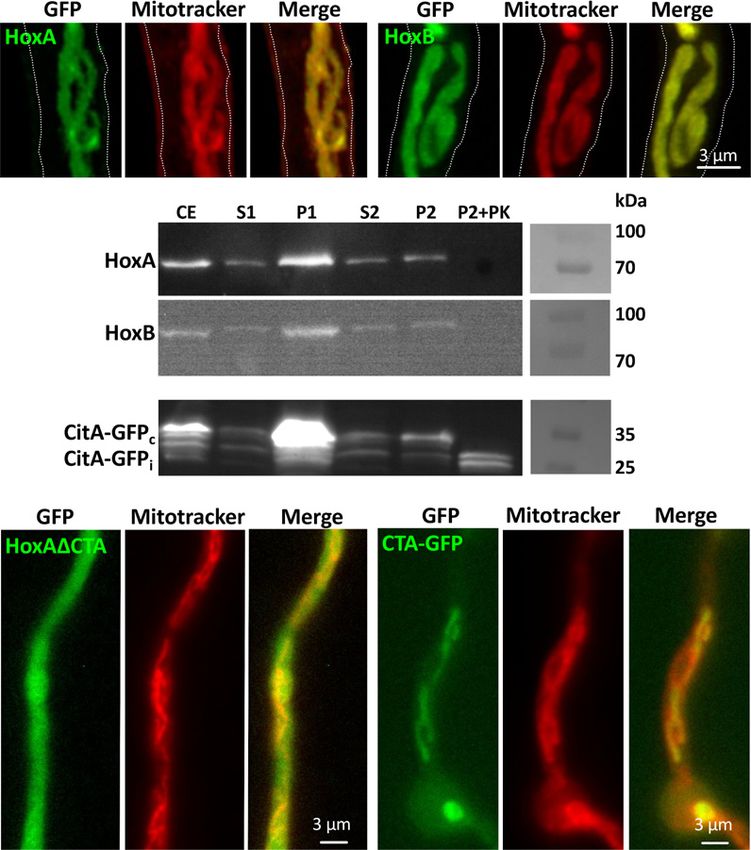
The EMBO Journal Christian Streng et al
Despite the well-established roles of phytochrome as light and mutant as compared to the hoxA or the fphA mutant. Taken
temperature sensor in A. nidulans, the biosynthesis of the chro- together, the results suggest that HoxA and HoxB could provide the
mophore remains enigmatic. In the genome of A. nidulans, no good chromophore for phytochrome.
heme oxygenase candidate can be identified using the cyanobacte- Most HO enzymes catalyze the conversion of heme to biliverdin
rial, bacterial, or mammalian heme oxygenases as bait. Here, we IXa or other isomers, depending on the cleavage site. Biliverdin IXa
studied two putative heme oxygenases from A. alternata. Alternaria is used by bacterial phytochromes as chromophore (Lamparter et al,
alternata contains a blue-light sensing system but used also phyto- 2003). To investigate whether HoxA and HoxB convert heme into a
chrome for light sensing (Igbalajobi et al, 2019; Igbalajobi et al, functional chromophore, several in vitro assays were performed.
2020). Surprisingly, deletion of either of the two putative heme HoxA was expressed in E. coli without the hydrophobic C-terminal
oxygenase genes caused a “blind” phenotype. They are able to form membrane anchor (HoxAΔCTA) and HoxB in Pichia pastoris. HoxB
homo- and heterodimers and physically interact with phytochrome. overexpression in E. coli rendered an inactive protein (Appendix Fig
Both enzymes together with phytochrome are attached to the mito- S2). Spectral analyses of the enriched proteins did not show any
chondrial outer membrane, suggesting metabolon formation. This evidence for heme binding already during the expression or the
could be a general strategy to separate heme linearization from subsequent purification. We then added hemin and tested for heme
heme degradation. Our study revealed a novel function of mitochon- binding. We started with a solution of 1 µM hemin and added HoxA
dria as chromophore-assembly platforms for phytochrome. or HoxB to a final concentration of 10 µM. The heme spectrum
changed slightly after protein addition, suggesting heme binding.
Afterward, the hemin concentration was stepwise increased up to
Results 8 µM. At each step, absorption spectra were recorded. Binding of
hemin by HoxA resulted in a peak at 408 nm. At 3 µM hemin, a
Two heme oxygenases are required for phytochrome function in shoulder appeared in the spectrum between 320 and 380 nm. This
A. alternata resembled free hemin, suggesting that not all HoxA molecules were
active or able to bind the chromophore which could be caused by
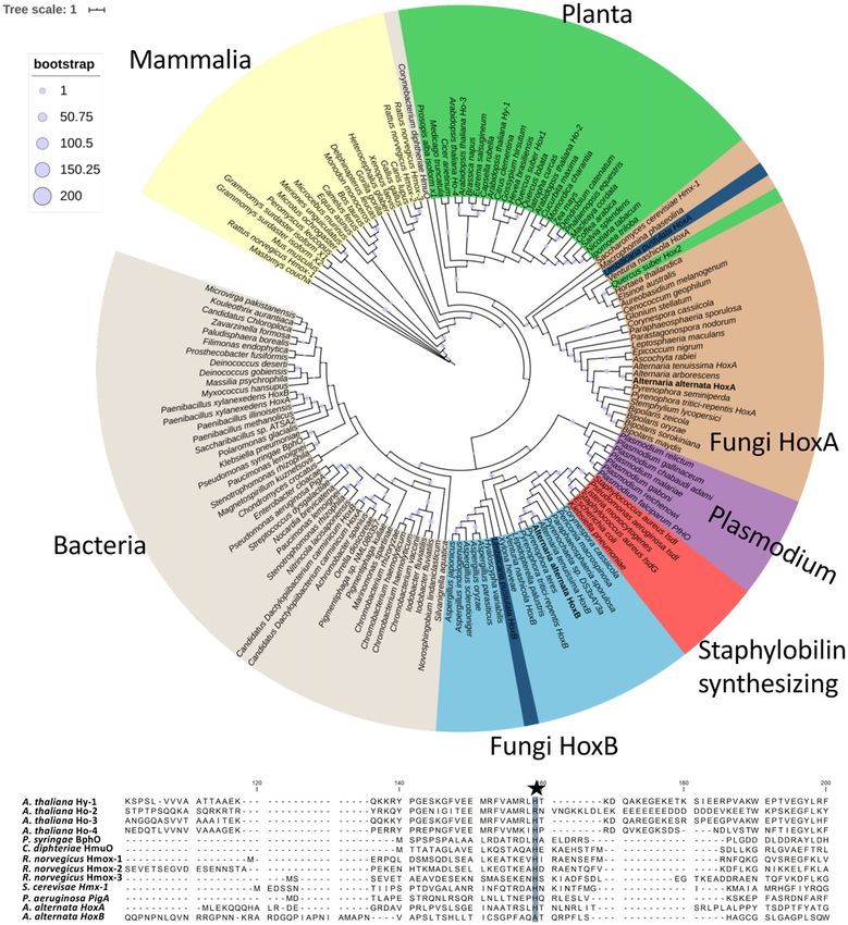
Here, we describe two HOs from the fungus A. alternata (Fig 1). partial oligomerization (Appendix Fig S3A). In the corresponding
Overall sequence similarities to other HOs are low, and comparison experiment with HoxB, the spectra resembled the spectra of HoxA
among fungal, plant, and animal HOs revealed two monophyletic (with a peak at 410 nm), but the shoulder in the spectrum was more
fungal clades represented by HoxA and HoxB from A. alternata (Fig pronounced and increased faster (Appendix Fig S3B). Hence, both
1A). These two clades are highly different to the clade of plants, proteins are able to bind heme. Stoichiometries for heme binding
bacteria, or mammals. In other heme oxygenases, a histidine were not determined as the protein preparations were not pure.
residue is conserved and involved in heme binding (Ito-Maki et al, We then characterized the catalytic activity of HoxA and HoxB in
1995). Whereas A. alternata HoxA contains this histidine, it is lack- the presence of ferredoxin as electron donor. 50 µM HoxA or HoxB,
ing in HoxB (Fig 1B). Nevertheless, in both proteins a HemeO-like or 25 µM of each protein combined in one reaction, was incubated
superfamily domain is predicted (Fig 2A). In addition, a hydropho- with 10 µM hemin and incubated for 10 min. When HoxA and HoxB
bic stretch of 21 amino acids was identified at the C-terminus of were combined, HPLC analyses of the reaction products revealed a
HoxA. Because this region is required for mitochondrial association, peak with a retention time 1–2.5 min larger than for the biliverdin
it was named C-terminal anchor (CTA) (see below). standard (Appendix Fig S3C). The small difference of the retention
To functionally characterize hoxA and hoxB in A. alternata, both time could point to minor modifications of the chromophore,
genes were deleted using CRISPR/Cas9 (Wenderoth et al, 2017; although the spectrum matched well with the biliverdin standard
Wenderoth et al, 2019) (Appendix Fig S1). In either deletion strain, (Appendix Fig S3D). In order to show that the produced biliverdin
the induction of the red-light regulated gene, ccgA, was drastically product is able to autocatalytically assemble with phytochrome, the
reduced, as much as in the phytochrome-deletion strain (Fig 2B). photosensory domain (PGP, 68 kDa) of FphA was added to the
To further link the deletion of hoxA or hoxB to the function of assay above and spectra recorded every 30 s (Appendix Fig S3E).
phytochrome (FphA), the stress behavior of the strains was tested. Whereas the Soret band decreased over time, the Q band increased,
Stress was applied by adding hydrogen peroxide (H2O2), mena- proving the functionality of the chromophore. The maxima of the
dione (generates free radicals and superoxide), or tert-butyl resulting Pr form was 702 nm and resembled the maxima of bili-
hydroperoxide (tBooH) (organic hydroperoxide) to the medium. verdin assembled FphA in the Pr form with 705 nm (Blumenstein
Similar to phytochrome mutants (Yu et al, 2016; Igbalajobi et al, et al, 2005). To test whether the in vivo activity of HOXs is the sum
2019), both hox-deletion strains were more resistant toward mena- of HoxA and HoxB activities, we co-expressed HoxA or HoxB alone
dione and H2O2 and more sensitive toward tBooH (Fig 2C). The or in combination along with the photosensory domain (PGP) of A.
higher stress resistance of the phytochrome mutant, and the hoxA nidulans FphA in E. coli. In all cases, low expression protein levels
and hoxB mutants, may be explained through the regulation of were maintained to avoid oligomerization and inactivation of HoxB.
stress-related genes, such as catalases, by phytochrome. It was As a control, we co-expressed the Pseudomonas aeruginosa HOX,
shown that they are upregulated in the absence of phytochrome BphO (Blumenstein et al, 2005) (Fig 3A). In all four cases, functional
(Igbalajobi et al, 2019; Igbalajobi et al, 2020). In addition, there is photosensory domains were obtained (Fig 3B–E). The covalent
evidence that under some stress conditions, human HO-1 is prote- insertion of the chromophore into PGP was further confirmed by
olytically processed, shuttles into nuclei, and fulfills an unknown Zn2+-induced red fluorescence after protein separation in a polyacry-
function there (Lin et al, 2007). Such an additional function in A. lamide gel (Fig 3A, inset). Only about 5% of the amount of assem-
alternata may explain the slightly higher sensitivity of the hoxB bled PGP was obtained when A. alternata HoxA in combination
2 of 11 The EMBO Journal 40: e108083 | 2021 ª 2021 The Authors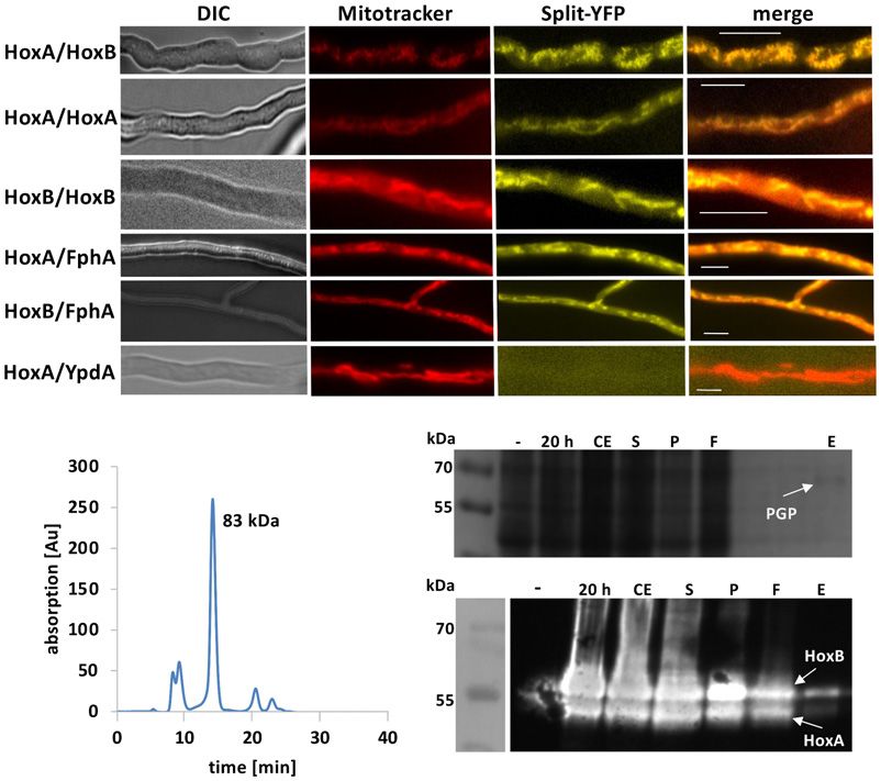
Christian Streng et al                                                                                                                          The EMBO Journal

               A

               B

  Figure 1. Phylogenetic analysis of HOs.
  A A phylogenetic tree of 161 different HO sequences was calculated. HOs were aligned with Clustal Omega. Afterward, the sequences were trimmed by trimAL with gap
    threshold 0.8 and conservation percentage of 70%. The tree was calculated with PhyML with 200 bootstrap cycles and the AIK parameter switched on. Visualization
    was done in iTOL. Within the two fungal clades, two lichen-derived sequences are shaded in dark blue.
  B Alignment of the heme-binding region of several HOs. The heme-coordinating histidine is indicated with a star. HoxB lacks this histidine, like HO-2 from A.
    thaliana.

with HoxB was used instead of the bacterial HO (Fig 3A). This                        Both heme oxygenases reside at the outer
amount was even lower, when only HoxA (1%) or HoxB (0.7%)                            mitochondrial membrane
was expressed, suggesting that the activity of HoxA and HoxB
together is higher than the sum of both. The reason for the much                     Next, the subcellular localization of A. alternata HoxA and HoxB
higher activity of the bacterial enzyme in comparison with the                       was investigated by heterologous expression of GFP-tagged
fungal enzymes may be the lack of appropriate electron donors for                    versions in A. nidulans (Fig 4A). The observed GFP-labeled struc-
the A. alternata enzymes.                                                            tures resembled mitochondria, as confirmed with the MitoTracker

ª 2021 The Authors                                                                                                  The EMBO Journal 40: e108083 | 2021     3 of 11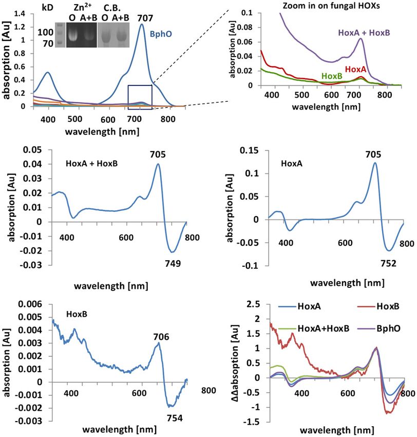
The EMBO Journal                                                                                                                                      Christian Streng et al

                                      A                                            C

                                      B

  Figure 2. Characterization of HoxA and HoxB from A. alternata.
  A Scheme of HoxA and HoxB. The open reading frame of hoxA is interrupted by one 67 bp long intron (RNA-seq data) and encodes a protein of 380 amino acids with a
    predicted mass of 42.5 kDa. The protein contains a heme-binding pocket, a HemeO-like superfamily domain and a C-terminal putative membrane anchor (CTA). In
    comparison, hoxB is interrupted by a 50 bp intron. The protein (509 amino acids, 55.9 kDa) contains only the HemeO-like superfamily domain.
  B Effect of the deletion of hoxA or hoxB on red-light induction of the ccgA gene. Alternaria alternata strains were grown for 36 h at 28°C. Samples were illuminated for 1
    h in red light, while controls were kept in the dark. The amount of ccgA transcript was quantified by qRT–PCR analyses with the histone 2B gene as housekeeping
    gene. Error bars represent the standard deviation (n = 3).
  C Comparison of wild-type colonies with phytochrome and hox-deletion strains in the presence of 0.12 mM menadione (MD), 7 mM H2O2 or 2 mM tert-butyl
    hydroperoxide (tBooH). The strains were incubated for 5 days at 28°C in the dark.

co-staining (Suelmann & Fischer, 2000). Therefore, we next tested                         C-terminus of HoxA. To test whether this C-terminal sequence is
whether the enzymes localize inside of mitochondria or are                                required for mitochondrial targeting, we removed the last 20
attached to the outer mitochondrial membrane (Fig 4B). Cells were                         amino acids from the protein and expressed it as GFP fusion
fractionated by two sequential centrifugation steps. After the first                      protein in A. nidulans. The truncated protein localized in the cyto-
low-speed centrifugation, mitochondria should be still present in                         plasm (Fig 4C). Next, we asked whether the C-terminal sequence is
the supernatant (S1), whereas after the second higher-speed                               sufficient for mitochondrial targeting and fused the sequence to
centrifugation, mitochondria should be enriched in the pellet (P2).                       GFP. Indeed, the GFP molecule labeled mitochondria. These results
This pellet fraction was used to test the sensitivity of the proteins                     suggest that the C-terminal sequence functions as an anchor for
toward proteinase K treatment (P2 +PK). HoxA-GFP and HoxB-GFP                             the protein.
were both detected in the mitochondrial fraction and were comple-
tely degraded after addition of proteinase K. As a control, we stud-                      Heme oxygenases form a complex with phytochrome
ied a mitochondrial matrix protein. We used the N-terminal part of
citrate synthase and fused it to GFP. The construct was used before                       In order to test homo- or heterodimer formation of A. alternata HOs,
for mitochondrial labeling (Suelmann & Fischer, 2000). The fusion                         bimolecular fluorescence complementation (BiFC, split-YFP) was
protein has a molecular mass of 41 kDa, although the apparent                             used. HoxA and HoxB were expressed as fusion proteins with the N-
molecular mass in the SDS–PAGE is less. After import, the protein                         terminal or the C-terminal half of YFP in A. nidulans. Interaction of
has a predicted molecular mass of 35 kDa. In crude extracts, both                         two proteins restores YFP fluorescence. We found that all three
bands, and in addition some degradation products, were detectable,                        combinations, HoxA-HoxA, HoxA-HoxB, and HoxB-HoxB, inter-
suggesting that not all proteins are imported into mitochondria.                          acted at mitochondria. Moreover, the same results were obtained
After proteinase K treatment of the second pellet fraction, the 35                        with the combination of HoxA or HoxB with phytochrome (Fig 5A).
kDa protein band remained, plus a smaller degradation product.                            As a negative control, YFP-N-YpdA (Yu et al, 2016) was tested with
The small protein could be free GFP. From these experiments, we                           either YFP-C-HoxA or YFP-C-HoxB. These combinations did not
conclude that HoxA is attached to the outer mitochondrial                                 result in reconstitution of the fluorescent YFP protein.
membrane.                                                                                     HoxA dimerization was confirmed by size-exclusion chromatog-
                                                                                          raphy (SEC) (Fig 5B). Calibration of the column was performed with
A C-terminal anchor (CTA) of HoxA is necessary and sufficient for                         cytochrome C, carbonic anhydrase, albumin, and alcohol dehydro-
mitochondrial targeting                                                                   genase (Appendix Fig S3F). For HoxB SEC failed, because of impuri-
                                                                                          ties after enrichment from P. pastoris. As a further proof for protein–
None of the HOs comprises an N-terminal mitochondrial targeting                           protein interaction, we co-expressed HoxA, HoxB, and the photosen-
sequence. However, we identified a hydrophobic region at the                              sory domain of FphA (Strep-tagged) in E. coli. After Strep-tag

4 of 11       The EMBO Journal 40: e108083 | 2021                                                                                                          ª 2021 The Authors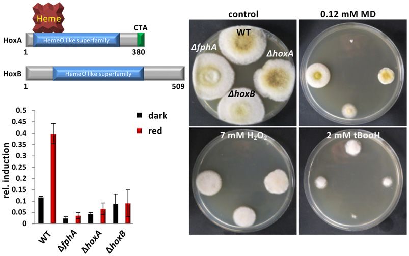
Christian Streng et al                                                                                                                              The EMBO Journal

                                 A

                                 B                                                      C

                                 D                                                      E

  Figure 3. Analysis of the HO enzymatic activities.
  A   UV–Vis spectra of the photosensory domain of A. nidulans FphA (PGP) after co-expression together with bacterial HO (BphO) or A. alternata HoxA, HoxB, or HoxA
      plus HoxB overnight at 20°C in the dark. PGP was purified via the Strep-tag system and concentrated before recording the spectra. Inset, left lane: Molecular mass
      markers stained with Coomassie Blue. Middle lanes: Zinc-induced red fluorescence (labeled zinc, Zn2+) of equal amounts of PGP expressed in E. coli along with
      bacterial BphO (O) or A. alternata HoxA plus HoxB (A + B). Right lanes: Coomassie Blue staining (C.B.) of the polyacrylamide gel used for the Zn2+ fluorescence.
  B–D Red/far-red light induced difference spectrum of purified PGP expressed with HoxA and HoxB or with HoxA or HoxB alone after large-scale batch fermentation and
      purification.
  E   Overlay of normalized (at 705 nm) difference spectra displayed in B-D plus a difference spectrum of the photosensory domain in the presence of the bacterial HO
      BphO as in A.
  Source data are available online for this figure.

purification, we identified HoxA and HoxB in the photosensory                           Discussion
domain fraction (Fig 5C). To assess whether insertion of the chro-
mophore into the photosensory domain influences its interaction                         Taken together our results, we propose the following model for the
with HoxA, which is proposed to be the key factor for assembly                          biosynthesis of functional phytochrome in fungi: The FphA apopro-
because of the membrane anchor, biolayer interferometry (BLI) was                       tein is translated in the cytoplasm and travels to the surface of mito-
used for the analysis of interactions of the purified proteins (Fig 6,                  chondria where it receives the chromophore (the electron donor for
Appendix Fig S4). Association and dissociation curves were moni-                        the heme oxidation is yet unknown). The holoprotein is then
tored, and the KD for the interaction of Apo-PGP and HoxA was                           released back into the cytoplasm where it interacts with the phos-
calculated to be 1.13 µM (Fig 6A) and for Holo-PGP 64.6 mM (Fig                         photransfer protein YpdA to induce the HOG pathway and
6B). The sudden increase of the signal after addition of the                            ultimately the transcription factor AtfA. In addition, a fraction of
chromophore-loaded PGP is due to the absorption of the chro-                            Holo-FphA is imported into the nucleus to control the activity of
mophore (Fig 6B). The 57,000 times weaker interaction of the holo-                      chromatin remodeling enzymes (Fig 7). Mitochondria can thus be
protein suggests immediate dissociation from HoxA after                                 considered as assembly platforms for phytochrome in fungi. Given
chromophore insertion.                                                                  that mitochondria arose in evolution before chloroplasts, our

ª 2021 The Authors                                                                                                     The EMBO Journal 40: e108083 | 2021       5 of 11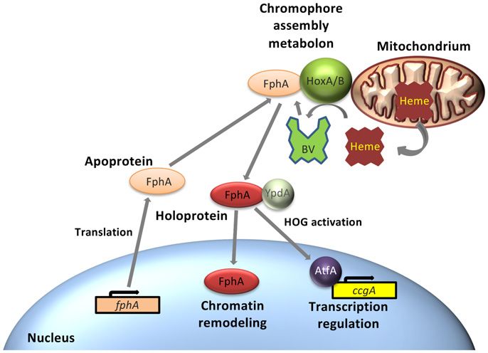
The EMBO Journal                                                                                                                                Christian Streng et al

                                        A

                                        B

                                        C                                            D

  Figure 4. Localization of A. alternata HoxA and HoxB in A. nidulans.
  A    Strains were grown overnight at 28°C. Mitochondria were stained for 30 min with MitoTracker Red CMOXRos. Microscopy was done with a LSM 900 Airyscan 2
       (Zeiss). Pictures were taken in the GFP and the RFP channel and overlaid (merge).
  B    HoxA-GFP and CitA-GFP (ca. 170 amino acids from the N-terminus of citrate synthase) were expressed overnight at 30°C in minimal medium with 2% threonine
       and 0.2% glucose. Mitochondria were incubated with 100 lg/ml proteinase K (PK) for 20 min on ice. Crude extract (CE), supernatant 1 (S1) containing
       mitochondria, pellet 1 (P1), supernatant 2 (S2), and pellet 2 (P2) with mitochondria were analyzed by Western Blot using anti-GFP antibodies. CitA-GFPc =
       cytoplasmic form and CitA-GFPi = imported version.
  C, D Role of the C-terminal anchor in HoxA. Microscopy was done as in A using a fluorescent microscope without the Airyscan technology. C, Expression of GFP-HoxA
       lacking the C-terminal anchor (ΔCTA) in A. alternata. D, Expression of GFP fused to the CTA motif.
  Source data are available online for this figure.

findings tempt us to speculate that chromophore biosynthesis at                       been shown that human HO-2 may have some chaperone functions
mitochondria is more ancient than in chloroplasts (Speijer et al,                     (Vanella et al, 2013; Vanella et al, 2016). In addition, it was shown
2020).                                                                                that human HO-1 fulfills functions in nuclei under certain stress
    In this work, we identified two HOs in A. alternata and showed                    conditions (Lin et al, 2007). Further work is required to unravel
catalytic activity for both. The activities when expressed in E. coli                 whether such a specialization of the two enzymes exists in fungi.
were quite low as compared to the activity of the bacterial HO used                      The fact that phytochrome chromophore assembly in A. alternata
in our experiments. This may reflect their real activities in A. alter-               occurs at the surface of mitochondria leads to the question why
nata, but it could also be that the HO activities in the fungus are                   there and not in the cytoplasm? Heme biosynthesis is initiation and
significantly higher. One reason could be the lack of an appropriate                  completed in mitochondria in animals and fungi. Glycine and
electron donor in E. coli and in the in vitro experiments. The fact                   succinyl-CoA are converted in mitochondria to 5-aminolevulinic
that HoxB undoubtedly produced active photosensory domains of                         acid, which is transported to the cytoplasm, further converted to
FphA when co-expressed in E. coli was surprising, because a                           coproporphyrinogen III, which in turn is re-imported into mitochon-
conserved histidine is lacking. This may explain the lower activity                   dria, where the final steps of heme biosynthesis occur. Heme is then
as compared to HoxA. However, we found evidence that, in addition                     either used in mitochondria for, e.g., cytochrome formation or
to its catalytic activity, HoxB could stimulate the HoxA activity. The                exported into the cytoplasm (Kim et al, 2012). In plants, the final
mechanism of this stimulation remains to be determined, but it has                    steps take place in chloroplasts. In the cytoplasm, heme is inserted

6 of 11       The EMBO Journal 40: e108083 | 2021                                                                                                    ª 2021 The Authors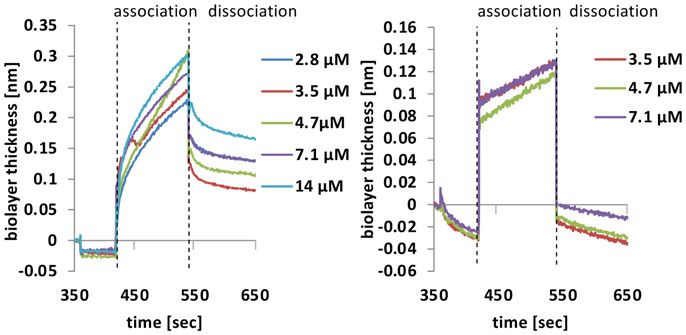
Christian Streng et al                                                                                                                                The EMBO Journal

                                        A

                                        B                                           C

  Figure 5. Characterization of the HO-phytochrome protein complex.
  A BiFC (split-YFP) analysis of HOs and phytochrome. Strains (SChS29-33) were grown overnight at 28°C minimal medium with 2% threonine and 0.2% glucose.
    MitoTracker Red CMOXRos was used for mitochondrial staining. The MitoTracker signal was observed in the RFP and the split-YFP signal in the YFP channel of the
    fluorescent microscope. Pictures in both channels were overlaid (merge). The left pictures show the same hyphae in differential interference contrast (DIC). The scale
    bar represents 10 µm.
  B HoxA was expressed in E. coli 20 h at 15°C. Purification was performed with the Strep-tag system. HoxA was analyzed by size-exclusion chromatography. Absorption
    was measured at 280 nm.
  C Co-expression of the HOXs along with the photosensory domain of A. nidulans FphA (PGP) overnight at 20°C. PGP was purified via the Strep-tag system and the eluate
    concentrated (upper panel). The lanes were loaded as follows: negative control before induction (), 20 h after induction (20 h) crude extract (CE), supernatant (S),
    pellet (P), flow through (F), and eluate (E). The fractions were analyzed by Western blot using the anti-His antibody (lower panel).
  Source data are available online for this figure.

                                                                                        into hemoproteins such as catalases. Likewise, HOs in A. alternata
A                                             B                                         could use cytoplasmic heme to produce the phytochrome chro-
                                                                                        mophore. HOs would thus compete for heme with the other
                                                                                        enzymes which need the incorporation of a heme molecule for func-
                                                                                        tion. However, in the case of HOs, heme is catalytically converted
                                                                                        and normally released for further degradation. Such degradation has
                                                                                        to be also postulated for the A. alternata cytoplasm, because heme
                                                                                        concentrations need to be controlled well, e.g., by high affinity
                                                                                        heme-binding proteins like peroxiredoxins, because free heme can
                                                                                        produce free radicals through Fenton chemistry (Fenton, 1894;
                                                                                        Gozzelino et al, 2010). Hence, A. alternata HoxA and HoxB would
                                                                                        largely interfere with heme homeostasis. One way of separating
                                                                                        heme degradation from chromophore biosynthesis is compartmen-
  Figure 6. Analysis of the interaction of HO and the phytochrome                       talization. In the case of heme oxidation for chromophore forma-
  photosensory domain in vitro.                                                         tion, further degradation of the linear tetrapyrrole has to be
  A, B Biolayer interferometry analysis with Apo-PGP A and holo-PGP B.                  prevented. This could be the reason for the observed complex
       Proteins were immobilized at a streptavidin sensor tip. Free binding sites       formation between HOs and phytochrome. This minimizes the
       were blocked with biotin. Association and dissociated kinetics were              chances that linearized heme is released and further degraded. This
       recorded at the indicated HoxA concentrations. Kd values were
                                                                                        could be an example for the recently postulated metabolon concept
       calculated with the manufacturer’s software using a global fit for both
       experiments.                                                                     (Piel et al, 2019). Metabolons are protein complexes of interacting
                                                                                        enzymes in metabolic pathways. The created microenvironments

ª 2021 The Authors                                                                                                      The EMBO Journal 40: e108083 | 2021        7 of 11
The EMBO Journal Christian Streng et al
interplay between the two HOs and to identify HOs for heme degra-
dation and unravel their interplay in heme homeostasis and chro-
mophore production.
Materials and Methods
Strains, plasmids, and culture conditions
Alternaria alternata ATC 66981 cultures were grown on modified
Czapek Doth broth (mCDB) agar and incubated 1–12 days at 28°C.
Supplemented minimal medium (MM) for A. nidulans was prepared
as reported, and standard strain construction protocols were used
(K€
afer, 1977). Growth of Pichia pastoris was performed in BMGY or
BMMY media according to the manufacturer’s protocol (Invitrogen).
All strains are listed in Appendix Table S1, oligonucleotides in
Appendix Table S2, and plasmids in Appendix Table S3.
Figure 7. Current model illustrating the role of mitochondria as
chromophore-assembly stations for phytochrome.
Gene structure and deletion of hoxA and hoxB in A. alternata
For details, see the Discussion.
The open reading frame of hoxA is interrupted by one 67 bp long
intron (RNA-seq data) and encodes a protein of 380 amino acids
facilitate substrate transport and specificity. In the case of phyto- with a predicted mass of 42.5 kDa. In comparison, hoxB is also inter-
chrome chromophore assembly, the protein complex could also be rupted by one intron (50 bp) but contains only the HemeO-like
important for limitation of the enzymatic activity to the needs. If superfamily domain. The hoxB gene encodes a protein of 509 amino
apo-phytochrome binds to the HO complex, heme should be acids with a predicted mass of 55.9 kDa. We used two 20 bp proto-
oxidized and transferred to form holo-phytochrome. In the absence spacers adjacent to a 30 AGG protospacer-adjacent motif (PAM) to
of apo-phytochrome though, HO should be inactive in order to target the beginning and the end of the gene. The protospacers were
prevent uncontrolled degradation of heme. This could be achieved introduced into plasmids pFC332 and pFC330 by PCR and cloning.
by modulation of the HO activity or through the accessibility of The resulting plasmids, which contain the Cas9-coding sequence
heme to the active center if phytochrome is missing from the meta- from Streptococcus pyogenes (codon optimized for Aspergillus niger)
bolon. Supporting our metabolon model, protein complex formation and the single-guide RNA (sgRNA) targeting the genes of interest
between HO and phytochrome was also shown in the bacterium (hoxA and hoxB), were used for transformation of A. alternata
Pseudomonas syringae (Shah et al, 2012). Furthermore, in the SMW24 (DpyrG in ATCC66981 wild type). The hygromycin resis-
pathogenic P. aeruginosa two heme oxygenases are expressed, one tance and the pyrG auxotrophy cassettes residing in a self-
of which produces the chromophore for phytochrome and another replicating plasmid (AMA plasmid) were used for selection. The
one is used for heme degradation and iron acquisition (Wegele et al, plasmid was constructed using PCR on PFC334 with primers, which
2004). Plants harbor also several HO with different catalytic activi- included the new protospacer. Subsequently, a builder reaction and
ties, perhaps also serving distinct functions besides chromophore the transformation was done as described, but in this case, two
production (Davis et al, 2001). protospacers were used to target the beginning and the end of the
Another reason for mitochondrial association of the phytochrome gene of interest (Wenderoth et al, 2019).
metabolon could be the need for an electron donor with a negative
redox potential, such as ferredoxin. Although mitochondria are opti- RNA isolation
mized organelles for electron flow, the localization of the phyto-
chrome metabolon at the outside of mitochondria raises the Alternaria alternata conidia were inoculated in 20–25 ml mCDB
question of how the electrons can be shuttled from the inner mito- containing uracil and uridine in a Ø 3.5 cm Petri dish and incubated
chondrial membrane toward the outside of the outer membrane. for 40 h in darkness at 28°C. Subsequently, mycelium was illumi-
This remains to be resolved. Alternatively, cytoplasmic electron nated 1h with red light and controls were kept in the dark. The
donors could be employed as it was shown for cytosolic iron/sulfur mycelia were harvested in dim-red light and frozen immediately in
cluster biogenesis (Zhang et al, 2014). One challenge for future liquid nitrogen. For the extraction, a "Fungal RNA Extraction Kit"
research will be to investigate whether the model proposed for from Omega was used. Disruption of the cells was performed by
fungal phytochrome biosynthesis may be generalized also to plant grinding in liquid nitrogen. To remove DNA contaminations, the
phytochrome. RNA was treated with TURBO DNA-free kit. After the treatment,
Our results suggest that the two HOs of A. alternata are special- RNA was diluted to 50 ng/ll with DEPC water. SensiFAST SYBR &
ized enzymes for chromophore assembly at mitochondria. This Fluorescein One-Step Kit from Bioline (Luckenwalde, Germany) was
special function may also explain the low enzymatic activity. One used for quantitative real-time PCR. Each reaction was carried out
can speculate that for heme degradation, higher activities are to be using 25 ll containing 0.2 lM primers and 100 ng RNA. The
expected. It will be the challenge of future research to decipher the program started with 10 min of the reverse transcription reaction at
8 of 11 The EMBO Journal 40: e108083 | 2021 ª 2021 The AuthorsChristian Streng et al The EMBO Journal
45°C, followed by 2.5 min at 95°C for the inactivation of the reverse set to 400 rpm, pH 7.5, and an aeration rate of 5 l air per min. For
transcriptase and 40 cycles of polymerase chain reaction (10 s at co-expression of HoxA and HoxB, we used the pACYC Duet plasmid.
95°C and then 30 s at 58°C). To assess the quality of the resulting The protein samples were illuminated 2 min with red or far-red light
PCR product, melting curve analyses were carried out (80 cycles, to photoconvert PGP between Pr and Pfr forms. Results are shown
95–58°C with 10 s per step). The h2b gene was used for normaliza- as difference spectra. This procedure doubled the yield of functional
tion. Each expression level is the average of three biological repli- PGP in comparison with the expression in flask cultures. HoxA
cates. The error is shown as standard deviation for the replicates. alone and the co-expression of HoxA and HoxB with PGP yielded
functional photoconvertible phytochrome, which suggests HoxA is
Expression of HOs in E. coli or P. pastoris sufficient.
HoxA was cloned into the plasmid pASK. pASK contains an Zinc-induced red fluorescence
anhydrotetracycline-inducible promoter (tet) and an ampicillin resis-
tance cassette. For the expression, E. coli was grown at 37°C until Covalent chromophore attachment has been verified by zinc-
OD600 0.8 was reached. Subsequently, the baffled flask was cooled induced red fluorescence as described previously (Berkelman &
down to 15°C and induction with 0.2 µg/ml AHT was performed for Lagarias, 1986). Free bilins as well as (denatured) biliproteins form
20 h. The pellet of 1 l was resuspended in 10 ml extraction buffer fluorescent complexes with zinc ions that can be visualized under
and the cells ruptured using a French press at 1,500 psi. Cell debris UV light. Sodium dodecyl sulfate–polyacrylamide gel electrophoresis
was pelleted by centrifugation prior to the application to the (SDS–PAGE) was performed as described (Laemmli, 1970). Tris-
column. Purification was done using the strep-tag system with glycine running buffer as well as Tris-buffers used for the 5% stack-
modified buffers: extraction buffer (50 mM Tris–HCl pH 8.0, 100 ing gel and the 10% separating gel contained 1 mM zinc acetate.
mM NaCl, 5 mM MgCl2, 0.005% Triton X-100, 1 mM PMSF, 1 pill Zinc fluorescence was visualized by transillumination with UV light.
for 10 ml pierce protease inhibitor cocktail EDTA free (Thermo Afterward, the same gel was Coomassie-stained using ROTIBlue
Scientific), washing buffer (137 mM NaCl, 2.7 mM KCl, 50 mM quick (Carl Roth, Karlsruhe). Protein concentrations were deter-
Na2HPO4, 5 mM K2HPO4, pH 7.4 was adjusted with H3PO4), and mined using ROTIQuant (Carl Roth, Karlsruhe) according to the
elution buffer (50 mM Tris–HCl, 3 mM Desthiobiotin). For better manufacturer’s instructions.
solubility, the C-terminal anchor was deleted. HoxB could not be
purified using this system, because HoxB aggregated. In order to Heme oxygenase assays
overcome this problem, HoxB-strep was cloned to pPIC3.5K.
pPIC3.5K is a vector containing a histidine auxotrophy marker and For the analysis of the catalytic activity of HoxA and HoxB, enriched
the methanol-inducible AOX1 promoter for P. pastoris. P. pastoris enzymes were used. 50 µM HoxA or HoxB or 25 µM of each protein
GS115 was transformed with SacI linearized vector to insert into the combined in one reaction was incubated with 10 µM hemin, 1.5 mg/
AOX1 locus. The transformants were screened for mut+ phenotype. ml BSA, 4.6 µM petF, 0.025 U/ml petH, 10 µM catalase, 5 mM tiron,
For protein expression, P. pastoris was grown in 50 ml BMGY over- 1.05 mM glucose-6-phosphate, 0.105 µM NADP+ and 0.15 U/ml
night at 30°C. After incubation, cells were pelleted and 400 ml glucose-6-phosphate-dehydroxygenase, spinach ferredoxin (PetF,
BMMY with OD600 1.0 were inoculated. Expression was performed Sigma) (4.6 µM), and ferredoxin reductase (PetH, Sigma) (4.6 µM).
for 2.5 days, while every 24 h methanol was added to maintain a Spectra from 300 nm to 1,100 nm were recorded every 30 s for 10
concentration of 0.5% (v/v). 1 l culture was pelleted and resus- min (Photometer: 8453 UV visible System (Agilent)). For assays
pended in 20 mM MES pH 6. Cells were lysed using glass beads. with phytochrome, 150 µg FphA was added per ml.
Purification was done using a Mono S FPLC column. The protein
eluted in a gradient ranging from 350 mM NaCl to 450 mM NaCl. HPLC analysis
HoxB was not as pure as HoxA after affinity chromatography.
Further purification using the Strep-tag failed. Hemeoxygenase assay products were pre-purified. To this end, the
sample was diluted 1:10 in 0.1% TFA immediately after the assay to
Expression of phytochrome (FphA) or its photosensory domain stop the reaction. Next, Sep-Pak C18 was equilibrated with subse-
(PGP) along with HOs in E. coli quent addition of 3 ml acetonitrile, 3 ml H2O, 3 ml 0.1% TFA, 3 ml
10% methanol in 0.1% TFA, 3 ml acetonitrile, 3 ml H2O, and 3 ml
We used the plasmids pASK for FphA PGP and pACYC Duet or pET 10% methanol in 0.1% TFA. The sample was added after the last
for the Hox. pACYC Duet contains two MCS, while pET contains one step, and the cartridge was washed with 6 ml 0.1% TFA, 6 ml
MCS with an IPTG-inducible T7 promoter. Cultures were grown in acetonitrile (20%):0.1%TFA (80%) and eluted with 1 ml acetoni-
500 ml LB at 37°C. After OD600 0.8 was reached, 1 mM IPTG was trile. After drying using a SpeedVac, the sample was dissolved in 10
added to the culture to induce the HO. After 1 h, the culture was µl DMSO and diluted in 110 µl acetone (50%):20 mM formic acid
cooled down to 20°C and 0.2 µg/ml AHT was added to induce the (50%). The sample was applied to the Ultracarb 5 U column
induction of FphA PGP. FphA PGP was purified using French press (Phenomenex). Bilins were detected at 350 and 650 nm.
and the strep-tactin system (IBA). Subsequently, the protein was
concentrated via vivaspin and the spectra were measured in the dark. Expression of HOs in A. nidulans
To improve the spectral results and in order to analyze the
photoconvertability of PGP, we used a 5 l fermenter (Bioflo 115, The expression of HoxA and/or HoxB in A. nidulans was achieved
Eppendorf) with the same protocol. Additionally, the fermenter was using the plasmid pMCB17apx as vector basis. The plasmid harbors
ª 2021 The Authors The EMBO Journal 40: e108083 | 2021 9 of 11The EMBO Journal Christian Streng et al
a cloning site using AscI and PacI restriction enzymes. The cloned system. We thank Gert Sonntag (Zeiss) for the opportunity and the help to use
gene is under the control of the alcA promoter, which can be the LSM 900 Airyscan Microscope and Natalia Requena for critically reviewing
repressed by glucose and derepressed by glycerol and induced by the manuscript.
threonine. It also encodes an N-terminal tag of GFP, YFP-C-
terminus, YFP-N-terminus, or HA. Author contributions
CS performed most of the experiments. JH and KL contributed to the in vitro
Microscopy experiments. NK and TL supervised and discussed the spectroscopy. NF-D
supervised the biochemical experiments. FW and MB provided the infrastruc-
For microscopic analysis, A. nidulans strains were grown 16 h at ture and support for the BLI experiments. ZY designed several experiments
28°C in MM containing 2% glycerol or 2% threonine with 0.2% and supervised the work with A. alternata. RF supervised the entire project and
glucose. Subsequently, the culture was stained for 30 min with Mito- wrote the manuscript.
Tracker Red CMXRos (M7512, Thermo Fisher) according to the
manufacturer’s protocol. Fluorescence microscopy was performed Conflict of interest
using AxioImager Z1 (Zeiss), the software AxioVision V4.5, and The authors declare that they have no conflict of interest.
Zen. Alternatively, we used the LSM 900 Airyscan 2 (Zeiss).
Mitochondrial fractionation References
Aspergillus nidulans protoplasts were applied to the “Yeast Mito- Berkelman TR, Lagarias JC (1986) Visualization of bilin-linked peptides and
chondria Isolation Kit” (SigmA) and mitochondria isolated as proteins in polyacrylamide gels. Anal Biochem 156: 194 – 201
described in the manufacturer’s protocol using detergent lysis Blumenstein A, Vienken K, Tasler R, Purschwitz J, Veith D, Frankenberg-Dinkel
(1:200 dilution). In the first centrifugational step, at 600× g mito- N, Fischer R (2005) The Aspergillus nidulans phytochrome FphA represses
chondria were obtained in the supernatant (S1). In the second sexual development in red light. Curr Biol 15: 1833 – 1838
centrifugational step, at 6,500× g mitochondria were sedimented in Corrochano LM (2019) Light in the fungal world. Ann Rev Genet 53:
the pellet (P2). Proteins were analyzed in a Western blot using anti- 149 – 170
GFP antibodies (Roche). For precipitation of the GFP fusion protein Davis SJ, Bhoo SH, Durski AM, Walker JM, Vierstra RD (2001) The heme-
(GFP trap), the protein extracts were incubated with the anti-GFP oxygenase family required for phytochrome chromophore biosynthesis is
antibody and protein G agarose (Roche). necessary for proper photomorphogenesis in higher plants. Plant Physiol
126: 656 – 669
BLI (biolayer interferometry) Davis SJ, Kurepa J, Vierstra RD (1999) The Arabidopsis thaliana HY1 locus,
required for phytochrome-chromophore biosynthesis, encodes a protein
For BLI assays, the BLItz system (FORTEBIO) was used. related to heme oxygenases. Proc Natl Acad Sci USA 96: 6541 – 6546
Streptavidin-coated sensor tips were coated with purified 30 mg/ml Fenton NJH (1894) Oxidation of tartaric acid in presence of iron. J Chem Soc
Apo- or Holo-PGP (tagged with streptavidin-binding protein) in 65: 899 – 910
analysis buffer (50 mM Tris–HCl, pH 7.8, 300 mM NaCl, 10% glyc- Froehlich AC, Noh B, Vierstra RD, Loros J, Dunlap JC (2005) Genetic and
erol, 0.05% Tween-20). Free binding sites were blocked with 5 mM molecular analysis of phytochromes from the filamentous fungus
biotin. Measurements were performed with purified HoxA at the Neurospora crassa. Eukaryot Cell 4: 2140 – 2152
indicated concentrations. The baseline was measured for 60 s, bind- Gisk B, Yasui Y, Kohchi T, Frankenberg-Dinkel N (2010) Characterization of
ing of PGP to the sensor tip was done for 120 s and blocking for 180 the haem oxygenase protein family in Arabidopsis thaliana reveals a
s. The resulting new baseline was measured for 60 s. Association diversity of functions. Biochem J 425: 425 – 434
and dissociation were measured for 120 s. For calculating associa- Gozzelino R, Jeney V, Soares MP (2010) Mechanisms of cell protection by
tion and dissociation rates, global fit corrections were activated in heme oxygenase-1. Ann Rev Pharmacol Toxicol 50: 323 – 354
the software. Resulting KD values are presented as mean of the Hedtke M, Rauscher S, Röhrig J, Rodriguez-Romvero J, Yu Z, Fischer R (2015)
values measured at the different concentrations. Light-dependent gene activation in Aspergillus nidulans is strictly
dependent on phytochrome and involves the interplay of phytochrome
and white collar-regulated histone H3 acetylation. Mol Microbiol 97:
Data availability 733 – 745
Hughes J, Lamparter T, Mittmann F, Hartmann E, G€
artner W, Wilde A,
This study includes no data deposited in external repositories. Boerner T (1997) A prokaryotic phytochrome. Nature 386: 663
Igbalajobi O, Gao J, Fischer R (2020) The HOG pathway plays different roles
Expanded View for this article is available online. in conidia and hyphae during virulence of Alternaria alternata. Mol Plant
Microbe Interact 33: 1405 – 1410
Acknowledgements Igbalajobi O, Yu Z, Fischer R (2019) Red- and blue-light sensing in the plant
This work was supported by the German Science Foundation (DFG Fi-459/19- pathogen Alternaria alternata depends on phytochrome and the white-
1). F.W. and M.B. were supported by the "Karlsruhe School of Optics and collar protein LreA. MBio 10: e00371 – e1319
Photonics, DFG GSC 21”. We thank Luis Raupach, Linda Schlegel, and Birgit Ito-Maki M, Ishikawa K, Matera KM, Sato M, Ikeda-Saito M, Yoshida T (1995)
Schreckenberger for their technical assistance. We are grateful to Matthias Demonstration that histidine 25, but not 132, is the axial heme ligand in
Mack, Hochschule Mannheim, for providing us the P. pastoris expression rat heme oxygenase-1. Arch Biochem Biophys 317: 253 – 258
10 of 11 The EMBO Journal 40: e108083 | 2021 ª 2021 The AuthorsChristian Streng et al The EMBO Journal
Jung JH, Domijan M, Klose C, Biswas S, Ezer D, Gao M, Khattak AK, Box MS, Schumacher J (2017) How light affects the life of Botrytis. Fungal Genet Biol
Charoensawan V, Cortijo S et al (2016) Phytochromes function as 106: 26 – 41
thermosensors in Arabidopsis. Science 354: 886 – 889 Schumacher J, Gorbushina A (2020) Light sensing in plant- and rock-
K€
afer E (1977) Meiotic and mitotic recombination in Aspergillus and its associated black fungi. Fungal Biol 124: 407 – 417
chromosomal aberrations. Adv Genet 19: 33 – 131 Shah R, Schwach J, Frankenberg-Dinkel N, G€
artner W (2012) Complex
Kim HJ, Khalimonchuk O, Smith PM, Winge DR (2012) Structure, function, formation between heme oxygenase and phytochrome during biosynthesis
and assembly of heme centers in mitochondrial respiratory complexes. in Pseudomonas syringae pv. tomato. Photochem Photobiol Sci 11:
Biochim Biophys Acta 1823: 1604 – 1616 1026 – 1031
Laemmli UK (1970) Cleavage of structural proteins during the assembly of Speijer D, Hammond M, Lukes J (2020) Comparing early eukaryotic
the head of bacteriophage T4. Nature 227: 680 – 685 integration of mitochondria and chloroplasts in the light of internal ROS
Lamparter T (2004) Evolution of cyanobacterial and plant phytochromes. challenges: timing is of the essence. MBio 11: e00955-20
FEBS Lett 573: 1 – 5 Suelmann R, Fischer R (2000) Mitochondrial movement and morphology
Lamparter T, Krauss N, Scheerer P (2017) Phytochromes from Agrobacterium depend on an intact actin cytoskeleton in Aspergillus nidulans. Cell Motil
fabrum. Photochem Photobiol 93: 642 – 655 Cytoskel 45: 42 – 50
Lamparter T, Michael N, Caspani O, Miyata T, Shirai K, Inomata K (2003) Ulijasz AT, Vierstra RD (2011) Phytochrome structure and photochemistry:
Biliverdin binds covalently to agrobacterium phytochrome Agp1 via its recent advances toward a complete molecular picture. Curr Opin Plant Biol
ring A vinyl side chain. J Biol Chem 278: 33786 – 33792 14: 498 – 506
Legris M, Klose C, Burgie ES, Rojas CC, Neme M, Hiltbrunner A, Wigge PA, Vanella L, Barbagallo I, Tibullo D, Forte S, Zappala A, Li Volti G (2016) The non-
Schafer E, Vierstra RD, Casal JJ (2016) Phytochrome B integrates light and canonical functions of the heme oxygenases. Oncotarget 7: 69075 – 69086
temperature signals in Arabidopsis. Science 354: 897 – 900 Vanella L, Li Volti G, Guccione S, Rappazzo G, Salvo E, Pappalardo M, Forte S,
Lin Q, Weis S, Yang G, Weng Y-H, Helston R, Rish K, Smith A, Bordner J, Polte Schwartzman ML, Abraham NG (2013) Heme oxygenase-2/adiponectin
T, Gaunitz F et al (2007) Heme oxygenase-1 protein localizes to the protein-protein interaction in metabolic syndrome. Biochem Biophys Res
nucleus and activates transcription factors important in oxidative stress. J Comm 432: 606 – 611
Biol Chem 282: 20621 – 20633 Vierstra RD, Zhang J (2011) Phytochrome signaling: solving the Gordian know
Muramoto T, Kohchi T, Yokota A, Hwang I, Goodman HM (1999) The with microbal relatives. T Plant Sci 16: 417 – 426
Arabidopsis photomorphogenic mutant hy1 is deficient in phytochrome Wegele R, Tasler R, Zeng Y, Rivera M, Frankenberg-Dinkel N (2004) The heme
chromophore biosynthesis as a result of a mutation in a plastid heme oxygenase(s)-phytochrome system of Pseudomonas aeruginosa. J Biol Chem
oxygenase. Plant Cell 11: 335 – 348 279: 45791 – 45802
Oh J, Park E, Song K, Bae G, Choi G (2020) PHYTOCHROME INTERACTING Wenderoth M, Garganese F, Schmidt-Heydt M, Soukup ST, Ippolito A, Sanzani
FACTOR8 inhibits phytochrome A-mediated far-red light responses in SM, Fischer R (2019) Alternariol as virulence and colonization factor of
Arabidopsis. Plant Cell 32: 186 – 205 Alternaria alternata during plant infection. Mol Microbiol 112: 131 – 146
Pfeiffer A, Nagel MK, Popp C, Wust F, Bindics J, Viczian A, Hiltbrunner A, Nagy Wenderoth M, Pinecker C, Voß B, Fischer R (2017) Establishment of CRISPR/
€fer E (2012) Interaction with plant transcription factors
F, Kunkel T, Scha Cas9 in Alternaria alternata. Fungal Genet Biol 101: 55 – 60
can mediate nuclear import of phytochrome B. Proc Natl Acad Sci USA Yu Z, Ali A, Igbalajobi OA, Streng C, Leister K, Krauss N, Lamparter T, Fischer R
109: 5892 – 5897 (2019) Two hybrid histidine kinases, TcsB and the phytochrome FphA, are
Pham VN, Kathare PK, Huq E (2018) Phytochromes and phytochrome involved in temperature sensing in Aspergillus nidulans. Mol Microbiol 112:
interacting factors. Plant Physiol 176: 1025 – 1038 1814 – 1830
Piel 3rd RB, Dailey Jr HA, Medlock AE (2019) The mitochondrial heme Yu Z, Armant O, Fischer R (2016) Fungi use the SakA (HogA) pathway for
metabolon: insights into the complex(ity) of heme synthesis and phytochrome-dependent light signaling. Nat Microbiol 1: 16019
distribution. Mol Genet Metab 128: 198 – 203 Yu Z, Fischer R (2019) Light sensing and responses in fungi. Nat Rev Microbiol
Purschwitz J, Mu €ller S, Kastner C, Schöser M, Haas H, Espeso EA, Atoui A, 17: 25 – 36
Calvo AM, Fischer R (2008) Functional and physical interaction of blue Zhang Y, Li H, Zhang C, An X, Liu L, Stubbe J, Huang M (2014) Conserved
and red-light sensors in Aspergillus nidulans. Curr Biol 18: 255 – 259 electron donor complex Dre2-Tah18 is required for ribonucleotide
Rauscher S, Pacher S, Hedtke M, Kniemeyer O, Fischer R (2016) A reductase metallocofactor assembly and DNA synthesis. Proc Natl Acad Sci
phosphorylation code of the Aspergillus nidulans global regulator VelvetA USA 111: E1695 – 1704
(VeA) determines specific functions. Mol Microbiol 99: 909 – 924
Rockwell NC, Lagarias JC (2020) Phytochrome evolution in 3D: deletion, License: This is an open access article under the
duplication, and diversification. New Phytol 225: 2283 – 2300 terms of the Creative Commons Attribution-
Schmidt A, Sauthof L, Szczepek M, Lopez MF, Escobar FV, Qureshi BM, NonCommercial-NoDerivs License, which permits use
Michael N, Buhrke D, Stevens T, Kwiatkowski D et al (2018) Structural and distribution in any medium, provided the original
snapshot of a bacterial phytochrome in its functional intermediate state. work is properly cited, the use is non-commercial and
Nat Commun 9: 4912 no modifications or adaptations are made.
ª 2021 The Authors The EMBO Journal 40: e108083 | 2021 11 of 11You can also read


