Scolymus hispanicus (Golden Thistle) Ameliorates Hepatic Steatosis and Metabolic Syndrome by Reducing Lipid Accumulation, Oxidative Stress, and ...
←
→
Page content transcription
If your browser does not render page correctly, please read the page content below
Hindawi
Evidence-Based Complementary and Alternative Medicine
Volume 2021, Article ID 5588382, 14 pages
https://doi.org/10.1155/2021/5588382

Research Article
Scolymus hispanicus (Golden Thistle) Ameliorates Hepatic
Steatosis and Metabolic Syndrome by Reducing Lipid
Accumulation, Oxidative Stress, and Inflammation in Rats under
Hyperfatty Diet

          Sihem Berdja ,1 Lynda Boudarene,2 Leila Smail,1 Samia Neggazi,1 Saliha Boumaza,1
          Abdelhamid Sahraoui,1 El-mehdi Haffaf,3 Ghouti Kacimi,4
          and Souhila Aouichat Bouguerra1
          1
            Laboratory of Cellular and Molecular Physiopathology, Faculty of Biological Sciences,
            University of Science and Technology Houari Boumediene, BP 32 DZ-16011 El Allia, Algiers, Algeria
          2
            Laboratory of Organic and Functionally Analysis, Faculty of Chemistry, USTHB, Algiers, Algeria
          3
            Laboratory of Nuclear Medicine of Central Hospital of Army, Algiers, Algeria
          4
            Laboratory of Biochemistry of Central Hospital of Army, Algiers, Algeria

          Correspondence should be addressed to Sihem Berdja; berdja.sihem@gmail.com

          Received 16 January 2021; Revised 22 June 2021; Accepted 3 July 2021; Published 12 July 2021

          Academic Editor: Fatima Martel

          Copyright © 2021 Sihem Berdja et al. This is an open access article distributed under the Creative Commons Attribution License,
          which permits unrestricted use, distribution, and reproduction in any medium, provided the original work is properly cited.
          Background. Lipotoxicity is characterized by a metabolic disturbance leading to the development of nonalcoholic fatty liver disease
          (NAFLD). Some medicinal plant extracts exert hepatoprotective activity by modulating oxidative stress, inflammation, and
          metabolic disorders. Scolymus hispanicus or the golden thistle can be considered an important natural source of antioxidants. In
          traditional medicine, the consumption of this plant is recommended for diseases of the liver and intestines. Objective. In this study,
          we aimed to determine the effects of Scolymus hispanicus on a hyperfatty diet- (HFD-) induced metabolic disorders, oxidative
          stress, and inflammation. Materials and Methods. Our experiment focused on the administration of an HFD (40%) in Rattus
          norvegicus for 2 months and treatment with the aqueous extract of Scolymus hispanicus at a rate of 100 mg/kg during the last eight
          days of experimentation. In this context, several aspects were studied: the evaluation of blood biochemical parameters, liver
          function such as lipids and glycogen, markers of oxidative stress (TBARS, carbonyl proteins, advanced oxidation proteins,
          catalase, and SOD) and inflammation (NO and NFkB), morphological study of hepatocytes in primary culture, and histological
          study of the liver. Results. Lipotoxicity induced metabolic disorders, both serum and tissue. HFD induced an increase in the total
          lipids and a decrease in glycogen reserve and an alteration in the oxidant-antioxidant balance. HFD induced an increase in
          markers of liver damage, which resulted in NAFLD, confirmed by histological study and hepatocytes cell culture. Scolymus
          appears to have lipid-lowering, hypoglycemic, anti-inflammatory and antioxidant properties. It improved glucose tolerance and
          the condition of fatty liver disease. Conclusion. Golden thistle improves glucose tolerance and hyperlipidemia and ameliorates
          hepatic steatosis by reducing oxidative stress, inflammation, and lipid accumulation. Its incorporation into a dietary program or as
          an aliment supplement would prevent hepatic complications associated with an HFD.

1. Introduction                                                           saturated fats and the increase in the availability of obeso-
                                                                          genic ultraprocessed foods combined with reduced physical
Overweight and obesity have become major global public                    activity have increased obesity rates threefold or more since
health problems. Increasing consumption of more energy-                   1980 [1]. Overnutrition leads to excess calories, which in-
dense, nutrient-poor foods with high levels of sugar and                  duce the installation of obesity, indicating an imbalance in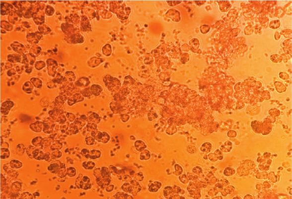
2                                                                   Evidence-Based Complementary and Alternative Medicine

the energy balance, which occurs when the calories ingested            Varieties of natural products have been proposed as a
are greater than those spent by the body. The intake will be       pharmacological treatment of MetS and T2D. Scolymus
higher and the storage lipids will therefore be increased. The     hispanicus, the golden thistle species, is food source and
increase in the storage of lipids and lipid derivatives leads to   can be considered an important natural source of anti-
the expansion of adipose tissue (hyperplasia and hyper-            oxidants. The golden thistle (Scolymus hispanicus), locally
trophy) and the installation of lipotoxicity, which has            known as “Guernina” or “Thaghadiwth,” is one of the
harmful effects resulting in nonalcoholic fatty liver disease       most popular plants in Algeria, Spain, and other Medi-
(NAFLD), which is associated with obesity [2].                     terranean countries [7]. In Algeria, we eat the petioles
    NAFLD was recently redefined as metabolic-associated            (“stems” of the leaf, or more exactly the main vein) cooked
fatty liver disease (MAFLD) to reflect better the patho-            in the broth (red sauce with meat) that accompanies
genesis [2]. NAFLD is the most common chronic liver                couscous.
disease that affects around 25% of the population. NAFLD                Scolymus hispanicus has been linked to many medicinal
encompasses a broad spectrum of diseases that include              properties such as diuretic, depurative, digestive, choleretic,
simple fatty infiltration nonalcoholic steatohepatitis              and lithiuretic properties [7]. Moreover, in traditional
(NASH), which is defined as the presence of fat leading to          medicine, consuming this plant in the green or cooked state
inflammatory damage to hepatocytes, fibrosis, and finally             is recommended for liver and intestines diseases [8]. The
cirrhosis. The importance of NAFLD lies in the possibility         flaky stems are used for digestive tract care, bronchitis, and
of its gradual progress to advanced fibrosis, cirrhosis, and        cold and have emmenagogic and antidiarrhoeal properties
hepatocellular carcinoma (HCC) [2, 3]. The overall prev-           [9]. The roots in decoction are recommended as an anti-
alence of NAFLD is growing in parallel with the global             diabetic. Consumption of the ribs (main veins) of this plant
epidemic of obesity [4]. The pathophysiology is complex            fresh or cooked is recommended for liver and intestinal
and involves multiple concurrent mechanisms in the                 diseases [8]. Other uses in the traditional medicine of golden
context of abnormal metabolic processes that arise mostly          thistle have been reported, such as in Malta fever and eye
in individuals with risk factors. Comorbidities associated         infection [10]; it can also be used as an appetizer and as a
with NAFLD include obesity, type 2 diabetes (T2D), ar-             hemostatic agent [11]. The antioxidant activity of Scolymus
terial hypertension, and dyslipidemia, as traits of metabolic      has been reported [10, 12].
syndrome (MetS) [3].                                                   Phytochemical analysis has demonstrated that the plant
    MetS is a clinical syndrome that includes obesity, dys-        contains many biologically active compounds and a high
lipidemia, arterial hypertension, and T2D [5]. NAFLD is            content of α-tocopherol and identified 3 flavonoids (cate-
strongly linked with all segments of MetS and it is in fact        chin, rutin, and tannic acid) and 13 phenolic acids, such as
liver manifestation of MetS. Some authors have suggested           gallic acid, pyrogallol, chlorogenic acid, p-hydroxybenzoic
that NAFLD could be defined as a fifth component of the              acid, vanillic acid, caffeic acid, syringic acid, p-coumaric
MetS [5]. Both conditions were related to insulin resistance       acid, ferulic acid, sinapic acid, salicylic acid, and rosmarinic
(IR), the main pathogenic factor underlying NAFLD and              acid resveratrol [13].
MetS. Abdominal fat overage is a fundamental determinant               In this context, the present study aims to evaluate the
in NAFLD pathogenesis due to its association with IR and a         effect of the aqueous extract of Scolymus hispanicus on HFD-
possible source of free fatty acids (FFA) [3, 6]. Trunk fat was    related metabolic disorders, steatosis hepatic, inflammation,
found to be indicative of elevated ALT, supporting the             and stress markers.
potential involvement of the metabolically active intra-ab-
dominal fat in increased liver injury [2].                         2. Materials and Methods
    Obesity is associated with an increase in adipose tissue
lipolysis, secretion of inflammatory, and fibrotic mediators,        2.1. Preparation of Aqueous Extract from Scolymus hispanicus.
which can reach the liver. The accumulation of inflamma-            The aerial part of Scolymus hispanicus or the golden thistle
tory/immune cells and the modification of the activities of         was harvested in Algiers in February 2019. The voucher
these cells in the adipose tissue contributed to chronic low-      specimen (INA/P/No 54) has been preserved in the her-
grade inflammation during obesity [2, 3]. This sustained            barium of the Botany Department, National Institute of
inflammation mediates IR and provides a contributing link           Agronomy (INA), Algiers, Algeria. The stems and leaves of
between its development and NAFLD [2, 3]. The accumu-              Scolymus were washed and separated from the roots, cut into
lation of hepatic diacylglycerol and the activation of in-         small slices, dried, then added to 1000 mL of water, and left
flammatory pathways are promoted. Diacylglycerols activate          to boil for 50 min on a thermostated stirrer. After the
protein kinase ε and inhibit insulin signaling, leading to         cooling, the extract was filtered through muslin. The filtrate
hepatic IR [2, 3]. The dysregulation of insulin-mediated           was centrifuged at 1500 rpm for 5 min and a second time at
control of hepatic production of glucose and lipids appears        2000 rpm for 10 min to obtain a homogeneous liquid. After
to be the main event in the development of NAFLD [3].              the centrifugation, all samples were filtered through filter
Normally, insulin impairs gluconeogenesis while promoting          paper (Whatman with a pore size of 11 μm). The collected
lipogenesis. There is a paradoxical situation in NAFLD,            aqueous extract was then lyophilized (Cryodos 80, −75°C,
especially in the context of T2D. IR results in a reduced          5 m3/h) to find an extract yield of 4.3%. The extract was
ability to inhibit gluconeogenesis but insulin-driven lipo-        stored in sealed glass vials at ± 4°C before being tested and
genesis still occurs and is even enhanced [3].                     analyzed.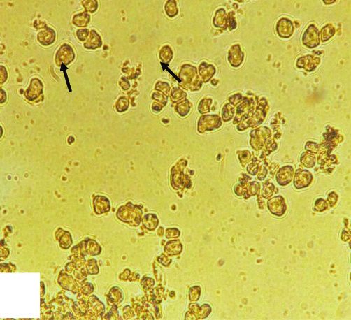
Evidence-Based Complementary and Alternative Medicine                                                                          3

2.2. Preparation of the Hyperfatty Diet. The hyperfatty diet at     phenolic content was evaluated from a standard calibration
40% was prepared in the cellular and molecular physiopa-            curve of gallic acid, and the results were expressed as mi-
thology team/BPO Laboratory/USTHB. According to the                 crograms of gallic acid (GA) equivalents (E) per milligram of
recommended nutritional intake, fats should not exceed 30%          extract (µg GAE/mg).
of the total daily energy intake to avoid unhealthy weight              (2) Determination of Total Flavonoids. The total flavo-
gain. In our study, we used a rate of 40% of lipids to confirm       noids were determined according to the modified method
the installation of obesity with metabolic dysfunctions in the      described by Lebreton et al. using quercetin as a reference
rats. The hyperfatty diet is based on cooked sheep fat; the         [15]. Four milliliters (4 mL) of dilution solution was mixed
cooking increases saturated fatty acids. The lipid intake in        with 4 mL of aluminum trichloride solution (2% in meth-
these rats is represented by 40 g of cooked sheep fat               anol). After 15 min of incubation, the absorbance was
equivalent of 360 kcal; this fat is added to 60 g of the standard   measured at 415 nm. Quercetin (Q) was used as a reference
laboratory food equivalent of 186 kcal to constitute 100 g of       compound to produce the standard curve. The results were
food equivalent of 546 kcal. A daily diet of 20 g of hyperfatty     expressed as μg QE/mg.
food provides 109.2 kcal/day.
                                                                    (3) Antioxidant Activity: Scavenging Effect on DPPH Radical.
                                                                    The 2,2-diphenyl-1-picrylhydrazyl (DPPH) free radical
2.3. Animals. This study was carried out on 28 female rats
                                                                    scavenging assay was carried out as described by Brand-
of the Rattus norvegicus with average weights of
                                                                    Williams et al. [16]. It is based on the degradation of the
111.33 ± 27.66 g, which were reared at the animal facility of
                                                                    DPPH radical dissolved in an 80% methanol/water mixture.
the Faculty of Biological Sciences, USTHB, with controlled
                                                                    An antioxidant will have the ability to donate an electron to
temperature (22 ± 1°C), lighting (12-hour dark/light cycle),
                                                                    the synthetic radical DPPH (purple coloration) to reduce it
and free access to food and water.
                                                                    to nonradical DPPH (yellow-green coloration). The aqueous
    The animals were divided into 4 groups:
                                                                    extract was dissolved in methanol. A sample of 25 μL of each
    (1) Control batch: seven control rats subjected to a            concentration (100, 200, 400, 600, 800, and 1000 μg/mL) was
        standard laboratory diet for 2 months of experi-            added to the DPPH methanol solution (60 μM, 975 μL).
        mentation. The feed was provided by the National            After 30 min of incubation at 25°C, the absorbance at 517 nm
        Animal Feed Office; the calories intake contained in          was measured by UV spectrophotometer (Jasco, V-530).
        20 g of food is 62 calories.                                Ascorbic acid and α-tocopherol were used as compounds
    (2) Control batch treated with the aqueous extract of           reference. The radical scavenging activity was then calcu-
        Scolymus hispanicus at a rate of 100 mg/kg of body          lated using the following equation: % of radical scavenging
        weight/day during the last eight days of experi-            activity � ((Abs control−Abs sample)/Abs control) × 100,
        mentation by intraperitoneal injection (7 animals).         where Abs control is the absorption of the blank sample and
                                                                    Abs sample is the absorbance of the tested extract.
    (3) Batch subjected to a hyperfatty diet (HFD) at 40% for
        two months with a daily intake of 20 g per rat. The
        calorie intake contained in 20 g of food was 109.2
                                                                    2.4.2. Biological Study
        calories.
    (4) Batch subjected to an HFD and treated with the              (1) Analytical Techniques. The animals were bled from the
        aqueous extract of Scolymus hispanicus at a rate of         retroorbital venous plexus; this technique eliminates using
        100 mg/kg of body weight/day during the last eight          anesthetic agents affecting measurements of biochemical
        days of experimentation by intraperitoneal injection        parameters. Blood, which was collected in dried tubes, was
        while maintaining the hyperfatty diet (7 animals).          centrifuged at 3000 rpm for 10 min and the sera were stored
                                                                    at −20°C. Blood glucose, triglycerides, cholesterol, and
2.4. Methods                                                        transaminase were measured by enzymatic colorimetric
                                                                    method using a test kit of Biosystem. Blood insulin was
2.4.1. Chemical Study                                               determined by radioimmunoassay using a CIS test kit (ORIS
                                                                    INDUS). The evaluation of the redox status was performed
(1) Total Phenolic Content. The content of total polyphenols        in the sera and erythrocytes by assaying the thiobarbituric
in the aqueous extract of Scolymus hispanicus was deter-            acid reactive substances (TBARs) and catalase.
mined using the Folin–Ciocalteu reagent according to the                (2) Oral Glucose Tolerance Test (OGGT). The oral glucose
method of Singleton et al., using gallic acid as a reference        tolerance test (OGTT) measures the clearance of glucose
[14]. An aliquot of the aqueous (0.2 mL) extract contains           from the body after its absorption from the intestinal tract.
1000 μg of Scolymus mixed with 46 mL of distilled water and         All rats were weighed one day before the test for the cal-
1 mL of Folin–Ciocalteu reagent in a volumetric flask. The           culation of the glucose solution to be administered. The
mixture was incubated for 3 min in the dark. After that, 3 mL       glucose solution (40%) was administered by intraperitoneal
of sodium carbonate solution (7.5%) was added to the                injection. Rats received 2 mg of glucose/g of body weight
mixture. After 2 hours of incubation in the dark, the ab-           [17, 18].
sorbance was measured at 740 nm in a spectrophotometer                  The rats were fasted for a period of 14 to 16 hours with
(Shimadzu 1800, Mulgrave, Victoria, Australia). The total           free access to water. A blood sample was taken from a small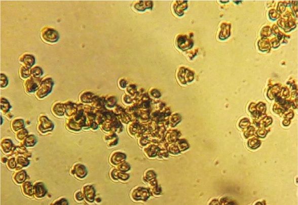
4                                                                 Evidence-Based Complementary and Alternative Medicine

incision in the tail using a scalpel to measure basal blood               contained in the supernatant in the presence of
glucose level (�time point 0) with the glucometer vital check             10% trichloroacetic acid reacted with TBA and
[17, 18]. Once basal glucose concentrations were measured                 caused the formation of a red complex estimated at
in all rats, the glucose solution was given to each animal by             532 nm.
intraperitoneal injection. The timer was immediately started         (iv) Protein Carbonyl Assay. Protein carbonyls (PC)
after the first administration of glucose to all rats. After               were measured in the liver of all animal groups
30 min, the blood glucose was measured using a glucometer                 according to the procedure described by Reznick
of each rat in the same order as they were injected. This                 and Packer [28] using dinitrophenylhydrazine
operation was repeated in 60, 90, and 120 min after glucose               (DNPH) reagent and spectrophotometric method.
administration [19, 20].                                                  The absorbance was measured at 370 nm. The re-
                                                                          sults were expressed as nanomoles of carbonyl
(3) Organs Harvesting. At the end of the experiment, animals              groups per milligram of protein using a molar ex-
were sacrificed after anesthesia by intraperitoneal injection              tinction coefficient of 22 000M−1 cm−1.
of urethane. The liver removed was divided into five frag-
                                                                      (v) Advanced Protein Oxidation Products Assay. The
ments, and each fragment was weighed. They were intended
                                                                          determination of advanced protein oxidation
for different assays, including total lipids where the fragment
                                                                          products (AOPP) levels was performed in the liver
is immersed directly in Folch solution. Another fragment
                                                                          by modifying the Witko-Sarsat method [29]. The
was bound in paraformaldehyde at 10%, and the other three
                                                                          absorbance of the reaction mixture was immediately
fragments were frozen directly in liquid nitrogen to evaluate
                                                                          estimated at 340 nm. AOPP concentrations were
redox status, inflammatory markers, and hepatic glycogen.
                                                                          expressed as micromoles/L of chloramine-T
Two animals from each batch were kept for the initiation of
                                                                          equivalents [30].
hepatocyte cell culture.
                                                                    (8) Measurement of Inflammation Markers.
(4) Histology of the Liver. After fixation in paraformaldehyde
                                                                     (i) Nuclear Factor-Kappa B (NFκB). The assessment was
at 10% for 24 h, the specimens of liver were dehydrated and
                                                                         determined by immunoenzymatic assay. Invitrogen
embedded in paraffin and cut at 5 μm. The sections were
                                                                         ELISA kits were used for measuring the levels of the
stained with Masson’s trichrome [21].
                                                                         NF-kB p65 in the liver of all groups. The estimation
    (5) Hepatic Glycogen. The principle of the method
                                                                         was made by Elisa reader at 450 nm (BioTek
consists in hydrolyzing the glycogen extracted from the liver
                                                                         Instruments).
of rats into glucose with an acid and determining the amount
of the formed glucose using the Folin and Wu method [22].           (ii) Nitrogen Monoxide Assay (NO). The determination
Concentrations were deduced from a standard curve pre-                   of nitrite and nitrate was evaluated from super-
pared with standard glucose solution and the amount of                   natants of the liver of different groups. The nitrite
glycogen was expressed per 100 g of liver.                               bearing in all samples, which were deproteinized and
    (6) Total Lipids. The extraction was carried out according           regenerated, was quantified after addition of Griess
to the method of Folch et al. [23]. The lipids were extracted            reagent (0.1% N-(1naphthyl) ethylenediamine
using chloroform/methanol (2 : 1 v/v). The total lipids were             dihydrochloride, 1% sulfanilamide, and 5% phos-
estimated in mg/100 g of liver.                                          phoric acid). Absorbance was measured at 543 nm
    (7) Oxidant and Antioxidant Activity.                                [31].
     (i) Catalase Activity Assay. The enzymatic activity of
         catalase was determined using the method of             (9) Perfusion and Isolation of Hepatocytes. This technique
         Claiborne [24]. The principle was based on the          was carried out using the modified method of Severgnini
         disappearance of H2O2 in the presence of the            et al. and Edwards et al. [32, 33]. All steps were performed
         enzyme source at 25°C. Catalase was evaluated in        under sterile conditions.
         sera, erythrocytes, and liver of all animal groups.         After anesthesia of the animals by intraperitoneal in-
         Absorbance was estimated at 240 nm in two time          jection with urethane, insert the cannula in the portal vein
         points, t0 and after two min. Erythrocytes and          and start the perfusion using a peristaltic pump containing
         liver were lysed, before all assays, in a lysis buffer   phosphate-buffered saline (PBS) at pH 7.2. As soon as the
         [25].                                                   infusion starts, immediately cut the hepatic vein to allow
                                                                 perfusate to run as waste.
    (ii) Superoxide Dismutase (SOD) Activity Assay. The              The flow was maintained at 5 mL/min for 15 to 20 min
         evaluation of the SOD activity was performed            thanks to the peristaltic pump to remove the blood com-
         according to the method of Giannopolitis and Ries       pletely in each lobe. A second solution at pH 7.4 containing
         [26].                                                   trypsin replaces PBS for tissue digestion. At this stage, the
    (iii) Thiobarbituric Acid Reactive Substances Assay          hepatic tissue was rapidly disaggregated.
          (TBARs). After the reaction with thiobarbituric            The liver was collected with a curved spatula and was
          acid (TBA) (Sigma) [27], the TBARs were mea-           transported in a sterile Petri dish containing DMEM + fetal
          sured in sera, erythrocytes, and liver. The MDA        calf serum (FCS), where we proceeded with the disruption of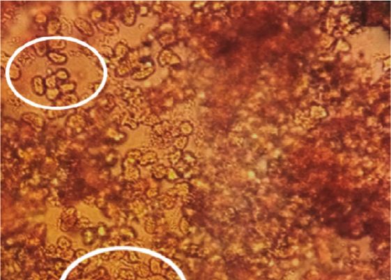
Evidence-Based Complementary and Alternative Medicine                                                                          5

the tissue proceeded using a scalpel. This step should be fast    metabolic disorders by reducing glycemia, insulinemia,
in order to avoid damage to hepatocytes.                          triglyceridemia, and cholesterolemia and amelioration of
    The medium containing the cells was recovered, followed       glucose tolerance compared to animals submitted to
by centrifugation at 600 rpm for 5 min at room temperature.       hyperfatty diet (HFD) (Table 2) and protected the animals
The supernatant was subsequently removed and the cells            against hepatic steatosis complication by decreasing trans-
were suspended a second time in 30 mL of Percoll cushion at       aminase levels.
37.5% for recovering viable cells. Another centrifugation was
effectuated for 3 minutes at 1000 rpm at room temperature.
                                                                  3.3. Scolymus Improved Glucose Tolerance after Eight Weeks of
   (i) Hepatocyte Culture and Microscopy. The viable cells        HFD Feeding. Experimental induction of hyperglycemia by
       recovered were suspended again in 2 mL of DMEM;            an intraperitoneal injection of glucose induced an increase in
       the hepatocytes are observed with an inverted mi-          blood glucose, revealing impaired glucose tolerance in the
       croscope after staining with trypan blue. The cells        HFD animal group (Figure 1). After the administration of
       were distributed in flasks, which were adjusted to          Scolymus, blood glucose levels were significantly lower at 30
       5 mL of DMEM supplemented with FCS, L-gluta-               and 60 min after glucose load in HFD rats treated with
       mine, and antibiotics, and they are incubated in a         Scolymus than that in the HFD group (Figure 1).
       CO2 incubator (Memmert) (5% CO2, 95% air) for the
       start of the primary culture. After 48 h of incubation,
       we noted the confluence of the cells. Trypsinization        3.4. Golden Thistle Ameliorates the Oxidant-Antioxidant
       was necessary to perform the first passage [32, 33].        Balance in Blood. The evaluation of redox statues in the
                                                                  blood (sera and erythrocytes) indicated a significant increase
                                                                  in lipid peroxidation (TBARs) with a decrease in the catalase
2.4.3. Statistical Analysis. Data were analyzed with ANOVA
                                                                  activity (Table 3). The treatment with Scolymus decreases
using STATISTICA version 6 and completed with HSD
                                                                  significantly the rate of TBARs and increases the catalase
Tukey’s test. The results were expressed as the mean-
                                                                  activity (Table 3).
± standard deviation. The differences at ∗ p < 0.05 were
considered to be statistically significant.
                                                                  3.5. Scolymus hispanicus Increased Hepatic Glycogen Storage
3. Results                                                        and Reduced the Hepatic Lipid Accumulation after Hyperfatty
                                                                  Diet. Hepatic glycogen was decreased in the animals sub-
3.1. Phytochemical Study of Scolymus hispanicus. The              jected to the hyperfatty diet; thus, the hepatic storage of
aqueous extract of Scolymus hispanicus showed a high              glycogen was altered. On the other hand, we noted a sig-
content of total polyphenols and flavonoids (Table 1). The         nificant increase in the quantity of total hepatic lipids,
antioxidant activity of the aqueous extract of Scolymus           characterizing the installation of hepatic steatosis (Table 4).
hispanicus was evaluated using the DPPH free radical                  As illustrated in Table 4, the administration of Scolymus
scavenging test. Our extract showed a very important              hispanicus to animals subjected to HFD induced a significant
antifree radical activity with an IC50 value of 0.0038 µg/ml,     increase in the liver glycogen content accompanied by a
which was extremely higher than the reference values BHA          decrease in total liver lipid. Scolymus ameliorates the storage
and BHT (21.18 ± 0.12 µg/mL and 12.66 ± 0.18 µg/mL, re-           of hepatic glycogen and reduces lipid accumulation.
spectively) (Table 1).

3.2. Scolymus hispanicus Improved the Metabolic Disorder and      3.6. Scolymus hispanicus Attenuated Oxidative Stress in Liver.
Reduced Body Weight Gain after Eight Weeks of Hyperfatty          The evaluation of liver catalase activity showed a decrease in
Diet. As illustrated in Table 2, baseline body weight and         the HFD group compared to control, accompanied by an
biochemical parameters were similar between the four study        increase in liver SOD activity. The evaluation of stress
groups of Rattus norvegicus. After eight weeks of hyperfatty      markers such as TBARs AOPP protein carbonyl showed a
diet, body weight was higher than that in normal diet-fed         significant increase compared to controls (Table 5).
animals (Table 2). In terms of biochemical parameters, the            As shown in Table 5, Scolymus hispanicus-treated rats
administration of hyperfatty diet induced an increase in          showed a decreased oxidative stress by increasing catalase
plasma levels of glycemia, insulinemia, total lipids, including   and SOD enzyme activity and reducing lipid peroxidation
triglycerides, and total cholesterol, characterizing the met-     (TBARs) and protein oxidation (PC, AOPP).
abolic syndrome known as insulin resistance (Table 2). The
evaluation of transaminase (AST and ALT) was found to be
significantly increased in terms of experimentation in an          3.7. Scolymus hispanicus Attenuated Hepatic Inflammation
animal group subjected to a hyperfatty diet compared to the       Induced by Hyperfatty Diet. The evaluation of two markers
control (Table 2).                                                of inflammation, such as total nitrite and NFκB, showed a
     The treatment with Scolymus hispanicus at a rate of          significant increase compared to the control group. The
100 mg/kg of body weight/day during the last eight days of        treatment with Scolymus reduces the inflammation by de-
experimentation by intraperitoneal injection corrected the        creasing the levels of total nitrite and NFκB (Table 6).
6                                                                                   Evidence-Based Complementary and Alternative Medicine

                       Table 1: Total phenolic and flavonoid contents and antioxidant activity of Scolymus hispanicus.
Extract/standards               Total phenolic content (µg GAE/ mg)                    Total flavonoids (ug QE/mg)                   DPPH (IC50) (µg/mL)
Aqueous extract                            270.321 ± 25.44                                     164.94 ± 9.45                              0.00383
BHA                                              n.a.                                              n.a.                                 21.18 ± 0.12
BHT                                              n.a.                                              n.a.                                 12.66 ± 0.18
Each value was expressed as means ± standard deviations for triplicate experiments. n.a.: not applied. Q: quercetin; QE: quercetin equivalents; GA: gallic acid;
GAE: gallic acid equivalents; BHA: butylhydroxyanisole; BHT: butylhydroxytoluene.

Table 2: Eight-week evolution of body weight and the plasma biochemical parameters after hyperfatty diet (40%) and normal diet feeding in
rats.
                                   Normal diet                  Hyperfat diet                         ND + Sh                             HFD + Sh
Baseline
Body weight (g)                       116 ± 4                116 ± 5 (p > 0.05)                  118 ± 3 (p > 0.05)                  115 ± 6 (p > 0.05)
Glucose (g/L)                        0.94 ± 0.5            0.91 ± 0.03(p > 0.05)               0.92 ± 0.04 (p > 0.05)              0.93 ± 0.05 (p > 0.05)
Triglycerides (g/L)                 0.45 ± 0.06            0.40 ± 0.05 (p > 0.05)              0.50 ± 0.09 (p > 0.05)              0.48 ± 0.08 (p > 0.05)
Total cholesterol (g/L)             0.70 ± 0.05            0.61 ± 0.11 (p > 0.05)               0.65 ± 0.1 (p > 0.05)              0.56 ± 0.06 (p > 0.05)
Total lipid (g/L)                   2.18 ± 0.12            1.93 ± 0.24 (p > 0.05)              2.11 ± 0.27 (p > 0.05)             1.92 ± 0.09 (p > 0.05)
TGO (ASAT) (UI/L)                 146.25 ± 13.79          141.24 ± 0.83 (p > 0.05)           145.08 ± 7.27 (p > 0.05)            134.38 ± 11.07 (p > 0.05)
TGP (ALAT) (UI/L)                  94.9 ± 6.65              97 ± 5.48 (p > 0.05)              81.6 ± 13.29 (p > 0.05)            84.54 ± 14.47 (p > 0.05)
Insulin (UI)                        76.2 ± 6.18             76 ± 5.24 (p > 0.05)                76 ± 5.55 (p > 0.05)                73 ± 5.83 (p > 0.05)
After 8 weeks
Body weight (g)                       134 ± 4                163 ± 8 (p > 0.05)                  135 ± 2 (p > 0.05)                  148 ± 8 (p > 0.05)
Glucose (g/L)                      0.91 ± 0.04                 1.17 ± 0.04 ∗∗∗∗                0.95 ± 0.07 (p > 0.05)              1.02 ± 0.11 (p > 0.05)
Triglycerides (g/L)                 0.56 ± 0.12                 2.1 ± 0.3∗∗∗∗                  0.43 ± 0.07 (p > 0.05)           1.57 ± 0.31∗∗∗∗ ; (p > 0.05)
Total cholesterol (g/L)            0.66 ± 0.04                 1.24 ± 0.03∗∗∗∗                  0.6 ± 0.11 (p > 0.05)           1.06 ± 0.13∗∗∗∗ , (p > 0.05)
Total lipid (g/L)                   2.20 ± 0.17                5.21 ± 0.28∗∗∗∗                 1.97 ± 0.33 (p > 0.05)               4.22 ± 0.31∗∗∗∗ ;+++
TGO (ASAT) (UI/L)                 143.13 ± 14.46              220.5 ± 10.77∗∗∗                147.9 ± 6.42 (p > 0.05)           145.74 ± 23.07 p > 0.05;+++
TGP (ALAT) (UI/L)                  99.48 ± 6.3                135.8 ± 10.15∗∗∗                  97 ± 5.48 (p > 0.05)             104.22 ± 14.9 p > 0.05;+++
Insulin (UI)                        80.4 ± 8.6                244.8 ± 98.42∗∗                   78.8 ± 7.5 (p > 0.05)               124.78 ± 9.33∗∗∗∗ ;+
Effect of Scolymus hispanicus at 100 mg/kg for eight consecutive days. Data were expressed as mean ± standard deviation (SD) (n � 7). ND: normal diet; HFD:
hyperfatty diet (40%) for eight weeks; ND + Sh: normal diet + Scolymus hispanicus extract (100 mg/kg of body weight/day during the last eight days of
experimentation); HFD + Sh: hyperfatty diet treated with aqueous extract of S. hispanicus. p > 0.05 was not statistically significant. Data were expressed as
mean ± standard deviation (SD) (n � 7). ND: normal diet; HFD: hyperfatty diet (40%) for eight weeks; ND + Sh: normal diet + Scolymus hispanicus extract
(100 mg / kg of body weight/day during the last eight days of experimentation); HFD + Sh: hyperfatty diet treated with aqueous extract of S. hispanicus.
p > 0.05 was considered not statistically significant. The symbol ∗ corresponds to the comparison between HFD versus ND and HFD + SH versus ND + SH; the
+ symbol corresponds to the comparison between ND + SH versus NS and HFD + SH versus HFD. ∗∗ p < 0.01; ∗∗∗ p < 0.001; ∗∗∗∗ p < 0.001 (HFD versus ND),
∗∗∗∗
     p < 0.001 (HFD + Sh versus ND + Sh), +++ p < 0.001; +++ p < 0.001 (HFD + Sh versus HFD).

3.8. Scolymus Alleviates Fatty Liver Disease after Hyperfatty                     macrovesicles, an improvement in cell structure, and a
Diet. As illustrated in Figure 2, Masson’s trichrome staining                     decrease in hypertrophy compared to the HFD group. We
showed a normal histological liver architecture formed of                         also noted the persistence of fibrosis and the absence of
hepatic lobules. Briefly, each lobule was made up of radiating                     inflammatory infiltration in the HFD + Sh group compared
plates. Strands of cells form a network around a central vein                     to the HFD group (Figure 2).
with myofibrils and muscle bundles in liver sections from the
normal diet-fed animals and NFD treated with Scolymus.
    After eight weeks of hyperfatty diet, we recorded                             3.9. Morphological Study of Hepatic Cells in Primary Culture:
structural alterations in the tissue compared to the controls                     Effect of Scolymus hispanicus after Hyperfatty Diet.
(ND and ND + Sh); these alterations were marked mainly by                         Observation of control hepatocytes (ND, ND + Sh) in pri-
lipid deposit accumulation within the hepatocyte under the                        mary culture revealed cells of small size, clear, round or oval,
form of lipid droplets. The latter are present in the form of                     mononuclear or binucleate (Figure 3).
lipid micro- and macrovesicles, marking the installation of                           Observation of Rattus norvegicus liver cells subjected to
hepatic steatosis. We also observed the infiltration of in-                        HFD in primary culture showed cells larger in size compared
flammatory cells associated with interstitial fibrosis marking                      to controls, irregular in shape, mononuclear or binucleate,
the onset of inflammation. In addition, we also noted the                          with lipid vesicles. Some cells have eccentric nuclei
hypertrophy of the hepatic cells, a disorganization of the                        (Figure 3).
cellular architecture, and the widening of the sinusoidal                             Treated with aqueous extract of Scolymus hispanicus
spaces (Figure 2).                                                                improved the cellular appearance of hepatocytes subjected to
    Treatment with Scolymus hispanicus induced attenuation                        HFD in primary culture marked by the reduction of lipid
of hepatic steatosis with a decrease in lipid micro- and                          droplets (Figure 3).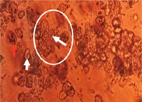
Evidence-Based Complementary and Alternative Medicine                                                                                                            7

                                        5

                                       4.5

                                        4

                                       3.5

                                        3
                      Glycemia (g/L)

                                       2.5

                                        2

                                       1.5

                                        1

                                       0.5

                                        0
                                                 0                    30               60                   90                    120
                                                                                   Times (min)
                                             ND                                               ND + Sh
                                             HFD                                              HFD + Sh
Figure 1: Oral glucose tolerance test in control and experimental groups. Effect of Scolymus hispanicus. Data were expressed as
mean ± standard deviation (SD) (n � 7). ND: normal diet; HFD: hyperfatty diet (40%) for eight weeks; ND + Sh: normal diet + Scolymus
hispanicus extract (100 mg/kg of body weight/day during the last 8 days of experimentation); HFD + Sm: hyperfatty diet treated with aqueous
extract of S. hispanicus. p > 0.05 was considered statistically not significant; p < 0.05 was considered statistically different.

   Table 3: Changes of redox states in the blood (sera and erythrocytes) in rats subjected to hyperfatty diet (40% for eight weeks).
Redox states                                    Blood              ND                   HFD                     ND + Sh                         HFD + Sh
                                                 Sera           0.17 ± 0.01       0.06 ± 0.004∗∗∗∗          0.35 ± 0.02++++             0.29 ± 0.06 p > 0.05;++++
Catalase (UI/mg protein)
                                             Erythrocytes       1.28 ± 0.03        0.89 ± 0.03∗∗∗∗          2.19 ± 0.06++++                4.05 ± 0.34∗∗∗ ,++++
                                                 Sera           65.6 ± 4.33        95.8 ± 2.59∗∗∗∗       67.6 ± 2.07 (p > 0.05)              76.2 ± 7.19∗ ;+++
TBARs (µM)
                                             Erythrocytes      102.8 ± 0.83       146.6 ± 3.85∗∗∗∗       100.4 ± 2.6 (p > 0.05)             94 ± 4.18∗∗ ;++++
Effect of aqueous extract of Scolymus hispanicus (Sh) at 100 mg/kg for eight consecutive days. Data were expressed as mean ± standard deviation (SD) (n � 7).
ND: normal diet; HFD: hyperfatty diet (40%) for eight weeks; ND + Sh: normal diet + Scolymus hispanicus extract (100 mg/kg of body weight/day during the
last eight days of experimentation), HFD + Sh: hyperfatty diet treated with aqueous extract of S. hispanicus. p > 0.05 was considered not statistically significant.
The symbol ∗ corresponds to the comparison between HFD versus ND and HFD + SH versus ND + SH; the + symbol corresponds to the comparison between
ND + SH versus NS and HFD + SH versus HFD. ++++ p < 0.0001 (ND + Sh versus ND), ∗∗∗∗ p < 0.001 (HFD versus ND), ∗ p < 0.05; ∗∗∗ p < 0.001, ∗∗∗∗ p < 0.0001
(HFD + Sh versus ND + Sh), +++ p < 0.001; ++++ p < 0.0001 (HFD + Sh versus HFD).

Table 4: Evaluation of glycogen, total lipids, and triglycerides in hepatic tissue in control and experimental groups and effect of Scolymus
hispanicus.
Hepatic tissue                                           ND                       HFD                          ND ± Sh                          HFD ± Sh
Glycogen (mg /100 g of liver)                         988 ± 87                704 ± 176∗∗∗∗                  1150 ± 88+                     1307 ± 405∗ ;++++
Total lipids (mg/100 g of liver)                   5330.83± 309.26         5803.83 ± 309.23∗∗∗∗      5137.87 ± 714.86 (p > 0.05)          6176.36 ± 372.81∗ ;+++
TG (mg/100 g of liver)                              906.72 ± 49.07         1751.74 ± 181.03∗∗∗∗       872.32 ± 120.83 (p > 0.05)          1047.36 ± 62.25∗ ;++++
Cholesterol (mg/100 g of liver)                    1769.64 ± 104.23        2804.54 ± 289.82∗∗∗∗      1706.22 ± 237.69 (p > 0.05)          2051.61 ± 124.30∗ ;+++
Data were expressed as mean ± standard deviation (SD) (n � 7). ND: normal diet; HFD: hyperfatty diet (40%) for eight weeks; ND + Sh: normal die-
t + Scolymus hispanicus extract (100 mg/kg of body weight/day during the last eight days of experimentation); HFD + Sh: hyperfatty diet treated with aqueous
extract of S. hispanicus. p > 0.05 was not statistically significant. The symbol ∗ corresponds to the comparison between HFD versus ND and HFD + SH versus
ND + SH; the + symbol corresponds to the comparison between ND + SH versus NS and HFD + SH versus HFD. + p < 0.05 (ND + Sh versus ND),
∗∗∗∗
     p < 0.0001 (HFD versus ND), ∗ p < 0.05 (HFD + Sh versus ND + Sh), +++ p < 0.001; ++++ p < 0.0001 (HFD + Sh versus HFD).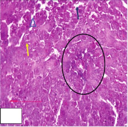
8                                                                                 Evidence-Based Complementary and Alternative Medicine

                                   Table 5: Effect of S. hispanicus on hyperfatty diet on redox states in liver.
Redox states                                             ND                   HFD                      ND + Sm                         HFD + Sm
Catalase (UI/100 mg of liver)                        0.72 ± 0.04         0.27 ± 0.07∗∗∗∗             0.88 ± 0.07++                 0.43 ± 0.04∗∗∗∗ ;++
SOD (UI/100 mg of liver)                             1.86 ± 0.53         4.22 ± 0.66∗∗∗          2.04 ± 0.17 (p > 0.05)               5.95 ± 1.1∗∗ ;+
TBARs (µmol/100 mg of liver)                         0.43 ± 0.09         1.01 ± 0.08∗∗∗∗         0.39 ± 0.04 (p > 0.05)          0.53 ± 0.15 p > 0.05;++
Protein carbonyls (pmol/100 mg of liver)              3.87 ±0.16          6.97 ± 1.8∗∗           3.93 ± 0.12 (p > 0.05)         3.58 ± 0.59 p > 0.05;++
AOPP (pmol/100 mg of liver)                         356.76 ± 29.01      953.63 ± 220.5∗∗       353.55 ± 29.66 (p > 0.05)       431.65 ± 99.96 p > 0.05;++
Data were expressed as mean ± standard deviation (SD) (n � 7). ND: normal diet; HFD: hyperfatty diet (40%) for eight weeks; ND + Sh: normal die-
t + Scolymus hispanicus extract (100 mg/kg of body weight/day during the last eight days of experimentation); HFD + Sh: hyperfatty diet treated with aqueous
extract of S. hispanicus. p > 0.05 was not statistically significant. The symbol ∗ corresponds to the comparison between HFD versus ND and HFD + SH versus
ND + SH; the + symbol corresponds to the comparison between ND + SH versus NS and HFD + SH versus HFD. ++p < 0.01 (ND + Sh versus ND), ∗∗ p < 0.01;
∗∗∗
    p < 0.001; ∗∗∗∗ p < 0.0001 (HFD versus ND), ∗∗∗ p < 0.001, ∗∗∗∗ p < 0.0001 (HFD + Sh versus ND + Sh), + p < 0.05 ; ++ p < 0.01; ++++ p < 0.0001 (HFD + Sh
versus HFD).

          Table 6: Changes of inflammatory markers in the liver of the rats subjected to hyperfatty diet (40% for eight weeks).
Hepatic tissue                                   ND                    HFD                          ND + Sh                            HFD + Sh
NO (Pmol/100 mg of liver)                     97 ± 3.54            146 ± 3.39∗∗∗∗                  92 ± 3.16+                     112 ± 3.16∗∗∗∗ ;++++
NFκ B P 35 (Pg/100 mg of liver)              92.17 ± 3.88        279.09 ± 31.12∗∗∗∗         84.77 ± 10.35 (p > 0.05)          101.5 ± 25.04 p > 0.05;++++
Effect of Scolymus hispanicus extract at 100 mg/kg for eight consecutive days. Data were expressed as mean ± standard deviation (SD) (n � 7). ND: normal
diet; HFD: hyperfatty diet (40%) for eight weeks; ND ± Sh: normal diet ± Scolymus hispanicus extract (100 mg/kg of body weight/day during the last eight days
of experimentation); HFD + Sh: hyperfatty diet treated with aqueous extract of S. hispanicus. p > 0.05 was not statistically significant. The symbol ∗ cor-
responds to the comparison between HFD versus ND and HFD + SH versus ND + SH; the + symbol corresponds to the comparison between ND + SH versus
NS and HFD + SH versus HFD. + p < 0.05 (ND + Sh versus ND); ∗∗∗∗ p < 0.0001 (HFD versus ND); ∗∗∗∗ p < 0.0001 (HFD + Sh versus ND + Sh); ++++ p < 0.0001
(HFD + Sh versus HFD).

4. Discussion                                                                    body weight. This is probably due to the duration of the
                                                                                 treatment. A period of 8 days would not, therefore, be
The present results showed that eight weeks of hyperfatty                        sufficient to have conclusive results. Our results were in
diet leads to a metabolic syndrome associated with several                       agreement with the work of Sayin et al. [38], which showed
structural changes in the liver characterized by lipid accu-                     that the treatment of animals subjected to a hyperfatty diet
mulation, inflammation, and oxidative stress in Rattus                            with ethanolic extract of Silybum marianum, belonging to
norvegicus. Moreover, the extract of Scolymus hispanicus                         the same family as Scolymus, for 4 months had no effect on
seems to have lipid-lowering, anti-inflammatory, and an-                          weight change, while the 11-month treatment reduced the
tioxidant properties in Rattus norvegicus subjected to HFD.                      body mass.
It improves the condition of fatty liver disease. The presence
of bioactive molecules in the extract of Scolymus hispanicus
could give this interesting antioxidant and hepatoprotective                     4.1. Metabolism Disorder. The hyper fatty diet caused
potential.                                                                       metabolism disorders in rats marked by the hyperglycemia
    The aqueous extract of Scolymus hispanicus showed a                          associated with hypertriglyceridemia, hypercholesterolemia,
high content of total polyphenols and flavonoids compared                         and hyperinsulinemia and impaired glucose tolerance at the
to other plants [25]. Our extract showed a very important                        end of the experiment. These results characterized the in-
antifree radical activity compared to the reference BHA and                      stallation of metabolic syndrome. Our work agrees with that
BHT. The content of the total polyphenols and flavonoids                          of Lee et al. [39]; this can be explained on the one hand by the
and the antioxidant activity of our extract are very high                        preferential oxidation of fatty acids, which leads to a defect
compared to the results found by Morales et al. [10] on                          in the use of glucose via the inhibition of the key enzymatic
Scolymus hispanicus in Spain. This variability is linked to the                  activities of glycolysis, which will block the entry of glucose
nature of the soil and to the geographical location.                             into the cells. Oxidation of fatty acids also provides key
    In our study, the 40% hyperfatty diet (HDF) induced the                      cofactors of gluconeogenesis, which increases the produc-
onset of obesity characterized by the increased body weight.                     tion of glucose by the liver and leads to an increase in
This result is in agreement with the study Hamalt et al. [34]                    circulating glucose. On the other hand, fatty acids interfere
and Harrat et al. [35], who showed that a 40% hyperfatty diet                    with insulin signaling pathways [40, 41]. Our work is in
induced an increase in the body weight in Wistar rats. It is                     agreement with the work of Murakami et al. [42]; the hy-
possible that the mechanisms regulating appetite respond                         perlipidemia observed can be explained by the high lipid
more slowly to fat than to protein and carbohydrates. Thus,                      content in the diet since the long-term administration of a
dietary fat has little effect on satiety and periodic exposure to                 hyperfatty diet causes both plasma and tissue disorders
hyperfatty diet and the increase in food density portion size                    [34, 43].
increased availability and promoted obesity [36, 37].                                 Treatment with Scolymus hispanicus extract led to a
    Treatment with the extract of the plant Scolymus his-                        decrease in the blood sugar level, decrease in insulin level,
panicus induced a decrease, however not significant, in the                       and improvement in glucose tolerance. Our results agree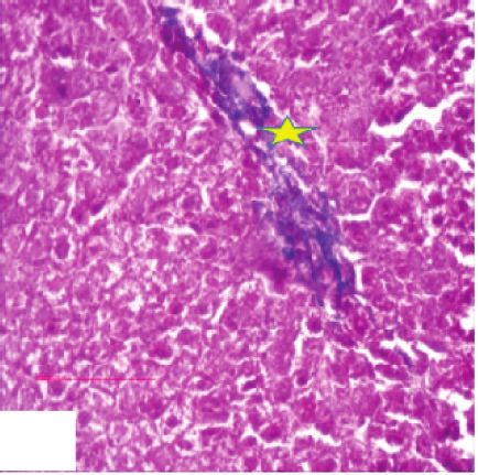
Evidence-Based Complementary and Alternative Medicine 9
A B C1 C2
C3 C4 C5 C6
D1 D2 D3 1 2 3
4 5
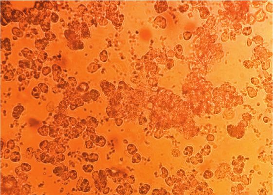
Figure 2: The effect of the Scolymus hispanicus extract on hepatic histological changes of the rats after Masson’s trichrome: (A) normal diet
group; (B) normal diet + Sh group; (C) hyperfatty diet group (40% during for 8 weeks); (D) hyperfatty diet + S. hispanicus at 100 mg/kg/day
for 8 days. (1, 2) extension of C2 figure showing, respectively, the infiltration of monocytes and the chemotaxis phenomenon, hepatic
steatosis marked by the accumulation of lipid droplets. Picture 3: enlargement of the zone of inflammation highlighting red blood cells and
adhesion inflammatory cells. (4) Enlargement of C5 figure showing a partial thrombus with initiation of chemotaxis represented by a green
arrow. (5) Enlargement of C6 figure showing the inflammatory process marked by the adhesion of monocytes to the vascular walls
(initiation of chemotaxis) represented by a green arrow. Yellow arrow: hepatic lipid droplets; yellow star: widening of the sinusoidal space;
blue triangle: disorganization of the cellular structure; green arrow: infiltration of inflammatory cells; black circle: inflammatory hem-
orrhagic focus marked by the presence of red blood cells (light blue arrow) and the inflammatory cells (monocyte and lymphocyte) (green
arrow); yellow circle: partial thrombus of the hepatic portal space; green star: perivascular fibrosis; yellow stars: persistence of fibrosis.
with the work of Feng et al. [44]. The polyphenols present in marianum. The latter had the regulatory role of the genes
the plants of the Asteraceae family improve the endocrine involved in the synthesis of fatty acids and in the metabolism
function of the pancreas and insulin sensitivity [38] and of hepatic triglycerides and cause also decreased expression
promote functional recovery of the insulin receptor sub- of the fatty acid synthase (FAS) gene and attenuation of
strate 1 [44]. Marmouzi et al. reported the relevant anti- acetyl-CoA carboxylase (ACC) activity. The latter two play a
oxidant effect of Scolymus hispanicus functional parts and role in inhibiting de novo lipogenesis [38, 45].
their antidiabetic activities via α-glucosidase and α-amylase
inhibition [13].
Scolymus induced a decrease in cholesterolemia and 4.2. Lipotoxicity and Hepatic Function. The hyperfatty diet
triglyceridemia, which is consistent with the work of Sayin induced an increase in glutamic oxaloacetic transaminases
et al. [38]. This is explained by the decrease in the tran- and glutamic-pyruvic transaminases in the rats subjected to
scription factor SREBP-1c under the effect of Silybum HFD, which confirms the work of Maximos et al. [46]. They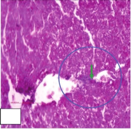
10                                                                          Evidence-Based Complementary and Alternative Medicine

           A                                    B                                    C1

           C2                                  D                                 1                                 2

Figure 3: The effect of the Scolymus hispanicus extract on hepatic cell changes in primary culture. (A) Cells from animals subjected to the
normal diet group; (B) cells from animals subjected to the normal diet + Sh group; (C1, C2) cells from animals subjected to the hyperfatty
diet group (40% during for 8 weeks); (D) cells from animals subjected to the hyper atty diet + S. hispanicus at 100 mg/kg/day for 8 days. (1)
Enlargement of C2 figure showing liver cells from rats subjected to HFD with lipid droplets and some cells with eccentric nuclei. (2)
Enlargement of C2 figure showing liver cells from rats subjected to HFD treated with Scolymus with clear mononuclear and binuclear cells.
Blue arrow: small clear mononuclear liver cells; red arrow: small clear binuclear liver cells; white circle and white arrow: lipid droplets; green
arrow: cells with eccentric nuclei.

are considered to be biomarkers of liver dysfunction and                   of genes involved in the inhibition of hepatic lipogenesis and
liver damage caused by hyperfatty diet. In obesity, the high               the regulation of lipoprotein metabolism by increasing the
amounts of toxic metabolites lead to the depletion of hepatic              synthesis of HDL and decreasing the synthesis of VLDL.
glutathione due to the increase of free radicals. These oxi-               Flavonoids treatment decreases the development of nonal-
dants cause necrosis in liver cells, inducing an increase in the           coholic fatty liver disease [52].
concentration of aminotransferases and their release into the                  Our results showed a decrease in liver glycogen in an-
serum [47].                                                                imals subjected to HFD, which does not agree with the
    Treatment with Scolymus extract resulted in a decrease in              studies by Auberval et al. [43]. According to Magnan [53],
the levels of transaminases compared to the HFD group,                     diets rich in lipids cause the intracellular increase in free fatty
which is in agreement with the work of Zhang et al. [48]. The              acids and induce, on the one hand, the direct inhibition of
polyphenols present in plants of the Asteraceae family in-                 the main enzymes of glucose metabolism.
duce an improvement in hepatic steatosis and inflammation                       An increase in hepatic glycogen was observed in an-
thanks to their hepatoprotective properties and by restoring               imals treated with the aqueous extract of Scolymus, which
hepatic markers [38].                                                      is consistent with the data reported by Guigas et al. [54].
    Our study showed an increase in total hepatic lipids in                This indicates the potent antihyperglycemic role of Sco-
animals on a 40% hyperfatty diet, which results in metabolic               lymus. A decrease in gluconeogenesis and glycogenolysis
dysfunction, leading to the accumulation of lipids in the                  associated with reduced hydrolysis of glucose-6-phos-
hepatocytes. This dysfunction affects all stages of lipid                   phate plays a key role in the regulation of hepatic glycogen
metabolism, which is primarily an excessive uptake of free                 metabolism [54].
fatty acids produced by lipolysis in adipose tissue, an ac-                    The hyperfatty diet caused macrovesicular steatosis,
cumulation of triglycerides, an increase in the hepatic li-                which results in ectopic accumulation of triglycerides in the
pogenesis concomitant with a decrease in ß-oxidation, and                  cytoplasm of hepatocytes and inflammatory infiltration of
finally a decrease in the secretion of VLDL [49].                           liver tissue by polymorphonuclear cells and mononuclear
    Golden thistle induces a decrease in the quantity of lipids            cells, which are mainly lymphocytes that led to the evolution
and triglycerides stored in the liver. Our results agree with              to steatohepatitis then to fibrosis, consistent with the work of
those carried out by Aoun et al. [50]. A number of studies                 Savard et al. [55]. This is explained, on the one hand, by the
have suggested that the polyphenols contained in milk thistle              metabolic disturbance that induced the increase in the influx
promote the use of stored fatty acid as an energy source,                  of free fatty acids and the activation of de novo lipogenesis
which would primarily explain this decrease [51]. Secondly,                and, on the other hand, by oxidative stress that induced a
the active compounds of milk thistle lead to the stimulation               decrease in hepatic ATP production and the production of
Evidence-Based Complementary and Alternative Medicine 11
proinflammatory cytokines, which trigger inflammatory production of free radicals or by maintaining the integrity of
necrosis leading to the progression of steatohepatitis [56]. mitochondrial electrons transport under stress conditions
Hepatic steatosis was alleviated after treatment with [38]. Polyphenols participate in the maintenance of an
golden thistle, which is in agreement with the work of Ni and optimal redox state of the cell by activating a range of an-
Wang [57]; this can be explained by the presence of poly- tioxidant enzymes. Polyphenols modulate the signaling
phenols, which reduce the accumulation of triglycerides and pathways Nrf2/Keap1/ARE and NFκB, resulting in an in-
improve the severity of fatty liver disease [58]. We have also crease in the expression of genes encoding cytoprotective
noted the persistence of fibrosis. This may be related to the molecules. In addition, a decrease in the expression of genes
short duration of treatment and the dose administered. modulated by NFκB would reduce the production of
The morphological study of hepatic cells in primary proinflammatory cytokines [65].
culture strengthens and approves the liver histological re- Our results indicated that the level of serum erythrocyte
sults obtained following treatment with Scolymus. and hepatic TBARs increases in the rats subjected to HFD,
which is in agreement with the work of Feng et al. [44]. The
40% hyperfatty diet is closely related to the onset of oxidative
4.3. Lipotoxicity, Redox Status, and Inflammation. Our re- stress and causes an increase in free radicals, which will cause
sults concerning the markers of the antioxidant status the oxidation of polyunsaturated lipids, which is a free
showed that there was a decrease in the antioxidant capacity radical chain reaction process known as lipid peroxidation
of catalase in the serum, the erythrocyte, and the liver in the that gives primary products such as hydroperoxides or
rats subjected to a hyperfatty diet of 40%. These results were terminal secondary products such as malondialdehyde
in agreement with the work of Furukawa et al. [59]. An (MDA) [66]. In addition, the treatment with Scolymus de-
increase in the flow of fatty acids to the liver, an excess of creases the level of TBARs in animals subjected to HFD. Our
hepatic synthesis, and an imbalance in the diet cause a results are in agreement with the work carried out by
decrease in catalase. CAT is an enzyme, which catalyzes the Frederico et al. [67].
disproportionation of hydrogen peroxide into oxygen and Prolonged consumption of HFD resulted in high levels
water. The initial excess of fatty acids leads to the accu- of carbonylated proteins and advanced oxidized protein
mulation of triglycerides in hepatocytes; the failure to me- products, which is consistent with the work of Feng et al.
tabolize triglycerides accumulated in hepatocytes leads to [44]. Oxidation can affect proteins and generate advanced
fatty liver disease. This process triggers an avalanche of many products of oxidized proteins, which are uremic toxins
factors such as an increase in the activity of cytochrome formed by the reaction of plasma proteins and, more par-
P450, an increase in the production of reactive oxygen ticularly, albumin with oxidants. These oxidants are pro-
species, lipid peroxidation, a deficiency in antioxidant de- duced specifically by myeloperoxidase (MPO) secreted by
fense, activation of proinflammatory cytokines and the activated neutrophils [68]. Any radical attack on an amino
nuclear transcription factor NFκB, and an increased ex- acid will cause the oxidation of certain residues resulting in
pression of PPAR receptors, initiating the transition from the appearance of carbonyl groups in the cleavage of peptide
steatosis to steatohepatitis. Fibrosis and subsequent cirrhosis chains and intra and interchain bityrosine bridges. Most of
are the final stages in this process [60]. the damage is irreparable and can lead to major functional
Our results showed an increasing superoxide dismutase changes [68].
activity in animals subjected to HFD. Our results agreed with Scolymus hispanicus induced a decrease in AOPP and PC
the work of Sfar et al. [61]; this can be explained by the body’s in the rats subjected to HFD, which is consistent with the
response to increased ROS in the prevention of alterations in work of Ramadan et al. [64] and Feng et al. [44]. This de-
cells, lipids, and proteins that are at the origin of various crease may be due to the active components of Scolymus
pathologies. These results confirm the fact that obesity is having a powerful antioxidant activity by scavenging free
associated with oxidative stress. Cellular adaptation to this radicals such as hydroxyl and superoxide anion, making
state is, therefore, the increase in the activity of SOD, which them inactive and stable thanks to their hydroxyl group [69].
is a metalloprotein possessing enzymatic activity catalyzing Our results showed an increase in inflammation markers
the dismutation of superoxide anions into dioxygen and “NO and NFκB” in the liver of animals subjected to a
hydrogen peroxide [62]. hyperfatty diet. Treatment with Scolymus extract decreases
Scolymus induced an increase in the catalase and the the levels of NFκB and nitrites. These results are consistent
SOD activity, which is consistent with the work of Her- with those in the work of Berdja et al. [25] and Smail et al.
menean et al. [63]. Our results suggest that golden thistle [70]. The lipophilic metabolites from Scolymus hispanicus
induces an antioxidant effect, which may be linked to the L. compounds exhibited anti-inflammatory activities in
richness of this plant in bioactive and antioxidant com- vitro, manifested by inhibition of NFκB p65 expression and
pounds, inhibiting the alterations caused by the excessive subsequent decrease of inflammatory cytokines: IL-6, IL-1b,
production of free radicals [64]. The extract of the Scolymus and TNF-α in PHA-stimulated with human PBMCs [71].
plant contains various flavonoids and polyphenol acids such
as “catechin, rutin and tannic acid, gallic acid, pyrogallol, 5. Conclusion
and chlorogenic acid,” which can contribute to antioxidant
defenses in different ways by direct scavenging of free In summary, we provide proof that Scolymus hispanicus
radicals and inhibition of enzymes responsible for the corrects metabolic disorders by exerting a hypoglycemic,You can also read