Review Article Remodeling of Macrophages in White Adipose Tissue under the Conditions of Obesity as well as Lipolysis
←
→
Page content transcription
If your browser does not render page correctly, please read the page content below
Hindawi
Oxidative Medicine and Cellular Longevity
Volume 2021, Article ID 9980877, 13 pages
https://doi.org/10.1155/2021/9980877

Review Article
Remodeling of Macrophages in White Adipose Tissue under the
Conditions of Obesity as well as Lipolysis

 Xiaohui Tong ,1 Lu Wei ,2 Tongsheng Wang ,1 and Rongchun Han 3

 1
 School of Life Sciences, Anhui University of Chinese Medicine, 350 Longzihu Road, Xinzhan District, Hefei 230012, China
 2
 School of Life Sciences, Hainan University, 58 Renmin Avenue, Meilan District, Haikou 570228, China
 3
 School of Pharmacy, Anhui University of Chinese Medicine, 350 Longzihu Road, Xinzhan District, Hefei 230012, China

 Correspondence should be addressed to Rongchun Han; hanr@ahtcm.edu.cn

 Received 25 March 2021; Revised 23 July 2021; Accepted 6 August 2021; Published 31 August 2021

 Academic Editor: Mateusz Maciejczyk

 Copyright © 2021 Xiaohui Tong et al. This is an open access article distributed under the Creative Commons Attribution License,
 which permits unrestricted use, distribution, and reproduction in any medium, provided the original work is properly cited.

 Adipose tissue macrophages (ATM) are a major source of low-grade inflammation in obesity, and yet reasons driving ATM
 accumulation in white adipose tissue (WAT) are not fully understood. Emerging evidence suggested that ATM underwent
 extensive remodeling in obesity. In addition to abundance, ATM in obesity were lipid-laden and metabolically reprogrammed,
 which in turn was tightly related to their functional alterations and persistence in obesity. Herein, we aimed to discuss that
 activation of lipid sensing signaling associated with metabolic reprogramming in ATM was indispensible for their migration,
 retention, or proliferation in obesity. Likewise, lipolysis also induced similar but transient ATM remodeling. Therefore, we
 assumed that obesity might share overlapping mechanisms with lipolysis in remodeling ATM. Formation of crown-like
 structures (CLS) in WAT was presumably a common event initiating ATM remodeling, with a spectrum of lipid metabolites
 released from adipocytes being potential signaling molecules. Moreover, adipose interlerkin-6 (IL-6) exhibited homologous
 alterations by obesity and lipolysis. Thus, we postulated a positive feedback loop between ATM and adipocytes via IL-6
 signaling backing ATM persistence by comparison of ATM remodeling under obesity and lipolysis. An elucidation of ATM
 persistence could help to provide novel therapeutic targets for obesity-associated inflammation.

1. Introduction scavengers. ATM would increase the uptake of lipid fluxes
 from adipocytes in mice in response to fasting or pharma-
The prevalence of Western dietary pattern and sedentary life- cologically induced lipolysis, while intra-abdominal ATM
style boosted the pandemic of obesity worldwide, which was depletion resulted in elevated fasting serum free fatty acid
closely associated with increased risks of various metabolic (FFA) levels in mice [5]. The fact that most of ATM were
disorders [1]. To tackle this problem, the development of localized within crown-like structures (CLS) in obesity [6]
new therapeutics for obesity is urgent. Strongly involved in indicated CLS might be a vital initiator for obesity-related
the pathogenesis of obesity was white adipose tissue (WAT) ATM recruitment. Indeed, adipocyte death per se can trig-
[2]. Expansion of fat in obesity was associated with adipose ger ATM infiltration [7] because ATM were important in
tissue macrophages (ATM) accumulation, which caused a digesting dead adipocytes within CLS via secretion of cyto-
state of chronic, low-grade inflammation, and insulin resis- kines or lysosomal lipases which activated lipolysis and
tance [3]. To date, reasons for the persistence of excessive breakdown of triglyceride (TG) in adipocytes [8, 9]. The
ATM in obesity have not been fully understood. Canonically, group of Becker identified a “metabolically activated”
it was known that inflamed adipose tissue could overexpress (MMe) phenotype of ATM in obese mice, which were bene-
chemokines such as monocyte chemoattractant protein-1 ficial by clearing dead adipocytes through lysosomal exocyto-
(MCP-1) which would lead to increased infiltration of sis at the late onset of obesity [10].
macrophages [4]. Despite the role in inflammation, ATM Obesity influenced not only ATM abundance but also
were also key players for systemic energy homeostasis as lipid their metabolic process, indicated by accumulated cellular
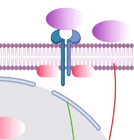
2 Oxidative Medicine and Cellular Longevity

lipid droplets (LD) in ATM [11, 12]. Mechanistically, ATM lipid-rich microenvironment where cells including ATM
in obese mice and humans overexpressed various cell surface were metabolically challenged. Would this metabolic
receptors to uptake lipids derived from adipocytes in CLS, challenge to ATM relate to their persistence in obesity?
many of which were indispensable for ATM accumulation (Figure 1).
in obesity [13, 14]. Thus, in this regard, it was postulated that The concentration of lipids in ATM in obesity was higher
adipocytes could crosstalk with ATM via secretion of diverse than that of the lean counterpart in mice [11, 12, 18]. In fact,
lipid metabolites and then activated lipid sensing signaling in obesity-associated ATM were metabolically remodeled to be
ATM, which could be another key mechanism underlining preferential of lipid handling. Firstly, stimulated by the lipid-
obesity-linked ATM accumulation. rich microenvironment, ATM boosted cellular lipid contents
 Compared to obesity, the process of adipocyte lipolysis by upregulation of lipid uptake in response to obesity in mice.
was also accompanied with CLS formation, lipid liberation This process was mediated by increased expressions of a
in adipocytes, and inflammation in WAT [6, 15, 16]. The variety of cell surface markers, including the ATP-binding
accumulation of ATM occurred in response to fat mobiliza- cassette transporters A1 (ABCA1), very low-density lipopro-
tion activated by fasting or catecholamine signaling activa- tein receptor (VLDLR), perilipin (PLIN) 2, and cluster of
tion in healthy lean mice, which would be restored later differentiation (CD)36 [13, 14] as well as the scavenger
possibly due to cease of FFAs fluxes from adipocytes [5]. receptor of macrophage receptor with collagenous structure
Moreover, ATM also became lipid-laden during lipolysis (MARCO) in mouse models of obesity [18]. Moreover, the
[5, 17, 18]. Therefore, lipolysis might share overlapping large quantity of FFAs released from apoptotic adipocytes
mechanisms with obesity to recruit and remodel ATM. activated proteins like nicotinamide adenine dinucleotide
Thus, we postulated that comparison of ATM remodeling phosphate (NADPH) oxidase 2 (NOX2), myeloid differenti-
in these two conditions would lead to discoveries of ation primary response 88 (MYD88) and toll-like receptor
related mechanisms for excessive ATM in obesity. 2 (TLR2) in ATM, and modeled ATM to increase the uptake
 Adipose interlerkin-6 (IL-6) appeared to be a potential of lipids through lysosomal exocytosis indicated by detection
shared molecule exhibiting similar upregulation by both of lysosomal associated membrane protein (LAMP)1 and
obesity and adipocyte lipolysis [16, 19, 20]. In obese mice, LAMP2 on cell surface of ATM in obese mice [10]. In the
IL-6/signal transducer and activator of transcription 3 process of lysosomal exocytosis, ATM formed close synapse
(STAT3) signaling played an important role in liberation of with dead adipocytes and digested them with lysosomal
FFAs from adipocytes [21]. Recently, Yu et al. demonstrated enzymes reported by Haka et al. using mouse models of obe-
that ATM in obesity could also crosstalk with adipocytes sity, human adipose tissue, and ex vivo cell model [26]. In
through secretion of IL-6 [22]. Therefore, in this review, addition, around three percent of ATM in WAT possibly
supported with current literature, we proposed the existence conjugated to form the multinucleated giant cells (MGCs)
of a positive feedback loop between adipocytes and ATM via in order to phagocytize oversized dead adipocytes in CLS in
IL-6 signaling in obesity and WAT lipolysis concerning ATM obese patients and mice [27]. Furthermore, biogenesis of
persistence, in hoping to provide new insights for studies of lysosome was driven in ATM to enable high rate of lipid
obesity-related inflammation. catabolism, because inhibition of lysosome function by chlo-
 roquine treatment resulted in accumulation of LD in ATM in
2. Remodeling of ATM in Obesity obese mice [12, 18]. Lipid metabolism in ATM was also
 altered by obesity. Petkevicius et al. demonstrated that
2.1. Metabolic Reprogramming of ATM by Obesity. The initial ATM isolated from obese mice and humans exhibited
event leading to infiltration, retention, or proliferation of increased rate of de novo phosphatidylcholine (PC) biosyn-
ATM under obesity was of limited understanding. The thesis [28]. Consistently, the cholesterol efflux capacity
phenomenon that up to 90% of ATM was localized within (CEC) was enhanced in ATM in obesity associated with
CLS in adipose tissue in obese db/db mice and obese increased expressions of the transporters like ABCA1 com-
humans [6] implied CLS might have a pivotal role in pared to that of lean mice [29].
obesity-associated ATM accumulation. By means of Taken together, the available data suggested that meta-
immunofluorescence, Haase et al. demonstrated that the bolic reprogramming of ATM featuring lipid handling and
microenvironment of CLS predominantly contributed to LD deposition occurred in obesity, which was likely related
a time-dependent increase in ATM proliferation in mice to CLS formation. We further set out to discuss in detail
fed with high fat diet (HFD) [23]. At the late state of obesity, the implications of metabolic reprogramming in ATM accu-
expansion of fat was dominated with CLS clustering and adi- mulation and inflammation in obesity.
pocyte death in obese mice [10, 24]. Meanwhile, the number
of ATM could rise up to 10-folds in adipose tissue in diet- 2.2. ATM Accumulation Driven by Lipid Sensing Signaling.
induced obesity (DIO) in mice [3]. Thus, obesity-linked The finding that the rate of adipocyte death in WAT coin-
CLS formation was possibly one of the events orchestrating cided with ATM accumulation in obese mice suggested a role
ATM accumulation. Then, what were the signaling molecules of ATM in adipose tissue remodeling [8]. Metabolically, the
within the loci of CLS that gave rise to obesity-associated lipid-laden macrophages in insulin-resistant adipose tissue
ATM abundance? It was well acknowledged that obesity were also regarded as a protective measure. Because selective
was featured by inefficiency of lipid storage and increased silencing of lipid handling genes including lipoprotein lipase
lipid liberation from adipocytes in WAT [25], creating a (LPL) or CD36 in ATM resulted in less LD formation in
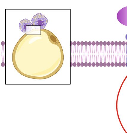
Oxidative Medicine and Cellular Longevity 3

 TG TG/ C16:0 Cholesterol Anionic/ Succinate
 ceramides zwitterionic
 lipids
 TG FFAs

 TLR2 NOX2 CD36 VLDLR CD9 PLN2 ABCA1 TREM2 SUCNRI MARCO

 MYD88
 P62 P13K MARCO

 NRF2 Glucose
 CD36
 PLN2 ABCA1
 LAL Autophagy
 Exocytosis
 FFAs Glycolysis
 PPAR
 OXPHOS

 Netrin-1

 sy ATP
 se
 ha
 I

 nt
 II III IV
 LAMP1 LAMP2
 ER stress ROS

 OXPHOS Fgr
 FFAs

 Lysosome ATM polarization
 Cytokine s , IL-6 e tc .

Figure 1: Integrative model of ATM remodeling in obesity. Representation of ATM surrounding apoptotic adipocytes in CLS in obesity, with
the small black square enlarging alterations of ATM in connection with dysfunctional adipocytes. The signaling pathways that link these
components by activation (green arrows) or inhibition (red inhibitory lines) are also indicated. Briefly, in obesity, ATM are dynamically
recruited to apoptotic adipocytes to form CLS and becoming lipid lading associated with increased synthesis of lysosome responsible for
catabolism of LD. This alteration might involve these following processes. Firstly, to digest dead adipocytes and facilitate FFA liberations,
exocytosis of lysosomal hydrolases from ATM is activated by NOX2 and TLR2/MYD88 signaling pathways with the detection of LAMP1
and LAMP2 on cell surface of ATM. Secondly, expressions of specific cell surface markers including CD36, VLDLR, CD9, PLN2, ABCA1,
TEM2, SUCNR1, and MARCO are boosted in ATM in response to stimulation of metabolites like TG, ceramides, cholesterol,
anionic/zwitterionic lipids, or succinate, contributing to increased lipid uptake and LD formation in ATM, which seems to be essential for
ATM migration and accumulation in adipose tissue by obesity. Mechanistically, metabolism of cellular TG to FFAs mediated by LIPA
activates PPARγ responsible for increased expressions of PLN2, CD36, and ABCA1, while activation of PI3K-NRF2 signaling leads
to increased MARCO expression. Finally, lipid accumulation in ATM might lead to metabolic reprogramming associated with ER
stress as well as shifting to aerobic glycolysis in which building up of ROS results in Fgr activation, mitochondrial complex II
activation, and complex I inhibition, all of which induce preferential production of inflammatory cytokines and ATM polarization.
By contrast, OXPHOS promoted by FFA metabolism and p62 activation due to reduced autophagy or PI3K signaling activation
exhibits anti-inflammatory functions.

ATM as well as elevated circulating serum FFAs levels in More importantly, activation of lipid handling signaling
mice fed with HFD [30]. On the contrary, enhancing the lipid was vital for macrophages to accumulate in WAT. In a study
storage capability of ATM by depletion of lysosome with of Sharma et al., myeloid-specific depletion of the neuroim-
chloroquine treatment decreased basal lipolysis and serum mune guidance cue molecule netrin-1 in mice attenuated
FFAs in obese mice [12]. In addition, HFD feeding to mice ATM accumulation in visceral WAT as well as obesity-
led to generation of a subset of metabolically beneficial associated metabolic dysfunctions, mainly due to improved
ATM defined by markers of F4/80, CD11b, CD11c, and lipid handling and metabolic function in residual ATM
CD206 (termed as FBC ATM), featuring increased lipid [31]. By genetic depletion of netrin-1 coding gene Ntn 1 in
uptake and catabolism mediated by the activation of phos- a mouse model of obesity, adipose tissue inflammation and
phoinositide 3-kinase- (PI3K-) nuclear factor erythroid insulin resistance were reduced, due to restored migratory
2-related factor 2- (NRF2-) MARCO signaling pathway. capacity and facilitated emigration of ATM [32]. Likewise,
Similarly, this lipid-buffering subset of ATM antagonized the group of Amit identified a subset of ATM with a special
obesity-associated metabolic disorders to some extent in transcriptional signature of triggering receptor expressed on
mouse models of obesity [18]. myeloid cell 2 (TREM2), lysosomal acid lipase (LIPA), LPL,
4 Oxidative Medicine and Cellular Longevity

Cathepsin B (CTSB), Cathepsin L (CTSL), fatty acid-binding A report employing transmission electron microscopy
protein (FABP)4, FABP5, lectin, galactoside-binding, soluble (TEM) showed that M1-like macrophages contained more
(LGALS)1, LGALS3, CD9, and CD36 and characteristic of LD than M2 phenotype [14], indicating lipid accumulation
lipid metabolism and phagocytosis, which they named as correlated with inflammation in ATM. In addition, within
“lipid-associated macrophages” (LAM) in human and mouse CLS, a novel subset of ATM defined by cell surface marker
adipose tissue [33]. Deletion of TREM2 abrogated the lipid CD9 promoted inflammation after becoming lipid-laden in
uptake and storage function of LAM, as well as their recruit- mice fed a HFD [42, 43]. In fact, inflammatory macrophages
ment to enlarged adipocytes for the assembly of CLS in obese would increase the uptake of lipids [44]. In a study of Cas-
mice, thereby enforcing weight gain, hypercholesterolemia, toldi et al., cellular TG was demonstrated to be indispensible
and glucose intolerance [33, 34]. TREM2 could sense a broad for macrophages’ inflammatory role [45]. Moreover, eleva-
array of anionic and zwitterionic lipids to sustain the micro- tion of VLDLR expression in ATM in obese mice increased
glial response in Alzheimer’s disease in mice [35]. However, the uptake of TG and C16:0 ceramides and rendered the
the specific species and source of lipids sensed by ATM via polarization of ATM to M1-like phenotype, thus promoting
TREM2 in obesity remained largely unexplored. Besides adipose tissue inflammation and glucose intolerance [46].
TG, unesterified free cholesterol was also deposited in adipo- These data suggested that cellular LD could be an important
cytes [36]. Cellular cholesterol efflux was initiated and prop- contributor to inflammation in ATM.
agated by ABCA1 and the ATP-binding cassette transporters Then, how is ATM-LD linked with inflammation? Based
G1 (ABCG1) whose expressions and functions might be rate- on the current literature, the answer might be partly related
limiting for cholesterol efflux [37]. During obesity in mice, to the metabolic reprogramming accompanied with the
expression of ABCG1 was reported to be essential for M2- formation of LD in ATM. The result of Na et al. suggested
like macrophages to migrate towards CLS for handling the that increased glycolytic capacity was required for the inflam-
unesterified cholesterol released from dead adipocytes matory phenotype of macrophages [47]. Specifically, the
[38]. Additionally, succinate, a tricarboxylic acid (TCA) reliance of aerobic glycolysis by M1-like macrophages led to
cycle intermediate which could serve as an activation signal production of mitochondrial reactive oxidative species
in macrophages stimulated by lipopolysaccharide (LPS) (ROS) and oxidative stress, which contributed to secretion
[39], was overproduced from adipose tissue in mice under of cytokines such as IL-6 and tumor necrotic factor-α
stressed conditions like hypoxia and hyperglycemia and (TNF-α) and adipose tissue inflammation [48–50]. Indeed,
induced upregulated chemotaxis in ATM towards CLS in patients with morbid obesity, disturbances in the antioxi-
through activation of succinate receptor 1 (SUCNR1) [40]. dant barrier and enhanced oxidative damage to proteins
 In sum, we postulated that dysfunctional adipocytes and lipids were observed. Although bariatric surgery
would release a distinguished spectrum of lipid species or improved redox homeostasis in obese patients without meta-
metabolites to signal ATM migration, retention, or prolifera- bolic syndrome, those with metabolic syndrome showed a
tion in obesity, which might be another important mecha- continuous decrease in the antioxidant status [51]. ROS
nism underlining obesity-associated ATM accumulation. accumulation would also lead to activation of Fgr kinase (a
The effects of these lipid species or metabolites on ATM were member from the Src family), which induced phosphoryla-
possibly mediated by expressions of specific lipid sensors on tion and activation of mitochondrial complex II and degra-
cell surface of ATM. If so, blockage of these lipid sensors dation of mitochondrial complex I, contributing to ATM
might be new therapeutic targets for obesity-related inflam- polarization and obesity-associated insulin resistance in
mation. Nevertheless, except results from genetically engi- mice [52].
neered mouse models, by far, there was no pharmacological Moreover, LD formation was often associated with
component targeting these sensors in ATM. Therefore, fur- endoplasmic reticulum (ER) stress. Exposure of RWA264.7
ther investigation is needed to characterize the spectrum of to saturated fatty acid palmitic acid led to cytoplasmic accu-
lipids or metabolites released from adipocytes during obesity, mulation of lipids, ER stress, and M1 polarization in vitro
as well as to elucidate relevant signaling pathways in ATM. [53], while attenuation of ER stress reduced M1 polarization
 of ATM and subsequent metabolic stress in mice fed by HFD
2.3. Involvement of ATM-LD in Inflammation. Previous stud- [53, 54]. On the contrary, in M2-like macrophages which
ies had demonstrated that inflammation of ATM compromised were less lipid-laden, PPARγ activation resulted in mito-
metabolic homeostasis in obese mice [3, 4, 21, 41]. ATM chondrial biogenesis and oxidative metabolism due to IL-4
comprised proinflammatory M1 and anti-inflammatory M2 stimulation and suppressed production of cytokines in mice
subsets. The M1-like ATM induced by T helper (Th) 1 medi- [41, 55]. Interestingly, M2-like ATM were not using the
ators such as LPS were responsible for increased production of LD-dependent TG hydrolysis pathway to generate FFAs for
proinflammatory cytokines. However, the M2-like phenotype fatty acid oxidation. Instead, uptake of TG substrates via
driven by Th2 mediators including IL-4 and IL-13 exhibited CD36 and subsequent lipolysis by LIPA was used to support
an anti-inflammation role with activation of the transcrip- elevated oxidative phosphorylation (OXPHOS), prolonged
tional factor peroxisome proliferator-activated receptor γ survival, and expressions of gene characteristic of M2 pheno-
(PPARγ) [41]. Obesity was associated with an increased ratio type in mice [14]. What was more, promoting lipid metabo-
of M1/M2-like ATM in WAT, favoring generation of cyto- lism via activation of PPARγ and p62/Sqstm1 by palmitate
kines such as IL-6 and promoting adipocyte lipolysis, ectopic partly limited inflammation in macrophages in adipose
lipid accumulation, and insulin resistance [21]. tissue, explaining why there was a low-grade sterile
Oxidative Medicine and Cellular Longevity 5

 LD contained vesicles

 Fasting
 catecholamine
 PLIN deficiency Lipolysis
 Cease of
 lipolysis
 FFAs

Figure 2: Scheme of transient ATM remodeling during lipolysis. Briefly, adipocyte lipolysis induced by fasting and catecholamine stimulation
or PLIN deficiency leads to ATM accumulation and cellular LD abundance in ATM, possibly stimulated by LD contained vesicles and FFAs
released from adipocytes. After lipolysis, remodeled ATM would return to normalization by unknown mechanisms.

inflammation in obese mice [13]. However, van Dierendonck dramatic but transient remodeling, which showed certain
et al. reported lipid contents in ATM were significantly similarities to that in the state of obesity (Figure 2).
reduced in mice with myeloid-specific deficiency of On the one hand, accumulation of ATM in WAT was
hypoxia-inducible lipid droplet-associated (HILPDA). But detected during lipolysis in normal lean mice. So what is
in DIO in mice, this decreased lipid storage in HILPDA- the mechanism behind this alteration? Similar to obesity,
deficient ATM did not alter inflammatory and metabolic lipolysis also resulted in the death of adipocytes and forma-
parameters. Thus, the data presented by van Dierendonck tion of CLS in normal mice [6, 15, 16]. Moreover, ATM accu-
et al. questioned the contribution of LD accumulation in mulation was colocalized within CLS during lipolysis. Kim
ATM to obesity-associated inflammation and metabolic et al. reported increased formation of F4/80+ macrophage
complications [56]. Xu et al. also reported that by depletion crown structure in gonadal WAT associated with augmented
of lysosome, lipid contents increased in ATM in mouse expression of CD11B, following activation of ADRB3 with
perigonadal fat in vitro. But this lipid accumulation was not isoproterenol treatment in mice [7]. While formation of
associated with a clear inflammatory induction in ATM [12]. CLS in obesity was known to persist for weeks or months,
 Taken together, the role of cellular LD in ATM inflam- CLS induced by ADRB3 activation would quickly resolve
mation merits further validation. Possibly, the cellular LD and be updated by newly differentiated adipocytes during
per se do not lead to polarization of ATM, but the metabolic lipolysis in vivo in mice [15]. Thus, these data suggested that
reprogramming shifting to aerobic oxidation under LD for- lipolysis-linked CLS formation might be one of the reasons
mation is crucial to ATM inflammation. A case in point responsible for the temporary recruitment of ATM in
was HILPDA deficiency which led to LD removal associated WAT. In a study by Lee et al., the result of triple staining of
with increased oxidative metabolism in ATM [56]. It was also macrophage galactose-C type lectin 1 (MGL1, a marker of
demonstrated that upregulation of oxidative metabolism in M2-like macrophages), LD enclosing protein PLIN1, and
ATM by stimulation with recombinant growth differentia- neutral lipid in whole-mount gonadal WAT showed that
tion factor 15 (GDF15) led to M2-like polarization, revers- M2-like macrophages accumulated and surrounded LD in
ing adipose inflammation as well as insulin resistance in adipocytes devoid of PLIN1 in response to CL-316,243 treat-
mice [57]. ment in mice, in order to function as a scavenger of dead
 In light of the finding that adoptive transfer of bone adipocytes [15]. Thus, adipocyte death in CLS following
marrow cells from VLDLR knockout mice into wild-type ADRB3 activation could be a trigger for lipolysis-related
mice relieved adipose tissue inflammation and improved ATM accumulation. Moreover, except the canonical manner,
insulin resistance in DIO in mice due to reduced TG and cer- adipocytes could get rid of stored lipids directly by exocytosis
amides uptake into ATM [46], certain subsets of ATM could of LD-contained vesicles during lipolysis in mice. These
be new pharmacological targets in obesity-related inflamma- lipid-laden vesicles, after being internalized, would then
tion and metabolic complications, which warrants further induce migration, differentiation, and accumulation of
investigation. ATM [59]. Unfortunately, the molecules and pathways
 involved were not well known by far.
 On the other hand, metabolic reprogramming of ATM
3. Reversal Remodeling of ATM during Lipolysis was also indicated by the results that lipolysis-derived FFAs
 or LD-containing vesicles eliciting neutral LD formation in
3.1. ATM Accumulating and Becoming Lipid-Laden during ATM [5, 17, 59]. Functionally, some subsets of ATM were
Lipolysis. Lipolysis induced by fasting resulted in mobiliza- hypothesized to act as lipid buffer to prevent plasma TG from
tion of TG in adipocytes to maintain metabolism of other soaring up too much during fasting in mice [59]. In parallel
organs in the body [5]. Canonically, activation of β3-adren- to DIO, ADRB3 signaling could also lead to activation of
ergic receptor (ADRB3) in adipocytes by epinephrine or the PI3K-NRF2-MARCO signaling pathway in myeloid,
norepinephrine stimulated hydrolysis of TG to FFAs and which contributed to accumulation of FBC ATM characteris-
glycerol mediated by cyclic adenosine monophosphate- tic of facilitated lipid uptake and catabolism in mice [18].
(cAMP-) dependent protein kinase A (PKA) activation and Whether lipolysis could lead to activation of the lipid sensing
subsequent phosphorylation of hormone sensitive lipase signaling pathway in ATM similar to that in obesity as dis-
(HSL) [58]. In the process of lipolysis, ATM also underwent cussed above is worthy of further investigation. Of note,
6 Oxidative Medicine and Cellular Longevity

unlike in obesity where LD persisted in ATM, cellular lipid M2-like macrophage increase indicated by CD206, CD301,
levels were restored in ATM post-lipolysis in mice, possibly and CD163 expressions. However, this dynamic switch to
due to ceasing of FFA flux. However, the specific mechanism M2 phenotype during nutrition deprivation was impaired
is far from understanding. Since CLS were postulated to be in obese mice [64]. Camell et al. also reported that the young
an event initiating ATM remodeling in obesity, whether fasted mice showed greater reduction in CD11c+CD206-
lipolysis-related temporary LD accumulation also associates ATM (M1 population) with corresponding increase in
CLS formation needs further validation. Meanwhile, it would CD11c+CD206+ ATM (M2 population) in comparison to
be intriguing to answer the question how ATM were oriented fed ones [65]. Likewise, the team of James showed that
to apoptotic adipocytes to form CLS in obesity or lipolysis. chronic treatment of CL-316,243 for one week in mice
The discovery that the composition of FFAs generated by increased M2 macrophages in gonadal WAT without
lipolysis in mice was similar to that released from dead affecting M1 macrophages [15]. The reason leading to
adipocytes in cultured adipose tissue explant ex vivo [7] indi- these inconsistent data could be the composition of ATM
cated the possibility that lipolysis might share with obesity a in WAT was more complex than the M1/M2 diagram
similar spectrum of adipocyte-secreted lipid metabolites in and distinct types of ATM dynamically adapted to differ-
signaling ATM recruitment. ent metabolic environments. Silva et al. found that a sub-
 In sum, the available data suggested that lipolysis was set of ATM, termed as vasculature associated macrophages
associated with ATM remodeling characteristic of accumula- (VAM), was tightly associated with blood vessels in epi-
tion and metabolic reprogramming, possibly related to the didymal WAT. Remarkably, both fasting and ADRB3
role of ATM in clearance of lipid metabolites from adipo- activation could cause rapid depletion of VAM which
cytes. Besides, the results also indicated the existence of would be restored by refeeding in healthy mice, confirming
mechanisms responsible for ATM renormalization postlipo- ATM subpopulations dynamically adapted to lipolysis-
lysis which is not yet well investigated. In the following, we induced metabolic stress [66].
will address the alterations and implications of ATM during It is now clear that ATM composition in fat depots
lipolysis in detail. dynamically and transiently changes during acute metabolic
 stress such as fasting. But how exactly ATM subpopulations
3.2. ATM Composition Altered by Lipolysis. Subpopulations respond to lipolysis requires further characterization. Pro-
of ATM were dynamically changed in lipolysis. As discussed found understanding of the mechanism responsible for
above, deficiency in LD storage in adipocytes might trigger a ATM renormalization after lipolysis is of vital importance
signal for macrophages accumulation in lipolysis. Irregular to prevent ATM accumulation in pathophysiological state-
and persistent lipolysis due to deficiency of PLIN1 in adipo- like obesity.
cytes initiated migration and M1-like polarization of mono-
cytes into WAT in mice, mediated by lipid metabolites such 3.3. Role of ATM in Lipid Storage Efficiency of Adipocytes. The
as prostaglandin (PG) secreted from adipocytes. In this transient recruitment of ATM by lipolysis gives rise to a
condition, macrophage clearance by clodronate alleviated question concerning the significance of ATM in adipocyte
inflammation in WAT in mice with PLIN1 deficiency [60]. lipolysis. According to the current literature, ATM could
Likewise, lipolysis in mice stimulated by CL-316,243 also both positively and negatively influence lipid storage effi-
led to PLIN1 deficiency in LD in adipocytes [15]. It was ciency of adipocytes.
assumable that ADRB3 signaling resulted in ATM accumula- On the one hand, certain subsets of ATM were indispen-
tion associated with M1 polarization. Intriguingly, during sible for maintaining lipid storage in adipocytes, as the
fasting in lean mice, Hu et al. reported that ATM accumu- demonstration that overall depletion of ATM by clodronate
lated but did not polarize, thus without altering the M1/M2 treatment promoted lipolysis and reduced fat weight in mice
ratio of ATM in WAT. Nevertheless, it was demonstrated [5]. Thus, there might be a paracrine effect of ATM on lipid
that adipocyte-derived prostaglandin E downstream of mobilization in adipocytes. Bu et al. revealed that CD11c+
phospholipase A2 α (PLA2α) and cyclooxygenase 2 (COX2) ATM-derived growth differentiation factor 3 (GDF3), func-
activation played an important role in mediating ATM infil- tioning as a ligand for activin receptor-like kinase 7 (ALK7)
tration during adipocyte lipolysis ex vivo [61]. Allison et al. receptor on the surface of adipocytes, antagonized lipolysis
also demonstrated that recruitment of macrophages into fat via inhibition of lipase in mice [67]. In line with this result,
depot was triggered by possible lipid metabolites derived age-associated impairment in fasting-induced lipolysis was
from adipocytes in lipolysis both ex vivo and in vivo, followed partly accounted by activation of the Nod-like receptor
by activation of the c-Jun N-terminal kinase (JNK)/nuclear (NLR) pathway and subsequent induction of GDF3 in
factor kappa light chain enhancer of activated B cells ATM in visceral WAT in aged mice [65]. Moreover, M2-
(NFκB)/COX2 axis [62]. In contrast, intermittent fasting like ATM also had an inhibitory effect on lipolysis, with the
which was associated with metabolic benefits was found to impairment in their differentiation leading to lipolysis,
increase the population of M2 macrophages without affecting diminished fat mass, and subsequent development of hyper-
M1 population in mice fed with HFD [63]. Additionally, triglyceridaemia as well as insulin resistance in mice, which
Asterholm et al. showed that 24 h fasting in healthy mice could be restored by supplementation of M2-like ATM
increased pan macrophages indicated by F4/80 expression [68]. Nevertheless, the underlying mechanism responsible
in WAT, with M1-like macrophage decrease indicated by for actions of M2-like ATM on lipolysis remains elusive. Fur-
CD11c and nitric oxide synthase 2 (NOS2) expressions and thermore, certain subsets of ATM in CLS could induce quick
Oxidative Medicine and Cellular Longevity 7

 GDF3 FFAs PGE IL-6 AIM GDF3
 IL-6
 ALK7 3-AR AIM

 JAK JAK
 PGE
 cAMP
 JNK
 COX2
 PKA pJNK

 HSL
 AP-1 NFkB
 FFAs
 PPAR
 Sphk1
 p38 pp38

 IL-6 STAT3

 Lipolysis SOCS3
 7
 P2

 PLIN
 FS

 IN
 PS FSP27
 P27
 FS
 PLIN

Figure 3: Scheme of relevant adipocyte alterations interacted with ATM in response to lipolysis. Representation of apoptotic adipocytes
surrounded by ATM in CLS in lipolysis, with the small black square enlarging alterations of adipocytes. The signaling pathways that link
these components by activation (green arrows) or inhibition (red inhibitory lines) are also indicated. Light yellow bar indicates secretome
of adipocytes, while light purple one indicates secretome of ATM. Briefly, ADRB3 activation mediated by the canonical cAMP/PKA/HSL
axis as well as the JNK/NFκB/COX2 axis liberates various lipid metabolites like FFAs and PGE from adipocytes, which would trigger
infiltration of lipolysis-associated ATM. In addition, ADRB3 signaling also leads to IL-6 production from adipocytes, through p38
activation stimulated by FFAs building up or downstream of the JNK/AP-1/SPHK1 signaling pathway. IL-6 could induce adipocyte
remodeling by activation of the JAK/STAT3/SOCS3 signaling, further enhancing catabolism and liberation of lipids from adipocytes. Also,
IL-6 is an important signal node for ATM accumulation. Bilaterally, ATM participate in affecting adipocyte lipolysis via secretion of
different molecules. On the one hand, ATM-derived GDF3 can inhibit lipolysis via activation of ALK on cell surface of adipocytes and
subsequent lipase inhibition. Additionally, AIM secreted from ATM leads to irregular and persistent lipolysis due to inhibition of PPARγ
and reduction of FSP27 and PLIN levels. On the other hand, M2-like ATM inhibit adipocyte lipolysis by undefined mechanisms.

differentiation of preadipocytes during lipolysis, since it was genes indispensable for LD coating and TG storage including
revealed that osteopontin (OPN) expressing M2-like ATM fat-specific protein 27 (FSP27) and PLIN [71]. Moreover,
in CLS in mice attracted and initiated the differentiation of activation of the chemokine (C-C motif) receptor 2-positive
platelet-derived growth factor receptor alpha- (PDGFRα-) (CCR2+) macrophages was required for adipocyte death-
positive adipocyte progenitor that had a receptor CD44 for triggered lipolysis in mice, associated with increased serum
OPN [15]. Finally, the subpopulation of ATM expressing levels of catecholamine [7].
estrogen receptor β (ERβ) improved lipid storage capability It is likely that ATM of different subsets can secrete an
of adipocytes and had a suppressive effect on CLS formation array of molecules that have bilateral roles in the regulation
via the axis of ERβ/OPN/hypoxia inducible factor-1α of lipid storage efficiency of adipocytes. Therefore, further
(HIF-1α), with depletion of ERβ in ATM increasing the interrogation into the secretome of diverse subsets of ATM
number of CLS in obese mice [69]. might be helpful for in-depth understanding of the crosstalk
 On the other hand, some subsets of ATM could also between adipocytes and ATM (Figure 3).
accelerate lipid depletion from adipocytes. It was well Compared to obesity, lipolysis is also associated with
acknowledged that M1-like phenotype promoted lipolysis ATM remodeling in terms of composition and metabolism.
through secretion of cytokines like IL-6 and TNF-α [70]. Future experiments on the comparison and characterization
Cytokines aside, ATM were able to secrete other molecules of ATM remodeling in these two conditions will help to
positively regulating lipolysis. Iwamura et al. reported that comprehend the persistence of ATM in obesity. Next, we
macrophage-derived soluble protein apoptosis inhibitor of decipher a potential molecule (IL-6), which showed homolo-
macrophage (AIM) would induce ablation of PPARγ activity gous alterations in WAT in both obesity and lipolysis and
in adipocytes in mice, leading to declined expressions of might have a role in ATM remodeling.
8 Oxidative Medicine and Cellular Longevity

4. Role of IL-6 in the Crosstalk between ATM At the early stage of obesity, IL-6 probably induces ATM
 and Adipocytes polarization and inflammation, whereas it antagonizes
 inflammation at the late onset of obesity when CLS forma-
As summarized above, dysfunctional adipocytes in obesity or tions are dominant. However, the mechanisms responsible
lipolysis might drive chemotactic response of ATM via a for elevated IL-6 in adipocytes or ATM under obesity remain
secretome of lipids/metabolites which induced activation of largely unexplored and merit further investigation.
lipid sensing pathways in ATM. If this assumption was true,
upstream molecules involved in regulation of this process 4.2. Effects of Adipocyte IL-6 on ATM in Fasting. Lipolytic
must exist. We focused on the role of adipose IL-6 in ATM activation also triggered a proinflammatory response in
remodeling in the context of obesity and lipolysis for the rea- WAT, with acute activation of ADRB3 eliciting expressions
sons as follows. First of all, both obesity and lipolysis induced of proinflammatory genes including IL-6 in WAT in mice
a surge in IL-6 in adipose tissue in mice [16, 19, 20]. Sec- [16, 20]. Moreover, ADRB3 was demonstrated to be neces-
ondly, both adipocytes and ATM can secrete IL-6 [72, 73]. sary and sufficient for acute stress-induced IL-6 production
The level of IL-6 in adipocytes and macrophages would be from brown adipocytes in mice [85]. In white adipocytes,
enhanced when coculturing these two cells ex vivo, which building up of FFAs was proved to be a trigger for lipolytic
would become more prominent in the presence of TLR4 signaling-induced IL-6 production through phosphorylation
ligand LPS [74], suggesting a crosstalk between adipocytes and activation of p38 downstream of HSL, with blockage of
and macrophages in IL-6 production [75]. Thirdly, emerging FFAs release minimizing the increased proinflammatory
evidence supported that IL-6 was associated with ATM accu- cytokines both in vivo and in vitro [86]. Furthermore, it
mulation in obesity as well as lipolysis [76–78]. Finally, IL-6 was reported that expression of sphingosine kinase 1
performed an important role in regulating lipid homeostasis (SPHK1) induced by activation of the JNK/activating
in adipocytes [79]. protein-1 (AP-1) signaling pathway was indispensible for
 IL-6 production by ADRB3 in mice. Functionally, this IL-6
4.1. Effects of Adipose IL-6 on ATM in Obesity. IL-6 expres- production was correlated with macrophage infiltration in
sion in adipose tissue was persistently elevated in obese mice epididymal fat during lipolysis in mice [76–78], which sug-
[19]. In fact, obesity was demonstrated to be a positive gested a possible role of adipocyte IL-6 in ATM remodeling
regulator of IL-6 and IL-6 receptor levels in subcutaneous during lipolysis. Moreover, it was reported that lipolysis-
WAT, which contributed to induction of inflammation and derived FFAs per se only elicited formation of neutral LD
obesity-associated metabolic dysfunctions in mice [80]. In but not inflammation in macrophages in vitro [17], support-
addition, IL-6 trans-signaling through the soluble IL-6 recep- ing the possibility that other molecules like IL-6 released
tor (sIL-6R) had a major proinflammatory effect, with circu- from adipocytes during lipolysis might trigger ATM recruit-
lating sIL-6R more closely associated with insulin resistance ment and inflammation.
status than waist-to-hip ratio or body mass index (BMI) in Therefore, lipolysis by ADRB3 activation could lead to
morbidly obese Taiwanese adults [81]. This inflammatory IL-6 production in adipocytes partly mediated by the
role of IL-6 possibly involved ATM, as it was demonstrated JNK/AP-1/SPHK1 signaling pathway (Figure 3). It would
that adipocyte IL-6 promoted local inflammation by increas- be fascinating to clarify whether obesity shares with lipolysis
ing ATM accumulation as well as M1 polarization in WAT, this mechanism to regulate IL-6 production in adipocytes.
with conditional depletion of IL-6 in adipocytes ameliorating
inflammation as well as glucose intolerance in mice fed a 4.3. Crosstalk between ATM and Adipocytes via IL-6. Except
HFD [73]. On the contrary, blocking IL-6 trans-signaling for its role in immunity, adipose IL-6 played a role in lipid
with its inhibitor sgp 130Fc prevented HFD-induced ATM homeostasis in both ATM and adipocytes. For one thing,
accumulation in mice [82]. Braune et al. reported that IL-6 IL-6 accelerated lipid removal from macrophages. IL-6 facil-
was also produced by M2-like ATM in obese mice fed a itated ABCA1-mediated cholesterol efflux from human
HFD [83]. Paradoxically, IL-6/STAT3 signaling was required macrophages to apolipoprotein AI (apoAI) via activation of
for M2-like ATM proliferation in obesity and could thus the Janus kinase (JAK)-2/STAT3 signaling pathway in vitro
retard obesity-related insulin resistance in mice [84]. The [87]. The class A macrophage scavenger receptor (MSR)
augmentation and proliferation of M2 polarization by IL-6 contributed to increased uptake of modified low density lipo-
were presumably due to upregulation of the IL-4 receptor α proteins (LDL) and transformation of macrophages into
(IL-4 Rα) expression via STAT3 activation. IL-6 signaling lipid-laden foam cells. IL-6 dose-dependently inhibited the
could act as a Th2 cytokine and antagonize inflammation expression of MSR and reduced the uptake of acetylated
via favoring proliferation of M2-penothype macrophages LDL in THP-1 macrophages in vitro [88]. For another, IL-6
and consequent clearance of dead adipocytes in obese mice was also implicated in regulation of lipid metabolism in adi-
[83, 84]. Taken together, adipose IL-6 signaling would exert pocytes [79]. In adipose tissue, IL-6 elevation could initiate a
diverse functions depending on where it stemmed from, with metabolic reprogramming. For example, in fasting state,
adipocyte-derived IL-6 inducing ATM accumulation and metabolism was transiently switched from carbohydrate to
inflammation in WAT, while IL-6 from M2-like ATM ame- lipid utilization in humans [89]. In this context, circulating
liorating inflammation by clearance of dead adipocytes. IL-6 level was increased, contributing to TG mobilization
 So what is the ultimate effect of adipose IL-6 on inflam- and increased oxidation of FFAs in adipose tissue in mice
mation in obesity? It might depend on the onset of obesity. [90]. In fact, exercise-induced IL-6 accelerated lipolysis and

Oxidative Medicine and Cellular Longevity 9

reduced visceral fat mass in humans [91]. Furthermore, IL-6 adipocytes to induce ATM remodeling, and ATM then in
accounted for increased lipolysis in mesenteric adipose tissue turn release more IL-6 enhancing its action on adipocytes.
in mice fed a HFD [21, 92]. Additionally, IL-6 was associated It should be noted that the majority of the reported informa-
with cachexia-related lipolysis and WAT waste, with anti-IL- tion came from animal models. To verify these findings and
6 receptor antibody inhibiting WAT lipolysis in mice [93]. develop new drugs, more data from human studies should
Mechanistically, IL-6 could induce the expressions of STAT3 be collected. Hopefully, investigation of adipose IL-6 signal-
and suppressor of cytokine signaling 3 (SOCS3) which inhib- ing might be helpful in the discovery of the clearance mech-
ited insulin signaling in adipocytes [94]. Likewise, vesicle- anism of ATM from adipose tissue.
contained IL-6 released from tumor would lead to activation
of the JAK/STAT3 pathway and resulted in lipolysis in mice Abbreviations
[95]. Hence, IL-6/STAT3 signaling had an important role in
liberating lipids from adipocytes in various conditions. It was ABCA1: ATP-binding cassette transporter A1
well known that ATM were major sources for increased sys- ABCG1: ATP-binding cassette transporter G1
temic IL-6 levels in obesity in mice [49]. In fact, exposure to ADRB3: β3-adrenergic receptor
lipids or lipid metabolites such as PGE2 stimulated produc- AIM: Apoptosis inhibitor of macrophage
tion of IL-6 in macrophages in vitro [96, 97]. As discussed ALK7: Activin receptor-like kinase 7
above, a specific spectrum of lipid species or metabolites AP-1: Activating protein-1
from adipocytes possibly functions as signals for remodeling apoAI: Apolipoprotein AI
and recruiting ATM during obesity or lipolysis. But the exact ATM: Adipose tissue macrophages
molecule signaling governing this process is scarcely known. BMI: Body mass index
Given the result that ATM surrounding dead adipocytes in cAMP: Cyclic adenosine monophosphate
CLS in obese mice expressed IL-6 [8], it is tempting to CD36: Cluster of differentiation 36
assume that ATM-derived IL-6 is one of the upstream mole- CEC: Cholesterol efflux capacity
cules manipulating the secretion of those lipids/metabolites CLS: Crown-like structures
from adipocytes. COX2: Cyclooxygenase 2
 It is likely that there is a positive feedback loop between CTSB: Cathepsin B
adipocytes and ATM via IL-6 signaling. Specifically, IL-6 CTSL: Cathepsin L
stimulates lipid/metabolites release from adipocytes, which DIO: Diet-induced obesity
consequently recruits and remodels ATM into lipid-laden ER: Endoplasmic reticulum
status. Then, ATM respond by releasing more IL-6 to get ERβ: Estrogen receptor β
rid of their own lipids and further facilitate lipid liberation FABP: Fatty acid-binding protein
from adipocytes, thus forming a vicious circuit to enable FFAs: Free fatty acids
ATM persistence. Rigorous investigation is needed to test FSP27: Fat-specific protein 27
this assumption. GDF3: Growth differentiation factor 3
 GDF15: Growth differentiation factor 15
5. Conclusions HFD: High fat diet
 HIF-1α: Hypoxia inducible factor-1α
ATM exhibited similar remodeling in terms of abundance HILPDA: Hypoxia-inducible lipid droplet-associated
and cellular LD contents in the contexts of obesity and lipol- HSL: Hormone sensitive lipase
ysis. Thus, comprehensive comparisons of ATM remodeling IL-6: Interlerkin-6
in these two situations might give rise to novel insight into JAK: Janus kinase
obesity-associated persistence of ATM. Metabolic repro- JNK: c-Jun N-terminal kinase
gramming associated with activation of lipid sensing signal- LAM: Lipid-associated macrophages
ing in ATM might be an important mechanism for ATM LAMP: Lysosomal associated membrane protein
accumulation in obesity. Based on the comparison of ATM LD: Lipid droplets
remodeling under obesity and WAT lipolysis, CLS formation LDL: Low-density lipoproteins
seemed to be an overlapping event that initiated ATM LGALS: Lectin, galactoside-binding, soluble
remodeling, with a spectrum of lipids or metabolites released LIPA: Lysosomal acid lipase
from dysfunctional adipocytes being potential signaling LPL: Lipoprotein lipase
molecules. Thus, it is assumable that blockage of this lipid LPS: Lipopolysaccharide
sensing signaling in ATM could antagonize obesity-related MARCO: Macrophage receptor with collagenous structure
inflammation. ATM composition dynamically changed in MCP-1: Monocyte chemoattractant protein-1
response to metabolic stress signals from adipocytes, and MGCs: Multinucleated giant cells
characterization of distinct subpopulations of ATM is MGL1: Macrophage galactose-C type lectin 1
necessary for pharmacological targeting. How the transient MSR: Macrophage scavenger receptor
abundance of ATM and their cellular LD contents are renor- MYD88: Myeloid differentiation primary response 88
malized postlipolysis is poorly understood. There could be a NADPH: Nicotinamide adenine dinucleotide phosphate
positive feedback loop between ATM and adipocytes via IL-6 NFκB: Nuclear factor kappa light chain enhancer of
signaling which generates liberation of lipid metabolites from activated B cells

10 Oxidative Medicine and Cellular Longevity

NLR: Nod-like receptor and hepatic steatosis in obesity,” The Journal of Clinical Inves-
NOX2: NADPH oxidase 2 tigation, vol. 116, no. 6, pp. 1494–1505, 2006.
NOS2: Nitric oxide synthase 2 [5] A. Kosteli, E. Sugaru, G. Haemmerle et al., “Weight loss and
NRF2: Nuclear factor erythroid 2-related factor 2 lipolysis promote a dynamic immune response in murine adi-
OPN: Osteopontin pose tissue,” The Journal of Clinical Investigation, vol. 120,
OXPHOS: Oxidative phosphorylation no. 10, pp. 3466–3479, 2010.
PC: Phosphatidylcholine [6] S. Cinti, G. Mitchell, G. Barbatelli et al., “Adipocyte death
PDGFRα: Platelet-derived growth factor receptor alpha defines macrophage localization and function in adipose tissue
PG: Prostaglandin of obese mice and humans,” Journal of Lipid Research, vol. 46,
PI3K: Phosphoinositide 3-kinase no. 11, pp. 2347–2355, 2005.
PKA: Protein kinase A [7] S. J. Kim, D. Feng, A. Guillot et al., “Adipocyte death preferen-
PLA2α: Phospholipase A 2α tially induces liver injury and inflammation through the
 activation of chemokine (C-C motif) receptor 2-positive mac-
PLIN2: Perilipin 2
 rophages and lipolysis,” Hepatology, vol. 69, no. 5, pp. 1965–
PPARγ: Peroxisome proliferator-activated receptor γ
 1982, 2019.
ROS: Reactive oxidative species
 [8] K. J. Strissel, Z. Stancheva, H. Miyoshi et al., “Adipocyte death,
SOCS3: Suppressor of cytokine signaling 3
 adipose tissue remodeling, and obesity complications,” Diabe-
SPHK1: Sphingosine kinase 1 tes, vol. 56, no. 12, pp. 2910–2918, 2007.
STAT3: Signal transducer and activator of transcription 3
 [9] R. K. Singh, V. C. Barbosa-Lorenzi, F. W. Lund, I. Grosheva,
SUCNR1: Succinate receptor 1 F. R. Maxfield, and A. S. Haka, “Degradation of aggregated
TCA: Tricarboxylic acid LDL occurs in complex extracellular sub-compartments of
TEM: Transmission electron microscopy the lysosomal synapse,” Journal of Cell Science, vol. 129,
Th: T helper no. 5, pp. 1072–1082, 2016.
TG: Triglyceride [10] B. R. Coats, K. Q. Schoenfelt, V. C. Barbosa-Lorenzi et al.,
TLR2: Toll-like receptor 2 “Metabolically activated adipose tissue macrophages perform
TNF-α: Tumor necrotic factor-α detrimental and beneficial functions during diet-induced obe-
TREM2: Triggering receptor expressed on myeloid cell 2 sity,” Cell Reports, vol. 20, no. 13, pp. 3149–3161, 2017.
WAT: White adipose tissue [11] A. Grijalva, X. Xu, and A. W. Ferrante Jr., “Autophagy is
VAM: Vasculature-associated macrophages dispensable for macrophage-mediated lipid homeostasis in
VLDLR: Very low-density lipoprotein receptor. adipose tissue,” Diabetes, vol. 65, no. 4, pp. 967–980, 2016.
 [12] X. Xu, A. Grijalva, A. Skowronski, M. van Eijk, M. J. Serlie, and
Conflicts of Interest A. W. Ferrante Jr., “Obesity activates a program of lysosomal-
 dependent lipid metabolism in adipose tissue macrophages
The authors declare no conflict of interest. independently of classic activation,” Cell Metabolism, vol. 18,
 no. 6, pp. 816–830, 2013.
Acknowledgments [13] M. Kratz, B. R. Coats, K. B. Hisert et al., “Metabolic dysfunc-
 tion drives a mechanistically distinct proinflammatory pheno-
This work was supported by the National Natural Science type in adipose tissue macrophages,” Cell Metabolism, vol. 20,
Foundation of China (Nos. U19A2009 and 32060169), no. 4, pp. 614–625, 2014.
Department of Human Resources and Social Security of [14] S. C. Huang, B. Everts, Y. Ivanova et al., “Cell-intrinsic lyso-
Anhui Province (No. 2019LCX028), Education Department somal lipolysis is essential for alternative activation of mac-
of Anhui Province (No. KJ2020A0385), and Hainan Provin- rophages,” Nature Immunology, vol. 15, no. 9, pp. 846–855,
cial Natural Science Foundation of China (No. 320RC503). 2014.
Figures in this article were created with http://Biorender [15] Y. H. Lee, A. P. Petkova, and J. G. Granneman, “Identification
.com. of an adipogenic niche for adipose tissue remodeling and res-
 toration,” Cell Metabolism, vol. 18, no. 3, pp. 355–367, 2013.
References [16] M. Varghese, C. Griffin, K. McKernan et al., “Sex differences in
 inflammatory responses to adipose tissue lipolysis in diet-
 [1] B. A. Swinburn, G. Sacks, K. D. Hall et al., “The global obesity induced obesity,” Endocrinology, vol. 160, no. 2, pp. 293–312,
 pandemic: shaped by global drivers and local environments,” 2019.
 Lancet, vol. 378, no. 9793, pp. 804–814, 2011. [17] S. Caspar-Bauguil, C. I. Kolditz, C. Lefort et al., “Fatty acids
 [2] M. Longo, F. Zatterale, J. Naderi et al., “Adipose tissue from fat cell lipolysis do not activate an inflammatory response
 dysfunction as determinant of obesity-associated metabolic but are stored as triacylglycerols in adipose tissue macro-
 complications,” International Journal of Molecular Sciences, phages,” Diabetologia, vol. 58, no. 11, pp. 2627–2636, 2015.
 vol. 20, no. 9, 2019. [18] J. S. Brunner, A. Vogel, A. Lercher et al., “The PI3K pathway
 [3] S. P. Weisberg, D. McCann, M. Desai, M. Rosenbaum, R. L. preserves metabolic health through MARCO-dependent lipid
 Leibel, and A. W. Ferrante Jr., “Obesity is associated with uptake by adipose tissue macrophages,” Nature Metabolism,
 macrophage accumulation in adipose tissue,” The Journal of vol. 2, no. 12, pp. 1427–1442, 2020.
 Clinical Investigation, vol. 112, no. 12, pp. 1796–1808, 2003. [19] M. I. Jonas, A. Kurylowicz, Z. Bartoszewicz et al., “Interleukins
 [4] H. Kanda, S. Tateya, Y. Tamori et al., “MCP-1 contributes to 6 and 15 levels are higher in subcutaneous adipose tissue, but
 macrophage infiltration into adipose tissue, insulin resistance, obesity is associated with their increased content in visceral
Oxidative Medicine and Cellular Longevity 11

 fat depots,” International Journal of Molecular Sciences, [35] Y. Wang, M. Cella, K. Mallinson et al., “TREM2 lipid sensing
 vol. 16, no. 10, pp. 25817–25830, 2015. sustains the microglial response in an Alzheimer's disease
[20] S. L. Buzelle, R. E. K. MacPherson, W. T. Peppler, L. Castellani, model,” Cell, vol. 160, no. 6, pp. 1061–1071, 2015.
 and D. C. Wright, “The contribution of IL-6 to beta 3 adrener- [36] Y. Zhu, C. Y. Chen, J. Li, J. X. Cheng, M. Jang, and K. H. Kim,
 gic receptor mediated adipose tissue remodeling,” Physiologi- “In vitro exploration of ACAT contributions to lipid droplet
 cal Reports, vol. 3, no. 2, article e12312, 2015. formation during adipogenesis,” Journal of Lipid Research,
[21] R. J. Perry, J. P. G. Camporez, R. Kursawe et al., “Hepatic acetyl vol. 59, no. 5, pp. 820–829, 2018.
 CoA links adipose tissue inflammation to hepatic insulin resis- [37] S. Frambach, R. de Haas, J. A. M. Smeitink, G. A. Rongen, F. G.
 tance and type 2 diabetes,” Cell, vol. 160, no. 4, pp. 745–758, M. Russel, and T. J. J. Schirris, “Brothers in arms: ABCA1- and
 2015. ABCG1-mediated cholesterol efflux as promising targets in
[22] H. Yu, S. Dilbaz, J. Coßmann et al., “Breast milk alkylglycerols cardiovascular disease treatment,” Pharmacological Reviews,
 sustain beige adipocytes through adipose tissue macrophages,” vol. 72, no. 1, pp. 152–190, 2020.
 The Journal of Clinical Investigation, vol. 129, no. 6, pp. 2485– [38] H. Wei, E. J. Tarling, T. S. McMillen, C. Tang, and R. C.
 2499, 2019. LeBoeuf, “ABCG1 regulates mouse adipose tissue macrophage
[23] J. Haase, U. Weyer, K. Immig et al., “Local proliferation of cholesterol levels and ratio of M1 to M2 cells in obesity and
 macrophages in adipose tissue during obesity-induced caloric restriction,” Journal of Lipid Research, vol. 56, no. 12,
 inflammation,” Diabetologia, vol. 57, no. 3, pp. 562–571, pp. 2337–2347, 2015.
 2014. [39] G. M. Tannahill, A. M. Curtis, J. Adamik et al., “Succinate is an
[24] J. Geng, X. Zhang, S. Prabhu et al., “3D microscopy and deep inflammatory signal that induces IL-1β through HIF-1α,”
 learning reveal the heterogeneity of crown-like structure micro- Nature, vol. 496, no. 7444, pp. 238–242, 2013.
 environments in intact adipose tissue,” Science Advances, vol. 7, [40] J. A. van Diepen, J. H. Robben, G. J. Hooiveld et al., “SUCNR1-
 no. 8, 2021. mediated chemotaxis of macrophages aggravates obesity-
[25] P. Arner and D. Langin, “Lipolysis in lipid turnover, cancer induced inflammation and diabetes,” Diabetologia, vol. 60,
 cachexia, and obesity-induced insulin resistance,” Trends in no. 7, pp. 1304–1313, 2017.
 Endocrinology and Metabolism, vol. 25, no. 5, pp. 255–262, [41] J. I. Odegaard, R. R. Ricardo-Gonzalez, M. H. Goforth et al.,
 2014. “Macrophage-specific PPARγ controls alternative activation
[26] A. S. Haka, V. C. Barbosa-Lorenzi, H. J. Lee et al., “Exocytosis and improves insulin resistance,” Nature, vol. 447, no. 7148,
 of macrophage lysosomes leads to digestion of apoptotic adi- pp. 1116–1120, 2007.
 pocytes and foam cell formation,” Journal of Lipid Research, [42] D. A. Hill, H. W. Lim, Y. H. Kim et al., “Distinct macrophage
 vol. 57, no. 6, pp. 980–992, 2016. populations direct inflammatory versus physiological changes
[27] J. Braune, A. Lindhorst, J. Fröba et al., “Multinucleated giant in adipose tissue,” Proceedings of the National Academy of Sci-
 cells in adipose tissue are specialized in adipocyte degrada- ences of the United States of America, vol. 115, no. 22,
 tion,” Diabetes, vol. 70, no. 2, pp. 538–548, 2021. pp. E5096–E5105, 2018.
[28] K. Petkevicius, S. Virtue, G. Bidault et al., “Accelerated phos- [43] C. Li, A. Menoret, C. Farragher et al., “Single cell transcripto-
 phatidylcholine turnover in macrophages promotes adipose mics based-MacSpectrum reveals novel macrophage activation
 tissue inflammation in obesity,” eLife, vol. 8, 2019. signatures in diseases,” JCI Insight, vol. 5, 2019.
[29] M. E. O'Reilly, S. Kajani, J. C. Ralston, Y. M. Lenighan, H. M. [44] J. Feng, A. Li, J. Deng et al., “miR-21 attenuates
 Roche, and F. C. McGillicuddy, “Nutritionally derived meta- lipopolysaccharide-induced lipid accumulation and inflam-
 bolic cues typical of the obese microenvironment increase matory response: potential role in cerebrovascular disease,”
 cholesterol efflux capacity of adipose tissue macrophages,” Lipids in Health and Disease, vol. 13, no. 1, 2014.
 Molecular Nutrition & Food Research, vol. 63, no. 2, [45] A. Castoldi, L. B. Monteiro, N. van Teijlingen Bakker et al.,
 p. 1800713, 2019. “Triacylglycerol synthesis enhances macrophage inflamma-
[30] M. Aouadi, P. Vangala, J. C. Yawe et al., “Lipid storage by adi- tory function,” Nature Communications, vol. 11, no. 1, 2020.
 pose tissue macrophages regulates systemic glucose tolerance,” [46] K. C. Shin, I. Hwang, S. S. Choe et al., “Macrophage VLDLR
 American Journal of Physiology. Endocrinology and Metabo- mediates obesity-induced insulin resistance with adipose tis-
 lism, vol. 307, no. 4, pp. E374–E383, 2014. sue inflammation,” Nature Communications, vol. 8, no. 1,
[31] M. Sharma, M. Schlegel, M. Schlegel et al., “Netrin-1 alters 2017.
 adipose tissue macrophage fate and function in obesity,” [47] Y. R. Na, G. J. Gu, D. Jung et al., “GM-CSF induces inflam-
 Immunometabolism, vol. 1, no. 2, 2019. matory macrophages by regulating glycolysis and lipid
[32] B. Ramkhelawon, E. J. Hennessy, M. Ménager et al., “Netrin-1 metabolism,” Journal of Immunology, vol. 197, no. 10,
 promotes adipose tissue macrophage retention and insulin pp. 4101–4109, 2016.
 resistance in obesity,” Nature Medicine, vol. 20, no. 4, [48] R. Watanabe, M. Hilhorst, H. Zhang et al., “Glucose metabo-
 pp. 377–384, 2014. lism controls disease-specific signatures of macrophage effec-
[33] D. A. Jaitin, L. Adlung, C. A. Thaiss et al., “Lipid-associated tor functions,” JCI Insight, vol. 3, no. 20, 2018.
 macrophages control metabolic homeostasis in a Trem2- [49] L. Boutens, G. J. Hooiveld, S. Dhingra, R. A. Cramer, M. G.
 dependent manner,” Cell, vol. 178, no. 3, pp. 686–698.e14, Netea, and R. Stienstra, “Unique metabolic activation of adi-
 2019. pose tissue macrophages in obesity promotes inflammatory
[34] C. Liu, P. Li, H. Li et al., “TREM2 regulates obesity-induced responses,” Diabetologia, vol. 61, no. 4, pp. 942–953, 2018.
 insulin resistance via adipose tissue remodeling in mice of [50] P. Chimin, M. L. Andrade, T. Belchior et al., “Adipocyte
 high-fat feeding,” Journal of Translational Medicine, vol. 17, mTORC1 deficiency promotes adipose tissue inflammation
 no. 1, p. 300, 2019. and NLRP3 inflammasome activation via oxidative stress and
You can also read