WISP2 - A Novel Adipokine Related to Obesity and Insulin Resistance
←
→
Page content transcription
If your browser does not render page correctly, please read the page content below
John Grünberg

 WISP2 – A Novel Adipokine
Related to Obesity and Insulin
          Resistance

                 John Grünberg

   The Lundberg Laboratory for Diabetes Research
   Department of Molecular and Clinical Medicine
               Institute of Medicine
  Sahlgrenska Academy at University of Gothenburg

                 Gothenburg 2015

                         1
WISP2 – A Novel Adipokine Related to Obesity and Insulin Resistance

Cover illustration: WISP2 decreases the lipid accumulation in mature
adipocytes (by John Grünberg)

WISP2 – A Novel Adipokine Related to Obesity and Insulin Resistance
© John Grünberg 2015
john.grunberg@gu.se

ISBN: 978-91-628-9283-8 (print)
ISBN: 978-91-628-9323-1 (epub)
http://hdl.handle.net/2077/37992
Printed by Ineko AB, Gothenburg, Sweden 2015

                                           2
John Grünberg
“I may not be there yet, but I'm closer than I was yesterday.”
3WISP2 – A Novel Adipokine Related to Obesity and Insulin Resistance
4John Grünberg
WISP2 – A Novel Adipokine Related
to Obesity and Insulin Resistance
John Grünberg
The Lundberg Laboratory for Diabetes Research
Department of Molecular and Clinical Medicine, Institute of Medicine
Sahlgrenska Academy at University of Gothenburg
Göteborg, Sweden
ABSTRACT
Type 2 diabetes mellitus (T2D) is increasing worldwide at an epidemic rate
and is expected to reach 592 million inflicted individuals by 2035 as
compared to 382 million in 2013. Obesity is a major risk factor for insulin
resistance, defined as an impaired cellular effect of insulin, and this precedes
the development of T2D. Around 85% of subjects with T2D are overweight
or obese. However, the obesity-associated insulin resistance is not a direct
consequence of an increased fat mass per se but rather a reduced ability to
recruit new subcutaneous adipose cells following weight gain and the
associated dysregulated, inflamed and insulin-resistant adipose tissue
characterized by enlarged adipose cells (hypertrophic obesity).
The adipogenic potential of human pre-adipocytes differs between donors
and this is related to cell size and maintained activation of WNT-signaling in
precursor cells. The canonical WNT pathway allows the mesenchymal stem
cells to proliferate and prevents them from committing to the adipocyte
linage. We identified a novel secreted “adipokine” induced by WNT
activation, WNT1 inducible signaling pathway protein 2 (WISP2). WISP2 is
preferentially expressed in mesenchymal precursor cells and links
hypertrophic obesity with canonical WNT-signaling. We found
transcriptional activation of WISP2 in the subcutaneous adipose tissue to be a
marker of the obesity-associated metabolic complications including degree of
insulin resistance, ectopic fat accumulation and hypertrophic obesity.
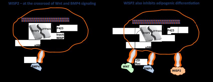
Mechanistically, we found canonical WNT signaling/WISP2 to regulate
adipogenic commitment and differentiation in two different ways; -
intracellular WISP2 retains the PPARγ transcriptional activator ZFP423 in a
cytosolic complex which, when dissociated by BMP4, allows nuclear entry of
ZFP423, induction of PPARγ and commitment into to the adipose lineage
and; - as a secreted molecule, WISP2 enhances cell proliferation and inhibits
5WISP2 – A Novel Adipokine Related to Obesity and Insulin Resistance
adipocyte differentiation by activating canonical WNT signaling and,
thereby, inhibiting PPARγ activation.
To investigate the effect of WISP2 in vivo, we generated a transgenic mouse
model overexpressing WISP2 in the adipose tissue under the aP2-promoter.
We found WISP2 to be secreted by the adipose tissue and present in serum.
The mice had a similar body weight but were characterized by improved
insulin sensitivity, increased circulating levels of adiponectin and the novel
FAHFA lipids and increased Glut4 in both adipose tissue and skeletal
muscle. They were also characterized by markers of increased mesenchymal
stem cell growth and development with a markedly expanded BAT, a
”healthy” hyperplastic subcutaneous adipose tissue and increased lean body
mass. Serum from the Tg mice also increased the proliferation of both brown
adipose precursor cells and the mesenchymal stem-like CH3T101/2 cells and
this was inhibited by adding specific anti-WISP2 monoclonal antibodies to
the serum.
Taken together, WISP2 is a novel secreted autocrine/endocrine regulator of
mesenchymal stem cell growth and proliferation as well as their adipogenic
commitment. There is important cross-talk between WISP2 and BMP4 in the
regulation of adipogenic commitment and differentiation and BMP4 is also a
regulator of WISP2 transcriptional activation. WISP2 is a novel target in
hypertrophic obesity and the Metabolic Syndrome.
Keywords: Adipose tissue; BMP4; Canonical WNT pathway; Insulin
Resistance; Obesity; PPARγ; Type 2 Diabetes; WISP2
ISBN: 978-91-628-9283-8 (print)
ISBN: 978-91-628-9323-1 (epub)
http://hdl.handle.net/2077/37992 Gothenburg 2015
6John Grünberg
SAMMANFATTNING PÅ SVENSKA
Typ 2 diabetes ökar med epidemisk hastighet världen över och förväntas nå 592
miljoner drabbade år 2035, jämfört med dagens 382 miljoner (6,4 % av den svenska
befolkningen).
Insulinresistens är ett förstadium till typ 2 diabetes och innebär en nedsatt förmåga att
svara på insulin som till exempel att ta upp socker från blodet. Insulinresistens
orsakas av ett samspel mellan genetiska och omgivningsfaktorer såsom övervikt,
ohälsosam livsstil och rökning.
En starkt bidragande orsak till insulinresistens och typ 2 diabetes är en allt mer
utbredd övervikt hos befolkningen. Idag är ca hälften av alla vuxna män, en tredjedel
av alla kvinnor och vart femte barn överviktiga eller obesa. Många upplever övervikt
som ett problem och det ökar även risken för flertalet sjukdomar såsom typ 2
diabetes . Ca 85 % av alla som drabbas av typ 2 diabetes är överviktiga eller obesa,
men ca 30 % av obesa individer är metaboliskt friska. Orsaken till varför vissa
överviktiga individer, men inte alla, drabbas av metabola komplikationer är bland
annat kopplat till en nedsatt mognad och funktion av fettvävnaden varvid det bildas
få, men stora fettceller, så kallad hypertrofisk fettansamling. Detta leder i sin tur till
att fett ansamlas på platser i kroppen som normalt inte lagrar fett, dvs ektopisk
fettansamling, vilket inkluderar fettansamling i buken, levern, skelettmuskel, hjärtat
och runt blodkärlen. Detta orsakar lipotoxicitet med flera metabola komplikationer,
inklusive insulinresistens.
För att motverka hypertrofisk fettansamling krävs nybildning av fettceller när
behovet att lagra fett ökar. Detta sker från så kallade mesenkymala stamceller som
kan ge upphov till både fettceller, muskelceller, benceller och broskceller. Första
steget kallas ”comittment”, och innebär att stamcellerna bara kan utvecklas till en av
dessa typer av celler. Nästa steg inom utvecklingen av fettceller innebär att de
omogna cellerna genomgår differentiering och utvecklas till mogna fettceller med
förmåga att lagra fettsyror och utsöndra olika proteiner, så kallade adipokiner. Dessa
processer är komplicerade och strikt reglerade genom ett samspel av flera olika
molekylära signaleringsvägar. WNT signalen är en signaleringsväg av stor betydelse
för fettcellernas differentiering och som måste stängas av för att tillåta att
mesenkymala stamceller mognar ut till fettceller.
Vi har nyligen identifierat ett nytt sekretoriskt protein, WISP2, som framför allt finns
och utsöndras av mesenkymala stamceller och som aktiveras av WNT-signaleringen.
Det faktum att WISP2 utsöndras till blodet gör att det kan vara en viktig
kommunikatör mellan fettväven och andra vävnader och därmed är extra intressant.
Vi har visat att förekomsten av WISP2 i fettväven kan kopplas samman med flera
riskfaktorer för typ 2 diabetes, att dess förekomst ökar i stora ”hypertrofa” fettceller
samt är relaterat till nedsatt insulinkänslighet.
Förmågan att differentiera human fettceller skiljer sig åt mellan individer och detta är
försämrat vid förekomsten av ”hypertrof” fetma och insulinresistens. Vi har nu visat
att en bibehållen aktivering av WNT-signalen och en ökad förekomst av WISP2 är
kopplat till en nedsatt mognadsgrad av fettcellerna. Vidare har vi visat att WISP2 kan
7WISP2 – A Novel Adipokine Related to Obesity and Insulin Resistance
förhindra fettcellsutmognaden och på så sätt bidra till den nedsatta fettvävsfunktion
som är kopplad till metabola komplikationer. Vi har funnit att det är två
underliggande molekylära mekanismer som regleras av WISP2 och som involverar
reglering av såväl ”comittmentsteget” som differentieringen via bland annat de
regulatoriska proteinerna ZNF432 och BMP4.
För att ytterligare studera effekten skapade vi en transgen musmodell med ett
fettvävsspecifikt överuttryck av WISP2. De transgena mössen visade sig vara
skyddade mot de negativa metabola förändringar som en högfett-diet med påföljande
fetma normalt leder till. Flera faktorer visade sig vara inblandade i den skyddande
effekten av WISP2, vilka till stor del verkar vara kopplade till en ökad förmåga hos
de transgena mössen att rekrytera och differentiera mesenkymala stamceller.
Förutom en ökad muskelmassa hade de transgena mössen väsentligt mer brunt,
energibrännande fett och en så kallad ”frisk” och väl differentierad vit fettväv med
många små celler (hyperplastisk fettväv) istället för den mer ofördelaktiga,
hypertrofa fettväven. Vidare fann vi att insulinkänsligheten och glukosupptaget var
förbättrat i både fettväven och skelettmuskulaturen och att detta var kopplat till en
ökad sekretion av adiponectin, ett protein som frisätts av fettväven och som tidigare
visats vara associerat med förbättrad insulinkänslighet, samt ett ökat uttryck av
glukostransportören GLUT4. Ytterligare ett intressant fynd som kunde påvisas var att
de transgena mössen uppvisade ökade nivåer av de nyligen identifierade gynnsamma
fettsyrorna FAHFA som både förbättrar insulinsekretion och insulinkänslighet och
som är anti-inflammatoriska.
Merparten av de förändringar som dokumenteras i de transgena mössen kan förklaras
genom en ökad förmåga att rekrytera mesenkymala stam celler. Denna hypotes
kunde vi bekräfta genom att serum från de transgena mössen ledde till en ökad
tillväxt av mesenkymala celler in vitro och att effekten försvann då vi tillsatte
blockerande, specifika WISP2 antikroppar.
Sammantaget, presenteras i denna avhandling bevis för att WISP2 är en ny adipokin
som utsöndras från fettväven och som har möjlighet att påverka celler i dess
omgivning med huvudmål att påverka mesenkymala stamcellers tillväxt och
utmognad. Detta leder också till en förbättrad och insulinkänslighet och metabolism.
WISP2s förmåga att reglera mesenkymala stamceller är en intressant upptäckt som
också kan leda till ny läkemedelsutveckling mot fetma och typ 2 diabetes.
8John Grünberg
LIST OF PAPERS
This thesis is based on the following studies, referred to in the text by their
Roman numerals:
I. Hammarstedt A, Hedjazifar S, Jenndahl L, Gogg S,
Grünberg JR, Gustafson B, Klimcakova E, Stich V,
Langin D, Laakso M, Smith U. WISP2 regulates
preadipocyte commitment and PPARγ activation by
BMP4
Proceedings of the National Academy of Sciences of the
United States of America 2013; 110(7): 2563-2568
II. Grünberg JR, Hammarstedt A, Hedjazifar S, Smith U. The
novel secreted adipokine WNT1-inducible signaling
pathway protein 2 (WISP2) is a mesenchymal cell
activator of canonical WNT
Journal of Biological Chemistry 2014; 289(10), 6899-6907
III. Grünberg JR, Hoffmann JM, Hedjazifar S, Nerstedt A,
Jenndahl L, Castellot J, Wei L, Movérare Skrtic S, Bäckhed
F, Syed I, Saghetelian A, Kahn B, Hammarstedt A, Smith U.
Increased brown fat and insulin sensitivity in obese mice
overexpressing WISP2 in the adipose tissue
Manuscript
9WISP2 – A Novel Adipokine Related to Obesity and Insulin Resistance
CONTENT
ABBREVIATIONS ............................................................................................ 11
1
INTRODUCTION ......................................................................................... 13
1.1
Prevalence of type 2 diabetes .............................................................. 13
1.2
Obesity, type 2 diabetes mellitus and insulin resistance ..................... 13
1.3
Metabolic syndrome & abdominal obesity ......................................... 14
1.4
Adipose tissue distribution and metabolic complications ................... 15
1.5
Adipose tissue ..................................................................................... 16
1.5.1
Precursor cells in the adipose tissue ............................................ 16
1.5.2
Adipogenesis ............................................................................... 17
1.5.3
Brown adipose tissue ................................................................... 18
1.5.4
Beige adipose tissue .................................................................... 18
1.6
Canonical WNT................................................................................... 19
1.6.1
Canonical WNT signaling ........................................................... 19
1.6.2
WISP2 .......................................................................................... 21
1.6.3
CCN-family & structure .............................................................. 21
1.6.4
WISP2 in human disease ............................................................. 22
2
AIM ........................................................................................................... 23
3
METHODS.................................................................................................. 24
3.1
Ethical statement ................................................................................. 24
3.2
Subjects and samples ........................................................................... 24
3.3
Isolation of adipocytes ........................................................................ 25
3.4
Cell culture experiments...................................................................... 25
3.5
Animal experiments ............................................................................ 27
3.6
Quantitative validation of mRNA and proteins................................... 31
3.7
Statistical Analyses ............................................................................. 33
4
SUMMARY OF RESULTS ............................................................................. 34
4.1
Paper I ................................................................................................. 34
4.2
Paper II ................................................................................................ 35
4.3
Paper III ............................................................................................... 37
5
DISCUSSION .............................................................................................. 39
5.1
WISP2 is associated with markers of Metabolic Syndrome ............... 39
5.2
WISP2 and adipogenesis ..................................................................... 40
5.3
WISP2 signaling .................................................................................. 41
5.4
WISP2 regulation ................................................................................ 42
5.5
WISP2 in vivo ..................................................................................... 43
6
CONCLUSION ............................................................................................ 47
ACKNOWLEDGEMENT .................................................................................... 48
REFERENCES .................................................................................................. 50
10John Grünberg
ABBREVIATIONS
2DOG 2[14C]deoxyglucose
2DOG-6P 2[14C]deoxyglucose-6-phosphate
aP2 Adipocyte protein 2
APC Adenomatous polyposis coli
α-SMA α -smooth muscle actin
BAT Brown adipose tissue
BCA Body composition analysis
BMI Body mass index
BMP Bone morphogenetic protein
BrdU Bromodeoxyuridine
CCN CTGF, Cyr61, Nov family
cDNA Complementary DNA
cEBP C/CAAT enhancer-binding protein
ChREBP Carbohydrate-responsive-element-binding protein
CTGF Connective tissue growth factor
Cyr61 Cysteine-rich angiogenic inducer 61
DEXA Dual energy X-ray absorptiometry
DKK1 Dickkopf 1
DNL De novo lipogenesis
DVL Dishevelled
EDL Extensor digitorum longus muscle
ELISA Enzyme-linked immunosorbent assay
ERK Extracellular signal-regulated kinases
eWAT Epididymal white adipose tissue
FABP4 Fatty acid binding protein 4
FAHFA Fatty acid esters of hydroxy fatty acids
FDR First-degree relative
FFA Free fatty acids
FZD Frizzled
GIR Glucose infusion rate
GLUT4 Glucose transporter type 4
GSK3β Glycogen synthase kinase 3 beta
GTT Glucose tolerance test
HFD High fat diet
HIF Hypoxia inducible factor
hMSC Human mesenchyme stem cells
IBMX Isobutylmethylxanthine
IHC Immunohistochemistry
IP Immunoprecipitation
ITT Insulin tolerance test
11WISP2 – A Novel Adipokine Related to Obesity and Insulin Resistance
JNK c-Jun N-terminal kinases
KRM Kremen 1/2
LFD Low fat diet
LRP5/6 Low-density lipoprotein-receptor-related protein-5 or -6
MAPK Mitogen-activated protein kinases
NFkB Nuclear factor kappa-light-chain-enhancer of activated B cells
NOV Nephroblastoma overexpressed
PDGF Platelet-derived growth factor
PPARγ Peroxisome proliferator activator receptor gamma
PTT Pyruvate tolerance test
qRT-PCR Quantitative real-time polymerase chain reaction
RQ Respiratory exchange quotient
SAT Subcutaneous adipose tissue
SDS Sodium dodecyl sulfate
sFRP Secreted Frizzled-related protein
SREBP-1 Sterol regulatory element-binding protein 1
SVF Stromal vascular fraction
sWAT Subcutaneous white adipose tissue
T2D Type 2 diabetes mellitus
TBX1 T-box protein 1
TCF/LEF T-cell factor/lymphoid enhancer factor
TCP7L2 Transcription factor 7-like 2
Tg Transgenic mice
TGFβ Transforming growth factor beta
TMEM26 Transmembrane protein 26
TNFα Tumor necrosis factor alpha
TSP1 Thrombospondin-1
TZD Thiazolidinedione
UCP-1 Uncoupling protein-1
UPR Unfolded protein response
VCO2 Carbon dioxide production
VO2 Oxygen consumption
VSMC Vascular smooth muscle cells
VWC von Willebrand factor type C domain
WAT White adipose tissue
WIF1 WNT inhibitory factor 1
WISP WNT1 inducible signaling pathway protein
WNT Wingless-type MMTV integration site family members
wt Wildtype mice
ZFP423 Zinc-finger protein 423
12John Grünberg
1 INTRODUCTION
1.1 Prevalence of type 2 diabetes
Type 2 diabetes mellitus (T2D) is increasing worldwide at an epidemic rate
and is expected to reach 592 million inflicted individuals by 2035 as
compared to 382 million in 2013 (6,4% of the Swedish population) and
where the vast majority lives in low- and middle-income countries. The
health cost for diabetes was expected in 2010 to be 12% of the world’s total
health expenditure and in Sweden the cost per person with diabetes was in
2010 predicted to be more than 4000 USD. The global health expenditures
for diabetes in 2030 will be 30-34% larger than those of 2010. These results
show that this epidemic imposes a major economical burden on the society
worldwide and prevention efforts are needed (1,2).
1.2 Obesity, type 2 diabetes mellitus and
insulin resistance
Obesity in both men and women is associated with a greater risk of
developing chronic diseases like T2D, hypertension, cancer and heart disease
and it is increased with increasing body mass index (BMI). World Health
Organization has categorized BMI as: underweight ≤18.5, healthy weight
range ≤ 24.9, overweight ≤ 29.9 and obese as ≥ 30. Compared with healthy
weight, overweight/obese men and women have a 3.5-4.6/10.0-11.2 relative
risk of developing diabetes over a 10-year period and more severe obesity
with a BMI > 35.0 has a 17.0-23.4 risk (3).
About 85% of patients with T2D are overweight or obese (4). However,
about 30% of obese individuals are metabolically healthy and, conversely,
around 20-30% of normal weight individuals develop these metabolic
abnormalities (5,6).
Insulin resistance is an essential component of T2D. Basically, this means
reduced insulin sensitivity, i.e., effect of insulin to lower plasma glucose by
suppressing hepatic glucose production and stimulating glucose
uptake/utilization in peripheral tissues. Insulin resistance leads to higher
glucose levels in the blood (hyperglycemia), which makes the glucose
sensing β-cells in the pancreas secrete more insulin to compensate for the
imbalance (hyperinsulinemia). Enhanced insulin secretion can compensate
for insulin resistance and enhanced insulin sensitivity can compensate insulin
secretory defect (7).
13WISP2 – A Novel Adipokine Related to Obesity and Insulin Resistance
Obesity is associated with toxic cellular effects such as increased
inflammation, ER-stress, production of reactive oxygen species,
mitochondrial dysfunction, ectopic accumulation of lipids/triglycerides and
activation of serine-threonine kinases.
The inflammation is a low-grade chronic inflammation that can be considered
as an abnormal immune reaction triggered by nutrients or other intrinsic cues.
Usually it is referred to as meta-inflammation or para-inflammation and it
occurs in metabolically important organs such as liver and adipose tissue.
Together, these responses contribute to the insulin resistance in the liver,
skeletal muscle, adipose tissue and β-cells. Obesity-induced metabolic
impairments then lead to a vicious cycle where excess nutrients trigger an
inflammatory response that enhances insulin resistance, placing a greater
demand on the β-cells. Eventually, this and other factors promote β-cell
dysfunction leading to insufficient insulin secretion and hyperglycemia (8-
10).
1.3 Metabolic syndrome & abdominal
obesity
The metabolic syndrome is a collection of risk factors that together are
associated with a higher risk for cardiovascular diseases and T2D. These risk
factors are elevated plasma glucose, dyslipidemia, hypertension, a
prothrombotic profile, and a state of inflammation. Elevated fasting or
postprandial plasma glucose fall under the range of either pre-diabetes or
diabetes. Dyslipidemia includes elevated very low-density lipoprotein-
triglycerides and decreased high-density lipoproteins. A prothrombotic
profile suggests impairments in procoagulant factors, anti-fibrinolytic factors,
platelet abnormalities and endothelial dysfunction. An inflammatory state is
illustrated by increased circulating cytokines and acute phase reactants
(11,12).
The major underlying risk factors of the metabolic syndrome are obesity and
insulin resistance. Risk association with obesity is measured as waist
circumference, and not primarily BMI, to assess visceral/abdominal obesity.
Therefore, it is not only degree of obesity that influences the risk of
metabolic disturbances but also where the fat is accumulated. Excess visceral
adipose tissue, reflected as increased abdominal girth or waist-to-hip ratio, is
an important factor for the correlation between metabolic aberrations and
obesity rather than the amount of subcutaneous abdominal fat (7,8,12,13).
This has raised the question about the difference between different adipose
tissue depots.
14John Grünberg
1.4 Adipose tissue distribution and
metabolic complications
Human adipocytes can expand about 20 fold in diameter and several 1000-
times in volume (5) and the subcutaneous cell size can differ markedly
between individuals with the same BMI and amount of fat. The number of
cells in the adipose tissue is set after puberty with a 10% annual replacement
and the turnover rate is lower in individuals with enlarged cells (14-16). The
subcutaneous adipose tissue has a limited expandability and when the
subcutaneous adipose tissue expands, due to excess energy intake, it can be
accomplished in two principally different ways; either by expanding the
existing adipocytes (hypertrophy) or by recruiting new cells (hyperplasia).
Hypertrophic obesity is associated with local inflammation and a
dysregulated and insulin- resistant adipose tissue. When the subcutaneous
adipose tissue storage capacity is exceeded, lipids will accumulate in non-
adipose organs, i.e., the so-called ectopic fat accumulation. This includes
intra-abdominal and visceral areas, liver, skeletal muscle, heart and around
vessels (5,12,16,17). Importantly, a genetic predisposition for T2D, defined
as being a first-degree relative (FDR) to individuals with T2D, is associated
with inappropriate hypertrophy of abdominal subcutaneous adipose cells
even in non-obese individuals indicating an impaired subcutaneous
adipogenesis (18).
The increased size of the adipose cells triggers release of stress signals and
hypoxia can occur when vascularization is insufficient for the growing
adipose tissue. ER-stress is triggered by hypoxia or excess nutrients, leading
to an unfolded protein response (UPR). In the ER, proteins are translated,
folded and checked for quality before they are released. These functions are
decreased during ER-stress, and the number of misfolded proteins increases.
This triggers the UPR, which activates genes involved in producing, folding,
modifying and degrading proteins to decrease the ER-stress. UPR also
triggers activation of stress and inflammatory pathways and production of
cytokines that alters the insulin-signaling pathway (5,9). Stressed and
enlarged adipocytes also attract different immune cells including
macrophages. This leads to a positive feedback loop where infiltrating
macrophages recruit more immune cells and introduce a chronic state of
inflammation, the meta-inflammation pathway. Dysfunctional, hypertrophic
adipose tissue produces more inflammatory cytokines such as tumor necrosis
factor alpha (TNFα) and interleukin-6. Some of these cytokines contribute to
the insulin resistance and defect adipose tissue function (5,9,10,12,19).
15WISP2 – A Novel Adipokine Related to Obesity and Insulin Resistance
Dysfunctional fat in hypertrophic obesity is also associated with increased
fibrosis of the adipose tissue, which also causes activation of stress-related
pathways, inflammation and ectopic lipid accumulation (20).
The visceral fat is less insulin-sensitive and lipolytically more active than
subcutaneous fat and is therefore considered to release more free fatty acids
(FFA). Elevated FFAs in the peripheral circulation can interfere with the
insulin signaling pathway in target tissues leading to increased insulin
resistance (7,12,13). However, it is probably not the FFA per se that enhance
insulin resistance but rather their metabolites such as long-chain acyl-
coenzyme A, diacylglycerol and ceramides (5).
Figure 1.
Adipocyte-hypertrophy-and-associated-characteris:cs-
Dyslipidemia-and-insulin-resistance-
Increased-lipid-storage- Perivascular-fat-
Reduced-
Ectopic-lipid-accumula:on-
-demand-
• Adipogenesis-
IR,-obesity,- • Glucose-uptake- Cardial-fat-
FDR- Increased-
• AT-insulin-resistance-
Liver-fat-
• Inflamma:on-
• Lipolysis-
Altered- Intra&abdominal-fat-
• Secre:on-of-adipokines-
Mesenchymal- - Muscle-fat-
precursor-recruitment-
Figure 1. Adipocyte expansion with a dysregulated subcutaneous adipose tissue
(SAT) promotes ectopic fat accumulation and the Metabolic Syndrome. Adipocyte
hypertrophy characterizes the SAT of insulin-resistant (IR) obesity and first-
degree relatives (FDR) of individuals with type 2 diabetes (Modified from(21)).
1.5 Adipose tissue
1.5.1 Precursor cells in the adipose tissue
The adipose tissue in mammals mainly consists of 2 types of fat: white
adipose tissue (WAT) and brown adipose tissue (BAT). There are also mixed
areas known as brown in white “brite” or beige adipose tissue. WAT and
BAT displays many similarities but WAT mainly stores excess energy,
whereas BAT generates heat through mitochondrial uncoupling of oxidation
(10,20,22).
The adipose tissue does not only consist of adipocytes and its precursor
preadipocytes, but also mesenchymal stem/precursor cells, immune cells,
fibroblasts and vascular cells. Adipocytes develop from multipotent
mesodermal stem cells residing in the adipose tissue. This process, i.e.
16John Grünberg
adipogenesis, can be divided into two related steps. Firstly, during
determination/commitment the mesenchymal stem cells loose their ability to
differentiate into other mesenchymal linages and become committed to
preadipocytes. They are now no longer able to transform into osteoblasts,
myocytes or chondrocytes. Secondly, the preadipocytes differentiate to
become mature adipocytes, acquiring lipids droplets and gain the ability to
respond to hormones such as insulin and catecholamines (20,23,24).
1.5.2 Adipogenesis
In order to commit mesenchymal stem cells to the adipocyte linage, the bone
morphogenetic protein (BMP) family member 4 plays a key role (25-27).
BMP is part of the transforming growth factor beta (TGFβ) superfamily and
signals through BMP receptors 1 and 2, which phosphorylate SMAD1/5/8.
Phosphorylated SMAD1/5/8 forms a complex with SMAD4 that translocates
to the nucleus and activates specific genes. BMPs have many cellular
antagonists and, for instance, Noggin can block differentiation to the
adipocytic linage by binding to BMP4 and prevent receptor activation
(22,25,28). BMP4 induces adipogenic commitment by binding to zinc-finger
protein 423 (ZFP423), a transcriptional activator of peroxisome proliferator
activator receptor gamma (PPARγ), via a SMAD-binding domain (29).
Committed preadipocytes have the potential to be terminally differentiated to
mature lipid-accumulating adipocytes. This process involves a series of well-
characterized steps which have been extensively studied in vitro. This
includes mitogenic clonal expansion followed by a well-coordinated
activation of several transcription factors and where cEBPβ and cEBPδ are
upregulated followed by PPARγ together with C/CAAT enhancer-binding
protein alpha (cEBPα). PPARγ activates the promoter of cEBPα, and vice
versa, which creates a positive feedback loop in order to maintain the
differentiated state. This is followed by expression of genes that characterize
the mature adipose phenotype and are involved in insulin sensitivity,
lipogenesis and lipolysis such as lipoprotein lipase, adipocyte protein 2 (aP2)
/ fatty acid binding protein 4 (FABP4), the insulin receptor and glucose
transporter type 4 (GLUT4). Sterol regulatory element-binding protein 1
(SREBP-1) is also activated by cEBPβ and cEBPδ, which regulates lipogenic
genes and can activate PPARγ by enhancing expression as well as promoting
production of endogenous ligand. However, the identification of a definitive
endogenous PPARγ agonist has not yet been successful (22,30-33).
Some inhibitors of adipogenesis are proinflammatory molecules such as
TNF-α (10,28,31), growth factors such as platelet-derived growth factor
(PDGF) and connective tissue growth factor (CTGF) (31,34). Many of these
inhibitory effects are mediated through mitogen-activated protein kinases
17WISP2 – A Novel Adipokine Related to Obesity and Insulin Resistance
(MAPK) including extracellular signal-regulated kinases (ERK) and c-Jun N-
terminal kinases. ERK1 is necessary for the proliferative phase of
differentiation but needs to be downregulated during the terminal
differentiation stage (30,31). MAPK are also regulated by the canonical
wingless-type MMTV integration site family members (WNT) signaling
(35).
1.5.3 Brown adipose tissue
BAT consists of highly specialized cells which waste energy through heat
production in the mitochondria. BAT contains many tightly packed
mitochondria where the BAT-specific protein, uncoupling protein-1 (UCP-1),
catalyzes a proton leak from the inner membrane which uncouples substrate
oxidation from ATP-synthesis.
The most powerful and physiological stimulus to activate BAT is cold
exposure or hormonal stimuli, like β-adrenergic agonists. (36-38).
BAT is present in all placental mammals and human babies have brown fat
depots which gradually reduce in size with aging. However, recent data
showed that classical BAT with UCP-1 positive cells does exist in the
supraclavicular and spinal regions of adult human. Human BAT consists of
both brown and white fat cells and data indicates that the brown adipose
tissue found in adult man has the molecular markers resembling murine beige
fat more closely than classical brown fat (37,39-41).
1.5.4 Beige adipose tissue
Within the white adipose tissue, a new type of adipose cells has been found.
They have been named beige or brite (brown in white) cells and have a
brown-like phenotype but probably a common origin with the white adipose
cells. Beige adipose cells have the characteristics of dissipating energy
through generation of heat like BAT, when stimulated. Beige cells are similar
to, but not identical to BAT cells, with lower levels of BAT genes such as
UCP-1, low uncoupled respiration and larger/unilocular lipid morphology
comparable to white adipose cells (20,41,42). Inducible browning effect
seems to be reversible; beige adipose cells can switch from being energy
storing to become energy-dissipating and back again, depending on
environmental conditions or stimuli (42-44).
18John Grünberg
1.6 Canonical WNT
1.6.1 Canonical WNT signaling
The canonical WNT pathway inhibits mesenchymal stem cells from
committing to the adipocyte lineage and terminal differentiation. WNT
signaling also restrains adipocyte differentiation by inhibiting the expression
of PPARγ and cEBPα. These transcription factors are induced directly by
cEBPβ and cEBPδ in response to adipogenic stimuli, which also serve to
switch off the canonical WNT pathway (23,45-47).
Canonical WNT signaling regulates mesenchymal stem cell fate. Activation
of WNT signaling promotes entry of mesenchymal precursor cells into the
myocyte and osteocyte lineages while suppressing commitment to the
adipocytic lineage and terminal differentiation (23,45,46,48).
Myoblasts
Insulin sensitivity
Lipid accumulation
Osteoblasts Adipogenic stimuli C/EBPs/PPARγ Adipokine secretion
+Wnt - Wnt
Commitment% Differen0a0on%
+ Wnt
Mesenchymal Pre-adipocytes Mature adipose cells
stem cells BMP4% BMP4%
Figure 2. Schematic figure over the regulation of adipogenesis from mesenchymal
stem cells and other uncommitted precursor cells. WNT silencing and BMP4
activation are required for adipogenic commitment and differentiation of
preadipocytes into mature adipose cells by activation of transcription factors of
the C/EBP family and the key regulator of adipogenesis, PPARγ (Modified from
(16)).
The name WNT comes from a discovery in Drosophila where it was found
that the polarity gene Wingless and the proto-oncogene Int-1 had a common
origin, the WNT signaling pathway. The WNT signaling cascade controls a
multitude of biological processes during development and adult life,
especially stem cell biology. An abnormal WNT signaling underlies a wide
range of pathologies in humans; for example cancer, osteoporosis and
metabolic diseases. Single nuclear polymorphisms in WNT5B, WNT10B and
transcription factor 7-like 2 (TCF7L2, formerly called TCF4) are all linked to
an increased risk for T2D; TCF7L2 plays an important role in downstream
signals of the canonical WNT pathway (49,50).
In the mammalian WNT family, there are nineteen members who are
expressed in both embryos and adults. The WNT proteins are about 40 kDa
19WISP2 – A Novel Adipokine Related to Obesity and Insulin Resistance
and are cytosine-rich glycoproteins that in their active form bind locally to
cellular receptors. WNT proteins are lipid-modified in order to be secreted
from the cell and bind to a receptor; one of these is palmitoleic acid, a mono-
unsaturated fatty acid attached to a conserved serine (49,51). WNT molecules
affect cell proliferation, survival, fate and behavior by signaling through
different “canonical” and “non-canonical” pathways. Cytosolic β-catenin is
fundamental for the canonical pathway and, therefore, it is often referred to
as “WNT/ β-catenin dependent” and non-canonical pathway as “WNT/ β-
catenin independent”. In this thesis, only the canonical pathway will be
discussed.
Cells can release or present WNT proteins in an autocrine or paracrine
manner by binding to the cell-surface receptors of the Frizzled (FZD) family
and the low-density lipoprotein-receptor-related protein-5 or -6 (LRP5/6) co-
receptors. In the absence of WNT molecules, cytoplasmic β-catenin is
recruited to a destruction complex consisting of Axin, adenomatous polyposis
coli (APC) protein and glycogen synthase kinase 3 beta (GSK3β). This
results in ubiquitination of cytosolic β-catenin followed by proteosomal
degradation.
Normally, the cellular β-catenin levels are low but when WNT proteins bind
to the cell-surface receptors of the FZD family and LRP5/6, Axin is recruited
to the phosphorylated cytoplasmic tail of LRP6. This phosphorylation is
regulated by GSK3 and CK1γ. Another cytoplasmic protein that is activated
is Dishevelled (DVL), which interacts with the cytoplasmic part of FZD and
Axin, facilitating an interaction between the LRP tail, Axin and FZD. This
leads to inhibition of the Axin/GSK/APC complex, also called β-catenin
destruction complex, resulting in higher concentrations of stabilized
cytoplasmic β-catenin and its nuclear translocation. In the nucleus, β-catenin
interacts with the T-cell factor/lymphoid enhancer factor (TCF/LEF) family
of transcription factors and promotes specific gene expression to regulate the
transcription of WNT target genes, many of which are associated with cell
proliferation and cell fate decision (23,35,47,49).
WNT-signaling can be inhibited by a number of extracellular antagonists
acting in different ways. Secreted Frizzled-related protein (sFRP) 1 and 2,
and WNT inhibitory factor 1 (WIF1) bind to the WNT proteins and, thereby,
inhibit activation. Dickkopf 1 (DKK1) prevents the formation of the
LRP/FZD receptor complex by binding with high affinity to LRP5/6 and
Kremen 1/2 (KRM). This leads to specific inhibition of canonical WNT
signaling by endocytosis and removal of the WNT receptors (52). WNT
signaling is highly activated in precursor cells and needs to be downregulated
for activation of adipogenesis (24,53,54). Inappropriate WNT activation is
related to a poor adipogenesis and seen in obese patients with hypertrophic
obesity (26).
20John Grünberg
WNT$signaling+
WNT Proteasomal+
OFF+ degrada4on+
LRP
Frizzled
Axin
DVL
B-catenin
APC
ON+ Target genes
B-catenin
Tcf/Lef
Figure 3. Canonical WNT-signaling.
1.6.2 WISP2
One of the many genes activated by the canonical WNT signaling is the
WNT1 inducible signaling pathway protein 2 (WISP2 also known as CCN5)
(55,56). Wisp2 has been shown to only be activated by the canonical WNT
and not non-canonical WNT signaling. WISP2 has a molecular weight of
27.5 kDa and the homology between mouse and human WISP2 is 73%
(57,58).
1.6.3 CCN-family & structure
Human and mouse WISP2 are homologues to the rat gene rCop1 and belong
to the CCN family of growth factors. The family consists of 6 members;
21WISP2 – A Novel Adipokine Related to Obesity and Insulin Resistance
CTGF or CCN1, cysteine-rich angiogenic inducer 61 (Cyr61 or CCN2),
nephroblastoma overexpressed (NOV or CCN3), and WISP 1-3 (CCN4-6).
The CCN family of proteins is essential for embryonic development and
plays important roles in inflammation, wound healing, and injury repair in
the adult. Many are considered to be involved in the pathogenesis of fibrosis,
artherosclerosis and cancers (57,59-65).
The CCN family of proteins contains, apart from WISP2, of 4 conserved
cysteine-rich domains which display homology to conserved regions in a
variety of extracellular proteins. All CCN proteins have an N-terminal
signaling peptide, important for secretion (66). Module 1 is an insulin-like
growth factor-binding domain and Module 2 is a von Willebrand factor type
C domain (VWC) that may participate in protein complex formation. Module
3; the thrombospondin-1 domain (TSP1), involved in the binding to sulfated
glycosaminoglycans either on the cell surface or in the extracellular matrix.
Module 4 does not exist in WISP2 but is a cysteine-knot-containing module
recognized by many growth hormones and may participate in dimerization
and receptor binding. Between the VWC domain (Module 2) and the TPS1
domain (Module 3) there is a non-conserved central hinge-region, suggesting
possible translational processing by proteolytic digestion. Considering the
structure of CCN proteins, it not much of a surprise that they are involved in
many essential biological functions (57,66-68).
1.6.4 WISP2 in human disease
WISP2 is a secreted protein (69) and has been reported to have various
effects in different cancers. It has both growth-promoting and growth-
arresting properties depending on cell types and environment of the cells.
WISP2 has also been suggested to have potential tumor-suppressive
properties in colorectal cancer, breast cancer and bone metastases (70-75).
Wisp2 expression is unaltered during the osteogenic and chondrogenic
differentiation of mesenchymal stem cells, but downregulated during
adipogenic differentiation (74,76).
Many of the studies of WISP2 have been focused on osteoblasts, myocytes or
chondrocytes. However, mRNA expression of both human and mouse tissues
showed that WISP2/Wisp2 is by far most highly expressed in the adipose
tissue (77) and activation of canonical WNT inhibits adipogenic
differentiation and upregulates Wisp2 (55,56). The secretome of the human
adipose tissue showed that WISP2 is an adipokine, i.e.; secreted by the
adipose tissue. Furthermore, WISP2 expression is increased in obesity and, in
particular, in the subcutaneous adipose tissue (78).
22John Grünberg
2 AIM
The overall aim of this thesis is to characterize effects and molecular
mechanisms for the novel “adipokine” WISP2 in the regulation of
mesenchymal precursor/stem cell growth and adipogenic commitment and
the association with hypertrophic obesity and its metabolic consequences,
i.e.; insulin resistance and Type 2 diabetes.
The specific aims:
Paper I. To investigate WISP2 in human hypertrophic adipose tissue and
its involvement in the regulation of adipogenic commitment by
BMP4.
Paper II. To characterize the signaling mechanisms for WISP2 and its
effects on adipogenesis and adipocyte differentiation.
Paper III. To evaluate the effects of WISP2 activation in the adipose tissue
in vivo using a transgenic mouse model.
23WISP2 – A Novel Adipokine Related to Obesity and Insulin Resistance
3 METHODS
3.1 Ethical statement
Informed consent was obtained from all subjects after the purpose and the
potential risks of the study were explained. The study protocols were
approved by the Ethics Committees of the University of Gothenburg, Charles
University (Prague), and the University of Kuopio and were in accordance
with the Declaration of Helsinki.
All animal experiments were performed after prior approval from the local
Ethics Committee for Animal Studies at the Administrative Court of Appeals
in Gothenburg, Sweden.
3.2 Subjects and samples
Nondiabetic subjects
Thirty-six healthy, nondiabetic subjects (Gothenburg cohort) were recruited.
Inclusion criteria were two first-degree relatives with type-2 diabetes or one
first-degree relative and two second-degree relatives, normal glucose
tolerance, fasting triglyceride concentration < 2.0mM, and no evidence of
hypertension, endocrine disease or metabolic disease. Subcutaneous adipose
tissue biopsies were obtained by needle aspiration from lower part of the
abdomen after local dermal anesthesia with lidocaine. Biopsies were
transferred to the laboratory for immediate processing.
Subcutaneous and Visceral Adipose Tissue Arrays of Subjects
Individuals scheduled to have abdominal surgery (Prague cohort) were
monitored and 53 women [age 21–66 y, body mass index (BMI) 17.3–48.5
kg/m2] were included. According to BMI and presence or absence of the
metabolic syndrome evaluated according to the International Diabetes
Federation criteria (79) participants were assigned into one of the four groups
(lean, overweight, obese, or obese with metabolic syndrome). A clinical
investigation was performed 7–14d before the surgery. Anthropometric
measurements and euglycemic–hyperinsulinemic clamps (80) were
performed at rest after an overnight fast. Body composition was evaluated
using bioelectrical impedance. Visceral and subcutaneous fat areas were
derived from computed tomography scans at the level of L4–5. During the
surgical procedures, paired samples of subcutaneous abdominal and visceral
24John Grünberg
adipose tissue biopsies were obtained and processed immediately. The
samples were stored at−80 °C until analyzed.
Type-2 diabetic subjects
Ten drug-naive type-2 diabetic patients (Kuopio cohort) with mild diabetes
(fasting plasma glucose ≤ 8.0 mmol/L), four men and six postmenopausal
women, were recruited. Exclusion criteria were evidence of peripheral
vascular disease or heart disease, blood pressure ≥160/85 mm Hg, and
treatment with calcium channel blockers or nonsteroidal anti-inflammatory
drugs on a regular basis. No prior anti-diabetic treatment was allowed.
Subcutaneous adipose tissue biopsies were obtained by needle aspiration
from lower part of the abdomen. The biopsies were stored at−80 °C until
analyzed.
3.3 Isolation of adipocytes
The adipose tissue from human (Paper I) or mice (Paper III) biopsies were
handled according to referenced method. Briefly, adipose tissue were cut into
smaller pieces and digested with collagenase type II in Hank’s balanced salts
medium (pH 7.4) complemented with 4% BSA for 45-60 min at 37 °C in a
shaking water bath.
The cells were then filtered through a 250 µm nylon mesh and washed 4
times in Hank’s medium (without glucose when glucose uptake was
measured (Paper III) and average cell diameter measured. Remaining
isolated adipocytes where either; snap frozen in liquid N2 or stimulated for 15
min with or without insulin (1000μU/ml) and stored in lysate buffer for
protein extraction or used for glucose uptake measurements.
3.4 Cell culture experiments
Cells
To study the effect of WISP2 on commitment to the adipogenic phenotype in
vitro, NIH-3T3 fibroblasts, which do not have activated endogenous PPARγ,
were used (81).
The well-characterized preadipocyte cell line 3T3-L1 was
used to study the effect of WISP2 on adipocyte differentiation and mature
adipocytes. 3T3 L1 cells are an immortalized subclone of mouse 3T3
fibroblasts (82). Both cell lines where cultured in DMEM supplemented with
10% fetal bovine serum, 2 mM glutamine and 1 % antibiotics.
Human mesenchyme stem cells (hMSC) were also used and were cultured in
Mesenchymal Stem Cell Growth Medium.
25WISP2 – A Novel Adipokine Related to Obesity and Insulin Resistance
Human stromal vascular cells were extracted from abdominal subcutaneous
adipose tissue biopsies. The first non-floating cell fraction from the isolation
procedure contains the stromal vascular fraction (SVF). SVF were washed
and centrifuged to remove red blood cells and cultured in DMEM/F12 media
supplemented with 10% FBS, 2mM glutamine, 1% antibiotics until
confluence. Inflammatory cells (CD14- and CD45-positive), and early
progenitor cells (CD 133-positive) were removed by magnetic immune
separation.
Cell transfection
The main purpose of cellular transfection is to study the function of a gene by
overexpression/inhibition or to produce recombinant proteins (83). Foreign
nucleic acids can be introduced in the cell either by stable (long-term) or
transient (short-term) methods. To introduce the foreign DNA/RNA,
liposome-mediated transfection was used. In this thesis we transfected 3T3-
L1 preadipocytes stably with a vector carrying Wisp2 shRNA (Paper I) to
conditionally study the effect of WISP2 depletion during adipocyte
differentiation. For short-term experiments, we used siRNA or different
overexpressing plasmids. Detailed information is presented in Papers I, II.
Cell differentiation
NIH3T3 fibroblasts and 3T3-L1 preadipocytes were grown to confluence and
remained quiescent for 48h before induction of differentiation. To
differentiate these cell lines, an adipogenic differentiation cocktail was added
containing DMEM with a combination of insulin, dexamethasone and
isobutylmethylxanthine (IBMX). Insulin increases the number of lipid
droplets (84), the glucocorticoid, dexamethasone, induces the expression of
C/EBPδ and inhibits the antagonist of adipogenesis, Pref-1 (85), and IBMX,
a phosphodiesterase, enhances differentiation through increased cAMP
levels. To activate PPARγ in the NIH-3T3 fibroblasts, rosiglitazone, a
thiazolidinedione (TZD), was added to the cocktail. After 8 days, the
preadipocytes had become mature adipocytes with lipid droplets.
hMSC were grown until confluence after which three cycles of
induction/maintenance were performed according to manufacture’s
instructions. rhWISP2 or WNT3a where added to supplemented adipogenic
induction medium/adipogenic maintenance medium.
Oil-Red O staining
To determine lipid accumulation, cells were fixed with paraformaldehyde,
stained with Oil Red O and washed with PBS. Quantification of optical
density was determined by dissolving the Oil Red O stained cells in 2-
propanol and measured at λ 510 nm.
26John Grünberg
Immunofluorescence staining
Cells were grown on glass slides and treated with respective agents. Cells
were fixed with 4% paraformaldehyde and permeabilized with 0.1% Triton
X-100. Samples were blocked with 20% FCS or goat serum and incubated
with specific antibodies. After washing in PBS, samples where incubated
with a secondary antibody conjugated with a fluorophore to visualize the
proteins and DAPI staining was used to visualize nuclei. Samples were
analyzed with a Leica SP5 confocal microscope.
Proliferation assay
To evaluate cell proliferation, a bromdeoxyuridine (BrdU) proliferation assay
kit was used. BrdU incorporates in newly synthesized DNA by replacing
thymidine. Briefly, C3H10 T1/2 mesenchymal cells and brown adipose
precursor cells were cultured with mouse serum from Tg and wt mice and
with or without anti-WISP2 antibody (58) for 48h. Cells were then fixed and
stained with BrdU antibody according to the manufacturer’s instructions.
3.5 Animal experiments
Generation and genotyping of transgenic mice overexpressing aP2-
Wisp2
The aP2-Wisp2 transgenic mice were generated as stated in Paper III created
in the laboratory of Fatima Bosch (Universitat Autònoma de Barcelona,
Spain) using microinjection of oocytes from C57Bl6/SJL mice (86). WISP2
transgenic (Tg) founders were then bred to generate F1 Tg mice and
subjected to PCR analysis and Southern blot to check the transgene
expression. The Tg F2 offspring was generated by backcrossing the F1 Tg
mice with wild type (wt) C57BL/6NTac mice (Taconic) for 10 generations
and then inbred for 4-7 generations (B6N.SJL/J-Tg(aP2-Wisp2)92Fbos ;
N10,F4-F7).
Animals
Only male mice were used for phenotyping. Animals were weaned at 3 weeks
of age and housed 2–5/cage in a temperature-controlled (21°C) facility with a
12-h light-dark cycle with free access to chow food and water. From the age
of 6 weeks, age-matched male transgenic mice and wild-type littermates were
fed either pelleted high-fat diet, HFD (45 kcal% fat), pelleted control low fat
diet, LFD (10 kcal% fat) or kept on chow diet, CD (16% protein). The HFD
had the same amount of proteins (20 kcal%) and minerals as the LFD. They
only differ in the ratio carbohydrates/fat; HFD 35/45 and HFD 70/10. Chow
27WISP2 – A Novel Adipokine Related to Obesity and Insulin Resistance
diet contained 22% calories from proteins, 12% from fat and 66% from
carbohydrates.
Body weight and blood sampling
Total body weight was recorded weekly during the period of either 11 weeks
on HFD or 17 weeks on HFD or LFD. Fasting glucose (4h food withdrawal)
was measured using an Accu-Check glucometer and blood sampling was
performed every 4th week. Blood for measuring glucose was taken from the
tip of the tail vein using a scalpel and blood for measuring metabolites was
taken from the submandibular vein using Goldenrod Animal Lancet (87).
Taking submandibular blood has the advantage of getting a large amount
very fast as well as reducing the stress on the animals. When small amounts
of blood is needed (fasting glucose) or repeated blood sampling is needed
(GTT/ITT/PTT); tail vein blood sampling is preferred.
Blood was stored in serum tubes until centrifuged. The supernatant serum
was stored in -80 for further analyses. Saline solution (9 mg/ml) was given as
fluid replacement.
Necropsy
The mice were euthanized using 5% isofluorane with a mixture of air. Blood
was collected by heart puncture for analysis of metabolites. Tissues and
organs were weighed and stored. Epididymal white adipose tissue (eWAT),
subcutaneous white adipose tissue (sWAT) and brown adipose tissue (BAT)
were placed in 37°C sterile Hank’s balanced salts medium without
collagenase II (pH 7.4), snap-frozen in liquid nitrogen and stored in -80 for
further analyses or stored in 4% paraformaldehyde (PFA) for 2 days and then
in 70% ethanol.
EDL muscle for glucose uptake was placed in 37°C sterile Hank’s medium
without collagenase II and glucose (See Glucose uptake).
Open field activity test
Activity test was performed to study locomotor activity and food intake. The
test lasted 23h; 11 h consisted of daylight (150 lux, 10:00–19:00 and 07:00–
09:00) and 12 h of nightlight (20 lux, 19:00–07:00). The equipment consists
of an opaque box (50×50×22.5 cm) that has a lower and a higher row of
infrared sensors built into the walls connected to a control unit for tracking of
the mouse. The mouse was placed in the center of the box, and the test was
performed for 23 h, which allowed the mouse to acquaint itself with the open
field test chamber for the first 3 h. Locomotion (increased by 1 every time the
animal breaks a new beam) was recorded. The amount of food was measured
before and after the test and daily food intake per animal was calculated.
28You can also read