Autoreactive T cells in type 1 diabetes - Journal of Clinical ...
←
→
Page content transcription
If your browser does not render page correctly, please read the page content below
The Journal of Clinical Investigation                                                                                                                                             REVIEW

Autoreactive T cells in type 1 diabetes
Alberto Pugliese
Diabetes Research Institute, Department of Medicine, Division of Endocrinology, Diabetes, and Metabolism, Department of Microbiology and Immunology, Miller School of Medicine,
University of Miami, Miami, Florida, USA.

             Type 1 diabetes (T1D) is a chronic autoimmune disease that causes severe loss of pancreatic β cells. Autoreactive T cells are
             key mediators of β cell destruction. Studies of organ donors with T1D that have examined T cells in pancreas, the diabetogenic
             insulitis lesion, and lymphoid tissues have revealed a broad repertoire of target antigens and T cell receptor (TCR) usage, with
             initial evidence of public TCR sequences that are shared by individuals with T1D. Neoepitopes derived from post-translational
             modifications of native antigens are emerging as novel targets that are more likely to evade self-tolerance. Further studies
             will determine whether T cell responses to neoepitopes are major disease drivers that could impact prediction, prevention, and
             therapy. This Review provides an overview of recent progress in our knowledge of autoreactive T cells that has emerged from
             experimental and clinical research as well as pathology investigations.

Introduction                                                                                          HLA types (7). The ability to detect and phenotype autoreactive T
Type 1 diabetes (T1D) is a chronic autoimmune disease resulting                                       cells in circulation, where they are present at extremely low fre-
in severe loss of pancreatic β cells (1) due to the targeting of islet                                quencies, has greatly improved. For example, HLA-II tetramers or
cell autoantigens. Autoantibody and T cell responses to autoanti-                                     HLA-I multimers/monomers allow measurement of autoreactive
gens are detected in at-risk individuals during the asymptomatic                                      T cells in the circulation ex vivo, without in vitro amplification that
period preceding T1D diagnosis and at clinical onset. Autoantibod-                                    might alter phenotypic features (8–10).
ies are robust predictive and diagnostic biomarkers (2); autoreac-                                        This Review integrates experimental, human pathology, and
tive T cells are considered the main effectors of β cell destruction.                                 clinical research studies and identifies key outstanding questions
Accordingly, 50%–60% of the genetic risk for T1D derives from                                         related to autoreactive T cells in T1D.
HLA alleles encoding molecules involved in the presentation of
antigen peptides to T cells (3). An individual’s HLA variants influ-                                  T cells in the T1D pancreas
ence peptide binding and signal transduction after T cell receptor                                    For decades, pathology studies relied on sporadic access to T1D
(TCR) engagement. These influences on antigen presentation are                                        pancreata (11). Early efforts to recover T1D pancreata include col-
key during thymic selection processes and peripheral activation of                                    lecting autopsy specimens from recently diagnosed patients in the
the immune response.                                                                                  United Kingdom (12) and limited percutaneous biopsies from liv-
     Strong predisposition for T1D derives from selected HLA class                                    ing patients in Japan (13). In the last decade, the DiViD study from
II (HLA-II) haplotypes, especially HLA-DRB1*04 (DR4), DQA1*03:                                        Norway (14) obtained laparoscopic biopsies from 6 adult patients
01-DQB1*03:02 (DQ8), HLA-DRB1*03:01 (DR3), and DQA1*05:                                               near diagnosis, and the JDRF Network for Pancreatic Organ
01-DQB1*02:01 (DQ2). Approximately 80%–90% of patients                                                Donors with Diabetes (nPOD) in the USA recovers pancreatic and
carry at least one high-risk haplotype, and 30%–50% have both                                         other tissues from T1D donors to support diabetes researchers
(4). The heterozygous genotype confers the strongest predispo-                                        worldwide and engage investigators in collaborative studies (15).
sition due to the formation of a trans-complementing HLA-DQ                                           nPOD enables examination of T1D pancreata from donors with a
heterodimer, consisting of the DQ2 α-chain (DQA1*05:01) and the                                       wide range of disease durations (16).
DQ8 β-chain (DQB1*03:02), which effectively presents autoanti-                                             The pathologic hallmark of T1D is insulitis, an inflammatory
gen epitopes to T cells (5). The HLA-DRB1 chain is also involved                                      lesion of the islet associated with β cell loss (17–19). Inflammatory
in the presentation of diabetogenic epitopes, but DRB1*04 vari-                                       cells are observed in the islet periphery (peri-insulitis) or within
ants differ in their disease association even when in cis with the                                    the islet parenchyma. Peri-insulitis is the predominant lesion in
high-risk DQA1*03:01-DQB1*03:02 (4). Selected HLA-I variants                                          the human pancreas (16, 20) and is less severe than insulitis in the
such as HLA-A2, HLA-A24, HLA-B39, HLA-B57, and HLA-B18                                                NOD mouse model (16, 20, 21). Insulitis is defined by at least 15
contribute to T1D risk (3, 6). HLA-A2 is also common in the gen-                                      CD45+ cells/islet present in three or more islets, with concomitant
eral population, being present in about 50% of individuals of                                         evidence of insulin-negative islets, dubbed pseudo-atrophic islets
European descent. Most studies of autoreactive CD4+ and CD8+                                          (18). Only 10%–30% of islets show insulitis at any time, even when
T cell responses in T1D have focused on those restricted by these                                     tissue is obtained at diagnosis, including in nPOD and DiViD
                                                                                                      specimens (14, 16, 17, 21). George Eisenbarth dubbed the lobular
Conflict of interest: The author has declared that no conflict of interest exists.
                                                                                                      and patchy distribution of insulitis “vitiligo of the pancreas” in his
Reference information: J Clin Invest. 2017;127(8):2881–2891.                                          Banting lecture (22). The lesion typically affects insulin-positive
https://doi.org/10.1172/JCI94549.                                                                     islets, suggesting that T cells leave islets after destroying β cells.

                                                                                                      jci.org   Volume 127   Number 8   August 2017                                2881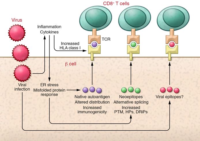
REVIEW                                                                                                                The Journal of Clinical Investigation

Figure 1. The natural history of islet autoimmunity. To illustrate the chronic nature and heterogeneity of islet autoimmunity and highlight critical gaps in
knowledge, this figure depicts two hypothetical patterns of T cell–mediated β cell destruction and loss of insulin secretion: early triggering of insulitis and
semi-linear β cell destruction (blue line); or late start of the autoimmune β cell destruction, closer to clinical diagnosis (red line). This is a simplified schematic
representation: loss of β cell mass and insulin secretion may not always correlate and may vary based on the relative importance of autoimmune destruction
and β cell dysfunction during disease progression in a given patient. Seroconversion for T1D-associated autoantibodies to native antigens is considered the
earliest step in the triggering of islet autoimmunity; it occurs in early life in children at genetic risk of T1D, but it may occur later in life in many individuals. As
only a minority of islets are affected at any given time, the process proceeds asynchronously. Residual β cell mass at onset may be a function of the severity
of autoimmune destruction and initial mass, which are impacted by age. It is often incomplete at diagnosis, but it continues afterward, sometimes for years.
Low levels of insulin secretion are detected in many patients even decades after diagnosis: some β cell regeneration occurs after diagnosis, and it has been
hypothesized that regeneration may occur during the preclinical stage. Critical questions remain unanswered: To what extent and when does the detection
of humoral and cellular responses in the circulation reflect insulitis and β cell destruction, and which responses are relevant? At what time are insulitis and β
cell destruction triggered during the progression of islet autoimmunity? Is there a preferred order for autoantibody and T cell responses to native antigens and
neoepitopes, and what are their relative roles in insulitis, β cell destruction, and disease onset (trigger/driver responses versus secondary responses)?

Eventually, most islets become pseudo-atrophic remnants of islet                       tive self-tolerance to β cell antigens and predisposing MHC variants
autoimmunity. In studies of 80 nPOD T1D donors (16, 23), 17                            remains controversial. Thus, HLA-II expression by β cells in the
donors with up to 12 years of disease duration exhibited insulitis;                    T1D pancreas should be reassessed with modern methodology
thus, islet autoimmunity may persist for years after diagnosis.                        and experimental models given the hypothetical implication that
Residual β cells were present in all T1D donors with insulitis, and                    β cells may directly present self-antigens to autoreactive CD4+ T
their β cell mass was higher than in T1D donors without insulitis.                     cells (Figure 3) (36).
T1D patients with more than 50 years of diabetes and residual
insulin secretion also displayed insulitis and residual β cells (24).                  T cells in the prediabetic pancreas
      CD8+ T cells are the predominant T cell population and most                      Clinical onset of T1D is preceded by an asymptomatic period last-
abundant inflammatory cell type in insulitis. Using HLA multim-                        ing months to years during which autoimmunity causes progres-
ers, investigators demonstrated the antigen specificity of autore-                     sive β cell destruction (Figure 1). Autoantibody screening identifies
active CD8+ T cells in insulitis lesions of nPOD T1D donors (23).                      individuals at risk for T1D among patients’ relatives; those with a
The infiltrating CD8+ T cells’ antigen repertoire increased in                         single autoantibody have low risk, while about 40%–80% of rela-
diversity with longer disease duration (23). Thus, antigen/epitope                     tives with multiple autoantibodies develop T1D within 5–10 years,
spreading occurs or continues after diagnosis (Figure 1).                              respectively (2). However, the extent to which humoral and cel-
      Hyperexpression of HLA-I molecules by endocrine cells in                         lular autoimmune responses in blood reflect ongoing pancreas
insulin-containing islets is another key feature of the T1D pancreas                   pathology during the prediabetic period are unknown, as is the rela-
(25). This observation was confirmed with various methodologies in                     tionship of autoantibody positivity with insulitis and the features of
multiple patient cohorts (26) (Figure 2). Hyperexpression of HLA-I                     islet-infiltrating T cells. Despite progress in identifying autoantibody-
molecules helps explain the predominance of CD8+ T cells in insu-                      positive non-diabetic donors (15, 37–39), recoveries have been too
litis. Earlier studies also reported aberrant expression of HLA-II                     few; we advocate that pancreata from these rare donors should be
molecules by β cells in 22 of 26 and 6 of 12 pancreata from patients                   allocated to research rather than transplant whenever possible (40).
with recent-onset and long-standing disease, respectively (25, 27,                          The limited data available from nPOD (16) and European
28). Expression of HLA-I/II molecules may be triggered by viruses                      studies (41, 42) suggest that insulitis is found only in donors with
linked to T1D (29, 30). Inflammatory cytokines (IFN-γ plus TNF-α                       high-risk HLA types and multiple autoantibodies. Higher T1D risk
or lymphotoxin) induce HLA-I/II molecules on β cells in vitro (31,                     associated with multiple autoantibodies may reflect ongoing insu-
32). The significance of HLA-II expression by cells was investigated                   litis, and low risk associated with a single autoantibody appears
using transgenic expression of MHC class II (MHC-II) molecules in                      consistent with lack of insulitis in pancreata of organ donors with
β cells, which did not drive immune infiltration of mouse islets (33–                  a single autoantibody (mostly against GAD65). Since these donors
35). However, these studies were not conducted in diabetes-prone                       are identified in the general population and many lack T1D-
mice, and the role of MHC-II expression in the presence of defec-                      predisposing HLA types (16, 43), only some may be representa-

   2882                jci.org   Volume 127   Number 8   August 2017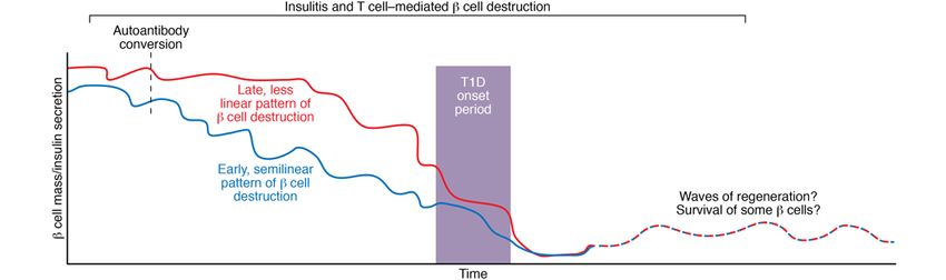
The Journal of Clinical Investigation                                                                                                            REVIEW

                                                                                                               efflux transporter ZNT8 (7, 81–85), chro-
                                                                                                               mogranin (86, 87), islet-amyloid polypeptide
                                                                                                               (IAPP) (8), and more. Many are present in
                                                                                                               secretory granules, and some are unique to
                                                                                                               β cells (insulin, IGRP, ZNT8), while some are
                                                                                                               expressed in other cells and tissues, including
                                                                                                               neuroendocrine cells (GAD65, IA-2) (88, 89).
                                                                                                                    Autoimmune responses to islet cell
                                                                                                               self-molecules evidence a specific loss
                                                                                                               of immunological self-tolerance. Most
                                                                                                               self-molecules, including those with tissue-
                                                                                                               restricted expression, are expressed in the
                                                                                                               thymus early in life to establish central tol-
                                                                                                               erance (90). Yet several mechanisms may
                                                                                                               determine suboptimal thymic expression
                                                                                                               and, in turn, imperfect self-tolerance. For
                                                                                                               T1D autoantigens, allelic variation and epi-
                                                                                                               genetic regulation affect the selection and
                                                                                                               levels of self-epitopes presented to develop-
                                                                                                               ing T cells during thymic selection. A poly-
Figure 2. CD8+ T cell responses to pancreatic β cells. Schematic representation of major autoantigen           morphic variable nucleotide tandem repeat
classes that may be targeted by CD8+ T cells, the predominant immune cell type in the insulitis. As            (VNTR) sequence at the 5′ of the insulin
described in the main text, these may include native antigens and neoepitopes. The generation of these
                                                                                                               gene influences thymic expression of insulin,
epitopes may be promoted by β cell inflammation and stress. Insulitis is often associated with an inter-
feron response with hyperexpression of HLA class I molecules, which may be induced by viral infections.        and alleles that confer resistance to T1D are
Hyperexpression of HLA class I molecules facilitates presentation of autoantigen epitopes to CD8+ T            associated with higher insulin thymic tran-
cells. Whether islet-infiltrating CD8+ T cells also target viral epitopes remains to be investigated.          scription than predisposing variants (91, 92).
                                                                                                               Epigenetic regulation may suppress thymic
                                                                                                               transcription of a parental copy of the insu-
tive of pre-diabetes. However, abnormalities are reported in the                      lin gene, especially for T1D-protective alleles associated with higher
pancreas of donors with single autoantibodies (15): accumula-                         transcription (91–93). These mechanisms may allow autoreactive
tion of proinsulin-containing vesicles in β cells observed in nPOD                    T cells to escape thymic selection, as reported for insulin in mouse
organ donors with single and multiple autoantibodies may reflect                      models (94–98) and patients (99, 100).
impaired proinsulin conversion, disrupted vesicular trafficking,                           T1D-predisposing HLA molecules may contribute to sub-
and possibly increased metabolic requirements and stress. Consis-                     optimal thymic presentation of autoantigens. Weak interactions
tent with the increases in size and number of β cells/islet observed                  between a preproinsulin peptide and HLA-A2 lead to suboptimal
in autoantibody-positive donors (44), there is no evidence of                         presentation to the TCR of responding CD8+ T cells (101), which
reduced β cell mass in independent analyses of autoantibody-                          may more easily survive thymic selection. Of note, HLA-A2 is
positive nPOD donors (44, 45). Based on these findings, Figure 1                      found also in about 50% of the general European-descent popu-
illustrates two theoretical patterns of β cell destruction: insulitis                 lation; the often-reported detection of circulating autoreactive
may begin early, with destruction proceeding in a semi-linear fash-                   T cells in healthy individuals (102) may derive from negative
ion; or insulitis and β cell destruction may occur closer to diagnosis.               selection processes that are inherently imperfect for islet cell self-
The latter fits the observations that (i) the proportion of infiltrated               molecules. However, autoreactive T cells in T1D patients are
islets is modest near diagnosis, and insulitis is only present in                     reportedly enriched in memory cells versus a naive phenotype in
non-diabetic donors with multiple autoantibodies (16, 21, 23); (ii)                   healthy subjects (103, 104). The positive effects of treatment with
β cell loss near diagnosis is partial in many patients (16, 21, 23),                  an anti–memory cell agent in patients with recently diagnosed T1D
and no appreciable reduction in β cell mass has been reported in                      supports the importance of memory autoreactive T cells in this dis-
autoantibody-positive donors (44, 45); and (iii) impaired glucose                     ease (105, 106). Memory autoreactive T cells were linked to T1D
metabolism, abnormalities of insulin secretion (46, 47) and insu-                     recurrence in transplanted patients, both in islet (107, 108) and
lin sensitivity (48), and increased β cell death (49) usually manifest                pancreas transplantation (109), despite immunosuppression.
shortly before diagnosis in longitudinal studies of at-risk relatives.                     Autoreactive T cells exhibit proinflammatory cytokine profiles
                                                                                      in T1D patients and regulatory profiles in healthy subjects (85, 110,
Loss of immunological tolerance to β cells                                            111). T1D patients exhibit defects in peripheral tolerance, including
Several islet cell molecules were identified as targets of autoimmu-                  impaired Treg function (112, 113) and effector T cell (Teff) resis-
nity in T1D, including native proteins and epitopes of proinsulin (9,                 tance to Treg suppression (114). Many T1D risk genes predispose
50–63), GAD65 (64–72), tyrosine phosphatase–like insulinoma-                          to impaired immune regulation (115); the best studied are PTPN22
associated antigen 2 (IA-2) (67, 73–75), islet-specific glucose-6-phos-               (encoding protein tyrosine phosphatase, non-receptor type 22),
phatase catalytic subunit–related protein (IGRP) (76–80), the cation                  CTLA4 (encoding cytotoxic T lymphocyte–associated protein 4),

                                                                                 jci.org   Volume 127   Number 8   August 2017                     2883
REVIEW The Journal of Clinical Investigation
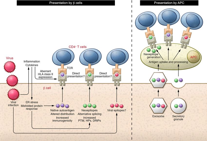
Figure 3. CD4+ T cell responses to pancreatic β cells. Schematic representation of major autoantigen classes that may be targeted by CD4+ T cells. Similar
to CD8+ T cells, these may include native antigens and neoepitopes, which may be formed under conditions of β cell inflammation and stress. β Cells
can produce several neoepitopes: for example, peptides originating from alternative splicing, insulin hybrid peptides (HP), and DRiP insulin peptides
are reportedly produced in β cells (for DRiPs, the evidence is from studies of cell lines). Many autoantigens are secretory granule proteins, which may
be released by β cells and acquired by APCs. Exosomes released by β cells also contain autoantigens. CD4+ T cells may react with antigens captured,
processed, and presented by APCs in the pancreatic lymph node and in the islets. The B:9–23 epitope is produced in the secretory granules, captured, and
presented by APCs, at least in the NOD mouse. The generation of neoepitopes in β cells further raises the question of whether the previously reported
aberrant expression of HLA class II molecules by β cells might allow CD4+ T cells to recognize antigens on their surface directly.
and IL2RA (encoding the IL-2 receptor α-chain, IL-2Rα), which (148). Dendritic cells’ efficiency at cross-presentation is important
control TCR signaling, inhibition of T lymphocyte responses, and for self-tolerance (149–151) and viral responses (152) but is also linked
Treg development and function (116, 117), respectively. to T1D in NOD mice (153–155). In the NOD mouse, self-antigen
Environmental exposures may promote loss of tolerance. Along cross-presentation to CD8+ T cells is also mediated by B lymphocytes
with their epidemiological associations (29, 30), enterovirus infec- and promotes disease progression (156).
tions of β cells impair insulin secretion (118), alter mRNA/miRNA
expression (119, 120), and induce interferon responses (121, 122) that Modified T cell autoantigens (neoepitopes)
promote β cell stress, dysfunction, and apoptosis (123). Enterovirus- Increasing evidence suggests that islet autoimmunity also targets
es may also trigger autoimmunity via presentation of self-molecules neoepitopes expressed by the target cells, which may not be avail-
in an inflammatory context (bystander activation) (124) and/or by able for negative thymic selection. Differential expression of IA-2
molecular mimicry (125–127). Cross-reactivities with viral proteins and IGRP mRNAs in thymus and pancreas (157, 158) may promote
were reported for autoantibody (128–134) and T cell responses (73, autoimmunity, as the immune system may not be tolerant to alter-
132, 135–144) with GAD65, IA-2, and insulin autoantigens. β Cell natively spliced variants expressed in pancreas but not thymus (159).
inflammation and ER stress promote β cell dysfunction and immu- Many autoantigens in autoimmune disease are post-translationally
nogenicity, and protein misfolding is associated with abnormal modified (PTM) (160). Inflammation and stress are likely factors
autoantigen presentation (145–147). Cytokine-induced ER stress in the generation of PTM antigens, which derive from both normal
enhances β cell release of exosomes loaded with autoantigens and and abnormal processes in cells. Predisposing HLA types are critical
immunostimulatory chaperones, which are taken up by antigen- for the presentation of these epitopes to T cells (161, 162). Below, the
presenting cells (APCs) and may be presented to T cells (Figure 3) major neoepitope classes associated with T1D are described.
2884 jci.org Volume 127 Number 8 August 2017The Journal of Clinical Investigation REVIEW
Neoepitopes generated by PTM. A PTM epitope exists in the insu- higher-affinity insulin-specific T cells in NOD mice (169). Regis-
lin A chain (A1–A13): T cell recognition requires oxidized cysteine ter 3 presentation may not occur in the thymus, possibly explain-
residues at A6 and A7, with the formation of a vicinal disulfide ing how insulin-specific CD4+ T cells escape negative selection.
bond between them (60). Other forms of PTM include citrullina- Despite unresolved differences related to registers used in these
tion (163) and transglutamination, which enhance GAD65 pep- studies, these observations support the concept that differences
tide binding to HLA-DRB1*04:01 and modulate recognition by in MHC register use are key to disease development and lead to
modifying amino acids at TCR contact positions (164). Memory T the presentation of native antigens as neoepitopes. Similar mech-
cells reacting with these PTM epitopes were detected in blood at a anisms may be operative in patients: HLA-DQ8–restricted CD4+ T
higher frequency in T1D patients than controls. The same T cells lymphocytes may target insulin peptides in a low-affinity binding
had weak or no reactivity toward the native peptides. Tissue trans- register similar to the NOD register 3 (170, 171). Ultimately, differ-
glutaminase mediates the deamidation reaction and generates PTM ences in HLA-peptide complex interactions can impact respond-
antigens in celiac disease, which shares HLA-II susceptibility with ing T cell activation and phenotypes and synergize with the
T1D (111). The HLA-DQ trans-dimer formed in DQ8/DQ2 hetero- reduced insulin expression in the thymus that is associated with
zygotes exhibits binding preference for negatively charged peptides predisposing insulin gene variants. Moreover, CD4+ T cells from
generated by deamidation. Van Lummel at al. (111) identified CD4+ T1D patients display abnormal immunological synapsis associ-
T cells against proinsulin peptides that preferentially bound to the ated with escape from negative selection and enhanced effector
HLA-DQ trans-dimer in DQ8/DQ2 heterozygotes after deamida- responses upon encounter with cognate antigen (172).
tion, and other deamidated peptides that preferentially bound to the Neoepitopes generated by peptide fusion. Neoepitopes are also
HLA-DQ8 cis-dimer. While healthy controls and T1D patients dis- formed by fusion of peptides from two proteins, termed hybrid
played similar reactivities, autoreactive T cells from most patients peptides, that are not genetically encoded and are high-affinity T
produced proinflammatory IFN-γ in response to cognate antigen cell targets (173). Hybrid peptides include those resulting from the
stimulation, in contrast to regulatory IL-10 responses in controls. fusion of proinsulin C-peptide fragments and other β cell secretory
Neoepitopes generated by differences in MHC binding registers. granule proteins, such as chromogranin, IAPP, or neuropeptide Y
Insulin is considered a key autoantigen in the NOD mouse model (NPY). Perhaps the formation of hybrid peptides explains the essen-
of autoimmune diabetes, as genetic manipulation to abolish tial role of insulin and chromogranin in diabetes development in
response to the insulin B chain B:9–23 peptide prevents diabe- NOD mice (165, 174). Hybrid peptides may result from proteolytic
tes (165). Unanue and Eisenbarth/Kappler/Michels defined key hydrolysis of peptide bonds in the presence of naturally occurring
molecular interactions of insulin peptides with the NOD mouse cleavage products. Insulin and other secretory proteins are pack-
I-Ag7 MHC-II molecule, which is remarkably similar to HLA-DQ8 aged together in secretory granules, which may favor reversed pro-
(166) in both sequence and binding features for insulin peptides teolytic transpeptidation. CD4+ T cells that react against hybrid pep-
(167). Unanue’s group (161, 162, 168) identified two registers in tides were identified in NOD mice as well as islet-infiltrating cells
which peptides encompassing the insulin B:9–23 epitope bind to isolated from deceased T1D patients (173, 175).
I-Ag7: register 1 (B:12–20) and register 2 (B:13–21). Type A CD4+ Neoepitopes generated by aberrant translational products. The
T cells recognize insulin presented by APCs, and these T cells are generation of defective ribosomal products (DRiPs) from the pro-
deleted in the thymus through presentation via register 2. Type B insulin gene is another recently discovered mechanism leading to
insulin-reactive T cells do not react with insulin protein processed production of T cell–targeted β cell neoepitopes. β Cells generate
by APCs but respond to soluble B chain peptide when weakly DRiP neoepitopes using an alternative initiation site for transla-
bound by I-Ag7 in register 1. Type B CD4+ T cells are not deleted tion and by translating the 3′ UTR (176). T cell reactivity against
in the thymus and become activated by APCs in islets (162). Thus, proinsulin DRiPs was tested in peripheral blood samples from
a single amino acid shift of the B chain peptide bound to I-Ag7 T1D patients, and proliferative responses were observed in most.
determines whether CD4+ T cells recognize peptides generated by Strong T cell responses to DRiPs were detected in individuals with
insulin processing and allows escape from negative selection. In predisposing HLA-DQ types, such as HLA-DQ8/DQ2, who can
essence, differences in MHC register usage determines whether express HLA-DQ trans-dimers (HLA-DQ8trans, DQA1*05:01/
a peptide is presented as a “neoepitope,” as the same peptide was DQB1*03:02 or HLA-DQ2trans, DQA1*03:01/DQB1*02:01). T
not presented in this register in the thymus. cell responses to DRiPs were not observed in rare patients carry-
Kappler’s group described a register 3 in which two peptides ing T1D-protective HLA-DQ or insulin gene haplotypes, consis-
containing the critical amino acid residues of insulin B:9–23 (B:9– tent with the concept that both genes impact thymic selection of
20 and B:9–21) bind poorly to the I-Ag7 molecule (86, 169). Accord- insulin-reactive T cells.
ing to these authors, most, if not all, NOD mouse CD4+ T cells The proinsulin UTR includes two SNPs in tight linkage dis-
reacting against B:9–23 target this peptide in the low-affinity regis- equilibrium with insulin VNTR variants associated with T1D risk
ter 3. These T cells can be divided into two types based on whether and the modulation of insulin gene transcription in the thymus (91,
their response is improved or inhibited by glycine substitution at 92). The presence of these SNPs in the UTR produces several DRiP
the B21 glutamic acid at the peptide’s p8 position. Modifying the variants. T cell responses against DRiPs containing amino acid
wild-type insulin B:9–23 peptide amino acid sequence at positions residues encoded by predisposing or protective haplotypes were
p8 and/or p9 (p8E/p9R) into p8G/p9E- and p8E/p9E-generated strongly correlated, suggesting that the UTR SNPs did not influ-
mimotopes that bind to I-Ag7 in register 3 increased the binding ence immunogenicity and may not be critical for HLA-DQ bind-
affinity with insulin-specific TCRs and favored the activation of ing. Dendritic cells processed and presented proinsulin DRiPs,
jci.org Volume 127 Number 8 August 2017 2885REVIEW The Journal of Clinical Investigation
preferentially via the HLA-DQ8trans, and patient T cells exclu- ity of the T cell responses, which may reflect differences in HLA
sively responded by producing IFN-γ and granzyme B. HLA-A2 restriction, environmental exposures, and disease stage. However,
can also present DRiPs, and T1D patients had higher frequencies a small fraction of islet-infiltrating T cell lines and clones reacted
of DRiP-specific CD8+ Teffs than healthy subjects. DRiP-specific to known autoantigen epitopes. Given the broad range of disease
CD8+ T cells killed HLA-A2–positive human islet cells in vitro, and duration, these findings match pathology studies showing that islet
pretreating the islet cells with high glucose and proinflammatory autoimmunity continues for years after diagnosis.
cytokines potentiated the cytotoxic action. Proinsulin DRiPs were Michels at al. (181) analyzed islet-infiltrating T cells from
formed under thapsigargin-induced experimental ER stress, a nPOD donors, focusing on proinsulin-specific CD4+ T cells. The
form of stress characterized by calcium depletion in the ER and study included 3 nPOD T1D donors overlapping with those of
increased cytoplasmic calcium levels, in contrast to classical ER Babon et al. (175) and nPOD donor 6323 examined by Seay et al.
stress caused by accumulation of misfolded proteins. Thus, in the (182), described below. These donors were 6, 14, and 19 years old,
T1D pancreas, ER stress and inflammation impair β cell function had T1D for 2, 3, and 6 years, respectively, and displayed insulitis in
by increasing production of autoantigens (147), and DRiPs could the pancreatic islets. Sequencing of infiltrating T cells’ TCR genes
trigger and enhance T cell–mediated killing of β cells (176). revealed diverse repertoires, but certain TCRs were repeatedly
detected, suggesting some level of clonal expansion. Many of these
Ex vivo studies of T cells in T1D-relevant tissues TCR sequences were found in multiple islets. TCRs expressed by
Investigations of antigen-specific T cells from T1D-relevant tis- islet-infiltrating T cells reacted against the insulin B:9–23 epitope
sues are rare. Earlier studies detected CD4+ T cells against insu- and were restricted by HLA-DQ8. Reactivity to a 19–35 epitope
lin and/or GAD65 in the pancreatic lymph nodes (PLNs) or islets of C-peptide reported in the islets of a previously discussed T1D
obtained from a few deceased T1D patients (57). GAD65-reactive donor (179) was reproduced. However, the majority of the TCRs
CD4+ T cells were detected in the circulation and lymph nodes examined did not respond to proinsulin/insulin epitopes, consis-
draining the transplanted pancreas in a T1D recipient who lost tent with the broad reactivity reported by Babon et al. (175).
graft function due to recurrent autoimmunity (109), and GAD65- Seay et al. (182) studied pancreas, PLN, spleen, irrelevant
reactive CD4+ T cells from pancreas transplant recipients killed lymph nodes, and peripheral blood from 18 nPOD donors diag-
cells when co-transplanted with human islets in immunodeficient nosed with T1D 4–32 years prior to their death at ages 11–44 years.
mice (109). Autoimmune β cell damage was reported in human- The researchers conducted high-throughput immunosequencing
ized mice grafted with immune cells from a T1D patient (177, 178). of the TCR β-chain (TRB) to investigate TCR repertoire diversi-
Proinsulin-specific (61, 63) and IGRP-reactive CD8+ T cells (109) ty; TCR sharing among blood and various tissues in T cell subsets
are also cytotoxic in vitro. Insulin-reactive CD4+ T cells from T1D (CD4+ conventional T [Tconv] cells; CD4+ Tregs; CD8+ T cells); TCR
donor islets targeted a PTM epitope in the insulin A chain (60), clonal expansion in selected compartments; and whether any T1D-
which was presented by HLA-DR4 (DRB1*04:01, but could also associated TCRs were public, or shared, among patients. TRB
be presented by *04:04 and *04:05). Of 53 CD4+ T cell clones sharing across compartments and TCR diversity were similar
(179), 47 unique clonotypes were identified, including 8 recogniz- in patients and controls, revealing limited evidence for receptor
ing proinsulin presented by HLA-DQ8; 26% of these clones tar- biases. Moreover, there was low CD3β sharing across tissues, and
geted 6 distinct but overlapping epitopes in the C-peptide of pro- shared sequences were not known to target T1D autoantigens. In
insulin. These were presented by the HLA-DQ trans-heterodimer both groups, there was minimal TCR overlap between PLN Tregs
that forms in HLA-DQ2/-DQ8 heterozygotes using DQA1*05:01 and Tconv cells, which may not support T-lineage instability and
and DQB1*03:02. More recently, collaborative efforts supported plasticity contributing to T1D through the interconversion of Tregs
by nPOD have allowed expanded analyses of islet-infiltrating T to Tconv effector cells. However, those TCRs matching sequences
cells from T1D organ donors. Islets were purified using standard with previously reported autoreactive T cells were highly enriched
isolation protocols and distributed to several laboratories, which in the T1D donors. These included autoreactive T cells directed
isolated islet-infiltrating T cells and examined antigen specifici- against epitopes of proinsulin C-peptide 19–35 previously identified
ties, phenotypic features, and TCR sequences. in the islets of a T1D donor (179) and several GAD65-reactive clones
Babon et al. (175) obtained islet-infiltrating T cells from 9 reported in blood of prediabetic individuals and patients (183).
donors between 6 and 30 years old with 2–20 years since T1D diag- These proinsulin and GAD65 TCR sequences were found in the
nosis. Representing the largest analysis to date of islet-infiltrating PLN of some T1D donors. CD4+ T cells targeting the GAD65 555–
T cells from T1D donors, the study explored the largest number 567 epitope were described in peripheral blood of patients (68, 184).
of antigen specificities. More than 250 T cell lines or clones were Seay et al. (182) described a TCR sequence matching a previ-
derived. Lines were obtained from all T1D donors, but islet- ously reported GAD65 clone (GAD4.13) that represented approx-
infiltrating T cells were not recovered from control donors, with imately 25% of the TCR sequences found in the PLN of a T1D
one exception. Thus, unlike autoreactive T cells detected in blood, donor. In this donor, the CDR3β sequence was the most abundant
lymphocytic islet infiltration is highly specific for T1D. Of 50 lines in PLN Treg and CD8+ T cell subsets, providing initial evidence
examined for autoreactivity so far, 19 reacted against a variety of of strong clonal dominance. The GAD4.13 CDR3β sequence was
known autoantigens, including PTM epitopes (180) and hybrid detected in several tissue compartments in about 40% of T1D
peptides (173). These autoreactive T cells secreted IFN-γ and other donors and in islet-infiltrating T cells in nPOD donor 6323. Approx-
inflammatory cytokines, consistent with an effector phenotype. imately 14% of 399 CD3β sequences identified in this donor’s islet-
This study’s major finding is the broad repertoire and heterogene- infiltrating CD8+ T cells were also in the PLN, and most of these
2886 jci.org Volume 127 Number 8 August 2017The Journal of Clinical Investigation REVIEW
shared sequences were detected in blood, yet there was minimal The ability to detect autoreactive T cells in the circulation has
overlap between the islet and PLN compartment and no overlap improved dramatically, but their common presence in healthy
with circulating CD4+ Tconv cells. Together with the data of Michels subjects requires assessing autoreactive T cell phenotypes and
et al. on the same donor (181), these findings support the existence of functions to aid in interpretation of positive results. Recent stud-
public TCRs shared among patients. Additional evidence for public ies of T1D and prediabetic organ donors have examined pathol-
TCRs derives from the observation that TCR sequences of GAD65 ogy and autoreactive T cells in the pancreas, focusing on islet-
autoreactive CD4+ T cells from a patient with T1D recurrence in the infiltrating T cells and those in lymphoid tissues. These stud-
transplanted pancreas (109) overlapped with sequences from a large ies provide insight into the T cells associated with insuli-
number of the nPOD T1D donors studied by Seay et al. (182). tis and allow comparisons with peripheral blood readouts.
TCR analysis in blood, pancreas, and lymphoid tissues may
Closing remarks help firmly establish whether detection of certain auto-
Autoreactive T cells targeting a broad repertoire of antigens and reactive T cells with particular phenotypes can be a bio-
epitopes are considered the main mediators of β cell death in T1D. marker of ongoing insulitis. Initial evidence for limited
Given the diversity of islet autoimmune responses and patient het- TCR sharing among patients could represent key responses and
erogeneity, it is challenging to discern whether any responses are perhaps therapeutic targets. Further studies investigating TCR
more pathogenic than others. Growing evidence supports the tar- sharing between antigen-specific Tconv cells and Tregs react-
geting of “neoepitopes” that result from post-translational modifi- ing to the same epitope with identical HLA restriction could
cation of native antigens, fusion of peptides derived from different help define to what degree Tregs compete with Tconv cells
proteins, alternative splicing, and defective ribosomal intermediate for binding to a shared cognate epitope. If TCR sharing is low,
products. T cells reacting with neoepitopes may have higher affin- opportunities may also open for selective therapeutic targeting
ity and stronger pathogenic potential if central tolerance mecha- of Teffs or Tregs. For example, there may not be natural Tregs
nisms do not apply to these epitopes, which may not be expressed specific for neoepitopes.
by tolerogenic thymic medullary epithelial cells or tolerogenic stro- Ongoing pathology studies of pancreata obtained near T1D
mal cells and APCs in peripheral lymphoid tissues (185–187). Many onset or afterward are revealing that only a proportion of islets
neoepitopes may only be expressed in the target organ; in particular, are affected at any given time. It remains unclear when T cells
neoepitopes involving insulin may be limited to β cells, and stress begin infiltrating the islets relative to the initiation of autoimmu-
and inflammation may influence their expression. The role of neo- nity as marked by autoantibody conversion. There is interest in
epitopes is a critical research area where we may learn more about defining which classes of autoimmune responses are detected
impaired tolerance mechanisms and whether responses to neoepi- first, under the hypothesis that initial responses are triggers and
topes are primary autoimmune responses and major disease driv- possibly major disease drivers. Perhaps responses to neoepi-
ers. If future studies can identify key disease driver epitopes, there topes represent the earliest and most pathogenic responses, with
would be major implications for disease prediction, prevention, and responses to native epitopes playing a secondary role. An alterna-
therapy, especially for antigen-specific therapies. tive hypothesis is that more benign, initial responses are directed
It is currently unknown if a proportion of autoreactive T toward native antigens, are fairly regulated, and produce modest
cells can react with viral epitopes, whether or not they cross- inflammation and little damage to β cells; however, persisting
react with islet antigens. Given the extensive literature associ- and increasing islet inflammation, β cell stress, and perhaps viral
ating T1D with enterovirus infection of the islets (30), the asso- infections could lead to the generation of neoepitopes and trig-
ciation of insulitis with hyperexpression of HLA-I molecules, ger more aggressive autoimmunity. Generation of novel reagents
and the predominance of CD8+ T cells in islet infiltrates, it is that track autoantibody and T cell responses against neoepi-
reasonable to hypothesize that a significant proportion of islet- topes will enable interrogation of archived blood samples from
infiltrating CD8+ T cells may react against viral epitopes (Figures at-risk relatives in natural history studies, some of which begin at
2 and 3). If so, CD8+ T cells against viruses may contribute to birth. Studies of clinical cohorts and expanded numbers of organ
T1D pathogenesis by eliminating infected β cells presenting viral donors will be critical to addressing outstanding questions about
epitopes in the context of HLA-I hyperexpression. New inves- autoreactive T cells in T1D.
tigations should attempt to identify CD8+ T cells against T1D-
associated viruses, especially for epitopes that are good binders Acknowledgments
for T1D-predisposing HLA molecules; determination of cross- Studies by the author related to this topic were supported by grants
reactivity with autoantigens should be also explored. Moreover, from the NIH (R01 DK070011), the JDRF (research grants 25-2013-
antiviral responses may be depressed in T1D patients, at least 268, 17-2011-594, 17-2012-3), the Leona M. and Harry B. Helmsley
temporarily. For example, impaired responses were linked to Charitable Trust (George Eisenbarth nPOD Award for Team Sci-
subsequent development of insulin autoimmunity in early life ence, 2015PG-T1D052), and the Diabetes Research Institute Foun-
(127). Perhaps responses to viral antigens that cross-react with dation, Hollywood, Florida.
islet autoantigens are more susceptible to regulation: weaker
antiviral responses may favor the persistence of viral infections. Address correspondence to: Alberto Pugliese, Diabetes Research
Addressing these major unanswered questions could impact our Institute, University of Miami Miller School of Medicine, 1450 NW
understanding of the potential role of viruses and overall dis- 10th Avenue, Miami, Florida 33136, USA. Phone: 305.243.5348;
ease pathogenesis, and design of future therapies. Email: apuglies@med.miami.edu.
jci.org Volume 127 Number 8 August 2017 2887REVIEW The Journal of Clinical Investigation
1. Atkinson MA, Eisenbarth GS, Michels AW. Type 1 19. Pugliese A. Insulitis in the pathogenesis of type 1 37. Gianani R, et al. Initial results of screening of
diabetes. Lancet. 2014;383(9911):69–82. diabetes. Pediatr Diabetes. 2016;17 Suppl 22:31–36. nondiabetic organ donors for expression of
2. Ziegler AG, et al. Seroconversion to multiple islet 20. Reddy S, et al. Analysis of peri-islet CD45-positive islet autoantibodies. J Clin Endocrinol Metab.
autoantibodies and risk of progression to diabe- leucocytic infiltrates in long-standing type 1 dia- 2006;91(5):1855–1861.
tes in children. JAMA. 2013;309(23):2473–2479. betic patients. Diabetologia. 2015;58(5):1024–1035. 38. Tauriainen S, Salmela K, Rantala I, Knip M,
3. Noble JA, et al. HLA class I and genetic suscep- 21. Krogvold L, et al. Insulitis and characterisation Hyöty H. Collecting high-quality pancreatic
tibility to type 1 diabetes: results from the Type of infiltrating T cells in surgical pancreatic tail tissue for experimental study from organ donors
1 Diabetes Genetics Consortium. Diabetes. resections from patients at onset of type 1 diabe- with signs of β-cell autoimmunity. Diabetes Metab
2010;59(11):2972–2979. tes. Diabetologia. 2016;59(3):492–501. Res Rev. 2010;26(7):585–592.
4. Erlich H, et al. HLA DR-DQ haplotypes and geno- 22. Eisenbarth GS. Banting Lecture 2009: an 39. Wasserfall C, et al. Validation of a rapid type
types and type 1 diabetes risk: analysis of the type unfinished journey: molecular pathogenesis 1 diabetes autoantibody screening assay for
1 diabetes genetics consortium families. Diabetes. to prevention of type 1A diabetes. Diabetes. community-based screening of organ donors to
2008;57(4):1084–1092. 2010;59(4):759–774. identify subjects at increased risk for the disease.
5. van Lummel M, et al. Type 1 diabetes-associated 23. Coppieters KT, et al. Demonstration of islet- Clin Exp Immunol. 2016;185(1):33–41.
HLA-DQ8 transdimer accommodates a autoreactive CD8 T cells in insulitic lesions 40. Burke GW, Posgai AL, Wasserfall CH, Atkinson
unique peptide repertoire. J Biol Chem. from recent onset and long-term type 1 diabetes MA, Pugliese A. Raising awareness: the need to
2012;287(12):9514–9524. patients. J Exp Med. 2012;209(1):51–60. promote allocation of pancreata from rare nondi-
6. Nejentsev S, et al. Localization of type 1 diabetes 24. Keenan HA, et al. Residual insulin production abetic donors with pancreatic islet autoimmunity
susceptibility to the MHC class I genes HLA-B and pancreatic ß-cell turnover after 50 years to type 1 diabetes research. Am J Transplant.
and HLA-A. Nature. 2007;450(7171):887–892. of diabetes: Joslin Medalist Study. Diabetes. 2017;17(1):306–307.
7. Roep BO, Peakman M. Antigen targets of type 1 2010;59(11):2846–2853. 41. In’t Veld P, et al. Screening for insulitis in adult
diabetes autoimmunity. Cold Spring Harb Perspect 25. Foulis AK, Farquharson MA, Hardman R. Aber- autoantibody-positive organ donors. Diabetes.
Med. 2012;2(4):a007781. rant expression of class II major histocompati- 2007;56(9):2400–2404.
8. Velthuis JH, et al. Simultaneous detection of cir- bility complex molecules by B cells and hyper- 42. Wiberg A, et al. Characterization of human organ
culating autoreactive CD8+ T-cells specific expression of class I major histocompatibility donors testing positive for type 1 diabetes-
for different islet cell-associated epitopes complex molecules by insulin containing islets associated autoantibodies. Clin Exp Immunol.
using combinatorial MHC multimers. Diabetes. in type 1 (insulin-dependent) diabetes mellitus. 2015;182(3):278–288.
2010;59(7):1721–1730. Diabetologia. 1987;30(5):333–343. 43. Oikarinen M, et al. Analysis of pancreas tissue in
9. Unger WW, et al. Discovery of low-affinity pre- 26. Richardson SJ, et al. Islet cell hyperexpression of a child positive for islet cell antibodies. Diabetolo-
proinsulin epitopes and detection of autoreactive HLA class I antigens: a defining feature in type 1 gia. 2008;51(10):1796–1802.
CD8 T-cells using combinatorial MHC multim- diabetes. Diabetologia. 2016;59(11):2448–2458. 44. Rodriguez-Calvo T, et al. Increase in pancreatic
ers. J Autoimmun. 2011;37(3):151–159. 27. Bottazzo GF, Dean BM, McNally JM, MacKay EH, proinsulin and preservation of β-cell mass in
10. Uchtenhagen H, et al. Efficient ex vivo analysis Swift PG, Gamble DR. In situ characterization of autoantibody-positive donors prior to type 1 dia-
of CD4+ T-cell responses using combinatorial autoimmune phenomena and expression of HLA betes onset. Diabetes. 2017;66(5):1334–1345.
HLA class II tetramer staining. Nat Commun. molecules in the pancreas in diabetic insulitis. 45. Diedisheim M, Mallone R, Boitard C, Larger E.
2016;7:12614. N Engl J Med. 1985;313(6):353–360. β-cell mass in nondiabetic autoantibody-positive
11. Rowe PA, Campbell-Thompson ML, Schatz DA, 28. Lernmark A, et al. Heterogeneity of islet pathol- subjects: an analysis based on the Network for
Atkinson MA. The pancreas in human type 1 dia- ogy in two infants with recent onset diabetes Pancreatic Organ Donors Database. J Clin Endo-
betes. Semin Immunopathol. 2011;33(1):29–43. mellitus. Virchows Arch. 1995;425(6):631–640. crinol Metab. 2016;101(4):1390–1397.
12. Foulis AK, Liddle CN, Farquharson MA, Rich- 29. Krogvold L, et al. Detection of a low-grade 46. Sosenko JM, et al. Acceleration of the loss of
mond JA, Weir RS. The histopathology of the enteroviral infection in the islets of Langerhans the first-phase insulin response during the
pancreas in type 1 (insulin-dependent) diabetes of living patients newly diagnosed with type 1 progression to type 1 diabetes in diabetes
mellitus: a 25-year review of deaths in patients diabetes. Diabetes. 2015;64(5):1682–1687. prevention trial-type 1 participants. Diabetes.
under 20 years of age in the United Kingdom. 30. Hyöty H. Viruses in type 1 diabetes. Pediatr 2013;62(12):4179–4183.
Diabetologia. 1986;29(5):267–274. Diabetes. 2016;17 Suppl 22:56–64. 47. Sims EK, et al. Elevations in the fasting serum pro-
13. Imagawa A, et al. Pancreatic biopsy as a pro- 31. Pujol-Borrell R, et al. HLA class II induction insulin-to-c-peptide ratio precede the onset of type
cedure for detecting in situ autoimmune phe- in human islet cells by interferon-gamma plus 1 diabetes. Diabetes Care. 2016;39(9):1519–1526.
nomena in type 1 diabetes: close correlation tumour necrosis factor or lymphotoxin. Nature. 48. Ferrannini E, Mari A, Nofrate V, Sosenko JM,
between serological markers and histological 1987;326(6110):304–306. Skyler JS, DPT-1 Study Group. Progression to
evidence of cellular autoimmunity. Diabetes. 32. Campbell IL, Oxbrow L, West J, Harrison LC. diabetes in relatives of type 1 diabetic patients:
2001;50(6):1269–1273. Regulation of MHC protein expression in pan- mechanisms and mode of onset. Diabetes.
14. Krogvold L, et al. Pancreatic biopsy by minimal creatic beta-cells by interferon-gamma and 2010;59(3):679–685.
tail resection in live adult patients at the onset tumor necrosis factor-alpha. Mol Endocrinol. 49. Herold KC, et al. β cell death and dysfunction
of type 1 diabetes: experiences from the DiViD 1988;2(2):101–107. during type 1 diabetes development in at-risk
study. Diabetologia. 2014;57(4):841–843. 33. Lo D, et al. Diabetes and tolerance in transgenic individuals. J Clin Invest. 2015;125(3):1163–1173.
15. Pugliese A, et al. The Juvenile Diabetes Research mice expressing class II MHC molecules in pan- 50. Keller RJ. Cellular immunity to human insulin
Foundation Network for Pancreatic Organ creatic beta cells. Cell. 1988;53(1):159–168. in individuals at high risk for the development
Donors with Diabetes (nPOD) Program: goals, 34. Böhme J, et al. Transgenic mice with I-A on islet of type I diabetes mellitus. J Autoimmun.
operational model and emerging findings. cells are normoglycemic but immunologically 1990;3(3):321–327.
Pediatr Diabetes. 2014;15(1):1–9. intolerant. Science. 1989;244(4909):1179–1183. 51. Rudy G, et al. Similar peptides from two beta
16. Campbell-Thompson M, et al. Insulitis and β-cell 35. Sarvetnick N, Liggitt D, Pitts SL, Hansen SE, cell autoantigens, proinsulin and glutamic acid
mass in the natural history of type 1 diabetes. Stewart TA. Insulin-dependent diabetes mellitus decarboxylase, stimulate T cells of individuals
Diabetes. 2016;65(3):719–731. induced in transgenic mice by ectopic expression at risk for insulin-dependent diabetes. Mol Med.
17. In’t Veld P. Insulitis in human type 1 diabetes: the of class II MHC and interferon-gamma. Cell. 1995;1(6):625–633.
quest for an elusive lesion. Islets. 2011;3(4):131–138. 1988;52(5):773–782. 52. Alleva DG, et al. A disease-associated cellular
18. Campbell-Thompson ML, et al. The diagnosis of 36. Bottazzo GF. Lawrence lecture. Death of a immune response in type 1 diabetics to an immu-
insulitis in human type 1 diabetes. Diabetologia. beta cell: homicide or suicide? Diabet Med. nodominant epitope of insulin. J Clin Invest.
2013;56(11):2541–2543. 1986;3(2):119–130. 2001;107(2):173–180.
2888 jci.org Volume 127 Number 8 August 2017The Journal of Clinical Investigation REVIEW
53. Durinovic-Belló I, Boehm BO, Ziegler AG. Pre- 2002;109(7):895–903. 86. Stadinski BD, et al. Chromogranin A is an
dominantly recognized proinsulin T helper cell 70. Reijonen H, et al. GAD65-specific CD4+ T-cells autoantigen in type 1 diabetes. Nat Immunol.
epitopes in individuals with and without islet cell with high antigen avidity are prevalent in periph- 2010;11(3):225–231.
autoimmunity. J Autoimmun. 2002;18(1):55–66. eral blood of patients with type 1 diabetes. Diabe- 87. Gottlieb PA, et al. Chromogranin A is a T cell
54. Narendran P, Williams AJ, Elsegood K, Leech tes. 2004;53(8):1987–1994. antigen in human type 1 diabetes. J Autoimmun.
NJ, Dayan CM. Humoral and cellular immune 71. Mallone R, et al. Differential recognition and acti- 2014;50:38–41.
responses to proinsulin in adults with newly diag- vation thresholds in human autoreactive GAD- 88. Takeyama N, et al. Localization of insulinoma
nosed type 1 diabetes. Diabetes Metab Res Rev. specific T-cells. Diabetes. 2004;53(4):971–977. associated protein 2, IA-2 in mouse neuroendo-
2003;19(1):52–59. 72. Chow IT, et al. Assessment of CD4+ T cell crine tissues using two novel monoclonal anti-
55. Astill TP, Ellis RJ, Arif S, Tree TI, Peakman M. responses to glutamic acid decarboxylase 65 bodies. Life Sci. 2009;84(19-20):678–687.
Promiscuous binding of proinsulin peptides to using DQ8 tetramers reveals a pathogenic role 89. Mally MI, Cirulli V, Otonkoski T, Soto G, Hayek
type 1 diabetes-permissive and -protective of GAD65 121-140 and GAD65 250-266 in T1D A. Ontogeny and tissue distribution of human
HLA class II molecules. Diabetologia. development. PLoS One. 2014;9(11):e112882. GAD expression. Diabetes. 1996;45(4):496–501.
2003;46(4):496–503. 73. Honeyman MC, Stone NL, Harrison LC. T-cell 90. Kyewski B, Klein L. A central role for central tol-
56. Ott PA, et al. T cells recognize multiple GAD65 epitopes in type 1 diabetes autoantigen tyrosine erance. Annu Rev Immunol. 2006;24:571–606.
and proinsulin epitopes in human type 1 diabetes, phosphatase IA-2: potential for mimicry with 91. Pugliese A, et al. The insulin gene is transcribed
suggesting determinant spreading. J Clin Immu- rotavirus and other environmental agents. Mol in the human thymus and transcription levels
nol. 2004;24(4):327–339. Med. 1998;4(4):231–239. correlated with allelic variation at the INS VNTR-
57. Kent SC, et al. Expanded T cells from pan- 74. Peakman M, et al. Naturally processed and IDDM2 susceptibility locus for type 1 diabetes.
creatic lymph nodes of type 1 diabetic sub- presented epitopes of the islet cell autoanti- Nat Genet. 1997;15(3):293–297.
jects recognize an insulin epitope. Nature. gen IA-2 eluted from HLA-DR4. J Clin Invest. 92. Vafiadis P, et al. Insulin expression in human
2005;435(7039):224–228. 1999;104(10):1449–1457. thymus is modulated by INS VNTR alleles at the
58. Toma A, et al. Recognition of a subregion of 75. Hawkes CJ, et al. T-cell lines reactive to an IDDM2 locus. Nat Genet. 1997;15(3):289–292.
human proinsulin by class I-restricted T cells in immunodominant epitope of the tyrosine 93. Vafiadis P, et al. Class III alleles of the variable
type 1 diabetic patients. Proc Natl Acad Sci U S A. phosphatase-like autoantigen IA-2 in type 1 dia- number of tandem repeat insulin polymorphism
2005;102(30):10581–10586. betes. Diabetes. 2000;49(3):356–366. associated with silencing of thymic insulin
59. Hassainya Y, et al. Identification of naturally 76. Lieberman SM, et al. Identification of the predispose to type 1 diabetes. J Clin Endocrinol
processed HLA-A2–restricted proinsulin beta cell antigen targeted by a prevalent pop- Metab. 2001;86(8):3705–3710.
epitopes by reverse immunology. Diabetes. ulation of pathogenic CD8+ T cells in auto- 94. Chentoufi AA, Polychronakos C. Insulin expres-
2005;54(7):2053–2059. immune diabetes. Proc Natl Acad Sci U S A. sion levels in the thymus modulate insulin-
60. Mannering SI, et al. The insulin A-chain epitope 2003;100(14):8384–8388. specific autoreactive T-cell tolerance: the mecha-
recognized by human T cells is posttranslationally 77. Mukherjee R, Wagar D, Stephens TA, Lee-Chan nism by which the IDDM2 locus may predispose
modified. J Exp Med. 2005;202(9):1191–1197. E, Singh B. Identification of CD4+ T cell-specific to diabetes. Diabetes. 2002;51(5):1383–1390.
61. Skowera A, et al. CTLs are targeted to kill beta epitopes of islet-specific glucose-6-phosphatase 95. Thébault-Baumont K, et al. Acceleration of type 1
cells in patients with type 1 diabetes through catalytic subunit-related protein: a novel beta diabetes mellitus in proinsulin 2-deficient NOD
recognition of a glucose-regulated preproinsulin cell autoantigen in type 1 diabetes. J Immunol. mice. J Clin Invest. 2003;111(6):851–857.
epitope. J Clin Invest. 2008;118(10):3390–3402. 2005;174(9):5306–5315. 96. Moriyama H, et al. Evidence for a primary islet
62. Abreu JR, et al. CD8 T cell autoreactivity to 78. Ouyang Q, et al. Recognition of HLA class autoantigen (preproinsulin 1) for insulitis and
preproinsulin epitopes with very low human leu- I-restricted beta-cell epitopes in type 1 diabetes. diabetes in the nonobese diabetic mouse. Proc
cocyte antigen class I binding affinity. Clin Exp Diabetes. 2006;55(11):3068–3074. Natl Acad Sci U S A. 2003;100(18):10376–10381.
Immunol. 2012;170(1):57–65. 79. Standifer NE, et al. Identification of novel 97. Fan Y, Rudert WA, Grupillo M, He J, Sisino G,
63. Kronenberg D, et al. Circulating preproinsulin sig- HLA-A*0201-restricted epitopes in recent-onset Trucco M. Thymus-specific deletion of insu-
nal peptide-specific CD8 T cells restricted by the type 1 diabetic subjects and antibody-positive lin induces autoimmune diabetes. EMBO J.
susceptibility molecule HLA-A24 are expanded at relatives. Diabetes. 2006;55(11):3061–3067. 2009;28(18):2812–2824.
onset of type 1 diabetes and kill β-cells. Diabetes. 80. Yang J, et al. Islet-specific glucose-6-phosphatase 98. Jarchum I, DiLorenzo TP. Ins2 deficiency aug-
2012;61(7):1752–1759. catalytic subunit-related protein-reactive ments spontaneous HLA-A*0201-restricted
64. Atkinson MA, et al. Response of peripheral-blood CD4+ T cells in human subjects. J Immunol. T cell responses to insulin. J Immunol.
mononuclear cells to glutamate decarboxy- 2006;176(5):2781–2789. 2010;184(2):658–665.
lase in insulin-dependent diabetes. Lancet. 81. Wenzlau JM, et al. The cation efflux transporter 99. Durinovic-Belló I, et al. Class III alleles at the
1992;339(8791):458–459. ZnT8 (Slc30A8) is a major autoantigen in insulin VNTR polymorphism are associated with
65. Panina-Bordignon P, et al. Cytotoxic T cells spe- human type 1 diabetes. Proc Natl Acad Sci U S A. regulatory T-cell responses to proinsulin epitopes
cific for glutamic acid decarboxylase in autoim- 2007;104(43):17040–17045. in HLA-DR4, DQ8 individuals. Diabetes. 2005;54
mune diabetes. J Exp Med. 1995;181(5):1923–1927. 82. Énée É, et al. ZnT8 is a major CD8+ T cell- Suppl 2:S18–S24.
66. Durinovic-Bellò I, Hummel M, Ziegler AG. Cellu- recognized autoantigen in pediatric type 1 diabe- 100.Durinovic-Belló I, Wu RP, Gersuk VH, Sanda S,
lar immune response to diverse islet cell antigens tes. Diabetes. 2012;61(7):1779–1784. Shilling HG, Nepom GT. Insulin gene VNTR gen-
in IDDM. Diabetes. 1996;45(6):795–800. 83. Scotto M, et al. Zinc transporter (ZnT)8(186-194) otype associates with frequency and phenotype
67. Harfouch-Hammoud E, et al. Identification of is an immunodominant CD8+ T cell epitope in of the autoimmune response to proinsulin. Genes
peptides from autoantigens GAD65 and IA-2 HLA-A2+ type 1 diabetic patients. Diabetologia. Immun. 2010;11(2):188–193.
that bind to HLA class II molecules predisposing 2012;55(7):2026–2031. 101. Bulek AM, et al. Structural basis for the killing of
to or protecting from type 1 diabetes. Diabetes. 84. Li S, et al. Identification of novel HLA-A human beta cells by CD8(+) T cells in type 1 dia-
1999;48(10):1937–1947. 0201-restricted cytotoxic T lymphocyte betes. Nat Immunol. 2012;13(3):283–289.
68. Reijonen H, et al. Detection of GAD65-specific epitopes from Zinc Transporter 8. Vaccine. 102. Mannering SI, Morris JS, Stone NL, Jensen KP,
T-cells by major histocompatibility complex class 2013;31(12):1610–1615. VAN Endert PM, Harrison LC. CD4+ T cell pro-
II tetramers in type 1 diabetic patients and at-risk 85. Chujo D, Foucat E, Nguyen TS, Chaussabel D, liferation in response to GAD and proinsulin in
subjects. Diabetes. 2002;51(5):1375–1382. Banchereau J, Ueno H. ZnT8-Specific CD4+ T healthy, pre-diabetic, and diabetic donors. Ann
69. Viglietta V, Kent SC, Orban T, Hafler DA. cells display distinct cytokine expression profiles N Y Acad Sci. 2004;1037:16–21.
GAD65-reactive T cells are activated in patients between type 1 diabetes patients and healthy 103. Monti P, Heninger AK, Bonifacio E. Differentia-
with autoimmune type 1a diabetes. J Clin Invest. adults. PLoS One. 2013;8(2):e55595. tion, expansion, and homeostasis of autoreactive
jci.org Volume 127 Number 8 August 2017 2889You can also read