G q-containing G proteins regulate B cell selection and survival and are required to prevent B cell-dependent autoimmunity
←
→
Page content transcription
If your browser does not render page correctly, please read the page content below
Published July 12, 2010
Ar ticle
Gq-containing G proteins regulate B cell
selection and survival and are required
to prevent B cell–dependent autoimmunity
Ravi S. Misra,1 Guixiu Shi,2 Miguel E. Moreno-Garcia,3 Anil Thankappan,4
Michael Tighe,4 Betty Mousseau,1 Kim Kusser,1 Shirly Becker-Herman,3
Kelly L. Hudkins,5 Robert Dunn,7 Marilyn R. Kehry,7 Thi-Sau Migone,8
Ann Marshak-Rothstein,9 Melvin Simon,10 Troy D. Randall,1 Charles E. Alpers,5
Denny Liggitt,6 David J. Rawlings,3 and Frances E. Lund1
1Department of Medicine, Division of Allergy, Immunology, and Rheumatology, University of Rochester, Rochester, NY 14642
2Division of Rheumatology, State Key Laboratory of Biotherapy, West China Hospital, Sichuan University, Chengdu 610041, China
The Journal of Experimental Medicine
3Department of Pediatrics, University of Washington and Seattle Children’s Research Institute, Seattle, WA 98101
Downloaded from jem.rupress.org on November 3, 2015
4Trudeau Institute, Saranac Lake, NY 12983
5Department of Pathology and 6Department of Comparative Medicine, University of Washington, Seattle, WA 98195
7Immunology, Biogen Idec, San Diego, CA 92122
8Clinical Immunology, Human Genome Sciences, Rockville, MD 20850
9Department of Medicine, Division of Rheumatology, University of Massachusetts Medical School, Worcester, MA 01655
10Department of Pharmacology, University of California, San Diego, School of Medicine, La Jolla, CA 92093
Survival of mature B cells is regulated by B cell receptor and BAFFR-dependent signals.
We show that B cells from mice lacking the Gq subunit of trimeric G proteins (Gnaq/ mice)
have an intrinsic survival advantage over normal B cells, even in the absence of BAFF. Gnaq/
B cells develop normally in the bone marrow but inappropriately survive peripheral tolerance
checkpoints, leading to the accumulation of transitional, marginal zone, and follicular B cells,
many of which are autoreactive. Gnaq/ chimeric mice rapidly develop arthritis as well as other
manifestations of systemic autoimmune disease. Importantly, we demonstrate that the develop-
ment of the autoreactive B cell compartment is the result of an intrinsic defect in Gnaq/
B cells, resulting in the aberrant activation of the prosurvival factor Akt. Together, these data
show for the first time that signaling through trimeric G proteins is critically important for
maintaining control of peripheral B cell tolerance induction and repressing autoimmunity.
CORRESPONDENCE Autoreactive B cells significantly contribute to BAFF (also known as BLyS), autoreactive B cells
Frances Lund: the morbidity and mortality associated with can pass the T1 checkpoint and enter the mature
frances_lund@urmc.rochester.edu many autoimmune diseases (Manjarrez-Orduño B cell pool (Thien et al., 2004). Thus, the appro-
OR
David Rawlings: et al., 2009). B cell tolerance is normally con- priate survival and selection of B cells in the
drawling@u.washington.edu trolled at several checkpoints in the BM and periphery appears to be dependent on a dynamic
periphery (Goodnow, 2007). In the periphery, integration of BAFFR and BCR signals.
Abbreviations used: Ab,
antibody; ANA, anti-nuclear BCR- and BAFFR-dependent signals are re- Engagement of either the BCR or BAFFR
Ab; EdU, 5-ethynyl-2- quired for the differentiation of immature tran- alone is insufficient to maintain mature B cell
deoxyuridine; FOB, follicular sitional B cells into mature B cells and the survival in the periphery (Cancro, 2009), as
B cell; GC, germinal center;
GPCR, G protein–coupled
continued maintenance of mature B cells BCR signaling is required to sustain NF-B–
receptor; MZ, marginal zone; (Cancro, 2009). B cells that express BCRs with dependent BAFFR signaling (Stadanlick et al.,
PNA, peanut agglutinin. intermediate affinity for autoantigens are less 2008). In addition to the NF-B–dependent
competitive than nonautoreactive B cells for cross talk between BAFFR and the BCR, both
access to the survival niches in the spleen and receptors can also activate PI3K (Fruman and
are eliminated at the transitional T1 stage of
development (Lesley et al., 2004; Thien et al., © 2010 Misra et al. This article is distributed under the terms of an
2004). However, in the presence of excess Attribution–Noncommercial–Share Alike–No Mirror Sites license for the first
six months after the publication date (see http://www.rupress.org/terms). After
six months it is available under a Creative Commons License (Attribution–
Noncommercial–Share Alike 3.0 Unported license, as described at http://creative-
R.S. Misra and G. Shi contributed equally to this paper. commons.org/licenses/by-nc-sa/3.0/).
The Rockefeller University Press $30.00 Supplemental Material can be found at:
J. Exp. Med. Vol. 207 No. 8 1775-1789 http://jem.rupress.org/content/suppl/2010/07/08/jem.20092735.DC1.html 1775
www.jem.org/cgi/doi/10.1084/jem.20092735
Published July 12, 2010
Bismuth, 2009) and its downstream target Akt (Pogue et al., RESULTS
2000; Patke et al., 2006), a serine threonine kinase which Gq regulates peripheral B cell homeostasis
functions as a prosurvival factor in many cell types (Manning Our laboratory previously showed that Gq regulates the in vitro
and Cantley, 2007). One recent study showed that BCR- chemotaxis of BM-derived neutrophils and DCs (Shi et al.,
dependent survival of mature B cells is highly dependent on 2007). Based on these data, we wanted to test whether Gq
PI3K (Srinivasan et al., 2009), and another study showed that was also required for B cell migration. However, mice lacking
activation of the PI3K pathway can rescue normally anergic Gq (Gnaq/ mice) are difficult to study as they are born
autoreactive B cells (Browne et al., 2009). The PI3K–Akt runted and exhibit motor defects (Offermanns et al., 1997).
signaling pathway is also engaged by activation of seven Therefore, to analyze the role of Gq specifically in immune
transmembrane-spanning G protein–coupled receptors cells, we generated BM chimeric mice by reconstituting lethally
(GPCRs; Yanamadala et al., 2009). GPCRs associate with het- irradiated C57BL/6J recipient mice with either C57BL/6 BM
erotrimeric G proteins in their GDP-bound state (Wettschureck (WT chimeras) or Gnaq/ BM (Gnaq/ chimeras). To deter
et al., 2004). Upon ligand binding to the GPCR, GDP is ex- mine whether Gq expressed by hematopoietic cells was re-
changed for GTP, which causes G protein release and the quired for the positioning of B and T cells within lymphoid
disassociation of the GTP-bound subunit and the dimer. tissues, we examined the splenic architecture of Gnaq/ and
Signal transduction is mediated by both the GTP-bound WT chimeras. Using antibodies (Abs) to B220, CD21, and
subunit and the dimer, but specialization and diversifica- CD90.2 (Thy1.2) we identified B220+ B follicles containing
Downloaded from jem.rupress.org on November 3, 2015
tion of the response is often mediated by the GTP-bound CD21+ follicular DC networks and CD90.2+ T cell zones in
subunits (Wettschureck et al., 2004). There are 16 subunits the spleens of WT and Gnaq/ chimeras (Fig. 1 A). However,
that fall into four classes, Gi, Gs, Gq/11, and G12/13, based on the numbers of CD21hi B cells within the splenic marginal
their downstream signaling targets. PI3K can be activated by the zone (MZ) appeared to be increased in the Gnaq/ chimeras
dimers released from Gi-coupled receptors (Wettschureck (Fig. 1 A). To address whether this alteration was a result of
et al., 2004). In contrast, Gq, a member of the Gq/11 family, changes in the migratory potential of the Gnaq/ B cells, we
normally inhibits PI3K activation and prevents activation of performed chemotaxis assays using spleen cells from WT and
Akt (Harris et al., 2006). In cardiomyocytes, Akt activation Gnaq/ chimeras. We found that the Gnaq/ B cells migrated
and cell survival is enhanced when the amount of active normally to CXCL13, CXCL12, CCL19, and S1P (Fig. 1 B).
GTP-bound Gq is low (Howes et al., 2006). However, when Furthermore, upon transfer of purified WT and Gnaq/
the amount of active Gq is increased in cardiomyocytes, Akt B cells to intact hosts, we found that the Gnaq/ B cells mi-
activity is inhibited (Ballou et al., 2003) and survival of the grated to both the B cell follicle and MZ of the spleen (Fig. 1 C).
cells upon stimulation is reduced (Howes et al., 2003). Exami- Therefore, the expansion of the MZ in the Gnaq/ chimeras
nation of cardiomyocytes from transgenic mice expressing did not appear to be a result of overt changes in the migratory
Gq in the cardiomyocytes indicated that the level of cardio- capacity of the Gnaq/ B cells.
myocyte apoptosis correlated directly with the amount of Previous experiments indicated that Gi-containing
active Gq expressed in the cells (Adams et al., 1998). Likewise, G proteins regulate BCR signaling, B cell activation, and parti-
increased expression levels of Gq are associated with changes tioning of B cells between the follicular and MZB cell subsets
in cardiomyocyte survival and in the development of cardiac (Dalwadi et al., 2003). We therefore tested whether Gq, like
disease in patients (Liggett et al., 2007; Frey et al., 2008). To- Gi, regulates B cell development or maturation in the BM of
gether, these data suggest that one major function of Gq is to chimeric mice (Fig. S1 A). We found that the total numbers
suppress the PI3K/Akt signaling axis and cell survival. Sur- of B220+ B cells in the BM of both groups of chimeras were
prisingly, despite that fact Gq is expressed ubiquitously in equivalent (Fig. 1 D). Likewise, the absolute numbers of pro–B,
B cells and myeloid cells (Wilkie et al., 1991), nothing is known pre–B (large and small), and immature B cells were indistin-
regarding the requirement for Gq-containing G proteins in guishable between WT and Gnaq/ chimeras (Fig. 1 D).
regulating Akt activity or hematopoietic cell survival. Next, we examined peripheral B cell development using
In this paper, we show that Gq regulates peripheral B cell CD93, IgM, and CD23 to identify the T1, T2, and T3 subsets
tolerance by suppressing the survival and selection of auto in the spleens of Gnaq/ and WT chimeras (Fig. S1 B). We
reactive B cells. In the absence of Gq, B cells constitutively found no differences between the numbers of T2 and T3 cells
express higher levels of activated Akt and preferentially sur- in the spleens of Gnaq/ and WT chimeras (Fig. 1 E).
vive BCR-induced cell death signals and BAFF withdrawal However, we reproducibly observed an increase in the per-
in vitro and in vivo. Most importantly, Gq-deficient mice rapidly centage (Fig. S1 B) and number (Fig. 1 E) of T1 cells in the
develop an autoreactive B cell repertoire and systemic auto- spleens of Gnaq/ chimeras. Interestingly, the percentages
immunity. Together, these data show that Gq-containing (Fig. S1 C) and absolute numbers (Fig. 1 E) of CD19+
G proteins, working in concert with the BCR and BAFFR splenic B cells, CD19+CD21hiCD23lo MZ B cells (MZBs),
signaling networks, regulate B cell development and periph- and CD19+CD21loCD23hi follicular B cells (FOBs) were
eral tolerance induction. In addition, the data provide the higher in the Gnaq/ chimeras compared with WT chimeras.
first example of G protein–dependent suppression of B cell– Likewise, the percentage of CD21hi B cells expressing the
mediated autoimmunity. MZB cell marker CD1dhi was also significantly elevated in
1776 Gq suppresses B cell–mediated autoimmunity | Misra et al.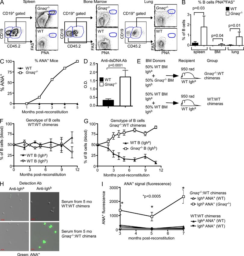
Published July 12, 2010
Ar ticle
Downloaded from jem.rupress.org on November 3, 2015
Figure 1. Altered B cell homeostasis in Gq-deficient chimeras. (A) Frozen sections of WT and Gnaq/ BM chimera spleens were stained with anti-CD90.2
to identify T cell zones (blue), anti-B220 to identify B cell follicles (green), and anti-CD21 (red) to identify follicular DC networks in the B cell follicle and CD21hi B cells
within the MZ. (B) Total B cells were isolated from WT and Gnaq/ chimera spleens and analyzed in in vitro chemotaxis assays using 300 ng/ml CXCL12, 500 ng/ml
CXCL13, 400 ng/ml CCL19, or 10 nM S1P as chemoattractants. Data are presented as the mean ± SD of triplicate wells. (C) Splenic B cells from CD45.1+ WT and
CD45.2+ Gnaq/ chimeras were purified. WT B cells were labeled with biotin and equivalent numbers of WT and Gnaq/ B cells were transferred into CD45.1+
hosts. Frozen sections were prepared from spleens 18 h after transfer and stained with SA-488 to visualize the transferred WT B cells (green), anti-CD45.2 to visual-
ize transferred Gnaq/ B cells (red), and anti–MOMA-1 (blue) to visualize the MZ. Bars, 50 µm. (D–F) BM and spleens were harvested from Gnaq/ and WT chime-
ras, stained with the indicated Abs to identify B cell subsets, and then analyzed by flow cytometry. (D) The absolute numbers of total BM B cells, pro–B, pre–B (large
and small), and immature B cells were determined. (E) The numbers of T1, T2, and T3 transitional B cells and the numbers of total splenic B cells, FOB cells, and MZB
cells were determined. (F) The numbers of splenic T2/FOII MZB precursors and T1 MZB precursors were determined. Data shown are the mean ± SD of five mice/group.
P-values were determined using an unpaired Student’s t test. Data in all panels are representative of three or more independent experiments.
the spleens of Gnaq/ chimeras (Fig. S1 D). However, incon- Interestingly, both the frequency (Fig. S1 G) and total num-
sistent with the phenotype of conventional MZB cells, 50% of ber (Fig. 1 F) of the T1-derived MZB precursors were
the CD21hiCD23lo MZ-like B cells found in the spleens of the significantly elevated in the spleens of Gnaq/ chimeras.
Gnaq/ chimeras also expressed high levels of IgD (Fig. S1 E). Collectively, the data suggest that Gq is not required for
To determine whether the expansion of MZ-like B cells B cell development in the BM. However, Gq does modulate
in the Gnaq/ chimera spleens was the result of changes peripheral B cell development and appears to control the
in the number of MZB precursors, we first examined the numbers of T1 cells, T1-derived MZB cell precursors, and
B220+CD93CD23hiCD21hi MZB cell precursors that are mature MZB and FOB cells.
thought to arise from the T2 or FOII population (Srivastava
et al., 2005; Cariappa et al., 2007). We found no difference Gq regulates B cell survival in vitro
between the percentages (Fig. S1 F) or numbers (Fig. 1 F) of Given the alterations in splenic B cell homeostasis in Gnaq/
these cells in spleens of Gnaq/ and WT chimeras. We next chimeras, we hypothesized that Gq may regulate B cell
examined the MZB precursors (B220+CD93+CD23IgMhi expansion or survival. To test this hypothesis, we cultured
CD21hi) found in the T1 population (Carey et al., 2008). splenic WT and Gnaq/ B cells in media alone or with
JEM VOL. 207, August 2, 2010 1777Published July 12, 2010
increasing amounts of anti-IgM F(ab)2 for 48 h, pulsed the survive in vitro in higher numbers than WT B cells at all stages
cells with 3H-thymidine for 5 h, and measured thymidine in- of transitional and mature B cell development.
corporation. No major differences in DNA replication were
observed between unstimulated and anti-IgM–stimulated Increased activation of BCR-mediated signaling
Gnaq/ and WT B cells (Fig. 2 A). Likewise, we did not ob- in Gnaq/ B cells
serve any differences in DNA replication between Gnaq/ GTP-bound Gq is reported to suppress activation of PI3K and
and WT B cells stimulated with agonistic anti-CD40 Abs Akt in nonhematopoietic cells (Bommakanti et al., 2000;
(Fig. 2 B). However, Gnaq/ B cells did proliferate more Ballou et al., 2003). Given the enhanced in vitro survival of
strongly (30% increase) in response to LPS than their WT Gnaq/ B cells, we next tested whether the loss of Gq leads
counterparts (Fig. 2 C), suggesting that either a higher pro- to enhanced Akt activation in B cells. We purified splenic
portion of Gnaq/ B cells are responsive to LPS or that B cells from WT and Gnaq/ chimeras, stimulated the cells
Gnaq/ B cells are hyperresponsive to TLR4 ligands. with anti-IgM F(ab)2 for 0–30 min, prepared total protein ly-
Because the loss of Gq only modestly affected the in vitro sates, and performed Western blot analysis. We found that the
proliferative responses of splenic B cells, we next examined basal levels of phospho-Akt (p-Ser473) were higher in Gnaq/
whether Gq regulates B cell survival. We cultured purified B cells than in WT B cells (Fig. 3 A). Furthermore, upon anti-
WT or Gnaq/ B cells in media alone or with anti-IgM F(ab)2 IgM stimulation phospho-Akt levels increased more rapidly in
and monitored viability. Surprisingly, a larger fraction of Gnaq/ B cells (Fig. 3 A), and the peak amount of phospho-
Downloaded from jem.rupress.org on November 3, 2015
Gnaq/ B cells remained viable in both unstimulated and anti- Akt was higher in the anti-IgM–stimulated Gnaq/ B cells
IgM–stimulated cultures (Fig. 2 D), suggesting that Gq plays a than in the anti-Ig–stimulated WT B cells (Fig. 3 B). We also
role in regulating the in vitro survival of mature B cells. To found that the levels of phosphorylated PLC2 (Y759) and
address whether Gq also modulates immature B cell survival, Erk were significantly increased in the anti-IgM–stimulated
we sort purified T1 B cells and T2/T3 B cells from the spleens Gnaq/ B cells relative to the WT B cells (Fig. 3, A and C).
of WT and Gnaq/ chimeras, cultured the cells in media alone Consistent with these results, we noted that several protein
or with anti-IgM F(ab)2 for 18 h, and measured B cell viabil species were hyperphosphorylated on either tyrosine (Fig. S2 A)
ity. As expected (Su and Rawlings, 2002), WT T1 B cells died or serine (Fig. S2 B) in the unstimulated Gnaq/ B cells rela-
rapidly after anti-Ig stimulation (Fig. 2 E), whereas WT T2/T3 tive to the WT B cells and were further phosphorylated as early
B cells were largely resistant to anti-IgM–induced cell death as 1 min after anti-IgM stimulation. Thus, several signaling sub-
(Fig. 2 F). Interestingly, significantly more of the Gnaq/ T1 strates, including Akt, are hyperphosphorylated in Gnaq/
(Fig. 2 E) and T2/T3 B cells (Fig. 2 F) survived the culture pe- B cells both before and after BCR ligation.
riod, regardless of whether the B cells were stimulated with To address whether the increased Akt phosphorylation
anti-IgM or cultured in media alone. Thus, Gnaq/ B cells observed in the anti-IgM–stimulated splenic Gnaq/ B cells
Figure 2. Gq-deficient B cells exhibit a prosurvival phenotype in vitro. (A–C) WT or Gnaq/ splenic B cells were stimulated with 0–25 µg/ml
anti-IgM F(ab)2 (A), 0–40 µg/ml anti-CD40 (B), or 0–10 µg/ml LPS (C) for 48 h and proliferative responses were measured by 3H-thymidine incorporation
after a 5-h pulse. Results represent the mean ± SD of triplicate cultures. (D–F) Purified total splenic B cells (D), sort-purified B220+CD93+CD23 T1 B cells
(E), or sort-purified B220+CD93+CD23+ T2/T3 B cells (F) were cultured in media alone or stimulated with 10–15 µg/ml anti-IgM F(ab)2 for 18–24 h. Viability
was measured by staining with propidium iodide. Data are shown as the mean ± SD of triplicate cultures. P-values were determined using an unpaired
Student’s t test. Data in all panels are representative of three independent experiments.
1778 Gq suppresses B cell–mediated autoimmunity | Misra et al.Published July 12, 2010
Ar ticle
was a result of the presence of more MZB cells in the Gnaq/ Next, to address whether the Gnaq/ B cells are more fit
spleens, we assessed phospho-Akt levels in CD43neg naive to survive than WT B cells, we prepared mixed BM chimeras
B cells and enriched populations of FOB and MZB cells. As by transferring equivalent numbers of WT (CD45.1+) and
expected, phospho-Akt levels were constitutively elevated in Gnaq/ (CD45.2+) BM cells into lethally irradiated WT
naive CD43neg Gnaq/ B cells and more rapidly reached CD45.1+ recipient mice (Fig. 4 D) and determined the rela-
peak levels after anti-IgM stimulation (Fig. 3 D). In contrast, tive composition of the splenic B cells after reconstitution.
the phospho-Akt levels in the enriched FOB cells were We found that the ratio of Gnaq/ B cells to WT B cells was
equivalent between the WT and Gnaq/ B cells both before already significantly increased by 1 mo after reconstitution
and after anti-IgM stimulation (Fig. 3 D). However, phospho- (Fig. 4 E). Furthermore, by 8 wk after reconstitution the ratio
Akt levels were constitutively elevated in the Gnaq/ MZB of Gnaq/ to WT B cells was 4:1, and within 6 mo the ratio
cells relative to the WT MZB cells (Fig. 3 D). Furthermore, was 19:1 in favor of the Gnaq/ B cells (Fig. 4 E). We found
phospho-Akt levels increased to peak levels more rapidly that the FOB and MZB compartments were both skewed
and were sustained for extended periods of time after BCR in favor of the Gnaq/ B cells at 8 wk after reconstitution;
stimulation in the Gnaq/ MZB cells (Fig. 3 D). Together, however, the MZB cell compartment was more highly
these data suggest that Akt activation after BCR engagement skewed than the FOB compartment toward the Gnaq/
is specifically enhanced in Gnaq/ MZB cells. B cells (Fig. 4 F). Likewise, all of the transitional B cells in
the spleen were already highly skewed toward the Gnaq/
Downloaded from jem.rupress.org on November 3, 2015
Gnaq/ B cells outcompete WT B cells for access to splenic B cells by 8 wk after reconstitution (Fig. 4 F). Interestingly,
niches in vivo the T1 MZB precursor population was almost entirely de-
Collectively, our data suggested that Gnaq/ B cells may rived from the Gnaq/ B cells within 8 wk after reconstitu-
survive better than WT B cells in vivo. To test this, we trans- tion (ratio of 20:1; Fig. 4 F). The skewing of the peripheral
ferred equivalent numbers of splenic B cells from WT or B cell compartments toward Gnaq/ B cells was not a result
Gnaq/ chimeras (CD45.2+) into intact congenic CD45.1+ of changes in the output of B cell progenitors from the BM,
WT hosts (Fig. 4 A). At various time points after transfer, we as the ratio of Gnaq/ to WT immature B cells in the BM
enumerated donor CD45.2+ B cells in the spleen by FACS. (fraction E) remained very close to 1:1 at 8 wk after reconsti-
As shown in Fig. 4 (B and C), the percentages and numbers tution (Fig. 4 F). However, the ratio of Gnaq/ to WT
of donor Gnaq/ B cells recovered at 1 wk after transfer B220+CD93+IgM+CD23+ transitional B cells in the BM
were significantly higher compared with those found in re- (Lindsley et al., 2007) increased slightly (2.0) and the re
cipients receiving WT B cells. At days 14 and 21 after transfer, circulating mature B cells in the BM were highly skewed
very few of the transferred WT B cells could be found in the toward the Gnaq/ genotype (10:1 ratio; Fig. 4 F).
congenic hosts (Fig. 4 B). However, the transferred Gnaq/ Finally, to evaluate whether the increase in Gnaq/ B cells
B cells were easily detectable and present at 10-fold higher relative to WT B cells in the spleen of the mixed chimeric mice
frequencies and numbers than the WT B cells (Fig. 4, B and C). was the result of increased proliferation of the Gnaq/ B cells,
Thus, the data suggested that Gnaq/ B cells survive longer we injected the 50:50 BM chimeras with 5-ethynyl-2-
in vivo than WT B cells. deoxyuridine (EdU) at 8 wk after reconstitution and then
Figure 3. Gq-deficient B cells are hyperresponsive to BCR triggering. (A–D) Splenic B cells from Gnaq/ and WT chimeras were stimulated with
anti-IgM F(ab)2 for 0–30 min. Protein lysates were prepared from negatively selected total B cells (A and D) or from enriched FOB and MZB cells (D) and
then analyzed by Western blotting. Akt (p-Ser473), PLC2, and Erk were probed with phospho-specific Abs and protein-specific Abs. Li-COR quantitative
densitometry was used to determine the fold changes in phospho-Akt (B) and phospho-PLC2 (C). The ratios of phospho-specific proteins versus total ERK
or PLC2 were obtained and data were normalized to WT B cells at time 0 (set as 1). Data in all panels are representative of two independent experiments.
JEM VOL. 207, August 2, 2010 1779Published July 12, 2010
Downloaded from jem.rupress.org on November 3, 2015
Figure 4. Gq-deficient B cells have a competitive survival advantage over WT B cells in vivo. (A–C) Splenic B cells were purified from WT and
Gnaq/ chimeras (CD45.2+) and transferred i.v. into CD45.1+ WT hosts (2.5 × 10⁷ cells/host). (B) Spleen cells from recipient mice were harvested at 1–3 wk
after transfer and analyzed by flow cytometry to identify donor B cells. Representative dot plots and the mean percentage ± SD of cells of donor origin are
shown. (C) The number of donor B cells recovered per recipient spleen was determined and is shown as the mean ± SD (n = 3–5 mice/group/time point).
Data are representative of two independent experiments. (D–G) Lethally irradiated CD45.1+ WT mice were reconstituted with a mixture of BM
consisting of 50% Gnaq/ (CD45.2+) BM and 50% WT (CD45.1+) BM. (E) Splenic B cells were collected from the 50:50 BM chimeras and flow cytometry
was used to identify WT and Gnaq/ B cells. Data are presented as the ratio of Gnaq/ to WT B cells (percentage of Gnaq/ B cells divided by percentage
of WT B cells) at each time point and are shown as the mean ± SD from five mice per group. Data are representative of more than three independent
experiments. (F) BM and spleen were collected from the 50:50 BM chimeras at 2 mo after reconstitution, and the ratio of Gnaq/ to WT B cells was
determined as described in E. Splenic B cells were subdivided into total mature (B220+CD93), FOB (B220+CD21loCD23hi), MZB (B220+CD21hiCD23lo), T1
(B220+CD93+CD23), T2/T3 (B220+CD93+CD23+), T1 MZB precursors (B220+CD93+IgMhiCD21hi), and T2/FOII MZB precursors (B220+CD93CD21hiCD23hi).
B cells in the BM were subdivided into immature (fraction E) B cells (B220+CD93+IgM+), transitional (B220+CD93+IgMhiCD23+), and recirculating
(B220hiIgM+IgD+). Data are shown as the mean ± SD of four to five mice/group. (G) 50:50 BM chimeras (2 mo after reconstitution) were injected with EdU
twice over a 12-h period. Spleen cells were isolated at 18 h, stained with Abs to CD19 and CD45.2 and the EdU detection reagent, and then analyzed by
flow cytometry. Representative FACS plots are shown and the percentage of B cells of each genotype that incorporated EdU is provided. Data shown are
the mean ± SD of five mice. Data in D–G are representative of two independent experiments.
1780 Gq suppresses B cell–mediated autoimmunity | Misra et al.Published July 12, 2010
Ar ticle
Figure 5. Gnaq/ B cells are more resistant to
BAFF neutralization in vivo and are more sensitive
to BAFF stimulation in vitro. (A–D) 50:50 mixed BM
chimeras were generated using Gnaq/ BM (CD45.2+)
and WT (CD45.1+) BM. 8 wk after reconstitution, chimeric
animals were injected i.p. with a blocking Ab to BAFF/
BLyS (clone 10F4) or an isotype control Ab. (B) Peripheral
blood samples were collected from Ab-treated chimeric
animals between 7 and 21 d and flow cytometry was
used to identify the percentage of B cells that were of
WT or Gnaq/ origin. Data are shown as the ratio
(±SEM) of Gnaq/ to WT B cells (percentage of Gnaq/
B cells divided by percentage of WT B cells; n = 5–10
mice). P-values were determined using an unpaired Stu-
dent’s t test. (C) Spleen cells from Ab-treated chimeras
were isolated, enumerated, and analyzed by flow cytom-
etry at 21 d after treatment. The numbers of mature
splenic B cells (B220+CD24lo) of WT and Gnaq/ origin
Downloaded from jem.rupress.org on November 3, 2015
were determined and are reported as the mean ± SD of
five mice/group. The fold depletion caused by BAFF neu-
tralization is shown above the bar graphs. (D) The ratio of
Gnaq/ to WT B cells in the spleen was calculated (per-
centage of Gnaq/ B cells divided by percentage of WT
B cells) and is shown as the mean ± SD. P-values were
determined using an unpaired Student’s t test. Data in
A–D are representative of three independent experiments.
(E) Splenic CD43neg naive B cells were isolated from
Gnaq/ and WT chimeras and enriched for CD21lo FOB
and CD21hi MZB cells. The total or enriched B cell subsets
were cultured in BAFF-free media overnight. Live cells
were recovered from the cultures and stimulated with
recombinant BAFF for 0–10 min. Protein lysates were
prepared and analyzed by Western blotting. Phospho-Akt
(p-Ser473), total Akt, and phospho-S6 ribosomal protein
were detected using phospho- and protein-specific Abs.
Data in E is representative of two experiments.
determined the proportion of WT and Gnaq/ B cells that B cells to WT B cells was 1.5 at 8 wk after reconstitution
proliferated in 18 h. As shown in Fig. 4 G, the percentage of (Fig. 5 B), indicating that Gnaq/ B cells were already over-
replicating B cells was not significantly different between represented in the blood at this time. The ratio of Gnaq/
Gnaq/ and WT B chimeras. Together, these data argue that B cells to WT B cells in peripheral blood modestly increased
the Gnaq/ B cells are not overtly more proliferative than over the next 3 wk in the chimeras that were treated with the
WT B cells but are able to efficiently outcompete WT B cells isotype control Ab (Fig. 5 B), which is consistent with the
for access to or retention within peripheral survival niches. gradual takeover by Gnaq/ B cells. However, the ratio of
Gnaq/ B cells to WT B cells increased even more rapidly
Gnaq/ B cells are more resistant to BAFF withdrawal in the anti-BAFF–treated chimeras (Fig. 5 B).
Because Gnaq/ B cells quickly outcompete WT B cells in Next, we determined the percentage (Fig. S3 B) and num-
the spleen, we next tested whether the Gnaq/ B cells are ber (Fig. 5 C) of mature WT (CD45.1+) and Gnaq/ (CD45.2+)
less reliant on survival factors such as BAFF. We generated B cells present in the spleens of the mixed BM chimeras
50:50 mixed BM chimeras by transferring equal amounts of on day 21 after treatment. As expected, approximately three
CD45.1+ WT BM and CD45.2+ Gnaq/ BM to irradiated times more mature Gnaq/ B cells were recovered relative
CD45.1 WT hosts. We treated the recipients with a neutral- to WT B cells in the control Ab–treated chimeras (Fig. 5 C).
izing anti-BAFF or isotype control Ab 8 wk after reconstitu- After BAFF blockade, we observed a significant reduction in
tion and then monitored WT and Gnaq/ B cells in the the number of total mature B cells, regardless of genotype
peripheral blood and spleen over the next 3 wk (Fig. 5 A). (Fig. 5 C). However, although the number of WT B cells
We determined the percentage of B cells in peripheral blood decreased by 23-fold after anti-BAFF treatment, the number
that were either Gq deficient or WT at each of the time of Gnaq/ B cells dropped by only 13.5-fold (Fig. 5 C).
points (Fig. S3 A) and then calculated the ratio of Gnaq/ Furthermore, the ratio of splenic Gnaq/ B cells to WT B cells
B cells to WT B cells. The ratio of peripheral blood Gnaq/ increased from 3.1 in the control Ab–treated chimeras to 5.0
JEM VOL. 207, August 2, 2010 1781Published July 12, 2010
Downloaded from jem.rupress.org on November 3, 2015
Figure 6. Gq-deficient B cells display an activated phenotype and are intrinsically prone to develop autoreactive specificities. (A and B) 50:50
mixed BM chimeras were generated using Gnaq/ BM (CD45.2+) and WT (CD45.1+) BM. 11 wk after reconstitution, cells from BM, spleen, and lungs
were harvested and B cells were analyzed by flow cytometry for expression of PNA and FAS. Representative FACs plots are shown (A) and the percentages
of WT or Gnaq/ B cells expressing a GC phenotype (PNAhiFAS+) were determined (B). Data are shown as the mean ± SD of four to five mice per
time point. P-values were determined using an unpaired Student’s t test. All data shown are representative of two independent experiments. (C) WT and
Gnaq/ BM chimeras were generated and aged for 5 mo. Serum samples were collected monthly. The presence of ANAs (ANA reactivity) in the serum
(diluted 1:100) was then determined by fluorescence microscopy using fixed HepG2 cells. The data are shown as the percentage of animals with detect-
able ANAs in the serum (n = 10 mice/group). (D) Serum samples were collected from female WT and Gnaq/ chimeras at 9 mo after reconstitution. Sam-
ples were evaluated for reactivity to dsDNA by ELISA. Data shown are the mean OD ± SD from a 1/200 dilution of the serum samples. n = 4–5 mice/group.
P-values were determined using an unpaired t test. Data in panels C and D are representative of two or more independent experiments. (E–I) 50:50 mixed
BM chimeras were made by reconstituting irradiated Igha hosts with 50% WT BM (Igha allotype) and 50% Gnaq/ BM (Ighb allotype; referred to as
Gnaq/:WT chimeras) or with 50% WT BM (Igha allotype) plus 50% WT BM (Ighb allotype; referred to as WT:WT chimeras). (F and G) Peripheral blood
from the WT:WT (F) and Gnaq/:WT (G) chimeras was analyzed by flow cytometry between 2 and 10 mo after reconstitution using allotype-specific Abs
to identify the B cells of either WT or Gnaq/ origin. Data are shown as the mean percentage of B cells of the different genotypes ± SD. n = 10 mice/
group. (H) Serum, isolated from WT:WT and Gnaq/:WT chimeras at 5 mo after reconstitution, was incubated (1:100 dilution) with fixed HepG2 cells.
ANA-reactive Abs were detected using allotype-specific Abs. Representative fluorescent images are shown. Bars, 50 µm. (I) The fluorescence signal de-
tected from each serum sample was determined and shown as the mean fluorescent signal ± SD (n = 9–10 mice/group). Data in E–I are representative of
two independent experiments. P-values were determined using an unpaired Student’s t test.
1782 Gq suppresses B cell–mediated autoimmunity | Misra et al.Published July 12, 2010
Ar ticle
in the anti-BAFF–treated chimeras (Fig. 5 D). This change in B cells from the same animal expressed PNAhiFas+ in spleen,
the ratio of splenic Gnaq/ B cells to WT B cells was not the lung, and BM (Fig. 6, A and B), suggesting that Gnaq/ B cells
result of an obvious difference in BAFFR expression levels make up most of the GCs present in these mice. In support
between WT and Gnaq/ B cells (Fig. S3, C and D). Thus, of this conclusion, we identified well defined B cell follicles in
Gnaq/ B cells are significantly more resistant to BAFF deple- the lungs of Gq-deficient chimeras, and some of the B cells
tion than their WT counterparts. within these follicles expressed the GC marker GL7 (Fig. S4 B).
BAFF signaling in B cells is reported to stimulate PI3K As these structures are not found in the lungs of naive normal
and induce activation of the Akt signaling pathway (Patke et al., mice (Moyron-Quiroz et al., 2004), the data suggest that Gnaq/
2006). Given the increase in constitutively phosphorylated B cells are being activated by endogenous self-ligands.
Akt observed in the Gnaq/ B cells and the increased
resistance of these B cells to BAFF depletion, we evaluated Intrinsic defects in Gnaq/ B cells result
whether Gnaq/ B cells are more responsive to BAFF sig- in the development of autoreactive B cells
nals than their WT counterparts. We isolated splenic CD43neg To address whether Gnaq/ B cells are self-reactive, we evalu-
naive B cells, FOB cells, and MZB cells from WT and Gnaq/ ated whether serum Abs from WT and Gnaq/ chimeras were
chimeras. We cultured the cells overnight in the absence of reactive in a standard anti-nuclear Ab (ANA) assay. As expected,
BAFF, purified the live cells, stimulated the BAFF-starved none of the serum samples from WT chimeras exhibited ANA
B cells with recombinant BAFF protein, and then monitored reactivity at any time point after reconstitution (Fig. 6 C).
Downloaded from jem.rupress.org on November 3, 2015
activation of the Akt signaling pathway by measuring phos- In contrast, 20% of the serum samples from Gnaq/ chimeras
phorylation of Akt and one of its downstream signaling targets, were ANA reactive within 2 mo after reconstitution. By 5 mo
S6 ribosomal protein. As shown in Fig. 5 E, phospho-Akt was after reconstitution, 100% of the serum samples from Gnaq/
observed at higher levels in BAFF-starved Gnaq/ CD43neg chimeras were ANA+ (Fig. 6 C). Interestingly, we observed
B cells than in WT CD43neg B cells. After BAFF stimulation, that some of the serum samples strongly stained the entire nu-
phospho-Akt levels increased more rapidly in the Gnaq/ cleus, whereas others exhibited a more punctate pattern of
B cells. This finding was recapitulated when either FOB or staining (Fig. S4 C), suggesting that multiple auto-Ags can be
MZB cells were analyzed (Fig. 5 E) but was most striking in recognized by Gnaq/ B cells. Indeed, we also identified sig-
the Gnaq/ FOB cell subset (Fig. 5 E). Interestingly, although nificantly higher levels of anti–double-stranded (ds) DNA reac-
Gq deficiency amplified BAFF-dependent Akt activation in tivity in some Gnaq/ chimera serum samples (Fig. 6 D).
both FOB and MZB cell subsets, phosphorylation of S6 ribo- To determine whether the development of autoreactive
somal protein was equivalent between BAFF-stimulated WT B cells in Gnaq/ chimeras is the result of an intrinsic defect
and Gnaq/ FOB cells (Fig. 5 E). However, phospho-S6 in the Gnaq/ B cells, we lethally irradiated WT Igha allotype
levels were constitutively higher in Gnaq/ MZB cells than congenic mice and reconstituted them with 50% WT Igha
in WT B cells and were induced to higher levels in the BM and 50% Ighb Gnaq/ BM (Gnaq/:WT chimera) or
Gnaq/ MZB cells after BAFF stimulation (Fig. 5 E). Together, with 50% WT Igha BM + 50% WT Ighb BM (WT:WT chi-
these data indicate that the Akt prosurvival pathway is con mera; Fig. 6 E). At various time points after reconstitution, we
stitutively more active in BAFF-deprived Gnaq/ B cells and collected serum and peripheral blood cells. As expected, pe-
that Gnaq/ B cells engage this pathway more efficiently than ripheral blood B cells from the WT:WT chimeras were evenly
WT B cells after BAFF stimulation. distributed, with 50% of the B cells expressing an Igha BCR
and the other half expressing Ighb BCRs (Fig. 6 F). In contrast,
Gnaq/ B cells are spontaneously activated in vivo in the Gnaq/:WT chimeras, the Ighb-expressing Gnaq/
Because Gnaq/ B cells are more resistant to BAFF depriva- B cells outcompeted the Igha-expressing WT B cells and
tion, we postulated that Gnaq/ B cells may survive peripheral eventually became the dominant B cell population present
tolerance checkpoints that normally eliminate B cells with self- in the peripheral blood (Fig. 6 G). We next evaluated the
reactivity. If this were true, then Gnaq/ B cells might be acti- serum samples for ANA reactivity using allotypic-specific Abs
vated and responding to self-antigens. To test this hypothesis, to detect the ANAs. As expected, neither Igha nor Ighb Abs
we made 50:50 mixed BM chimeras by reconstituting CD45.1+ from the serum of WT:WT chimeras exhibited ANA reactiv-
WT recipients with an equal mixture of CD45.1+ WT BM and ity (Fig. 6 H). However, analysis of serum from the Gnaq/:
CD45.2+ Gnaq/ BM. We isolated cells from spleen, BM, and WT chimeras, revealed that the Ighb Abs (derived from the
lung at 3 mo after reconstitution and used FACS to phenotypi- Gnaq/ B cells) efficiently stained the nucleus of HepG2 cells
cally characterize the B cells. Gnaq/ B cells from the mixed (Fig. 6 H). In contrast, the Igha Abs (derived from the WT
BM chimeras expressed statistically higher levels of CD80, B cells present in the same host) did not stain HepG2 nuclei
CD40, and MHC class II than the WT B cells isolated from the (Fig. 6 H). Indeed, ANA reactivity of the WT Igha Abs from
same animals (Fig. S4 A). Next, using peanut agglutinin (PNA) the Gnaq/:WT chimeras did not rise above background and
and FAS to identify B cells with a germinal center (GC) pheno- was equivalent to that observed for the Igha Abs from WT:WT
type (PNAhiFas+), we found only a very small proportion of chimeras (Fig. 6 I). This was not the result of a lack of Igha
WT B cells with this phenotype in any tissue (Fig. 6, A and B). Ab in the Gnaq/:WT serum samples, as nonautoreactive
However, significantly higher proportions of the Gnaq/ Igha Abs were present at low, but still detectable, levels in
JEM VOL. 207, August 2, 2010 1783Published July 12, 2010
Downloaded from jem.rupress.org on November 3, 2015
Figure 7. Gq-deficient chimeras develop B cell–dependent systemic autoimmunity. (A and B) Arterial blood from WT and Gnaq/ BM chimeras (8 mo
after reconstitution) was isolated and complete blood cell counts were performed. Mean red blood cell counts (A) and hemoglobin concentration (B) are reported.
Data shown are the mean ± SD of four mice/group and are representative of two independent experiments. P-values were determined using an unpaired Student’s t
test. (C) The diameters of ankle and wrist joints were measured in WT and Gnaq/ chimeras at 5 mo after reconstitution. Data are represented as the mean ± SD of
8–10 mice/group. Similar results were seen in three experiments. P-values were determined using an unpaired Student’s t test. (D–K) Histological analysis (hema-
toxylin and eosin [H&E]) of knee and ankle joints from WT and Gnaq/ chimeras was performed at 5 mo after reconstitution. (D) Normal knee joint from a WT
chimera showing femur (F) and Tibia (T). (E) Knee joint from a Gnaq/ chimera with focal soft tissue inflammation (arrow) adjacent to a joint space (S) where the
synovial lining is disrupted. A higher power view (F) demonstrates a focus of mixed inflammatory cells (arrow) and necrosis of the overlying synovium. (G) The ar-
ticular surface of the distal femur of a Gnaq/ chimera is irregular, thinned, and covered by fibrous pannus (arrows). (H) Normal ankle joint from a WT chimera.
(I) Ankle from a Gnaq/ chimera with marked inflammation of soft tissues accompanied by exostotic bone (arrows) and accumulation of fibrin within the joint space (S).
(J) Bone from the foot of a Gnaq/ chimera with proliferative periostitis overlying an area of bone lysis represented as scalloping of the bone surface (arrows).
(K) Higher power of bone from the foot shows multinucleated osteoclasts within resorptive foci (arrows). Images are from one representative animal out of 5–10
mice/group. Bars: (D–J) 100 µm; (K) 20 µm. Two independent experiments were performed. (L) Survival of Gnaq/ and WT chimeras was tracked for 6 mo after BM
reconstitution. Data are shown as the percentage of survival of each group (n = 10 mice/group). P-values were determined using the Wilcoxon test. Data are repre-
sentative of three independent experiments. (M) Gq-deficient and WT chimeras were treated with anti-CD20 (clone 18B12) or control Ab at 2 mo after reconstitu-
tion and survival was tracked over the next 11 mo. The experiment was performed once with 10 mice/group. P-values were determined using a Log-rank test.
the Gnaq/:WT chimera serum samples (Fig. S4 D). Together, Gq suppresses the development of systemic autoimmunity
the data indicate that the development of autoreactive B cells Given the propensity of Gnaq/ B cells to secrete autoreactive
is dependent on the loss of Gq expression in B cells. Further- Abs, we predicted that Gnaq/ chimeras would develop auto
more, the loss of Gq in other hematopoietic cells is not suffi- immune disease. To test this, we determined whether immune
cient to drive normal B cells to become autoreactive. complexes were present in the kidneys of WT and Gnaq/
1784 Gq suppresses B cell–mediated autoimmunity | Misra et al.Published July 12, 2010
Ar ticle
chimeras. No Ab-containing immune complexes were detected control or anti-CD20 Ab (Fig. 7 M). Gnaq/ chimeras
in the WT kidney sections (Fig. S5, A–C). In contrast, IgG2a- treated with the control Ab had high rates of mortality, with
and IgG2c-containing immune complexes were reproducibly 60% of the chimeras dying within 10 mo of reconstitution
observed in kidney sections from Gnaq/ chimeras (Fig. S5, (Fig. 7 M). In striking contrast, only 10% of the anti-CD20–
D–F). However, despite immune complex deposition in the treated Gnaq/ chimeras died during the study (Fig. 7 M).
kidney, kidney function, as assessed by measuring protein Thus, Gq expression by B cells is necessary for normal pe-
concentration in the urine, was normal in the Gnaq/ chime ripheral B cell development and tolerance induction and also
ras (unpublished data). Histopathologic analysis of the kidneys plays a role in preventing early onset mortality in mice.
from Gnaq/ chimeras showed mild to moderate mesangiolysis
(thrombotic microangiopathy) with splitting of the basement DISCUSSION
membranes in the glomeruli and increased mesangial matrix It is estimated that >800 seven-transmembrane GPCRs are
within the glomeruli (Fig. S5, G–J). Although this pathology encoded in the mammalian genome. B lymphocytes, like other
is atypical for the most common forms of immune complex– hematopoietic cells, express GPCRs, some of which play im-
mediated disease, thrombotic microangiopathy can be a mani- portant roles in regulating B lymphocyte adhesion and traffick-
festation of disease in Lupus patients with anti-coagulant, anti- ing (Wettschureck et al., 2004). In this paper, we show that Gq
phospholipid, or anti-endothelial Abs (Tektonidou, 2009). regulates B cell repertoire selection by controlling B cell survival
In addition to changes in the kidney, the chimeras exhib- during transitional cell differentiation. Aberrant regulation at this
Downloaded from jem.rupress.org on November 3, 2015
ited other symptoms consistent with autoimmune disease. point allows inappropriate survival of autoreactive B cells, ulti-
For example, we noted that the BM from the older Gnaq/ mately resulting in the development of autoimmune disease.
chimeras appeared to have reduced numbers of RBCs. To test Gq is expressed in B lineage cells throughout develop-
whether the Gnaq/ chimeras develop anemia, we performed ment (Wilkie et al., 1991). However, Gq appears to have
complete blood counts on arterial blood from WT and little to no effect on the survival or expansion of BM B cell
Gnaq/ chimeras. We found that the number of RBCs progenitors. Instead, Gq exerts its effects at the earliest T1
and the concentration of hemoglobin were significantly stage of transitional B cell development and extends its influ-
reduced in the Gnaq/ chimeras (Fig. 7, A and B). These ence to all transitional and mature B cells subsets. Interest-
symptoms are consistent with a diagnosis of autoimmune he- ingly, the loss of Gq particularly favors the development of
molytic anemia as a result of the presence of anti-RBC Abs, MZB-like cells. At least three precursors for the mature MZB
or they could also be a consequence of the autoimmune kid- cells have been identified: the T2 MZB precursor (Srivastava
ney thrombotic microangiopathy (Tektonidou, 2009). et al., 2005), the T1-MZB precursor (Carey et al., 2008), and
We also monitored joint swelling in WT and Gnaq/ the FOII precursor (Cariappa et al., 2007). Interestingly, only
chimeras, as we observed that many of the older Gnaq/ the T1-MZB precursor population was expanded in the
chimeras were less mobile than their WT counterparts. Sig- Gnaq/ chimeras and this population was the most highly
nificant differences in the size of the joints of Gnaq/ chi- skewed in favor of Gnaq/ B cells in the mixed BM chimeras.
meras was observed within 3.5 mo of reconstitution and Therefore, we think it likely that the Gnaq/ MZB-like cells
the penetrance of disease in the Gnaq/ chimeras was 100% develop directly from the T1 MZB precursors.
(unpublished data). By 5 mo after reconstitution, significant The MZB compartment contains B cells that are posi-
swelling was observed in both ankle and wrist joints of Gnaq/ tively selected from a subpopulation of proliferating T2 B cells
chimeras compared with the WT chimeras (Fig. 7 C). Histo- (Meyer-Bahlburg et al., 2008) as well as potentially autoreac-
logical analysis of knees, ankles, and portions of feet from tive B cells that escape deletion at the T1 to T2 checkpoint
Gnaq/ chimeras revealed a variety of arthritic changes includ (Thien et al., 2004). T1 B cells with low affinity for self-antigens
ing chronic/active areas of inflammation within the joints, normally up-regulate BAFFR, colonize the B cell niches of
evidence of synovitis and bone resorption, exostotic bone devel- the spleen, and enter the mature B cell pool (Thien et al.,
opment, and osteolytic activity (Fig. 7, D–K). 2004). In contrast, B cells of intermediate affinity for self-antigen
Collectively, our data suggest that Gnaq/ chimeras de- are excluded from the splenic niches and rapidly die (Cyster
velop systemic autoimmune disease with multiorgan involve- et al., 1994). However, when BAFF is readily available, the
ment. Consistent with this, we noted a significant difference intermediate affinity autoreactive B cells are rescued and
between the survival of Gnaq/ and WT chimeras with up positively selected into the MZ compartment (Thien et al.,
to 50% of the Gnaq/ chimeras dying within 6 mo of recon- 2004). These results indicate that B cells with a higher affinity
stitution (Fig. 7 L). To test whether the loss of Gq in B cells for self-antigen compete less effectively than the lower affin-
is responsible for the increased mortality observed in the ity B cells for a limiting pool of BAFF. It is clear from our
Gnaq/ chimeras, we depleted B cells in WT and Gnaq/ data that the Gnaq/ B cells can outcompete WT B cells under
chimeras at 8 wk after reconstitution using anti–mouse CD20 homeostatic conditions and that Gnaq/ B cells are less
(Hamel et al., 2008; Yu et al., 2008). We then monitored dependent on BAFF for survival than WT B cells. This would
survival in the anti-CD20 and control Ab–treated chimeras permit relaxed stringency of negative selection for the
over the next several months. As expected, mortality rates Gnaq/ B cells and allow for the entry of potentially autore-
were very low in WT chimeras treated with a single dose of active cells into the mature MZB compartment. However, it
JEM VOL. 207, August 2, 2010 1785Published July 12, 2010
is important to note that the T1 compartment, which is initially increased active PI3K, resulting in the augmented phosphory
BAFF unresponsive, is also substantially expanded in the Gnaq/ lation of Akt, activation of the mTOR pathway, and an in-
chimeras. Thus, either the loss of Gq affects more than just crease in the MZB compartment. Our experiments indicate
BAFF responsiveness or a proportion of the T1 cells can become that the increased Akt activity observed after BCR engagement
BAFF responsive as they transit this developmental window. is most pronounced in the MZ-like B cells found in Gnaq/
Regardless, our combined data strongly imply that loss of Gq chimeras. This selective enhanced engagement of the pro
facilitates the inappropriate survival of B cells in the periphery. survival Akt pathway may explain why these MZB cells are
The data presented in this paper suggests that Gq is an im- present in larger numbers in the Gnaq/ chimeras. Interest-
portant regulator of B cell homeostasis and survival. Although ingly, activation of Akt was very similar in the anti-IgM–
the neuronal defects in Gnaq/ mice make it impractical for stimulated FOB cells from Gnaq/ and WT chimeras. This
us to monitor the long-term development of clinical auto result was initially unexpected given that both MZB and
immunity without generating BM chimeras, B cell homeostasis FOB cell subsets are expanded in the Gnaq/ mice. However,
was identical in the Gnaq/ mice and the Gnaq/ chimeras. Akt is also constitutively more activated in BAFF-deprived
Therefore, our results are not an aberrant manifestation of irra- Gnaq/ B cells and is increased more strongly after BAFF
diation and reconstitution but are a result of the loss of Gq spe- stimulation. This enhanced response to BAFF was observed
cifically in hematopoietic cells. In addition, our data strongly in both the FOB and MZB cells from Gnaq/ chimeras.
argue that the Gnaq/ B cells are intrinsically defective and are Furthermore, BAFF-induced phosphorylation of S6, a down-
Downloaded from jem.rupress.org on November 3, 2015
more resistant than the WT B cells to cell death–inducing signals stream target of the Akt–mTOR pathway, was evident in both
such as BAFF withdrawal or strong BCR signals. Although the MZB and FOB subsets, although it was most affected in the
we do not yet know all the mechanistic details surrounding Gnaq/ MZB cells. Collectively, these data suggest that Gq
Gq-mediated suppression of B cell survival, we do know that deficiency confers both FOB and MZB cells with an enhanced
unstimulated and BCR- or BAFF-activated Gnaq/ B cells ability to activate the PI3K–Akt pathway.
have higher levels of active Akt as measured by phosphorylation Although we have made progress in identifying the pro-
of Ser473, a phosphorylation site which is under PI3K control. survival signaling pathway that is normally suppressed by Gq,
Prior work studying fibroblast cell lines (Bommakanti et al., we do not yet know which GPCR or ligand mediates this
2000; Ballou et al., 2003) and cardiomyocytes (Howes et al., dampening signal in peripheral B cells. Our preliminary tran-
2003) showed that Gq normally represses Akt activation. Fur- scriptome analysis indicated that >100 different GPCRs, in-
thermore, overexpression of Gq in cardiomyocytes leads to car- cluding chemokine receptors, are expressed by mature B cells.
diac hypertrophy as well as cardiomyocyte apoptosis (Adams Our data suggest that B cell chemotaxis to CXCR4, CXCR5,
et al., 1998). Based on these data, it is speculated that low-level Edg1, and CCR7 ligands is not abrogated by the loss of Gq.
Gq activity may lead to PI3K-dependent Akt activation and Although these data do not exclude the possibility that che-
cell survival, whereas high or sustained Gq activity results in mokine receptors play a separate Gq-dependent role in regu-
PI3K/Akt repression and cell death (Hubbard and Hepler, 2006). lating B cell survival, it is equally likely that one of the many
Although the active GTP-bound form of Gq interacts non-chemokine receptor GPCRs expressed by B cells regu-
with different signaling partners, including PLC- and Btk lates B cell survival. Unfortunately, surprisingly little is known
(Hubbard and Hepler, 2006), published data indicate that regarding the role for these other GPCRs in B cells and more
Gq mediates its pro-apoptotic effect in myocytes by bind- work will be needed to evaluate their potential roles in B cell
ing to p110 and suppressing the production of PIP3 by development and selection.
PI3K (Bommakanti et al., 2000; Ballou et al., 2003). This The autoimmune disease that develops in Gnaq/ chimeras
Gq-dependent inhibition of PI3K activity leads to reductions is similar, but not identical, to the disease that develops in BAFF
in Akt activation and suppression of the prosurvival mTOR Tg mice (Mackay et al., 1999). For example, both strains of mice
pathway (Manning and Cantley, 2007). Interestingly, expres- have increased numbers of MZB cells, develop auto-Abs, and
sion of activated p110 in B lymphocytes is sufficient to ensure exhibit signs of lupus-like disease. However, kidney failure did
survival of the mature B cell population, even when the BCR not occur in Gnaq/ chimeras, despite evidence of immune
has been deleted (Srinivasan et al., 2009). Furthermore, deletion complex deposition and mesangiolysis. Instead, the Gnaq/
of PTEN, an inhibitor of PI3K, also promotes mature B cell chimeras develop arthritis and become anemic. Furthermore,
survival (Srinivasan et al., 2009) and can rescue autoreactive the Gnaq/ chimeras develop inflammatory lesions and ectopic
B cells from anergy (Browne et al., 2009). Interestingly, the lymphoid tissues in multiple organs, including the lung. Given
autoreactive prone MZB cell compartment is also expanded the speed with which autoimmune disease manifests in the
in mice expressing activated p110 or lacking PTEN (Anzelon Gnaq/ chimeras and the number of affected tissues, we believe
et al., 2003; Srinivasan et al., 2009). Finally, B cells isolated that the animals suffer from a widespread autoimmune syndrome
from multiple models of autoimmunity are reported to ex- that affects multiple organ systems and likely involves autoreac-
press elevated levels of phospho-Akt (Wu and Mohan, 2009), tive B cells of different specificities. Importantly, the increased
and perturbations in the PI3K/Akt axis can lead to the devel- mortality observed in Gnaq/ chimeras is dependent on B cells,
opment of autoimmunity (Wu and Mohan, 2009). Based on as a single round of B cell depletion significantly delayed the
these data, we speculate that the loss of Gq in B cells leads to onset of morbidity and mortality. Although we do not yet know
1786 Gq suppresses B cell–mediated autoimmunity | Misra et al.Published July 12, 2010
Ar ticle
whether Gq deficiency in B cells alone is sufficient to induce anti-BAFFR-AF647, and CD45.2-APC-AF750 were produced by eBioscience,
and IA/IE Pacific Blue and CD80-AF647 were obtained from BioLegend. All
autoimmune disease, we do know that the loss of Gq specifi-
other Abs were purchased from BD. To analyze in vivo cell proliferation, chime-
cally within B lineage cells is required for the development and ras were injected i.p. twice with the nucleic acid homologue EdU (Invitrogen)
survival of the autoreactive B cells. Together, our results indicate 18 h before harvest. Splenocytes were then stained with Abs to CD19 and CD45.2,
that the loss of Gq in other hematopoietic lineage cells, includ- fixed with 1% neutral buffered formalin, permeabilized with saponin-based buffer,
ing T cells, is not sufficient to drive the selection of normal and incubated with the manufacturer-provided Pacific blue–conjugated EdU
B cells into the autoimmune repertoire. detection agent. Flow cytometry data were collected using FACSCalibur,
Canto II, or LSR II instruments (BD) from the University of Rochester Flow
Although Gnaq/ T cells are not sufficient to induce
Cytometry Core and were analyzed using FlowJo (Tree Star, Inc.).
B cell autoreactivity, we cannot exclude the possibility that
Gnaq/ T cells contribute to the disease. Indeed, the presence Chemotaxis assays. Spleen cells from 50:50 mixed BM chimeras were isolated
of GCs containing Gnaq/ B cells in the spleen, lung, LNs, and stained with Abs to B220 and CD45.2 to identify WT (CD45.2neg) and
and BM suggests that autoantigen-specific T cells are present Gnaq/ (CD45.2+) B cells. 3 × 10⁵ total spleen cells were added to the top well
of a transwell with a 5-µm pore size polycarbonate filter (Costar), and the indi-
and able to drive the GC reaction. Interestingly, a recent pub-
cated amount of chemokine was added to the bottom well. Cells were recovered
lication reported that Gq regulates TCR signaling (Ngai et al., from the bottom well 2 h later, fixed, and then analyzed and counted on a flow
2008). Preliminary unpublished experiments from our group cytometer to determine the number of B cells of each genotype that migrated to
indicate that Gnaq/ T cells outcompete normal T cells in the the bottom of the transwell. Data are reported as the chemotaxis index, which
thymus of mixed BM chimeras. The competitive advantage represents the fold change in the number of cells that migrated in response to the
Downloaded from jem.rupress.org on November 3, 2015
of Gnaq/ T cells is observed at the double-negative stage of chemokine versus the number of cells that spontaneously migrated in the ab-
sence of chemokine. S1P was purchased from Avanti Polar Lipids and CXCL12,
thymocyte development and persists throughout thymic and CXCL13, and CCL19 were purchased from R&D Systems.
peripheral T cell development. However, the Gnaq/ T cells
found in the LNs and spleen of the mixed chimeras are not In vivo B cell migration assay. B cells were purified from the spleens of
constitutively activated as measured by expression levels of CD45.2+ Gnaq/ and CD45.1+ WT chimeras by negative selection (>90%
CD44, CD62L, or CD25. Thus, although Gq plays a role in purity) using Abs to CD3, CD11b, and GR1 (MACS; Miltenyi Biotec). WT
B cells were incubated at room temperature for 15 min in a solution of 80 µg/
T cell development, it is still not clear whether the loss of Gq ml biotin in PBS. Gnaq/ B cells were incubated at room temperature in PBS
expression on T cells will be necessary for the initiation of the only. Cells were washed three times in PBS, resuspended in FACs buffer, and
autoimmune disorders observed in the Gnaq/ chimeras. counted. The cells were mixed at a 1:1 ratio and the volume was adjusted to
In conclusion, our work illustrates a critical intrinsic role 5 × 10⁷ cells/ml. CD45.1+ recipient B6 mice (n = 4 mice) were then injected
for Gq in the maintenance of peripheral B cell immunological i.v. with the cell mixture (107 cells/recipient). Spleens from the recipient mice
were harvested 18 h later and frozen sections were prepared. Sections were
tolerance and shows that deficiencies in Gq expression contrib-
stained with PE-labeled anti-CD45.2 (to detect transferred Gnaq/ B cells)
ute to the development of autoimmune disease. These data and a rat anti–mouse Ab to MOMA-1 (to identify the MZ). Sections were
also provide the first demonstration that the survival and selec- washed and then stained with SA-488 to detect transferred biotinylated WT
tion of B cells into the mature B cell repertoire is not only de- B cells and an Alexa Fluor 647–labeled anti–rat Ab to detect the MOMA-1+
pendent on NF-B activation in response to BCR and BAFFR cells. Slides were viewed on a fluorescent microscope (Axio Observer.Z1; Carl
engagement but is also controlled by G protein signals. Zeiss, Inc.) using a 10× objective (for 100× total magnification). Images were
collected using AxioVision 4.5 software and converted into TIFFs.
MATERIALS AND METHODS
Mice, BM chimeras, and in vivo Ab treatments. All experimental proce- B cell purification and in vitro proliferation and survival assays. Total
dures involving mice were approved by the Trudeau Institute Institutional B cells were purified by either positive (CD19+) or negative (CD3, CD4, CD8,
Animal Care and Use Committee (IACUC) and the University of Rochester CD43, CD11b, and CD11c negative) selection by MACS (Miltenyi Biotec).
Committee on Animal Resources. C57BL/6J (B6), Gnaq/ (n > 5 backcross to T1 and T2/T3 B cells were sort purified using a FACSAria sorter in the University
C57BL/6J), CD45.1+ congenic B6, and B6.Cg-Igha Thy1a Ptrpca Pep3b/Boy of Rochester Flow Cytometry Core. Purity of the B cells was >90% in all ex-
(Igha) mice were bred in the University of Rochester or Trudeau Institute ani- periments. To measure proliferation, purified B cells (5 × 10⁵/ml) were cultured
mal facilities. BM chimeras were generated by irradiating recipient mice with a in complete media for 2 d with 0–25 µg/ml of polyclonal goat anti–mouse anti-
split dose of 950 Rads from a 137Cs irradiator and then reconstituting the recipi- IgM F(ab)2 (SouthernBiotech), 0–40 µg/ml of monoclonal rat anti–mouse
ents with 1–2.5 × 10⁶ BM cells from B6 or Gnaq/ donors. In some experi- CD40 (clone 1C10), or 0–10 µg/ml LPS (Escherichia coli 055:B5) followed by
ments, CD45.1+ recipients were reconstituted with a mixture of 50% WT and the addition of 1 µCi 3H-thymidine for 5 h. Thymidine incorporation was
50% Gnaq/ BM (50:50 chimeras). In other experiments, BAFF was depleted measured using a scintillation counter. For in vitro survival experiments, 106
by injecting 50:50 BM chimeras i.p. with 250 µg of anti–mouse BAFF/BLyS B cells/ml were cultured in media alone or were stimulated with 10–15 µg/ml
(clone 10F4; Human Genome Sciences) or control hamster IgG (BioExpress) in of anti-IgM F(ab)2. Cells from the cultures were collected at 18–24 h and
PBS 1 time at 8 wk after reconstitution. CD20+ B cells were depleted by inject- stained with propidium iodide to determine the percentage of live cells.
ing Gnaq/ and WT chimeras i.p. with 200 µg of mouse anti–mouse CD20
(clone 18B12 IgG2a; Hamel et al., 2008) or with an isotype control Ab (clone B cell transfers. Splenic B cells from WT and Gnaq/ chimeras (both CD45.2+)
2B8) one time at 9 wk after reconstitution. B cell–depleted and nondepleted were purified by positive selection using MACS. 2.5 × 10⁷ donor B cells were
chimeras were monitored for health and survival. Mice that appeared hunched transferred i.v. into individual CD45.1+ hosts. Spleens from host mice were iso-
with ruffled fur and/or had difficulty moving were euthanized in accordance lated at 7, 14, and 21 d after transfer and the percentage and total number of trans-
with IACUC guidelines. ferred donor B cells were determined using flow cytometry.
Flow cytometry and EdU assays. B220 APC-AF750 and CD23-APC were Western blot analysis. Total naive B cells were purified (negative selection
obtained from Invitrogen. SA–Pacific orange, SA-APC-AF750, and 5-AF488 with biotinylated CD4, CD8, CD11c, and F4/80 Abs and SA microbeads or
were purchased from Invitrogen. CD19 Pacific blue, CD93 biotin, anti-CD43 beads; Miltenyi Biotec). CD43neg B cells were further separated
JEM VOL. 207, August 2, 2010 1787You can also read