Biofilm-isolated Listeria monocytogenes exhibits reduced systemic dissemination at the early (12-24 h) stage of infection in a mouse model - Nature
←
→
Page content transcription
If your browser does not render page correctly, please read the page content below
www.nature.com/npjbiofilms

                  ARTICLE                 OPEN

                  Biofilm-isolated Listeria monocytogenes exhibits reduced
                  systemic dissemination at the early (12–24 h) stage of infection
                  in a mouse model
                  Xingjian Bai1,2, Dongqi Liu1,2, Luping Xu1,2, Shivendra Tenguria1,2, Rishi Drolia                      1,2
                                                                                                                               , Nicholas L. F. Gallina1,2, Abigail D. Cox        3
                                                                                                                                                                                      ,
                  Ok-Kyung Koo4,5 and Arun K. Bhunia 1,2,3 ✉

                      Environmental cues promote microbial biofilm formation and physiological and genetic heterogeneity. In food production facilities,
                      biofilms produced by pathogens are a major source for food contamination; however, the pathogenesis of biofilm-isolated sessile
                      cells is not well understood. We investigated the pathogenesis of sessile Listeria monocytogenes (Lm) using cell culture and mouse
                      models. Lm sessile cells express reduced levels of the lap, inlA, hly, prfA, and sigB and show reduced adhesion, invasion,
                      translocation, and cytotoxicity in the cell culture model than the planktonic cells. Oral challenge of C57BL/6 mice with food, clinical,
                      or murinized-InlA (InlAm) strains reveals that at 12 and 24 h post-infection (hpi), Lm burdens are lower in tissues of mice infected
                      with sessile cells than those infected with planktonic cells. However, these differences are negligible at 48 hpi. Besides, the
                      expressions of inlA and lap mRNA in sessile Lm from intestinal content are about 6.0- and 280-fold higher than the sessle inoculum,
1234567890():,;

                      respectively, suggesting sessile Lm can still upregulate virulence genes shortly after ingestion (12 h). Similarly, exposure to
                      simulated gastric fluid (SGF, pH 3) and intestinal fluid (SIF, pH 7) for 13 h shows equal reduction in sessile and planktonic cell counts,
                      but induces LAP and InlA expression and pathogenic phenotypes. Our data show that the virulence of biofilm-isolated Lm is
                      temporarily attenuated and can be upregulated in mice during the early stage (12–24 hpi) but fully restored at a later stage (48 hpi)
                      of infection. Our study further demonstrates that in vitro cell culture assay is unreliable; therefore, an animal model is essential for
                      studying the pathogenesis of biofilm-isolated bacteria.
                      npj Biofilms and Microbiomes (2021)7:18 ; https://doi.org/10.1038/s41522-021-00189-5

                  INTRODUCTION                                                                             the vacuole for an extended period prompting latent infection15.
                  Listeria monocytogenes (Lm) is a Gram-positive facultative intra-                        The protein regulatory factor (PrfA) regulates expression of
                  cellular pathogen causing listeriosis, notorious for its high fatality                   virulence genes (hly, plc, actA) located on the Listeria pathogeni-
                  (20–30%) among immunocompromised individuals, such as the                                city island necessary for intracellular survival and spread16 while
                  elderly (65 and older), pregnant women, infants, and the AIDS                            stress response regulator, SigB, regulates virulence genes and
                  patients1. A recent study also showed individuals with damaged                           other accessory genes required for bacterial survival in the harsh
                  intestinal microbiota due to antibiotics or chemotherapy are at                          environment of food and the host gut17,18.
                  higher risk since the commensal microbes are considered the first                            Lm existence is ubiquitous in water and earth and can form
                  line of defense against Lm infection2. During foodborne infection,                       biofilm on the food-contact surface and food production
                                                                                                           environment; thus, biofilm serves as a potential source for
                  Lm crosses the gut barrier utilizing Listeria adhesion protein (LAP),
                                                                                                           contamination and threatens public food safety19–23. Evolutiona-
                  Internalin A (InlA), and M cells3,4. Lm LAP interacts with its cognate
                                                                                                           rily, Lm is well equipped to make the transition from soil/plant/
                  epithelial receptor, heat-shock protein 60 (Hsp60)5–7, and activates
                                                                                                           environment-living saprophytic lifestyle to an infective intracel-
                  NF-κB and myosin light chain kinase (MLCK) to disrupt epithelial                         lular lifestyle in the human host24.
                  tight junction barrier for bacterial passage into the lamina propria                        Biofilm formation is an essential survival strategy for bacteria by
                  during the early stage (24–48 h) of infection3,8. The pathogen also                      which they manage to colonize on a solid surface, absorb
                  uses InlA for epithelial cell invasion and gut barrier crossing by                       nutrients, proliferate, and communicate with other species
                  transcytosis9, which plays a significant role possibly at the later                       through quorum sensing25–27. Furthermore, biofilm formation is
                  stage of infection (72–96 h) on a mouse model of infection4,10.                          also associated with the majority of human infections28,29. Biofilm
                  Another invasion protein, InlB, also promotes Lm invasion of                             is generally made up of bacterial cells and extracellular polymeric
                  hepatic and intestinal epithelial cells11. After cell invasion, the                      substances composed of polysaccharide, protein, eDNA, and other
                  vacuole-trapped bacterium escapes into the cytoplasm with the                            inorganic molecules30,31. In a biofilm, bacteria are physically
                  aid of listeriolysin O (LLO, encoded in hly) and phospholipases                          protected from harmful environmental factors, for instance,
                  (PlcA and PlcB), suppresses cellular proinflammatory response                             antibiotics, acid or alkali, UV radiation, and osmotic stress32,33.
                  using internalin C (InlC), and moves from cell-to-cell by                                Not only surviving in the niche, but bacteria could also be released
                  polymerizing host actin protein (ActA)12–14. Lm also survives in                         from biofilms after they are matured30. Therefore, as long as Lm

                  1
                   Molecular Food Microbiology Laboratory, Department of Food Science, Purdue University, West Lafayette, IN, USA. 2Purdue Institute of Inflammation, Immunology and
                  Infectious Disease, Purdue University, West Lafayette, IN, USA. 3Department of Comparative Pathobiology, Purdue University, West Lafayette, IN, USA. 4Department of Food and
                  Nutrition, Gyeongsang National University, Jinju, Republic of Korea. 5Institute of Agriculture and Life Science, Gyeongsang National University, Jinju, Republic of Korea.
                  ✉email: bhunia@purdue.edu

                  Published in partnership with Nanyang Technological University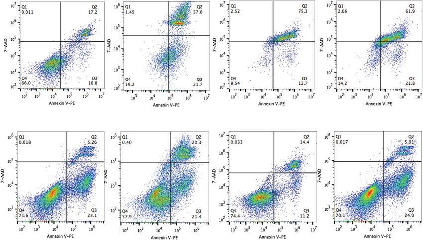
X. Bai et al.
                  2
                      forms a biofilm on a food-contact surface, it could become a                planktonic Lm cells, two human intestinal epithelial cell lines,
                      consistent contamination source. It has been reported that Lm              Caco-2 (colonic cells) and HCT-8 (Ileocecal junctional cells) were
                      strains with the same pulsotypes have been isolated from a food            used. Sessile cells of all five strains (F40, F45, F33, F4244, and
                      processing plant multiple times throughout a year25. Previously,           10403S) tested showed a significantly (P < 0.05) decreased
                      multiple studies have observed significant differences in gene              adhesion to Caco-2 (Fig. 2a) and HCT-8 (Fig. 2b) cells than that
                      expression between sessile and planktonic Lm cells34–36 especially,        of their planktonic counterparts. Likewise, sessile cells showed
                      the reduced expression of InlA, InlC, and LLO in biofilm cells35,37.        significantly lower invasion than the planktonic cells into Caco-2
                      However, none of them examined the pathogenicity of biofilm                 (Fig. 2c) and HCT-8 (Fig. 2d) cells. Sessile cells also showed
                      cells using cell culture or animal models. Therefore, the question         significantly (P < 0.05) lower transepithelial translocation than the
                      arose - how infective are these Lm sessile cells from the biofilm, if       planktonic cells in a Transwell setup (Fig. 2e). Altogether,
                      a food is consumed immediately after being contaminated with               reduction in adhesion, invasion, and transepithelial migration
                      these cells? In addition, can a conventional mammalian cell                between sessile and planktonic cultures was over 50%. Addition-
                      culture model38 that is used routinely in the laboratory predict the       ally, planktonic cells of wild-type (WT) Lm F4244 strain showed
                      nature of infectivity of biofilm isolates accurately? Is there any          significantly higher adhesion and invasion than that of the
                      direct correlation of in vitro infectivity data for biofilm-isolated        planktonic cells of an isogenic lap─ or ΔinlA mutant strains used as
                      cells with in vivo animal experimental data?                               controls (Fig. 2). Note, during this experiment, the growth of both
                         To date, various studies have reported the persistence and              sessile and planktonic Lm cells were negligible in mammalian cell
                      resistance of Lm cells in the biofilm to environmental stress and           culture medium (D10F; Dulbecco’s modified Eagle’s medium
                      significant change in global gene expression39,40; however, the             (DMEM) with 10% fetal bovine serum) after 3 h and there is no
                      exact virulence attributes of Lm isolated from biofilm has not been         significant difference (P > 0.05) between two cultures (Supple-
                      fully elucidated. The objective of this study was to assess and
                                                                                                 mentary Fig. 2), suggesting the differences in bacterial interaction
                      compare the pathogenicity of biofilm-isolated and planktonic Lm
                                                                                                 with mammalian cells are not influenced by their growth during
                      cells using in vitro intestinal epithelial cell culture model and an
                                                                                                 the assay period. Taken together, these in vitro results suggest
                      in vivo mouse (C57BL/6) model at different stages of infection (12,
                                                                                                 that the biofilm-isolated Lm strains have impaired ability to
                      24, and 48 h). Besides, we also analyzed the expression of key
1234567890():,;

                                                                                                 adhere, invade, or translocate across the epithelial cells than that
                      virulence proteins (LAP and InlA) that are involved during the early
                                                                                                 of their planktonic counterparts thus possibly have reduced
                      stage of infection4 and the regulatory genes, prfA and sigB. The
                      knowledge gained would help understand the pathogenesis and                virulence potential.
                      develop an intervention strategy for controlling biofilm-forming
                      bacterial pathogens from causing infection.                                Biofilm-isolated L. monocytogenes were less cytotoxic to
                                                                                                 Ped-2E9 and Caco-2 cells than the planktonic bacteria
                                                                                                 To characterize pathogenic attributes of biofilm-isolated cells, Ped-
                      RESULTS                                                                    2E9 (a hybrid murine B lymphocyte line)-based in vitro cytotoxicity
                      Food-isolated L. monocytogenes strains have higher                         assay was conducted44,45. Ped-2E9 has been established to be a
                      biofilm-forming capability than clinical isolates                           sensitive model to respond to the apoptosis triggered by Lm46–48.
                      The biofilm-forming capability of over 100 Lm isolates of food and          We used Annexin V and 7-AAD labeling method to distinguish
                      clinical origin on polystyrene surface was assessed after 48 h using       Ped-2E9 cells in the early or late apoptosis and analyzed them
                      crystal violet (CV) staining41 to choose for representative food and       using flow cytometry and fluorescence microscopy. Biofilm-
                      clinical strains to investigate the pathogenic potential in in vitro       isolated and planktonic cells of Lm strains F4244 and 10403S
                      cell culture and in vivo mouse models. Biofilm-forming capacity             and their corresponding isogenic mutant strains were analyzed
                      varied widely among the strains at 48 h. We found food isolates            (Fig. 3a). The damage caused by bacteria to Ped-2E9 cells was
                      (65 strains) had significantly (P < 0.05) higher biofilm-forming             quantitatively compared by the sum percentage of Annexin V-
                      capability than the clinical isolates (46 strains) (Fig. 1a–c).            positive events, which include cells in the early and the late stage
                      Furthermore, isolates of serotype 1/2a and 1/2c (Lineage II) had           (dead) of apoptosis (Supplementary Fig. 3). Firstly, all planktonic
                      greater biofilm-forming capabilities than serotypes 1/2b or 4b              WT strains (F4244, and 10403S) caused significantly more cell
                      (Lineage I) (Fig. 1a, c). Isolates were arbitrarily grouped into high,     damage than the corresponding biofilm-isolated cells (Fig. 3a, b).
                      moderate, and weak biofilm-producing groups (Fig. 1b) and                   Secondly, the microscopic analysis confirmed that the planktonic
                      representative strains with high (F40 and F45) or moderate (F33,           strains (F4244, 10403S, and F45) are responsible for more
                      F4244, and 10403S) biofilm-forming capabilities were chosen for             apoptotic or dead Ped-2E9 cells than the sessile cells. Thirdly, as
                      further characterization. These isolates (two clinical: F4244 (4b)         expected, planktonic 10403S cells caused significantly more
                      and 10403 S 1/2a) and three food: F40 (4b), F45 (1/2b), and F33            damage than the isogenic ΔprfA mutant strain whose virulence
                      (4b)) represent serotypes 4b, 1/2b, and 1/2a (Supplementary Table          cannot be upregulated by the major regulator, PrfA16 (Fig. 3a, b).
                      S1) which are responsible for a majority of human listeriosis              We used the ΔprfA mutant as a negative control since hly, plcA,
                      cases1,42. Light microscopic images revealed the formation of              and plcB are regulated by PrfA and whose gene products are
                      typical honeycomb-like structures of biofilms consistent with the           responsible for Ped-2E9 cell membrane damage and cytotoxi-
                      previous observation43 with varying sizes of the biofilm clusters           city48,49 Fourthly, F4244 ΔinlA and lap─ mutant strains did not
                      (Supplementary Fig. 1a). Furthermore, there is no apparent                 show a significant difference in cell death than that of the WT
                      difference in individual cell lengths between sessile and plank-           planktonic cultures, suggesting the cytotoxicity reduction in
                      tonic cells (Supplementary Fig. 1b). Collectively, these data reveal       biofilm-isolated bacteria was not affected by InlA or LAP (Fig. 3a,
                      that food-isolated Lm strains have higher biofilm-forming                   b). Finally, L. innocua F4248, a nonpathogenic strain, did not
                      capabilities than clinical isolates.                                       induce any cytotoxicity (Fig. 3a, b). In addition, using Caco-2 cells
                                                                                                 as a second model, we observed planktonic cultures of five WT Lm
                      Biofilm-isolated L. monocytogenes has attenuated adhesion,                  strains (F45, F4244, F40, F33, and 10403S) to induce a significantly
                      invasion, and translocation capability to intestinal epithelial            more lactate dehydrogenase (LDH) release than the sessile
                      cell lines in vitro                                                        cultures, suggesting that planktonic Lm cells are more cytotoxic
                      To compare the bacterial adhesion, invasion, and transmigration            than the sessile cells (Fig. 3c). In sum, Ped-2E9 and Caco-2-based
                      characteristics of 48-h-old biofilm-isolated sessile and 24-h-old           in vitro cytotoxicity data were consistent with the observation that

                      npj Biofilms and Microbiomes (2021) 18                                             Published in partnership with Nanyang Technological University
X. Bai et al.
3
1234567890():,;
Fig. 1 Quantification of L. monocytogenes biofilm formation and morphological analysis. a The biofilm-forming capabilities of over 100
food-(top panel) or clinical-isolated (bottom panel) L. monocytogenes (Lm) strains were tested using crystal violet staining assay. Arrows
indicate the strains selected for further characterization. b Assemblage (strong, moderate, and weak) of isolates based on their ability to form
biofilms. c Comparison of biofilm formation by food and clinical isolates and isolates of lineage I and II. Food isolates have significantly higher
biofilm-forming capability than the clinical isolates, and isolates of lineage II also have a significantly higher capacity than isolates of lineage I.
Mann–Whitney test was used for statistical analysis. **P < 0.005; *P < 0.05.
biofilm-isolated bacteria have attenuated virulence compared to bacterial cells could be asymmetrically distributed in cytosol and
the planktonic bacteria on cultured cell lines. cell wall, and the virulence molecules expressed on cell surface are
responsible for interacting with epithelial cells. Therefore, we
Key virulence factors were downregulated in biofilm-isolated specifically compared the amount of those proteins in different
bacteria on both transcription and translation levels cellular fractions. In cell wall and intracellular fractions, the amount
Next, to unravel the underlying reduced in vitro adhesion, of InlA and LAP were all significantly reduced in biofilm-isolated
invasion, translocation, and cytotoxic phenotypes in sessile cells, cells compared to those in the planktonic cells (Fig. 4b). At the
we assessed the expression of mRNA and protein of key virulence same time, biofilm-isolated cultures also secreted significantly (P <
factors using reverse transcriptional PCR and western blot in two 0.05) lower LLO in the supernatant compared to the planktonic
representative strains: F4244 (clinical) and F45 (food). Western blot cultures for strains F4244 and F45 (Fig. 4c). As a control for the
data showed that in biofilm-isolated cells, LAP, and InlA levels anti-LLO antibody, the planktonic culture of 10403S showed a
were all downregulated in the whole cells (Fig. 4a). Proteins in positive reaction with secreted LLO while an isogenic Δhly strain
Published in partnership with Nanyang Technological University npj Biofilms and Microbiomes (2021) 18X. Bai et al.
4
Fig. 2 Adhesion, invasion, and translocation characteristics of biofilm-forming sessile and planktonic cells on the cultured cell line.
Comparison of adhesion (a, b) and invasion (c, d) in Caco-2 and HCT-8 cells and transepithelial translocation across Caco-2 cells (e) between
L. monocytogenes biofilm-isolated sessile and planktonic cells on Caco-2 and HCT-8 cells. The percentage was calculated by dividing the
amounts of adhered, invaded, or translocated bacteria by the amounts of bacteria in the inoculum. Data are the average of at least three
independent experiments performed in triplicate. All error bars represent SEM. Pairwise Student’s t-test was used for statistical analysis.
****P < 0.0001; ***P < 0.0005; **P < 0.005; *P < 0.05.
was negative (Fig. 4c). The protein samples were standardized In mouse bioassay, biofilm-isolated Lm showed reduced tissue
with bicinchoninic acid assay method before loading onto the burden at the early stage of infection (12–24 h), but similar to
SDS-PAGE gel (Supplementary Fig. 4a). planktonic bacteria after 48 h post-infection
To quantify the transcription of the major virulence genes, we In vitro cell culture experiments suggest that biofilm-isolated Lm
first generated the standard curves for each gene’s copy number irrespective of food or clinical sources have a significantly lower
and Ct values and validated the specificity of qPCR primers. capacity to adhere, invade, and translocate across the intestinal
Standard curves for genes with copy numbers of approximately epithelial cells and lower cytotoxicity on B-lymphocytes and Caco-
101–109 copy/μL and Ct values were generated with each qPCR 2 cells than the planktonic bacteria. However, the actual virulence
primer sets (Supplementary Table 2). All the standard curves had of Lm in these two physiological states (sessile vs planktonic)
R2 values greater than 0.99 and a similar slope between −3.04 cannot be accurately compared without observing their patho-
and −3.40 (Supplementary Fig. 4b, c). Besides, each primer pairs genicity in vivo. Therefore, we orally infected 8–10 weeks old male
amplicon showed a sharp and single-peak melting curve and female C57BL/6 mice with both biofilm-isolated or planktonic
(Supplementary Fig. 4d) suggesting that the qPCR primers are Lm strains representing clinical (F4244) and food (F45) isolates
suitable for quantifying the target genes. with 1 × 10⁹ CFU/mouse and analyzed intestinal and extra-
The gene-specific mRNA expression analysis in F4244 and intestinal tissues for bacterial dissemination at 12, 24, and 48 h
post-infection (hpi).
F45 strain showed dramatic downregulation of both lap and inlA
At 12 hpi, the bacterial burden in mice tissues was below the
(~95–99%) in biofilm-isolated cells than that of the planktonic cells
detection limit when a standard plating method was used.
while both regulatory genes, prfA, and sigB, were downregulated Therefore, we enriched the tissue samples in buffered Listeria
by about 25% in the biofilm-isolated cells, except for the prfA in enrichment broth (BLEB for 24 h) followed by isolation on the
F45 (Fig. 4d). Compared to planktonic F45 cells, the amount of prfA Modified Oxford (MOX) agar plate to determine the presence or
mRNA is almost similar in biofilm-isolated F45 cells (Fig. 4d). These absence of Lm in mice tissues. The select isolates were further
data suggest that the attenuated adhesion, invasion, transepithe- verified by qPCR assay (Supplementary Table 3). To test the
lial translocation, and cytotoxicity of biofilm-isolated Lm cells were sensitivity of the detection method, we inoculated Lm (F4244 or
possibly due to low expression of corresponding virulence genes F45) at 1.4 ± 0.2 or 1.2 ± 0.2 CFU/ml BLEB, respectively, and
at both mRNA and protein levels. incubated at 37 °C for 24 h. Aliquots (10 µl) of each culture was
npj Biofilms and Microbiomes (2021) 18 Published in partnership with Nanyang Technological UniversityX. Bai et al.
5
Fig. 3 Cytotoxicity assessment of biofilm-forming sessile and planktonic L. monocytogenes. a Flow cytometric analysis of Ped-2E9 (B cell
hybridoma) cells treated with biofilm-isolated (B) and planktonic (P) cells of L. monocytogenes (Lm) F4244 and 10403S and corresponding
mutant strains at MOI 10. Annexin V-PE-positive and 7-AAD-negative events (Q3) were identified as cells in the early phase of apoptosis.
Events with both Annexin V-PE and 7-AAD positive (Q2) or both negative (Q4) were identified as dead or live cells, respectively. L. innocua (Lin)
F4248 was also tested as a nonpathogenic negative control. b Quantitative comparison of overall damage of Ped-2E9 caused by bacteria. Each
bar represents the percentage of Annexin V-PE-positive events, which included early apoptosis (Q3) or dead (Q2) cells. Biofilm-isolated
bacteria of both strains were significantly less cytotoxic than their planktonic counterparts. Bars marked with different letters are significantly
different at P < 0.05. c Lactate dehydrogenase (LDH) released from Caco-2 cells (a colorectal adenocarcinoma) treated with both sessile or
planktonic cells. Bacteria were incubated with cells at MOI 10 at 37 °C for 2 h. Data are the average of at least three independent experiments
performed in triplicate. All error bars represent SEM. A pairwise Student’s t-test was used for statistical analysis. *P < 0.05.
streaked on MOX plates, and colonies with the black center were F4244 sessile cells were negative while 16–50% mice (n = 6) were
further verified by PCR, suggesting the two-step selective positive when infected with the planktonic cells (Table 1).
enrichment combined with PCR can detect approximately 1 Similarly, all mice receiving F45 sessile cells were negative in
CFU/ml of Lm in BLEB (Supplementary Fig. 5). None of the jejunal extra-intestinal organs, while 20–60% mice were positive when
or ileal tissues of mice (n = 4–6) were positive for Lm after receiving planktonic cells (Table 1). None of the blood or kidney
challenge with the sessile or planktonic cells (Table 1). However, samples were positive when infected with both sessile or
only one of five (20%) cecum or colonic tissues were positive planktonic cells for both Lm strains at this early stage of infection.
when mice were challenged with F4244 sessile cells compared to Nevertheless, these data indicate that bacterial intestinal invasion
50–100% positive when mice were challenged with the planktonic and subsequent systemic dissemination was lower for sessile cells
cells (Table 1). Likewise, no Lm cells were detected from the cecum than the planktonic cells for both Lm strains in mice after 12 hpi.
and colon of mice when challenged with F45 sessile bacteria, At 24 hpi, we were able to enumerate Lm in most mice organs
whereas 80% (4/5) and 100% (5/5) were positive when challenged and tissues by a standard plating method. In the intestinal tissues,
with planktonic bacteria, respectively (Table 1). Analysis of extra- there were no significant differences in bacterial counts between
intestinal organs/tissues; mesenteric lymph nodes (MLN), liver, and sessile and planktonic cells-challenged mice with an exception of
spleen of mice revealed that all the animals (n = 5) receiving the cecum, where sessile cells had significantly (P < 0.05) higher
Published in partnership with Nanyang Technological University npj Biofilms and Microbiomes (2021) 18X. Bai et al.
6
Fig. 4 Comparison of virulence protein expression in biofilm-isolated (B) and planktonic (P) L. monocytogenes cells. a, b Immunoblot (top
panel) and densitometry (bottom panels) of InlA, and LAP, in the whole cell (a), cell wall, and intracellular fractions (b) of biofilm-isolated and
planktonic cultures of Lm F4244 and F45. Immunoblots are representative of three independent experiments. c Immunoblot of LLO in the
secreted protein fraction of biofilm-isolated and planktonic Lm F4244 and F45. Immunoblots are representative of three independent
experiments. d Relative mRNA expression of virulence genes (inlA and lap) and virulence regulators (prfA and sigB) in biofilm-isolated and
planktonic cells of F4244 and F45 by RT-PCR. The Student’s t-test was used for statistical analysis. ***P < 0.0005; **P < 0.005; *P < 0.05.
Table 1. Listeria monocytogenes strains (F45, F4244, and F4244 InlAm) translocation in C57BL/6 mice organs/tissues 12 h after oral infection.
Source Tissues Number of mouse tissues positive for L. monocytogenes/# mouse tested (%)a
F4244 (WT) F45 (WT) F4244 (InlAm)b
Biofilm Planktonic Biofilm Planktonic Biofilm Planktonic
Intestinal Jejunum 0/5 (0) 0/4 (0) 0/5 (0) 0/5 (0) 3/9 (33) 5/9 (56)
Ileum 0/5 (0) 0/4 (0) 0/5 (0) 0/5 (0) 1/9 (11) 2/9 (22)
Cecum 1/5 (20) 4/4 (100) 0/5 (0) 4/5 (80) 1/9 (11) 6/9 (67)
Colon 1/5 (20) 2/4 (50) 0/5 (0) 5/5 (100) 2/9 (22) 5/9 (57)
Extra-Intestinal MLN 0/5 (0) 3/6 (50) 0/5 (0) 2/5 (40) 0/9 (0) 2/9 (22)
Liver 0/5 (0) 3/6 (50) 0/5 (0) 2/5 (20) 2/9 (22) 5/9 (56)
Spleen 0/5 (0) 1/6 (16) 0/5 (0) 3/5 (60) 0/9 (0) 5/9 (56)
Kidney 0/5 (0) 0/6 (0) 0/5 (0) 0/5 (0) 0/9 (0) 0/9 (0)
a 9
Mice (both male and female) were orally gavaged with 1 × 10 CFU/mouse. Mouse tissue samples were enriched in buffered Listeria enrichment broth for 24 h,
plated on modified Oxford agar plate for 48 h, and 1–2 colonies per sample were verified by qPCR (see Supplementary Table 2).
b
These animals received streptomycin (5 mg/ml) in water for 32 h, followed by 16 h antibiotic-free water before oral gavage with Lm.
colonization than the planktonic cells (Fig. 5a–d). However, in the undetectable in these organs as determined by a plating method,
extra-intestinal organs (MLN, spleen, and liver) planktonic cells suggesting that the sessile cells were either unable or translocated
exhibited significantly (P < 0.05) higher bacterial burdens than and/or disseminated in blood/lymphatic circulation at levels that
the sessile cells (Fig. 5e–g). In fact, F4244 sessile cells were are below our detection limits at 24 hpi. In the kidney, counts for
npj Biofilms and Microbiomes (2021) 18 Published in partnership with Nanyang Technological UniversityX. Bai et al.
7
Fig. 5 Mouse bioassay to compare the pathogenesis of biofilm-isolated sessile and planktonic L. monocytogenes cells. Lm burden in
intestinal (a–d) and extra-intestinal tissues (e–h) after oral inoculation of mice (C57BL/6, male–female, 8–10 weeks old) with 1 × 10⁹ CFU/
mouse of sessile (B) and planktonic (P) cells of F4244 or F45 at 24 and 48 hpi. i, j Comparison of the number of bacteria in all intestinal and
extra-intestinal tissues at 24 and 48 hpi. Bars represent the median values of each group (B or P). Dashed lines indicate detection limits by a
plating method. Mann–Whitney test was used for statistical analysis. **P < 0.005; *P < 0.05.
both sessile and planktonic cells were below the detection limit (food isolate) indicates that F4244 had 1–2 log higher counts,
with an exception of one mouse, which was showing the hence it is more invasive than F45 in a mouse model of infection
planktonic burden of about two logs (Fig. 5h). Altogether, (Fig. 5i, j). These data further reveal that while biofilm-isolated
planktonic F4244 cell-challenged mice had significantly (P < cells are in the process of translocating through the intestinal
0.005) higher total Lm burden than the sessile cell-challenged tissues, planktonic Lm cells have already disseminated to the
mice in the extra-intestinal organs while there was no significant extra-intestinal sites at 24 hpi.
difference in total bacterial burden in whole intestinal tissues At 48 hpi, F4244 cell burden in both intestinal and extra-
combined at 24 hpi (Fig. 5i). Likewise, total Lm burdens in the intestinal tissues for both sessile cell- and planktonic cell-
intestine and extra-intestinal organs of mice challenged with challenged mice were alike (Fig. 5a, b, d, e, f, h) except for the
sessile or planktonic cells of strain F45 are similar to F4244- cecum (Fig. 5c) and spleen (Fig. 5g) where planktonic counts
challenged mice. We did not observe any significant difference were significantly (P < 0.05) higher than the sessile cells. Compar-
in intestinal Lm counts for F45 strain; however, significantly (P < ing the total bacterial burden in the whole intestine and extra-
0.05) more planktonic cells were found in extra-intestinal organs intestinal tissues, no significant difference in total bacterial burden
than the sessile cells (Fig. 5j). In particular, significantly (P < 0.05) in extra-intestinal tissues was observed between planktonic or
more planktonic F45 than biofilm-isolated bacteria were sessile bacteria-challenged mice (Fig. 5i). Likewise, in F45 infected
detected in the cecum, but not in other sections of the intestine mice, the burden of planktonic or sessile cells had no significant
(jejunum, ileum, and colon) (Fig. 5a–e). Whereas in the extra- difference in all intestinal or extra-intestinal organs examined at
intestinal organs, infection by planktonic F45 resulted in 48 hpi (Fig. 5a–j).
significantly (P < 0.05) more Lm counts in MLN and spleen than Collectively these data demonstrate that the biofilm-isolated Lm
in the liver (Fig. 5e-g). Furthermore, the presence of biofilm- has temporarily attenuated capacity to translocate across the gut
isolated Lm in MLN of three mice and spleen of one mouse could barrier and/or to disseminate in the blood/lymphatic circulation
not be detected even after culture enrichment followed by qPCR during the early phase of infection (12–24 h), while both
(Fig. 5e, g). Comparing the overall Lm burden in the intestine or planktonic and biofilm-isolated Lm were able to disseminate to
extra-intestinal organs between F4244 (clinical isolate) and F45 extra-intestinal tissues similarly at 48 hpi.
Published in partnership with Nanyang Technological University npj Biofilms and Microbiomes (2021) 18X. Bai et al.
8
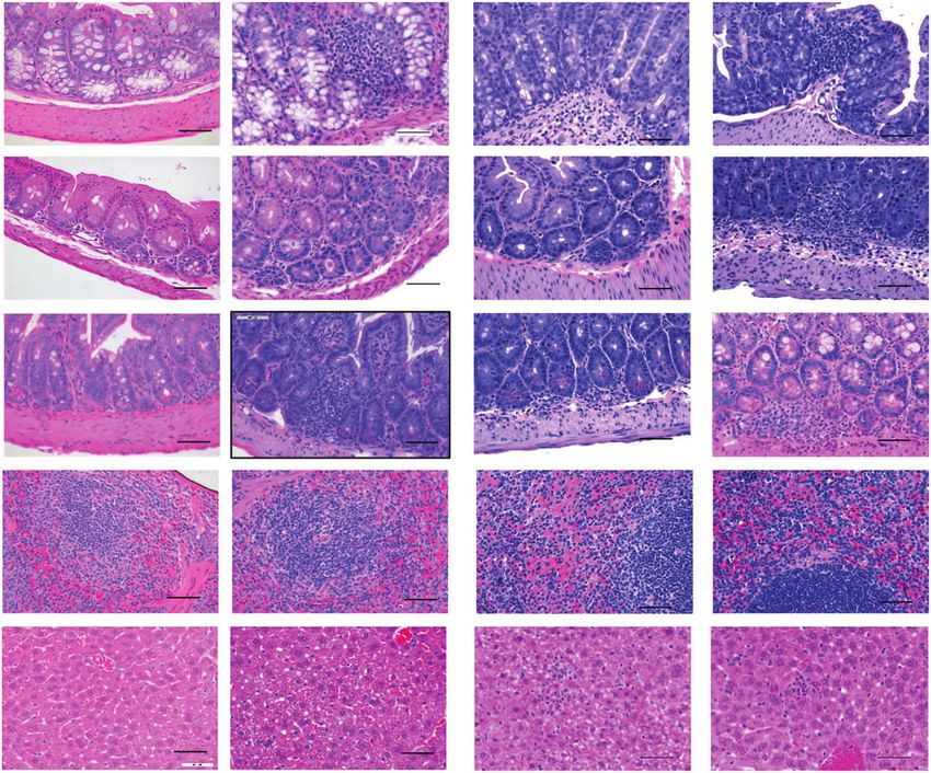
Fig. 6 Histopathology analysis of mouse tissues for inflammation. Representative images of hematoxylin and eosin-stained tissue sections
of mice challenged with 1 × 10⁹ CFU of F4244 sessile (B) or planktonic (P) cells at 24 and 48 hpi (a) and a graph representing histopathological
inflammation scores at 24 hpi (b, left panel) and 48 hpi (b, right panel). Scale bars represent 50 μm.
Histopathology shows the increased inflammatory response Sessile and planktonic Lm with murinized internalin A (InlAm)
for planktonic cells than the sessile cells showed similar pathogenicity and systemic dissemination as
At 24 hpi, histopathological analysis of planktonic F4244 infected the wild-type strain
intestinal tissues revealed more polymorphonuclear and mono- To verify the role of InlA in Lm pathogenesis in biofilm-isolated
nuclear cells infiltrating villi in mice than the sessile bacteria- sessile cells in the mouse model, we created murinized inlA (InlAm)
infected tissues (Fig. 6a). At the same time, an increased amount in F4244 by substituting two specific amino acids, S192N and
of single-cell necrosis and higher inflammation scores were Y369S (Fig. 7)10. The inlAm gene sequencing (Supplementary Fig.
observed in the liver and spleen of planktonic F4244-challenged 7a–d and Fig. 7a), Western blotting (Fig. 7b) and ELISA (Fig. 7c)
mice, suggesting planktonic bacteria caused more inflammatory confirmed the expression of InlA in the InlAm strain. Besides, InlAm
lesions in extra-intestinal organs than the sessile bacteria at 24 hpi strain also showed significantly (P < 0.05) higher invasion into
(Fig. 6a, b). At 48 hpi, a similar inflammatory lesion was observed intestinal epithelial HCT-8 cells than the WT (F4244) strain (Fig. 7d)
in both intestinal and extra-intestinal organs of mice challenged consistent with the results reported for Caco-2 cells10. In the
with either planktonic or sessile cells of F4244 (Fig. 6a). The sessile mouse experiment, InlAm strain also showed significantly (P < 0.05)
and planktonic cells of the F45 strain also showed similar results as higher invasion of large intestinal tissues and translocation to the
F4244 but the overall inflammatory response was much lower liver after 96 hpi compared to the WT strain (Fig. 7e) as observed
than F4244 (Supplementary Fig. 6). Overall inflammation scores before10.
showed that sessile bacteria caused much more lesions in the We then examined adhesion and invasion of planktonic (PM)
spleen and liver at 48 hpi compared to 24 hpi, which is consistent and biofilm-forming sessile cells (BM) of InlAm strain in vitro, and
with the increased bacterial burdens in these organs (Fig. 6). the planktonic cells showed significantly higher adhesion and
npj Biofilms and Microbiomes (2021) 18 Published in partnership with Nanyang Technological UniversityX. Bai et al.
9
Fig. 7 In mouse bioassay, biofilm-isolated and planktonic L. monocytogenes with murinized InlA (InlAm) display differential tissue
distribution. a PCR confirmation of the insertion of inlAm gene in the chromosome of Lm F4244 ΔinlA using primers inlA.up.5 and inlA.down.3
(Supplementary Table 2). WT (F4244) was used as a positive control. b Immunoblots showing expression profile of InlA, and LAP in whole-cell
extracts of WT, inlAm, and ΔinlA. c ELISA showing the positive reaction of anti-InlA mAb to whole-cell preparation of WT, inlAm, and reduced
reaction with ΔinlA. d Percent invasion of WT, inlAm, and ΔinlA to HCT-8 cells. Bars represent mean, and a pairwise Student’s t-test was used for
statistical analysis. e Lm WT, inlAm and ΔinlA strain burdens in the large intestine, MLN, spleen, and liver of mice (n = 5–6) 96 h after oral
challenge (5 × 10⁹ CFU/mouse). Mann–Whitney test was used for statistical analysis. f Percent adhesion and invasion of biofilm-isolated and
planktonic cells of InlAm strain to HCT-8 cells. Bars represent mean, and a pairwise Student’s t-test was used for statistical analysis. g, h Lm
burdens in tissues of mice (C57BL/6, male and female, 8–10 weeks old) challenged with murinized InlAm (1 × 10⁹ CFU/mouse) strain of biofilm-
isolated (BM) or planktonic (PM) cells at 24 (g) or 48 (h) hpi. Mice were pretreated with streptomycin (5 mg/ml) in drinking water for 32 h
followed by 16 h in antibiotic-free water before the Lm challenge. i Comparison of tissue (spleen and liver) burden between WT and InlAm
strain for biofilm-isolated (BWT vs BM) and planktonic (PWT vs PM) cells at 48 hpi. Data for WT were taken from Fig. 5. Bars represent median
values, and the Mann–Whitney test was used for statistical analysis in e, g, h. ****P < 0.0001; ***P < 0.0005; **P < 0.005; *P < 0.05; ns,
no significance.
invasion into HCT-8 cells than the sessile cells (Fig. 7f) similar to At 24 hpi, planktonic cells showed significantly (P < 0.05) higher
WT F4244 cells (Fig. 2). Next, aiming to observe increased invasion into the cecum and spleen than the sessile cells. While in
invasion and Lm tissue burdens in the mouse model of the colon and liver there were no differences (Fig. 7g). These data
infection, we pretreated the mice with streptomycin (5 mg/ml) further demonstrate that even though InlA-dependent invasion
for 32 h in drinking water50 before oral challenge with sessile was restored in the mouse model, the sessile cells still showed
and planktonic InlAm strains at 1 × 10⁹ CFU/mouse. The delayed invasion and tissue distribution.
sensitivity of InlAm strain to streptomycin was tested before At 48 hpi, there was no statistical difference in planktonic and
animal administration and was determined to be 2.5 µg/ml sessile cells of InlAm strain in the mouse intestinal and extra-
(Supplementary Fig. 7e). intestinal tissues (Fig. 7h). We also compared the tissue distribu-
At 12 hpi, as before, Lm could not be enumerated by the plating tion patterns of sessile and planktonic cells of both WT (data from
method; hence, the tissue samples were tested for the presence or Fig. 5) and InlAm strain (data from Fig. 7h) at 48 hpi and no
absence of Lm. In the intestinal tissue samples, only 11–33% of significant differences were observed between these two strains in
mice (n = 9) were positive when challenged with sessile cells the spleen and liver except for planktonic cells of InlAm strain in
while 22–67% of mice (n = 9) were positive for planktonic cells. In the liver which showed higher (P < 0.05) invasion (Fig. 7i). Overall
the extra-intestinal tissues, sessile cells were isolated only from the these data show a consistent trend in tissue invasion for sessile
liver of two mice (22%) while all other tissues (MLN & spleen) were and planktonic cells of InlAm and the WT strain confirming the
negative. In contrast, 22–56% of mice were positive when attenuation of translocation of biofilm-forming sessile cells during
challenged with planktonic cells (Table 1). the early stage (12–24 h) of infection.
Published in partnership with Nanyang Technological University npj Biofilms and Microbiomes (2021) 18X. Bai et al.
10
Fig. 8 Survival and virulence of biofilm-isolated and planktonic L. monocytogenes strain suspended in simulated gastrointestinal fluid.
a Survival of sessile and planktonic Lm F4244 and F45 after sequential exposure to simulated gastric fluid (SGF, pH 2) for 1 h and simulated
intestinal fluid (SIF, pH 7) for 12 h. b–d Comparison of adhesion (b), invasion (c), and translocation (d) rates on Caco-2 cells of SGF (pH 3) and
SIF (pH 7)-treated biofilm-isolated and planktonic Lm F4244 and F45. e, f Immunoblot showing LAP and InlA expression in sessile
and planktonic cells after exposure to SGF (pH 3) and SIF (pH 7). Immunoblots are representative of three independent experiments. g Relative
mRNA expression of virulence genes (inlA and lap) and virulence regulators (prfA and sigB) in biofilm-isolated InlAm from mice intestinal
chymus at 12 or 48 hpi and the same cells before infection using RT-PCR. UD, Undetectable. A pairwise Student’s t-test was used for statistical
analysis. ***P < 0.0005; **P < 0.005; *P < 0.05.
LAP and InlA expression were significantly upregulated in exposure to SGF and there was no significant difference in cell
planktonic cells than the sessile cells after exposure to viability between the two (Fig. 8a). We then examined the
simulated gastrointestinal fluids for 13 h adhesion, invasion, and translocation properties of these bacterial
In vivo data revealed late dissemination of sessile cells to extra- cells through Caco-2 cells and analyzed the expression of LAP and
intestinal tissues; therefore, we hypothesized that the sessile cells InlA proteins. Interestingly, SGF and SIF-exposed sessile cells of
are either more susceptible to intestinal conditions than that of both F4244 and F45 strains showed significantly increased
the planktonic cells or the intestinal condition may suppress the adhesion, invasion, and transepithelial translocation through
expression of key virulence factors in sessile cells. To verify the first Caco-2 cells compared to the sessile cells that are not exposed
event, we tested the survivability of both biofilm-isolated and to simulated gastrointestinal fluids (Fig. 8b–d). These results
planktonic Lm cells exposed to simulated gastric fluid (SGF) and further indicate that exposure to gastrointestinal conditions
simulated intestinal fluid (SIF). Both sessile and planktonic cell increased virulence attributes in Lm sessile cells. In contrast,
viability was decreased by about three logs after 60 min of planktonic cells showed mixed results showing slightly decreased
npj Biofilms and Microbiomes (2021) 18 Published in partnership with Nanyang Technological UniversityX. Bai et al.
11
or the same levels of adhesion, invasion, and translocation with or have demonstrated that mixed-species biofilms possibly facilitate
without exposure to SGF and SIF (Fig. 8b–d). Overall, planktonic the persistence of such weak biofilm-forming pathogens56,57.
cells displayed significantly higher adhesion, invasion, and Biofilm-forming cells experience stress and exhibit physiological
translocation than that of the sessile cells, thus supporting the and genetic heterogeneity52; thus, we were curious about their
hypothesis that virulence of planktonic bacteria is significantly dynamics of infectivity in cell culture and mouse models. Five Lm
higher in the gastrointestinal environment than the sessile strains of food and clinical origins representing the major
bacteria at the very early (12–24 h) stage of infection (Fig. 8c, d). outbreak causing serotypes with diverse biofilm-forming pheno-
Immunoblot analysis confirmed a significant increase (2–3-fold) types were selected for in vitro cell culture experiments. All
in the expression of LAP and InlA in sessile cells in both F4244 and biofilm-isolated cells we tested irrespective of food or clinical
F45 strains after exposure to SGF and SIF (Fig. 8e, f). Furthermore, origins were less adhesive, invasive, and cytotoxic and showed
significantly increased expression of these proteins was also reduced ability to traverse across the Caco-2 epithelial barrier than
observed in planktonic cells of the F4244 strain but not in the the planktonic cells, suggesting these cells are less virulent
F45 strain. Overall, the expression of these proteins was compared to the planktonic cells (Figs 2 and 3). We analyzed the
significantly higher in planktonic cells than in the sessile cells in expression of key virulence proteins (LAP, InlA, and LLO) that are
F4244 strains (Fig. 8f). Taken together, these data show that overall responsible for Lm invasion, paracellular translocation, and
reduced expression of LAP and InlA in sessile cells relative to the intracellular persistence. We observed significantly reduced
protein expression by planktonic cells may be responsible for expression of these proteins in sessile cells at the transcriptional
decreased adhesion, invasion, and transepithelial migration during and translational levels (Figs 3 and 4), which may explain the
the very early stage (12–24 h) of infection. reason for reduced virulence of sessile cells in in vitro cell culture
To validate the hypothesis that sessile cells upregulate virulence experiment. However, the contribution of other virulence factors
genes after oral infection, we quantified the transcriptional including ActA cannot be ignored. ActA, a PrfA, and SigB regulated
expression of virulence genes in sessile InlAm cells from mice protein known to contribute to biofilm formation and intestinal
intestinal chymus 12 and 48 hpi and compared them with the colonization58,59 may also be affected in sessile cells for the
expression in the same cells before infection. We observed a delayed invasion and tissue distribution in mice. Furthermore,
fivefold higher inlA expression in InlAm at both 12 and 48 hpi reduced inlA expression in sessile cells is in agreement with others
compared to that of control (InlAm cells before infection) (Fig. 8g). who also observed similar reduced InlA expression in biofilm-
Interestingly lap expression was 250- and 70-fold higher at 12 and isolated cells36,37. Besides, mRNA of gene sigB, coding a stress
48 hpi, respectively (Fig. 8g). The sigB expression was 10-fold response regulator, was also downregulated to around 25% in
higher at 12 hpi and maintained at a similar level at 48 hpi sessile cells of both Lm strains compared to their planktonic
counterparts (Fig. 4). SigB has been implicated in Lm biofilm
(Fig. 8g). In contrast, prfA expression remained unchanged at 12
formation39 and it also regulates InlA expression60. The observed
hpi, and it was below the detection limit at 48 hpi (Fig. 8g). These
suppression of SigB and consequent InlA expression in sessile cells
data indicate that the delayed invasion of sessile cells in mice at
possibly is responsible for reduced Lm adhesion and invasion into
24 hpi was possibly because of their lower expression of virulence
the intestinal epithelial cells, which was further supported by a
factors, LAP and InlA, than their planktonic counterparts. Besides,
proteomic analysis that indicated downregulation of SigB-
the intestinal environment positively upregulates lap and inlA
regulated proteins36.
expression in sessile cells in the intestine, which could allow the Interestingly, prfA mRNA in Lm F45, a strong biofilm-former, was
sessile cells to be as invasive as planktonic cells at 48 hpi. expressed at a similar level for both sessile and in planktonic cells;
however, its level was downregulated by 25% in Lm F4244 (a
DISCUSSION moderate biofilm-former) sessile cells than that of the planktonic
cells (Fig. 4). These observations differ from a previous study
The biofilm-forming ability gives Lm the advantage of persistence where PrfA is reported to positively regulate biofilm formation61
even for many years on various surfaces in a food processing/ and a strain (Lm10403S) overexpressing prfA showed higher
production environment, which presumably serves as a primary biofilm-forming ability than the WT. Our data further imply that
source for food contamination19–22. Lm has been routinely isolated PrfA-regulated biofilm formation may vary from strain to strain
from meat and dairy51 processing plants. The persistence of which requires further investigation. Although PrfA is a key
pathogens on the abiotic surface is facilitated by their ability to regulator for the expression of multiple virulence factors including
form a biofilm, in which cells experience a wide range of stress InlA62, our qRT-PCR results further suggest that decreased
thus show physiological and genetic heterogeneity allowing them expression of inlA in sessile cells is not always coupled with
to be more resistant to antimicrobials, and to survive in limited decreased expression of prfA (Figs 3 and 4) since InlA can be
nutrient and oxygen tensions27,52,53. Although several studies expressed independently of PrfA regulation63.
have reported reduced expression of virulence genes in sessile To confirm in vitro cell culture results in a mouse model, we
cells, the pathogenic potential of these cells has not been tested challenged mice orally with 48-h-old sessile cells or 24-h-old
using either in vitro or in vivo models. Therefore, we studied the planktonic cells of moderate biofilm-forming clinical strain (F4244)
virulence of biofilm-isolated sessile cells of Lm using both cell and a strong biofilm-forming food isolated strain (F45). At 12–24
culture and animal models, and the expression of virulence genes hpi in mice, sessile cell burden in intestinal and extra-intestinal
to support the observed phenotype, especially between 12 and tissues was undetectable or very low while the planktonic burden
48 hpi. was significantly high and infectivity was comparable to the
The biofilm-forming capability of over 100 Lm strains was in vitro cell culture data indicating sessile cells are less invasive.
screened and all formed biofilms of varying degrees on a However, at 48 hpi, burdens of both sessile and planktonic cells in
polystyrene surface, and food isolates, in general, had significantly mouse tissues were comparable, suggesting that sessile cells are
higher biofilm-forming capacity than the clinical isolates (Fig. 1). equally invasive as planktonic cells after the early stage (12–24
These observations agree with the previous studies27,54,55. Among hpi) of the gastrointestinal phase of infection; however, the rate of
the different serotypes examined, isolates of serovar ½a and ½c bacterial tissue distribution and disease progression was variable
(Lineage II) are stronger biofilm formers than the isolates of ½b (Figs 5 and 6). To explain such discrepancy in intestinal epithelial
and 4b (Lineage I), which are in accordance with another study55. cell invasion in the early stage (12–24 hpi) of infection, we
We also observed that many strains were weak biofilm-former and hypothesized that possibly sessile cells are highly susceptible to
their persistence on surfaces may be doubtful; however, studies antimicrobials present in intestinal fluids or expression of
Published in partnership with Nanyang Technological University npj Biofilms and Microbiomes (2021) 18X. Bai et al.
12
adhesion and invasion-related proteins are suppressed in intest- antibiotic used for microbiota disruption. For example, van der
inal fluids. Therefore, we examined survival and protein expression Waaij et al.72 demonstrated that the dissemination and persis-
in sessile cells suspended in SGF (pH 3) and SIF (pH 7.0) that tence of infectious E. coli in mice was facilitated only when the
contain HCl, enzymes, and bile salts64. In SGF + SIF, we did not bacterium is resistant to the pre-exposed antibiotics. Similarly,
observe any significant difference in Lm viability between sessile Hentges et al.73 reported that the burdens of clindamycin-
and planktonic cells but observed differential expressions of LAP sensitive Pseudomonas aeruginosa in MLN and liver of
and InlA, the two key virulence factors that are responsible for Lm clindamycin-treated mice were lower than the burdens in
translocation across the gut epithelial barrier3,4,9. Though the untreated mice. Further experiments may be necessary to validate
expression of both LAP and InlA were significantly upregulated in the antibiotic effect of our Lm strain (F4244) invasion by using a
sessile cells in SGF and SIF, overall expression in planktonic cells streptomycin-resistant strain which we plan to investigate in the
was significantly higher than the sessile cells (Fig. 8e). These future.
findings suggest that the gastrointestinal environment may help LLO is an important virulence factor required for Lm persistence
the sessile cells to quickly transition to a fully virulent state and during intracellular lifestyle74 and is also responsible for epithelial
may also explain the observed similar intestinal and extra- and lymphocyte apoptosis46,47,49,75. In addition, LLO has been
intestinal tissue burdens for both biofilm and planktonic cells implicated to aid Lm dissemination from the gastrointestinal tract
at 48 hpi. to extra-intestinal tissues76. In this study, we observed reduced
Our hypothesis is also supported by the observation that inlA LLO expression in sessile cells (Figs. 3 and 4), which agrees with
and lap mRNA in sessile cells were upregulated after they arrive in one study36, but contradicts with other77, where researchers
the mouse intestine for 12 h (Fig. 8g). However, the expression of report that biofilm formation does not affect bacterial ability to
inlA mRNA maintained at a similar level and the expression of lap produce LLO. Interestingly, in another pathogen (Bacillus cereus),
even decreased at 48 hpi (Fig. 8g), suggesting the expression of researchers77 observed reduced expression of Hemolysin BL and
the virulence genes may not continue to increase with increasing other enterotoxins (CytK and EntC) in biofilm cells and conse-
residence time in the intestine. During this period, expression of quently reduced cytotoxicity on both HeLa and MDA cells78.
regulatory genes, prfA, was unaffected while the sigB level Collectively, these data imply that impaired toxin synthesis in
increased several-fold consistent with a previous report, which biofilm cells affects bacterial virulence.
showed SigB-mediated upregulation of several virulence genes, In summary, our data indicate that sessile cells are less invasive
including inlA, is critical for Lm to switch global transcription from in cultured cell lines and during the early stage (12–24 h) of
saprophytism to virulence while residing in the intestine65. This infection in an animal model possibly due to reduced expression
study further reinforces the importance of sigB in virulence of regulatory proteins (PrfA and SigB) and virulence factors (LAP,
gene expression in sessile cells during the intestinal phase of InlA, and LLO). However, both sessile and the planktonic cells
infection. Although the gastrointestinal environment is known to showed similar extra-intestinal tissue burdens at 48 hpi and sessile
upregulate both LAP66–68 and InlA expression36,37,69, here, we cells are equally infective as planktonic cells but the dynamics of
provide evidence for the expression of these two proteins in infection may vary between sessile and planktonic cells with
biofilm-isolated cells in the mouse intestine. possible differential disease onset or incubation period. Further-
In mice, InlA-mediated transcytosis is absent due to a lack of more, in vitro cell culture experiment routinely used for virulence
interaction between InlA and its cognate receptor, E-cadherin70; potential determination is found to be unreliable for assessing the
thus, LAP-mediated Lm translocation is considered the predomi- pathogenic potential of biofilm-forming cells because it measures
nant gut-barrier crossing mechanism in mice during the early the pathogenic event over a short period (1–2 h). On the other
(12–24 h) stage of infection3,4,8. Hence, the observed reduction in hand, an animal model provides comprehensive pathogenic
LAP expression in sessile cells is considered a major contributory events over a prolonged period in physiologically relevant
factor towards impaired Lm translocation in the intestinal conditions and, thus, is most reliable for studying the pathogen-
and extra-intestinal tissues early in the infection process esis of biofilm-isolated cells.
(12–24 h) (Fig. 5).
To further investigate the role of InlA in sessile cell infection in
the mouse model, we generated InlAm strain10 and the intestinal METHODS
and extra-intestinal tissue distribution of sessile and planktonic Bacterial strains
cells of InlAm surprisingly followed the same trend as the WT strain Food (64) and clinical (46) isolates and several mutant Lm strains were used
at 12, 24, and 48 hpi (Table 1 and Fig. 7). InlAm strain still did not in this study (Supplementary Table 1). Cultures were stored in brain heart
show increased tissue distribution of either sessile or planktonic infusion broth (Acumedia) with 25% glycerol at −80 °C. To revive cells, the
cells at 48 hpi compared to the WT. This result was expected since frozen stock cultures were first streaked on a tryptic soy agar plate
previous studies have shown that differential tissue distribution of containing 0.6% yeast extract (TSAYE; Becton Dickinson, Franklin Lakes, NJ),
InlAm and WT strains occur only after 72–96 h infection in mice10,71 and incubated at 37 °C overnight. Then a single colony was inoculated into
and this was again verified in our study (Fig. 7e). 4 ml tryptic soy broth supplemented with 0.6% yeast extract (TSBYE),
Furthermore, this experiment was conducted with mice that which was further incubated at 37 °C for 16–18 h to obtain fresh cultures.
The cultures of Lm 10403 S ΔprfA and F4244 ΔinlA were prepared in the
were even pretreated with streptomycin for 32 h in the drinking
same way, while lap͞ strain in a medium containing erythromycin
water to disrupt resident microflora2 and we still did not observe (10 µg/ml)79 (Supplementary Table 4).
increased tissue distribution of either sessile or planktonic cells of
InlAm strain at 48 hpi compared to the WT (Fig. 7i). The failure to
observe increased tissue distribution is believed to be due to Development of Lm F4244 expressing murinized Internalin A
increased sensitivity of F4244 strain to streptomycin (MIC, 2.5 µg/ (InlAm)
ml) (Supplementary Fig. 7e) used in the drinking water, thus Murinization of InlA in F4244 (4b) was accomplished by following a
possibly affected its survival and tissue distribution. On the other method described before10 and outlined in Supplementary Fig 7. Briefly,
the sequence between nucleotide 494 and 1485 of the inlA gene in Lm
hand, the previous study2 used the 10403S strain which is highly
F4244 (GenBank: CP015508.1)80 including mutations for the two amino
resistant to streptomycin (~1 mg/ml) (Supplementary Fig. 7e), thus acids substitution was synthesized by GenScript and amplified using Q5
ensuring its survival and increased tissue dissemination (2–4 log) high-fidelity polymerase (New England BioLabs). The other two fragments,
in the animal pretreated with streptomycin. To study bacterial upstream 800 base pairs to inlA nt 493 and inlA nt 1486 to downstream 800
invasiveness in an antibiotic-pretreated animal model, it is base pairs, were individually amplified using Lm F4244 gDNA as the
imperative to use a pathogen that is resistant to the same template. Then, a mixture of the three segments was used as the
npj Biofilms and Microbiomes (2021) 18 Published in partnership with Nanyang Technological UniversityX. Bai et al.
13
templates, amplified, and combined into a whole fragment by PCR. The cells by 0.1% Triton X-100. The amount of invaded Lm was enumerated by
whole fragment and a temperature-sensitive suicide plasmid, pHoss1 plating on BHIA. Adhesion and invasion rates were calculated by dividing
(Addgene), were digested by NcoI and SalI and ligated using T4 ligase (New bacterial cell numbers from lysed cells by the number of inoculums.
England BioLabs). Ligated pHoss1::inlAm was transformed and maintained Translocation assay was performed using Caco-2 cells seeded in a
in E. coli DH5α (Invitrogen). Purified pHoss1::inlAm was electroporated into Transwell setup for 14–21 days as described before3,84. Briefly, washed
electrocompetent Lm F4244 ΔinlA at 2000 V (BTX Electroporation System), bacterial cells were added to the apical well at an MOI 100 and incubated
and transformants were selected on BHI agar plates supplemented with at 37 °C for 4 h in a CO2 incubator. The liquid (500-µl aliquot) from the basal
10 μg/ml erythromycin81. The inlAm knock-in mutant strain was selected as well was removed, diluted, and plated on BHIA for enumeration.
before82 and the specific mutation in inlAm ORF was confirmed by Sanger The transepithelial electrical resistance (TEER) values of each well were
sequencing (Eurofin) using four primers of both directions (Supplementary measured before and after translocation experiments to monitor the
Fig. 7). Colony PCR to screen the knock-in mutant was conducted using the integrity of cell monolayers. The wells with TEER values between 400 and
Platinum II Hot-Start PCR Master Mix (Invitrogen). The expression of InlAm 600 (200 Ω/cm2) were used for experiments.
in the whole-cell extract was verified in western blot by using anti-InlA For cytotoxicity assay, freshly grown Ped-2E9 cells were counted by
mAb-2D12 (ref. 83). The surface expression of InlAm was also confirmed by Trypan blue staining and resuspended in 500 µl D10F with the final cell
whole-cell ELISA as before7. concentration of approx. 106 cell/ml. Bacteria were added to achieve an
MOI 10 and incubated at 37 °C for 2 h48. Ped-E9 cells were stained with
Annexin V-PE and 7-AAD (BD, Franklin Lakes, NJ) following the vendor’s
Mammalian cells protocol. The Ped-2E9 cells which were viable, in the early apoptosis phase
Caco-2 cell line (ATCC, Manassas, VA) was seeded and incubated in T-25 or dead were recognized as both Annexin V and 7-AAD negative, Annexin
tissue culture flasks (TPP, Switzerland) with high glucose DMEM (HyClone, V positive and 7-AAD negative, and both positive Annexin V and 7-AAD
Logan, UT) and 10% (vol/vol) fetal bovine serum (D10F; Atlanta Biologicals) positive, respectively. Labeled cells were analyzed by BD Accuri™ C6 that
at 37 °C with 7% CO2, and 95% relative humidity until the confluence was detects Annexin V-PE in FL-2 and 7-AAD in FL-3, and at least 10,000 events
achieved84. The cell monolayer in the flask was trypsinized (Hyclone) and were collected from each sample. As blank controls, two samples of Ped-
about 1–2 × 10⁴ cells were seeded in each well of 24-well tissue culture 2E9 cells went through all the labeling procedures the same as the testing
plates (TPP, Switzerland) or Transwell inserts with 3.0 μm pores, samples but without bacterial infection. The percentage of Annexin V-
respectively, for 14–21 days until 95% confluence and polarization were positive events of each sample was calculated by subtracting the average
achieved3,84. HCT-8 (ATCC) human ileocecal cell line was prepared with percentage of Annexin V-positive events in blank controls from the same
the same procedures with minor adjustments. Cells (4.5 × 10⁴/500 μl) were experiment. To confirm proper labeling, bacteria-treated Ped-2E9 cells
seeded in each well of a 48-well tissue culture plate (TPP, Switzerland) and were also examined under a fluorescence microscope (Leica, Buffalo Grove,
incubated for 4–5 days6. Ped-2E9 cell line, a B cell hybridoma44, was IL) after staining with Annexin V-FITC and 7-AAD, respectively48,86.
cultured in the same conditions and incubated in the 75 cm2 flask (TPP, Cytotoxicity was also tested on a Caco-2 cell line by assessing
Switzerland) for 3–4 days before being used for experiments44,47. intracellular LDH release87. Caco-2 cells in 24-well plates were incubated
with sessile or planktonic Lm at MOI 10 for 2 h at 37 °C. Released LDH in the
Biofilm assay and the preparation of biofilm and planktonic supernatant was quantified using Pierce LDH Cytotoxicity Kit (Thermo
culture Scientific, Frederick, MD). Supernatant from non-treated cell monolayers
and cells lysed with 0.1% Triton X-100 were used as negative and positive
The microtiter plate biofilm assay41 was followed to quantify biofilm controls to determine 0 and 100% cytotoxicity, respectively.
formation with slight modification. Optical density of overnight grown Lm
TSBYE culture was measured at 595 nm and standardized to 1.2. Then, the
culture was diluted with Modified Welshimer’s Medium (MWB, Himedia) by Protein extraction and Western blotting
1 to 40 and 150 µl MWB was aliquoted into six wells on a 96-well tissue The whole-cell protein, secreted protein, cell wall protein, and intracellular
culture-treated microtiter plate (Corning, NY) and incubated at 30 °C for proteins were extracted and analyzed separately using Western blot to
48 h55. To quantify the formation of biofilm in each well, supernatant compare the expression of key virulence genes in biofilm-isolated and
media was pipetted out and each well was washed thrice with 10 mM planktonic Lm66.
sterile phosphate-buffered saline (PBS) to remove loosely attached cells. Briefly, to extract whole-cell protein, approximately 1 × 10⁸ to 1 × 10⁹
After air-drying microtiter plate for 15 min, 150 μl of 0.1% CV solution was biofilm-isolated or planktonic Lm cells were harvested, washed, and
added to each well to stain the biofilm cells and incubated for 45 min at resuspended into 100 μl PBS. Then, the bacterial cultures were kept in ice
room temperature. Further, each well was washed three times with sterile and sonicated for three 15 s cycles. After centrifugation (14,000g for 10 min
water to remove residual CV stain, and, after air-drying wells for 15 min, an at 4 °C), whole-cell protein in the supernatant was collected. Protein from
aliquot of 200 μl of 95% ethanol was added into each well and incubated the cell wall, intracellular, and supernatant fractions were also extracted66
for 15 min under ambient temperature to destain the biofilms. Finally, the and quantified using BCA (Thermo Scientific) to standardize loading
ethanol solution from each well was transferred to a fresh flat-bottom amount. To collect supernatant protein secreted by Lm in biofilm, biofilm
microtiter plate to measure absorbance at 595 nm. was incubated in tissue culture-treated Petri dish (TPP) for 24 h, rinsed to
To collect sessile bacteria, the MWB culture (40 ml) was prepared in the remove loosely attached bacteria and added with the same volume of
same way but inoculated into a tissue culture-treated petri dish with fresh MWB medium for another 16 h at 30 °C. Secreted protein in the
60.1 cm2 growth surface area (TPP, Switzerland). After growth at 30 °C, each supernatant from biofilm cells was extracted from the MWB medium after
plate was rinsed with 5 ml sterile PBS twice and 5 ml PBS was added before 48 h while from planktonic cells after 24 h at 30 °C. After standardizing the
detaching biofilm by 15 min sonication in a water bath (iSonic, Chicago, IL). loading amount, quality, and quantity of protein samples were analyzed
Planktonic bacteria were prepared by incubating the MWB culture in test using sodium dodecyl sulfate-polyacrylamide gel electrophoresis (SDS-
tubes under the same conditions (at 130 r.p.m. for 24 h). PAGE; 10% acrylamide). Proteins were transferred to a hydrophobic
membrane (PVDF) and immunoprobed with antibodies to LAP, and InlA (all
In vitro bacterial adhesion, invasion, translocation, and from our lab, Supplementary Table 4) and LLO (Cat # ab200538; Abcam,
Cambridge, UK). All blots/gels derive from the same experiment and were
cytotoxicity assays processed in parallel. See Supplementary Information for original blots.
Bacterial adhesion and invasion were examined using Caco-2 (colon) and
HCT-8 (ileocecal) cell lines. Lm planktonic and biofilm cultures were prepared
as described above, washed thrice with sterile PBS, and diluted in D10F84 to RNA extraction and quantitative reverse transcription PCR
proper concentrations. Both sessile and planktonic bacteria in D10F were Isolation and quantification of mRNA from biofilm-isolated Lm from the
diluted and added to epithelial cell monolayers in a well at a multiplicity of intestinal chymus were performed as described before88 with some
infection (MOI) 10. For adhesion assay, monolayers were washed once after modifications. Chymus from the ileum, cecum, and colon were collected
30 min infection5 and bacteria were released from mammalian cells into into a 2 ml screw-cap tube (BioSpec) pre-filled with 1 ml cold PBS and 2
500 µl sterile 0.1% Triton X-100, serially diluted, and enumerated by plating glass beads (5 mm diameter) (BioSpec), and homogenized using FastPrep
on BHI agar plates (BHIA). For invasion assay, cells were incubated for 2 h85, 5 G (2 cycles of 6 m/s treatment for 40 s; MP Biomedicals) to release
washed, and then monolayers were incubated with D10F containing 50 µg/ bacteria. The homogenate was then combined into 9 ml of cold PBS and
ml gentamicin to eliminate extracellular bacteria before lysing mammalian centrifuged (250g, 5 min) and the supernatant containing bacteria was
Published in partnership with Nanyang Technological University npj Biofilms and Microbiomes (2021) 18You can also read