Rhinovirus and Cell Death - Review - MDPI
←
→
Page content transcription
If your browser does not render page correctly, please read the page content below
viruses
Review
Rhinovirus and Cell Death
Shannic-Le Kerr † , Cynthia Mathew † and Reena Ghildyal *

                                          Faculty of Science and Technology, University of Canberra, Canberra 2617, Australia;
                                          shannic.kerr@canberra.edu.au (S.-L.K.); Cynthia.Mathew@canberra.edu.au (C.M.)
                                          * Correspondence: Reena.Ghildyal@canberra.edu.au
                                          † These authors contributed equally to this work.

                                          Abstract: Rhinoviruses (RVs) are the etiological agents of upper respiratory tract infections, partic-
                                          ularly the common cold. Infections in the lower respiratory tract is shown to cause severe disease
                                          and exacerbations in asthma and COPD patients. Viruses being obligate parasites, hijack host cell
                                          pathways such as programmed cell death to suppress host antiviral responses and prolong viral
                                          replication and propagation. RVs are non-enveloped positive sense RNA viruses with a lifecycle fully
                                          contained within the cytoplasm. Despite decades of study, the details of how RVs exit the infected
                                          cell are still unclear. There are some diverse studies that suggest a possible role for programmed
                                          cell death. In this review, we aimed to consolidate current literature on the impact of RVs on cell
                                          death to inform future research on the topic. We searched peer reviewed English language literature
                                          in the past 21 years for studies on the interaction with and modulation of cell death pathways by
                                          RVs, placing it in the context of the broader knowledge of these interconnected pathways from other
                                          systems. Our review strongly suggests a role for necroptosis and/or autophagy in RV release, with
                                          the caveat that all the literature is based on RV-A and RV-B strains, with no studies to date examining
                                          the interaction of RV-C strains with cell death pathways.

                                          Keywords: rhinovirus; cell death pathways; lifecycle; apoptosis; necrosis; necroptosis; autophagy


Citation: Kerr, S.-L.; Mathew, C.;
Ghildyal, R. Rhinovirus and Cell
Death. Viruses 2021, 13, 629.
                                          1. Introduction
https://doi.org/10.3390/v13040629               Programmed cell death is a key component of the host antiviral response, but picor-
                                          naviruses, including rhinoviruses (RVs), are able to modulate cell death at different stages
Academic Editor: Gary McLean              of virus lifecycle; inhibition of apoptosis early in infection facilitates virus survival, while
                                          induction of apoptosis later in infection may aid virus release. In addition to apoptosis,
Received: 5 February 2021                 we now know that there are several kinds of programmed cell death, e.g., necroptosis,
Accepted: 29 March 2021                   pyroptosis, ferroptosis and parthanatos; all of which are implicated in one or more virus
Published: 7 April 2021
                                          infections [1–4]. With our increasing understanding of the complexities of cell death path-
                                          ways, and the host–virus interactions during RV infection, it is becoming clear that RV
Publisher’s Note: MDPI stays neutral      components interact with, interrupt or modulate several cell-signaling cascades in the
with regard to jurisdictional claims in
                                          infected cell to support the virus lifecycle [1–5].
published maps and institutional affil-
                                                In this review we aim to summarize current literature on the interaction of RV with
iations.
                                          cell death pathways to enable replication and virus release in order to integrate diverse
                                          studies to inform future research.

                                          2. Search Strategy
Copyright: © 2021 by the authors.               PubMed, Scopus and Google Scholar were extensively searched for research papers
Licensee MDPI, Basel, Switzerland.
                                          published between 2000 and 2021 using a MeSH database search strategy. The keywords
This article is an open access article
                                          used for the search were ‘rhinovirus’ and ‘apoptosis’ or ‘necrosis’ or ‘necroptosis’ or ‘cell
distributed under the terms and
                                          death pathways’ or ‘cell death mechanism’. Furthermore, a small number of additional
conditions of the Creative Commons
                                          articles were identified from the reference lists. The search was limited to papers that were
Attribution (CC BY) license (https://
                                          available in English and in full text.
creativecommons.org/licenses/by/
4.0/).

Viruses 2021, 13, 629. https://doi.org/10.3390/v13040629                                                      https://www.mdpi.com/journal/viruses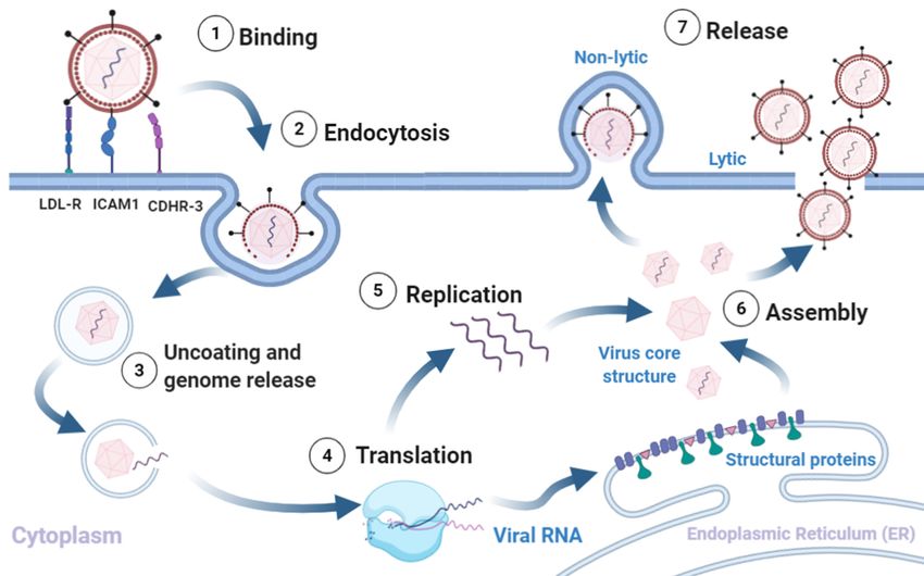
Viruses 2021, 13, 629                                                                                            2 of 16

                        3. Virion Structure and Lifecycle
                             RV is the leading cause of upper respiratory infections globally, affecting millions of
                        people every year [6–9]. RV infections can be asymptomatic, cause mild upper respiratory
                        tract symptoms or lead to severe lower respiratory tract infections and exacerbations in
                        asthma and COPD [10,11]. RV is a positive sense RNA virus belonging to the Enterovirus
                        genus in the Picornaviridae family [6,12]. RVs are highly diverse and to date, 170 strains
                        have been described based variously on serotyping and/or genetic sequencing. RVs can
                        be classified into three species (A, B, C) based on gene sequences. RVs belonging to A
                        and B species can be further subclassified into major or minor groups based on their
                        receptor usage [13,14].

                        3.1. Virion Structure
                             RVs are non-enveloped single-stranded(+) RNA viruses. The structural proteins
                        VP1, VP2, VP3, and VP4 form an icosahedral capsid that encases the RNA genome [15].
                        The capsids are composed of 60 copies each of VP1-4. The VP1-3 confer strain-specific
                        properties of immunogenicity, receptor binding and drug susceptibility to each RV isolate.
                        The short VP4 proteins localize inside the capsid, attached to the genome. The single
                        stranded RV genome (approximately 7200 bp) consists of a single open reading frame
                        with a 50 untranslated region and a short viral priming protein (VPg) that acts as a primer
                        for replication [13–15].

                        3.2. Viral Replication, Assembly and Release
                              RV infection typically involves the following sequence (Figure 1). (1) Binding of
                        the virus to the respective cognate receptors on the plasma membrane facilitated by VP1.
                        Majority of known RV strains (most RV-A and all RV-B) target ICAM1, a type 1 trans-
                        membrane protein that mediates cell to cell adhesion and immune reactions by binding
                        to the integrin lymphocyte function agent (LFA)-1 and macrophage-1 antigen [16–18]. A
                        minority of RV-A strains use Low-Density Lipoprotein Receptor (LDLR) [19], and all RV-C
                        strains characterised so far use the Cadherin Related Family Member 3 (CDHR3), as recep-
                        tor [17,20,21]. (2) Uptake of the virion into the cell goes through different endocytic routes,
                        such as macropinocytosis or clathrin-dependent or -independent endocytosis, dependent
                        on receptor usage. (3) The virus undergoes a conformational change within the endosome,
                        and the acidic environment (preferred method for minor-group RV’s), to form hydrophobic
                        subviral particles [18,22–24]. Though the exact mechanism of release is unknown, it is
                        suggested that the viral proteins rupture the endosome to release the genome into the
                        cytoplasm [13,14]. (4) Translation. Once the genome and VP4 enter the cytosol, the host
                        cell ribosomes translate the (+)ss-RNA into a polyprotein [18,25,26]. The genome is trans-
                        lated into a single polyprotein (P0) that is co-translationally cleaved in cis and trans by
                        viral proteases (2A protease, 2Apro and 3C protease, 3Cpro) into 11 proteins. The initial
                        cleavage mediated by 2Apro results in the release of P1 from the polyprotein. P1 is cleaved
                        to form the viral capsid proteins 3Cpro is probably responsible for all the other polyprotein
                        cleavages; releasing P2 and P3, and subsequently cleaving them to form the non-structural
                        proteins 2Apro, 2B, 2C, 3A, 3B (VPg), 3Cpro, 3D (RNA-dependent RNA polymerase) [27].
                        (5) RNA replication takes place within virus-induced membranous replication organelles
                        where 3Dpol synthesises (−)ssRNA, resulting in the formation of dsRNA, an important
                        pathogen associated molecular pattern (PAMP). The full length (−)ssRNA then functions
                        as the template for synthesis of new (+)ssRNA genomes that either enter another round of
                        replication or get packaged into virions [13–15]. Finally, (6) assembly and release of new
                        infectious virions. Recent studies have shown an important role for myristoylation of the
                        capsid protein VP0 in capsid assembly [15,18,22]. The RV polyprotein is co-translationally
                        modified at the N-terminus by myristoylation. Release of myristoylated P1 by 2Apro
                        is followed by cleavage into VP0, VP3, VP1 by 3Cpro and formation of a protomer [28].
                        Protomers assemble into pentamers, that form an icosahedral capsid, enclosing the RV
                        genome [28]. Exactly how the process ensures specificity (RV (+)ssRNA) and number of the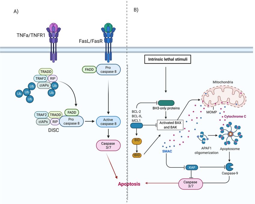
Viruses 2021, 13, x FOR PEER REVIEW 3 of 18
Viruses 2021, 13, 629 3 of 16
capsid, enclosing the RV genome [28]. Exactly how the process ensures specificity (RV
RNA moieties being encapsidated is unclear, but VP4 domain may have a role. (7) The final
(+)ssRNA) and number of the RNA moieties being encapsidated is unclear, but VP4 do-
step in virion maturation is the cleavage of VP0 into VP4 and VP2, by an as yet an unknown
main may have a role. (7) The final step in virion maturation is the cleavage of VP0 into
mechanism. The release of infectious virions from the cell probably varies between strains,
VP4 and VP2, by an as yet an unknown mechanism. The release of infectious virions from
as various pathways have been shown to be required, including cell lysis and release in
the cell probably varies between strains, as various pathways have been shown to be re-
membrane encased structures [13,14].
quired, including cell lysis and release in membrane encased structures [13,14].
Figure
Figure1.1.Rhinovirus
Rhinoviruslifecycle.
lifecycle.(1)
(1)Binding
Bindingof ofthe
thevirus
virusto
toreceptors
receptorson
onthe
theplasma
plasmamembrane
membraneinitiates
initiates (2)
(2) endocytosis.
endocytosis. The
The
intake
intake of the virion is followed by (3) uncoating of the capsid and release of the (+)ss-RNA into the host cytoplasm,which
of the virion is followed by (3) uncoating of the capsid and release of the (+)ss-RNA into the host cytoplasm, which
is (4) translated and processed into structural and non-structural proteins. The viral RNA dependent RNA polymerase
is (4) translated and processed into structural and non-structural proteins. The viral RNA dependent RNA polymerase
converts the viral genome into (-)ss-RNA, that is (5) replicated into new (+)ss-RNA genomes. The new genomes become
converts the viral genome into (−)ss-RNA, that is (5) replicated into new (+)ss-RNA genomes. The new genomes become
the template for translation into new viral proteins and replication into new genomes. The final stages involve (6) assembly
thethe
of template for translation
structural proteins andintoRNA
newgenome
viral proteins and replication
into capsids into newofgenomes.
and (7) release The final
new infectious stagesvia
virions involve (6)non-lytic
lysis or assembly
of the structural
mechanisms. proteins
Adapted fromand RNA genome
[5,13,16,28]. Createdintowith
capsids and (7) release
BioRender.com of new
accessed on infectious
12 Decembervirions
2020.via lysis or non-lytic
mechanisms. Adapted from [5,13,16,28]. Created with BioRender.com, accessed on 12 December 2020.
4. Cell Death Pathways
4. Cell Death Pathways
Regulated cell death is a conserved mechanism to maintain homeostasis in physio-
logicalRegulated cell death
and pathological is a conserved
settings. Complexmechanism
intra cellulartosignaling
maintainpathways
homeostasis in physio-
underpin the
logical and
observed pathological changes
morphological settings. and
Complex intra cellular
are triggered signaling
in response pathways underpin
to physiological signalsthe
or
observed
injury andmorphological
infection. Cell changes and are
death models aretriggered
classifiedinaccording
response to to physiological signals
their morphological
or injury andenzymatic
appearance, infection. criteria,
Cell deathor models are classified
immunological according [29].
characteristics to their
Themorphological
major path-
ways with known involvement in the lifecycle of viruses are described below. pathways
appearance, enzymatic criteria, or immunological characteristics [29]. The major
with known involvement in the lifecycle of viruses are described below.
4.1. Apoptosis
4.1. Apoptosis
Apoptosis is characterized by cell shrinkage, nuclear condensation and plasma mem-
braneApoptosis is characterized
blebbing with by cell shrinkage,
little or no inflammation. nuclear condensation
A characteristic feature of and plasma
apoptosis is mem-
pyk-
nosis (irreversible condensation of chromatin in the nucleus of a cell) followed by exten-is
brane blebbing with little or no inflammation. A characteristic feature of apoptosis
pyknosis (irreversible condensation of chromatin in the nucleus of a cell) followed by ex-
sive membrane blebbing and then karyorrhexis ultimately resulting in the separation of
tensive membrane blebbing and then karyorrhexis ultimately resulting in the separation of
cell fragments [29]. Initiated via extrinsic or intrinsic pathways, apoptosis does not involve
cell fragments [29]. Initiated via extrinsic or intrinsic pathways, apoptosis does not involve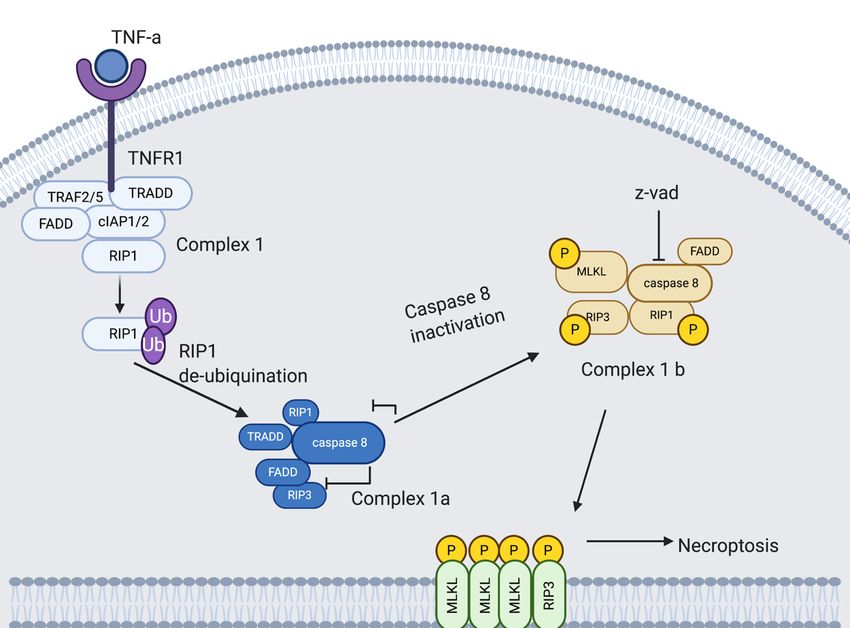
Viruses 2021, 13, 629                                                                                            4 of 16

                        inflammation as the cell contents are contained within the membrane bound apoptotic
                        bodies that are immediately phagocytosed.
                              The extrinsic pathway (Figure 2) involves transmembrane receptor-mediated inter-
                        actions. This pathway involves death receptors, proteins that contain an extracellular
                        cysteine-rich domain and a cytoplasmic death domain. Activation of the death domain
                        initiates a signal from the cell surface to the intracellular signaling pathways to induce
                        apoptosis. The best characterised extrinsic pathways are the Fas Ligand/Fas Receptor
                        (FasL/FasR) and Tumour Necrosis Factor-alpha/Tumour Necrosis Factor Receptor (TNF-
                        α/TNFR1) pathways [30–35].
                              Once ligand-binding occurs (Figure 2A), cytoplasmic adaptor proteins are recruited
                        and bind to respective death domains of the receptor proteins. Fas-associated protein with
                        death domain (FADD), is recruited to the FasL/FasR cytoplasm domain. Bound FADD
                        associates with pro-caspase 8, which is activated by the action of death-inducing signalling
                        complex (DISC). Tumour necrosis factor receptor type 1-associated death domain protein
                        (TRADD), FADD and receptor-interacting protein (RIP) are recruited to TNF-α/TNFR1
                        complex. Bound FADD associates with pro-caspase 8 leading to the formation of DISC,
                        resulting in the activation of caspase 8. Activation of caspase-8 initiates the execution phase
                        of apoptosis [5,30,36,37].
                              The intrinsic pathway of apoptosis, also known as the mitochondrial pathway, in-
                        volves a variety of non-receptor-mediated stimuli that generate mitochondrial-initiated
                        intracellular signals [5]. Positive or negative signals such as absence of certain hormones, cy-
                        tokines or growth factors, presence of hypoxia, toxins, reactive oxygen species or radiation
                        can initiate the intrinsic pathway. These signals (Figure 2B) alter the inner mitochondrial
                        membrane, leading to opening of the mitochondrial permeability transition (MPT) pores,
                        loss of transmembrane potential and release of two groups of pro-apoptotic proteins from
                        the intermembrane space into the cytosol [5,36].
                              The first group consists of proteins (cytochrome c, Smac/DIABLO, and the serine pro-
                        tease HtrA2/Omi) and activates the caspase-dependent mitochondrial pathway [30,38–40].
                        Cytochrome c binds and activates Apaf-1 as well as procaspase-9, forming an “apoptosome”
                        in the cytoplasm [41,42]. This complex then cleaves and activates the executioner caspases-
                        3/6/7, resulting in apoptosis. The second group of pro-apoptotic proteins, namely AIF,
                        endonuclease G and CAD, are released from mitochondria in the later stages of apoptosis.
                        AIF and endonuclease G are caspase-independent proteins that translocate to the nucleus.
                        They induce condensation of chromatin followed by DNA fragmentation [43,44]. CAD
                        is subsequently released from the mitochondria and translocates to the nucleus where,
                        after cleavage by caspase-3, it completes the condensation and fragmentation process [45].
                        The cascade of events results in destruction of the nuclear proteins and cytoskeleton,
                        crosslinking of proteins and the formation of apoptotic bodies [30,32].

                        Rhinovirus Can Modulate Apoptosis
                              Infection with RV-B14 in HeLa and 16HBE14o− bronchial epithelium cells exhibited
                        typical apoptotic morphological alterations similar to those induced by puromycin [36]. The
                        apoptotic morphological alterations, such as cell contraction and nuclear condensation, co-
                        incided with high-molecular-weight DNA fragmentation, cytochrome c translocation from
                        the mitochondria to the cytoplasm, activation of caspase-9 and caspase-3 and poly(ADP–
                        ribose) polymerase cleavage. Apoptosis did not affect RV14 replication per se, but it
                        facilitated the release of newly formed virions from cells [36].
                              The RV 3Cpro targets several cellular proteins, probably to support virus replication
                        and limit the innate antiviral response, including apoptosis. RV 2Apro, 3Cpro, and 3CDpro
                        have variously been shown to cleave proapoptotic adaptor proteins and nucleoporins
                        resulting in downregulation of apoptosis and disruption of nucleocytoplasmic trafficking
                        pathways involved in apoptosis, respectively. Nuclear transport of caspases is key to the
                        nuclear consequences of apoptosis and it is possible that the disruption of nuclear transport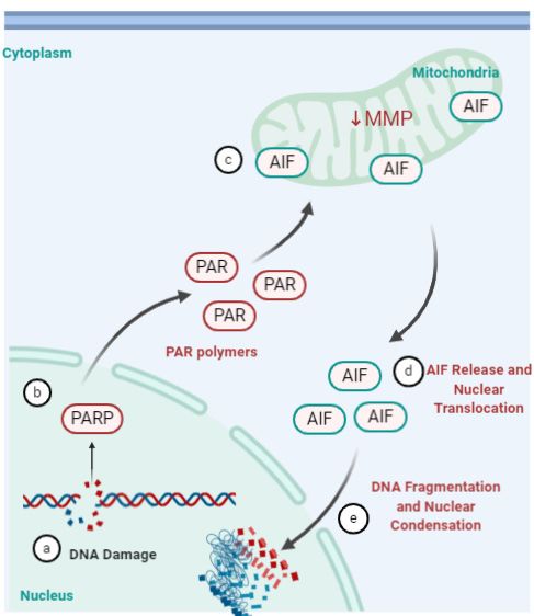
Viruses 2021, 13, 629 5 of 16
in
Viruses 2021, 13, x FOR PEER REVIEW RV infected cells [48,49] may serve to inhibit the progression of apoptosis; however,5 of
this
18
remains to be investigated [50].
Figure 2.
Figure 2. Extrinsic
Extrinsic and
and Intrinsic
Intrinsic apoptosis.
apoptosis. (A) Extrinsic
Extrinsic apoptosis.
apoptosis. Ligand binding results in in activation
activation of
of cytoplasmic
cytoplasmic
adapter proteins
adapter proteins and
and recruitment
recruitment ofof death
death domains
domains on onFasL/FasR
FasL/FasRororTNF-α/TNFR1.
TNF-α/TNFR1. This activates
This FADD,
activates FADD,which cancan
which di-
rectly activate pro-caspase-8 or can form a complex with TRADD and RIP, bind to TNF-α/TNFR1 and then
directly activate pro-caspase-8 or can form a complex with TRADD and RIP, bind to TNF-α/TNFR1 and then induce auto- induce auto-
catalytic activation of procaspase-8. Activated caspase 8 initiates the execution phase of apoptosis. (B) Intrinsic apoptosis.
catalytic activation of procaspase-8. Activated caspase 8 initiates the execution phase of apoptosis. (B) Intrinsic apoptosis.
Initiated by non-receptor-mediated stimuli that generate mitochondrial-initiated intracellular signals. These signals initi-
Initiated by non-receptor-mediated stimuli that generate mitochondrial-initiated intracellular signals. These signals initiate
ate a cascade of events resulting in the formation of an apoptosome. The apoptosome activates the executioner caspases-
a3/6/7
cascade
and of eventsapoptosis.
initiates resulting in the formation
Adapted of an apoptosome.
from [5,46,47]. TheBioRender.com
Created with apoptosome activates theon
accessed executioner
12 Decembercaspases-3/6/7
2020.
and initiates apoptosis. Adapted from [5,46,47]. Created with BioRender.com, accessed on 12 December 2020.
Rhinovirus Can Modulate Apoptosis
RV 3Cpro cleaves RIP1, a central player in pro-inflammatory, pro-survival and cell
deathInfection
pathways.with RV-B14
RV-A16 in HeLainand
infection 16HBE14o−
HeLa bronchial
cells results epithelium
in activation cells exhibited
of caspase 8 which
typical apoptotic
cleaves morphological
RIP1, committing the cellalterations
to apoptosis.similar to thosesubsequent
However, induced bycleavage
puromycin [36].
of RIP1
The3Cpro
by apoptotic
haltsmorphological
the progressionalterations,
of apoptosis. such as cell contraction
Significantly, and nuclear
3Cpro mediated condensa-
cleavage was
tion, coincided with high-molecular-weight DNA fragmentation, cytochrome
observed in cells infected with representative RV-A, -B, major and minor strains (Sarah c transloca-
tionCroft,
N. from personal
the mitochondria to the cytoplasm,
communication), suggesting activation of caspase-9
that it may and caspase-3
be conserved and
across most
RV strains. Intriguingly, markers of a necrotic cell death pathway were observed late se,
poly(ADP–ribose) polymerase cleavage. Apoptosis did not affect RV14 replication per in
but it facilitated
infection, the release
along with of newly
membrane formed
rupture [51].virions
Sudden from cells of
release [36].
calcium ions from their
Thelocation
storage RV 3Cpro targets
in the several cellular
Endoplasmic Reticulum proteins, probably
(ER) can induceto supportapoptosis.
intrinsic virus replication
The RV
andprotein
2B limit the innate
inserts into antiviral response,
the intracellular includingand
membranes apoptosis. RV 2Apro,
disrupts calcium 3Cpro,
stores, and
probably
3CDpro
via haveofvariously
leakage calcium ionsbeeninto
showntheto cleave proapoptotic
cytoplasm, reducing the adaptor proteins
calcium and nucleo-
ions available to
porins resulting in downregulation of apoptosis and disruption
translocate to the mitochondria, resulting in apoptosis inhibition [52–54]. of nucleocytoplasmic traf-
ficking pathways involved in apoptosis, respectively. Nuclear transport of caspases is key
to the nuclear consequences of apoptosis and it is possible that the disruption of nuclearViruses 2021, 13, 629 6 of 16
4.2. Autophagy
Autophagy is the term given to any pathway in which cytoplasmic material is trans-
ported to the lysosome [55,56]. It is a lysosomal-dependent process which is based on the
degradation of the mitochondrial and other intracellular structures. Autophagy is used to
maintain cell homeostasis under stressful conditions; this is achieved through the removal
of misfolded protein, damaged organelles or intracellular pathogens, e.g., viruses [55].
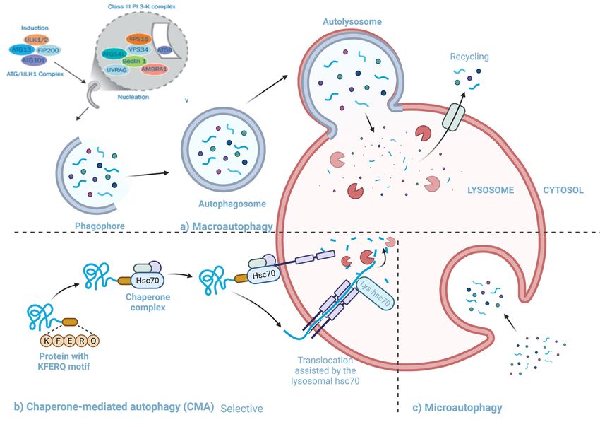
There are three types of autophagy (Figure 3), micro-autophagy, chaperone-mediated
autophagy and macro-autophagy. Micro-autophagy involves cytosolic components being
directly taken up by the lysosome itself through invagination of the lysosomal membrane,
and unlike macro-autophagy, engulfment in micro-autophagy is by both non-selective
and selective mechanisms [57]. Chaperone-mediated autophagy involves proteins being
translocated to the lysosomal membrane by chaperone complexes [57]. Chaperone proteins
(i.e., heat shock cognate 70(HSC70)) and co-chaperones recognize proteins that contain
KFERQ-like pentapeptides. Once the chaperones and proteins are bound, the whole
complex is translocated into the lysosomal lumen and where it binds with Lysosomal-
associated protein 2A (LAMP-2A) [55–57]. LAMP-2A, a transmembrane protein, acts as
the receptor in which proteins bind for unfolding and degradation [55].
Macro-autophagy is the best characterized among the known types of autophagy
(henceforth will be referred to as autophagy) [58,59]. This pathway involves the transfer of
cytosolic material into lysosomes using double membrane-bound vesicles 7which
Viruses 2021, 13, x FOR PEER REVIEW of 18 fuse to
the lysosomes [57].
Three3.autophagy
Figure 3.Figure pathways
Three autophagy (a)(a)
pathways Macroautophagy: cytosolic
Macroautophagy: cytosolic material
material is transported
is transported to the lysosome
to the lysosome using double
using double
membrane-bound vesicles which engulf the material and then fuse to the lysosome (b) Chaperone
membrane-bound vesicles which engulf the material and then fuse to the lysosome (b) Chaperone mediated autophagy:mediated autophagy:
proteins proteins
bound to bound to HSC70 being translocated to the lysosomal membrane to be unfolded and degraded. (c) Microautoph-
HSC70 being translocated to the lysosomal membrane to be unfolded and degraded. (c) Microautophagy:
agy: cytosolic components being directly taken up by the lysosome. Adapted from [60,61]. Created with BioRender.com
cytosolicaccessed
components being directly taken up by the lysosome. Adapted from [60,61]. Created with BioRender.com, accessed
on 25 January 2020.
on 25 January 2020.
4.2.1. The Pathway and Mechanisms of Autophagy
The primary mechanism involved in autophagy can be divided into four stages: (i)
induction and nucleation, (ii) elongation, (iii) cargo recruitment and (iv) fusion with the
lysosome and breakdown. (i) Induction and nucleation: Autophagy is initiated when Unc-
51 like kinase-1 (ULK1) complex interacts with the mechanistic target of rapamycin com-
plex 1 (mTORC1), and AMP-activated protein kinase (AMPK) [62]. The ULK1 complex
phosphorylates a class III phosphoinositide 3-kinase (P13K) complex, comprised of au-Viruses 2021, 13, 629 7 of 16
4.2.1. The Pathway and Mechanisms of Autophagy
The primary mechanism involved in autophagy can be divided into four stages: (i) in-
duction and nucleation, (ii) elongation, (iii) cargo recruitment and (iv) fusion with the
lysosome and breakdown. (i) Induction and nucleation: Autophagy is initiated when
Unc-51 like kinase-1 (ULK1) complex interacts with the mechanistic target of rapamycin
complex 1 (mTORC1), and AMP-activated protein kinase (AMPK) [62]. The ULK1 com-
plex phosphorylates a class III phosphoinositide 3-kinase (P13K) complex, comprised of
autophagy-related 14L (ATG14L), Beclin 1, vacuolar protein sorting 34 (VSP34), VSP15,
VPS15, and autophagy and beclin 1 regulator 1 (AMBRA-1) [62,63]. AMBRA1 is phospho-
rylated by ULK1 after which it separates from the complex and gets translocated to the ER
where it binds to Beclin-1. During the formation of the phagophore VPS34, generates PI3P,
which is vital for phagophore growth [64]. The activity of VPS34- Beclin-1 complex is regu-
lated in a Beclin-1 dependent manner by proteins like Barkor and UV resistance-associated
gene (UVRAG) [64]. (ii) Elongation: Two protein conjugation systems are involved during
elongation and formation of the autophagophore. One system requires the formation of the
Atg12–Atg5-Atg16 complex, this system uses Atg7, Atg10 and Atg16L1. The complex is
anchored onto phosphoinositol 3-phosphate on emerging autophagosomal membranes, the
ATG5-ATG12-ATG16L1 complex adds to the curvature of the phagophore and is essential
for the microtubule-associated protein 1A/1B-light chain 3 (LC3) lipidation [55]. The other
system is the LC3-phosphatidylethanolamine (LC3-PE) system that includes ATG7 and
ATG3 conjugate, LC3 (ATG8), and Atg4B [64]. ATG4 cleaves LC3 which results in the
formation of LC3-1, which is then conjugated to the lipid phosphatidylethanolamine (PE)
on the surface of the emerging autophagosome by ATG3 and ATG7 [62]. The final stage
is the formation of LC3B-I-PE conjugate. The LC3B-I-PE conjugate elongates and seals
the phagophore. LC3B-II remains associated with the autophagosomal membrane until
the fusion with lysosome takes place [64]. Both systems result in a double-membraned
autophagosome. (iii) Cargo recruitment: During the formation of the autophagosome,
cargo is collected for degradation. Protein p60 binds to any ubiquitinated proteins and
makes them a target for degradation [64]. p60 and its bound protein bind to LC3II present
on the inner and outer surface of the autophagosomal membrane where it interacts with
the constitutively expressed adaptor molecule P62/SQSTM1 [64]. P62/SQSTM1 and NBR1
are cargo receptors that contain a ubiquitin-binding domain [62,64]. As the autophago-
some is formed, the target proteins and organelles become engulfed in the newly formed
autophagosome [63]. Once the membrane closes around its cargo, LC3-II is cleaved from
the outer membrane of this structure. (iv) Fusion and cargo breakdown: The final step
in the autophagy pathway is the least understood [59]. Fully formed and cargo-bound
autophagosomes are transported on microtubules to the perinuclear region where the outer
membrane fuses with the lysosome to form an autolysosome [59,62]. The full fusion of
autophagosomes occurs when lysosomal hydrolases degrade the inner membrane and ex-
pose the contents to the lysosome lumen [55], UVRAG, which associates with the PtdIns3K
complex, can activate the GTPase RAB7, that promotes fusion with the lysosome [59]. In
some instances, the autophagosome may fuse with an endosome and form an amphisome
involving VTIlB protein, prior to fusing with the lysosome [59]. Rab7 effector protein,
PLEKHM1, regulates the fusion of autophagosome and lysosome through HOPS complex
and LC3/GABARAP protein [64].
4.2.2. The Interactions between Autophagy and Rhinovirus Infection
The interactions between RV and autophagy are relatively understudied compared to
other cell death pathways. However, it has gained attention in recent years. RV infection
has been shown to induce the formation of autophagosome-like structures [65]; however,
not all RV strains use autophagy in the same way or at all.
A study has suggested that RV-A2, a minor group RV, induces and uses autophagy for
more efficient replication, although it is still unclear as to what autophagy machinery is
involved [65]. RV-A2 infected HeLa-cells showed autophagosome formation indicated bothViruses 2021, 13, 629 8 of 16
by punctate green fluorescent protein (GFP)-LC3 signal colocalized with LAMP1 staining,
and an increase in LC3-II, during the later stages of infection (4 and 6 h post infection).
These two points of data confirm that RV-2 infection stimulates autophagic induction [65].
RV-A2 use of autophagy pathways was further confirmed by the demonstration that
chemical inhibitors of autophagy or deletion of critical autophagy genes inhibited RV-A2
replication [65]. In contrast, an earlier study [66] found that RV-A2 did not induce the
synthesis of LAMP-2- and LC3-positive compartments and modification of autophagy does
not result in increased viral synthesis [66]. Klein and Jackson attribute the discrepancy
in the two studies to improved understanding of autophagy in the time between the two
studies as well as the types of cells and autophagosome formation markers used. However,
these studies need to be further validated for different RV strains.
Two studies by Wu et al. [67,68] have shown that RV-A16 utilizes autophagy during
its replication in the context of interleukin-1 receptor associated kinase M (IRAK-M). LC3
II/LC3 I protein, an indicator of autophagosome formation, was increased during RV-
A16 infection in a cell line over expressing IRAK-M. Using beclin-1 to inhibit autophagy,
the authors observed a marked decrease of RV-A16 RNA levels and viral particles, sug-
gesting that autophagy may facilitate RV-A16 replication in cells in which IRAK-M is
over-expressed [67]. Additionally, trehalose-induced autophagy directly inhibited Inter-
feron lambda-1 (IFN-λ1) expression and promoted RV-A16 infection in normal human
primary airway epithelial cells. This effect was reversed when ATG5 was knocked out. Tre-
halose is a natural glucose disaccharide which functions in the prevention of LPS-mediated
inflammatory response [69] and induces autophagy in various cells by promoting the
recruitment of LC3 II into the forming autophagosome membrane in an ATG5-ATG12-
dependent manner [70]. Interestingly, RV-A16 infection was shown to result in reduced
levels of p62/SQSTM1 late in infection, probably due to its cleavage by 3Cpro [51]; effects
on autophagy were not investigated in the study.
RV-B14 infection may also induce autophagy as shown by co-localization of LAMP1
with punctate GFP-LC3 in MCF-7 cells. Further, monodansylcadaverine, a fluorophore
retained in autophagosomes under mild cell fixation conditions, showed a punctate pat-
tern after infection with RV-B14 and suggesting the formation of autophagosome-like
vesicles [65].
In contrast, RV-A1a does not induce or use autophagic signaling or autophagosomes [65].
In the same study in which RV-A2 was found to induce LC3 modification, simulating au-
tophagy, RV-A1a did not induce LC3 modification or simulate autophagy [65]. No other RV
strains have been investigated for their exploitation and/or modulation of autophagy.
4.3. Necrosis
Necrosis follows an energy-independent mode of death that occurs in an unpro-
grammed state in cells induced by a number of external triggers such as infection, trauma,
or excessive stress. It is a pathological event characterised by cytoplasmic swelling, irre-
versible membrane damage and organelle breakdown, with characteristic cellular content
leakage into the extracellular environment. Leaked cellular content triggers an increase in
the secretion of pro-inflammatory cytokines from activated macrophages [71].
Necrosis triggered by DNA damage leads to the activation of poly (ADP-ribose) poly-
merase 1 (PARP-1) to catalyze the hydrolysis of NAD+ into nicotinamide and poly-ADP
ribose, causing NAD depletion. This results in cellular energy failure and a caspase- in-
dependent death. N-methyl-N-nitro-N-nitrosoguanidine (MNNG)- induced necrosis is
dependent on RIP-1 and TNFR associated factor (TRAF)2, which function downstream of
PARP-1 and lead to c-Jun N-terminal kinase (JNK) activation. JNK affects mitochondrial
membrane integrity which allows for the release of proteins into the mitochondrial inter-
membrane space. The specific way JNK induces this is unclear however, either modification
of Bcl-2 family or caspase-independent JNK-mediated processing of bid may be at play [72].Viruses 2021, 13, 629 9 of 16
Modulation of Necrosis by Rhinovirus
Limited studies have looked into the modulation of necrosis by RV’s. Lötzerich et al.,
2018 have shown 3Cpro is able to suppress apoptosis and trigger necrosis [51]. Montgomery
et al., 2020 have shown a direct correlation between RV infection, increased expression
of IL-1 and increased necrotic events [73]. RV was shown to induce necrosis in airway
epithelial cells collected from children with and without cystic fibrosis accompanied by
higher levels of inflammatory mediators IL-1α, IL-1β and IL-8 [73], suggesting necrosis as
a mechanism by which RVs induce mucin accumulation and inflammation in early lung
disease. Whether necrosis has a role in virus assembly or release is unclear.
4.4. Necroptosis
Necroptosis is a programmed variant of necrosis triggered by interactions of death
ligands and death receptors in the context of caspase inhibition. Necroptosis and necrosis
share morphological features such as early destruction of membrane integrity, cell and
intracellular organelle swelling, oxidative bursts and cell content spillage. Necroptosis
leads to reduced damage to the cells by regulation of a series of signals [74]. Necroptosis,
unlike apoptosis, causes local inflammation, infiltration and activation of inflammatory
cells. This inflammatory process mediates the release of damage-associated molecular
patterns (DAMPs), which results in the recruitment of pro-inflammatory cell types to the
sites of infection. The necroptotic pathway has three key players; RIP1, RIP3 and mixed
linkage kinase domain-like protein (MLKL) [75].
4.4.1. Necroptosis Pathway
Necroptosis is induced by a class of death receptors that include TNFR1, TNFR2 and
Fas. Upon binding with their respective agonists, the cell can be directed down a cell death
pathway. Most research into the necroptotic pathway has been done on the TNFa/TNFR1
induced pathway (Figure 4). TNFa bound to the extracellular domain of TNFR1 creates
allosteric changes in the cytoplasmic region of TNFR1, triggering downstream signaling
by forming complex I with TRADD, FADD, RIP1, and E3 ubiquitination ligases, TRAF2/5
and cellular inhibitor of apoptosis protein-1 (cIAP1/2) [68].
RIP1, a serine/threonine kinase, plays a role in both necroptosis and apoptosis (in ad-
dition to necrosis), and is initially recruited to complex I by TNFR1 and is polyubiquitinated
by TRAF2/5, cIAP1/2 on lysine at position 63; this drives the cell down a pro-survival
pathway. This pathway progresses via Inhibitor of Kappa B Kinase (IKK) and NF-kappa
B Essential Modulator (NEMO) recruitment and activation of NF-kB pathway. However,
when RIP1 is deubiquitinated, it inhibits the NF-kB pathway, which directs the cell towards
death [68,76].
Deubiquitinated RIP1 is transferred from complex I to the cytoplasm and prompts the
recruitment of complex II. TRADD is also released from complex I at this time. Complex
II, or DISC, comprises TRADD, FADD, RIP1 and caspase 8. Caspase 8 is inhibited in
complex II, allowing RIP1 to bind to RIP3. Bound RIP3 recruits and phosphorylates
MLKL at serine 358 and threonine 357 resulting in the formation of the necrosome [77].
Activated MLKL then binds to phosphatidylinositol phosphates (PIPs), Cardiolipin (CL)
and phosphatidylglycerol (PG), allowing the necrosome to move through the cell and target
the membrane where MLKL disrupts the membrane leading to permeabilization, swelling
and rupturing [78]. The final or propagation stage of necroptosis involves an inflammatory
wave of DAMP release [76]. DAMP production is a crucial contributor to chronic and
acute inflammation, specifically induction of cytokine and other chemo-attractants, for
the recruitment of primary immune cells to the necroptotic site [76]. The steps between
complex II and the formation of the necrosome as well as the downstream signaling in the
necroptotic pathway are still under-researched.Viruses13,
Viruses 2021, 2021,
62913, x FOR PEER REVIEW 11 of 1810 of 16
Figure 4. Necroptopic signaling pathway. TNFa is bound to the extracellular domain of the TNFR1. TNFR1 triggers down-
4. Necroptopic
Figurestream signaling pathway. TNFa is bound to the extracellular domain of the TNFR1. TNFR1 triggers downstream
signaling by forming complex I with TRADD, FADD, RIP1, and several E3 ubiquitination ligases, TRAF2/5 and
signaling by forming
cIAP1/2. complexRIP1
Deubiquitinated I with TRADD, FADD,
is transferred RIP1, Iand
from complex several
to the E3 ubiquitination
cytoplasm and prompts the ligases, TRAF2/5
recruitment and cIAP1/2.
of complex Ia.
Complex IaRIP1
Deubiquitinated is comprised of TRADD,
is transferred from FADD,
complex RIP1
I toand
the caspase 8. Caspase
cytoplasm 8 is inhibited
and prompts in complexof
the recruitment Ib,complex
allowingIa.
forComplex
RIP1 Ia
to bind of
is comprised to RIP3.
TRADD, Bound RIP3 recruits
FADD, RIP1 and and phosphorylates
caspase 8. CaspaseMLKL. Activated in
8 is inhibited MLKL disrupts
complex the membrane
Ib, allowing leading
for RIP1 to per-
to bind to RIP3.
meabilization, swelling and rupturing. Adapted from [51,79–82]. Created with Biorender accessed on 15 January 2021.
Bound RIP3 recruits and phosphorylates MLKL. Activated MLKL disrupts the membrane leading to permeabilization, swelling
and rupturing. Adapted from [51,79–82]. Created with BioRender.com, accessed on 15 January 2021.
4.4.2. Potential Exploitation of Necroptosis by Rhinovirus
The pathway used by RV to exit from a cell after replication is still unknown however
4.4.2. Potential
there Exploitation
are several of Necroptosis
lines of direct by Rhinovirus
and indirect evidence to suggest that necroptosis may be
Theduring
used pathway used by Recent
RV infection. RV to exit from
studies a cellhave
[36,37] afteridentified
replication
thatisRV still unknown
infection however
inhibits
caspase-dependent
there are several linescell of death
directpathways
and indirectlike apoptosis
evidencethrough the that
to suggest cleavage of RIP1 by
necroptosis may be
3Cpro,
used duringcoinciding with impaired
RV infection. recruitment
Recent studies of active
[36,37] caspase 8 that
have identified [37,51].
RVInhibition
infection of inhibits
caspase 8 activity would
caspase-dependent be expected
cell death pathwaysto commit the cell to necroptosis.
like apoptosis through the Whether the 3Cpro
cleavage of RIP1 by
cleavage product of RIP1 is able to interact with RIP3, an essential
3Cpro, coinciding with impaired recruitment of active caspase 8 [37,51]. Inhibition step in necroptosis, is of
unknown. IL-33, a key necroptotic inflammatory factor, has been shown to be released
caspase 8 activity would be expected to commit the cell to necroptosis. Whether the 3Cpro
during RV infection. RV infection triggers the production of several cytokines and chem-
cleavage product of RIP1 is able to interact with RIP3, an essential step in necroptosis, is
okines (including IL-1, IL-6, IL-8, GM-CSF, eotaxins, and regulated upon activation nor-
unknown. IL-33, a key necroptotic inflammatory factor, has been shown to be released dur-
mal T-cell expressed and secreted (RANTES), TNF-α, IFN-γ, and macrophage inflamma-
ingtory
RV infection. RV infection
protein (MIP)–1a [83–85]).triggers
Amongthe production
these, TNF-α and of several
IL-1 arecytokines
known to and inducechemokines
the
(including
necroptotic pathway [86]. This surge in expression of cytokines and chemokines leads us T-cell
IL-1, IL-6, IL-8, GM-CSF, eotaxins, and regulated upon activation normal
expressed andthat
to speculate secreted (RANTES),
RV-induced TNF-α,may
inflammation IFN-γ, andnecroptosis
trigger macrophage inflammatory
within the cell. How- protein
(MIP)–1a [83–85]).
ever, further Among
studies these, TNF-α
are required and IL-1
to address are known
this link in moretodetail.
induce the necroptotic
Elevated cytosolic path-
way [86]. This
calcium surge induring
is observed expression of cytokines
RV infection, probablyandduechemokines leads
to the actions us protein
of 2B to speculate
(see that
above). Accumulation
RV-induced inflammation of may
cytosolic calcium
trigger ions led within
necroptosis to inhibition or deficiency
the cell. However,of caspase
further studies
are8required
in neuroblastoma cells,this
to address along
linkwith
inCa2+/calmodulin-dependent
more detail. Elevated cytosolic protein kinase
calcium II (CaMK
is observed
II) phospholrylation
during and necroptosis
RV infection, probably due to[54].
the The RV viroporin
actions (2B) disrupts
of 2B protein the calcium
(see above). ion
Accumulation
homeostasis within the host cell by discretely affecting calcium release from
of cytosolic calcium ions led to inhibition or deficiency of caspase 8 in neuroblastoma cells, the ER [50].
This suggests that necroptosis may be a potential viral release mechanism for RV-A2
along with Ca2+/calmodulin-dependent protein kinase II (CaMK II) phospholrylation and
through the accumulation of cytosolic Ca2+ within infected cells [87]. Finally, a study has
necroptosis [54]. The RV viroporin (2B) disrupts the calcium ion homeostasis within the
host cell by discretely affecting calcium release from the ER [50]. This suggests that necrop-
tosis may be a potential viral release mechanism for RV-A2 through the accumulation of
cytosolic Ca2+ within infected cells [87]. Finally, a study has shown that chemical inhibition
of membrane channels characteristic of necroptosis inhibits RV-A2 release [88]. Together,
the evidence provides a strong incentive to investigate the possible role of necroptosis inViruses 2021, 13, 629 11 of 16
RV release. Recently it has been seen that a deficiency in IFN-β, a multipurpose cytokine,
during asthma exasperations promotes necroptosis. Primary cells from asthmatics show a
deficient ability to produce IFN-β during RV infection, and RV induced asthma exacerba-
tions have been linked to increased levels of LDH [89]. Results from experimentation on
mice deficient in IFN-β suggest that necroptosis may contribute to high LDH levels due to
the observed increase in pMLKL [90]. Data from this paper implies that RV infection may
result in a more harmful cell death response in asthmatics with reduced IFN expression.
However, further studies should be conducted into the role of IFN-β as a regulator of
necroptosis during RV infection.
4.5. Parthanatos, Stress and Rhinovirus
Parthanatos is a relatively new addition to cell death pathway characterized in recent
years. The pathway is regulated by poly(ADP-ribose) polymerase 1 (PARP1), an enzyme
that catalyzes DNA base excision repair [91]. PARP1 is activated in response to severe
or prolonged DNA damage, oxidative stress, hypoxia, hypoglycemia or inflammation.
This triggers a series of cytotoxic effects such as NAD+ and ATP depletion and accumu-
lation of poly(ADP-ribose) polymers (PAR) and poly(ADP-ribosyl)ated proteins (PARP)
at mitochondria (Figure 5) [92,93]. RV-1B infection of human primary bronchial epithelial
cells (pBECs) exposed to cigarette smoke or other oxidative stressors reduced mitochon-
drial respiration, increased proton leak and subsequently increased pro-inflammatory
cytokines [94]. The oxidative stress caused by the external stimuli, including RV infection,
lead to increased expression of cytochrome C [94]. Exposure of RV-A16 infected human
bronchial epithelial cells and the BEAS-2B cells to cigarette smoke increased the expression
of CXCL8 (linked to mRNA stabilization) compared to either stimulus by themselves.
Cigarette smoke was shown to inhibit RV-16-induced expression of CXCL-10 vis transcrip-
tional regulation [94,95]. This suggests parthanatos could be one of the mechanisms by
which RV infections, aggravated by external stimuli such as cigarette smoke, can induce
exacerbations in asthma and chronic obstructive pulmonary disease (COPD) [94,95]. This
could potentially explain increased susceptibility of smokers, asthma and COPD patients
Viruses 2021, 13, x FOR PEER REVIEW 13 of 18
to respiratory infections and contribute to exacerbations.
Figure 5. Parthanatos pathway. (a) Severe or prolonged DNA damage, oxidative stress, hypoxia, hypoglycemia or inflam-
Figure
mation (b) triggers the activation Parthanatos
of the5.enzyme pathway.
poly(ADP-ribose) (a)(PARP).
polymerase Severe or prolonged
(c) This triggers a series DNA damage, oxidative stress, hypoxia,
of cytotoxic
effects such as NAD+ and ATP depletion and accumulation
hypoglycemia of poly(ADP-ribosyl)ated
or inflammation proteins
(b) triggers the(PAR) at the mitochondria.
activation of the enzyme poly(ADP-ribose) poly-
(d) PAR bind to apoptosis-inducing factor (AIF), decrease in mitochondrial membrane potential (MMP) and prompts re-
merase
lease of AIF into the cytoplasm. (e) AIF(PARP). (c) This
then translocates triggers
to the nucleus a serieslarge-scale
and mediates of cytotoxic effects such
DNA fragmentation and as NAD+ and ATP depletion
chromatin condensation [91–93,96]. Created using Biorender accessed on 31 January 2021.
and accumulation of poly(ADP-ribosyl)ated proteins (PAR) at the mitochondria. (d) PAR bind to
apoptosis-inducing
5. Conclusions factor (AIF), decrease in mitochondrial membrane potential (MMP) and prompts
release of AIF into the
RVs have significant cytoplasm.
impact (e) AIF
on society being then translocates
the primary torespiratory
agent for upper the nucleus and mediates large-scale
tract infections globally. The indications range from mild (common cold) limited to the
DNA fragmentation and chromatin condensation [91–93,96]. Created using BioRender.com, accessed
upper respiratory tract, to chronic or severe illnesses (such as, asthma exacerbations, bron-
on 31 January
chiolitis, and otitis2021.
media) [9]. Vaccine design against rhinoviruses is unlikely due to its
high mutation rates and over 100 serotypes while existing non-specific antiviral treat-
ments are generally ineffective. Characterization of how the virus subverts cell signalling
pathways, especially cell death, could potentially drive targeted drug development [97].
Most of the work to date has focussed on the interplay between RVs and apoptosis, with
some studies examining necrosis and autophagy. The review of existing direct and indi-
rect evidence presented here clearly shows that RVs have a very complex relationshipViruses 2021, 13, 629 12 of 16
5. Conclusions
RVs have significant impact on society being the primary agent for upper respiratory
tract infections globally. The indications range from mild (common cold) limited to the
upper respiratory tract, to chronic or severe illnesses (such as, asthma exacerbations, bron-
chiolitis, and otitis media) [9]. Vaccine design against rhinoviruses is unlikely due to its high
mutation rates and over 100 serotypes while existing non-specific antiviral treatments are
generally ineffective. Characterization of how the virus subverts cell signalling pathways,
especially cell death, could potentially drive targeted drug development [97]. Most of the
work to date has focussed on the interplay between RVs and apoptosis, with some studies
examining necrosis and autophagy. The review of existing direct and indirect evidence
presented here clearly shows that RVs have a very complex relationship with several cell
death pathways, opening up several possible lines of future research investigation.
That RVs manipulate cell death pathways to favour viral replication and propagation,
is clear [32,98]. This manipulation may correlate with the severity of disease and exacer-
bations of pre-existing lung conditions [10,20,97]. Our understanding of RV interaction
with and modulation of cell death pathways has evolved along with our knowledge of
these complex pathways. Accumulating literature shows that RVs inhibit apoptosis early
in infection via cleavage of RIP1 and by disrupting nucleocytoplasmic trafficking; although
the latter has not been directly linked with apoptosis inhibition. Recent studies implicate
necroptosis as a potential mechanism by which RVs exit the host cell, accompanied by re-
lease of proinflammatory chemokines and cytokines. RIP1, which is cleaved in RV infection,
is a master regulator, positioned to commit the cell to prosurvival, apoptotic or necroptotic
pathway. RV infection induces caspase 8 dependent apoptotic pathway, which is quickly
inhibited by cleavage of RIP1, possibly committing the cell to necroptosis. This supposition
is supported by the observation of necrotic markers in the absence of late-stage necrosis in
RV infected cells. RVs appear to modulate autophagy in a strain, maybe species, specific
manner. The limited literature raises the question—does the autophagy pathway facilitate
RV release, albeit inside a membrane vesicle? Chronic damage to DNA caused by extensive
oxidative stress is shown to induce parthanatos during RV infections. Parthanatos is one of
the mechanisms by which RV infections may induce exacerbations in asthma and COPD.
Intriguingly, cell death related targets of 3Cpro identified so far (e.g., p62/SQSTM1) are
involved in multiple pathways, possibly suggesting a fine modulation of several pathways
by RVs that may vary through the lifecycle to enable optimal virus replication, assembly
and release. A limitation for our review was the lack of studies on modulation of cell death
pathways by RVs belonging to genotype C. However, review of available literature on RV-A
and RV-B strains strongly suggests the role of necroptosis and autophagy in RV release.
Author Contributions: Conceptualization, R.G., C.M. and S.-L.K.; methodology, C.M. and S.-L.K.;
writing—original draft preparation, C.M., S.-L.K.; writing—review and editing, C.M. and S.-L.K.;
visualization, C.M. and S.-L.K.; supervision, R.G. All authors have read and agreed to the published
version of the manuscript.
Funding: This research received no external funding.
Institutional Review Board Statement: Not applicable.
Informed Consent Statement: Not applicable.
Data Availability Statement: Not applicable.
Conflicts of Interest: The authors declare no conflict of interest.
References
1. Zhang, T.; Yin, C.; Boyd, D.F.; Quarato, G.; Ingram, J.P.; Shubina, M.; Ragan, K.B.; Ishizuka, T.; Crawford, J.C.; Tummers, B.; et al.
Influenza Virus Z-RNAs Induce ZBP1-Mediated Necroptosis. Cell 2020, 180, 1115–1129. [CrossRef] [PubMed]
2. Kuo, C.-Y.; Chiu, V.; Hsieh, P.-C.; Huang, C.-Y.; Huang, S.J.; Tzeng, I.S.; Tsai, F.-M.; Chen, M.-L.; Liu, C.-T.; Chen, Y.-R.
Chrysophanol attenuates hepatitis B virus X protein-induced hepatic stellate cell fibrosis by regulating endoplasmic reticulum
stress and ferroptosis. J. Pharmacol. Sci. 2020, 144, 172–182. [CrossRef]Viruses 2021, 13, 629 13 of 16
3. Suwanmanee, S.; Luplertlop, N. Immunopathogenesis of Dengue Virus-Induced Redundant Cell Death: Apoptosis and Pyroptosis.
Viral Immunol. 2017, 30, 13–19. [CrossRef]
4. Kim, J.H.; Kim, J.; Roh, J.; Park, C.S.; Seoh, J.Y.; Hwang, E.S. Reactive oxygen species-induced parthanatos of immunocytes by
human cytomegalovirus-associated substance. Microbiol. Immunol. 2018, 62, 229–242. [CrossRef]
5. Croft, S.N.; Walker, E.J.; Ghildyal, R. Picornaviruses and Apoptosis: Subversion of Cell Death. mBio 2017, 8, e01009-17. [CrossRef]
[PubMed]
6. Zlateva, K.T.; de Vries, J.J.C.; Coenjaerts, F.E.J.; van Loon, A.M.; Verheij, T.; Little, P.; Butler, C.C.; Goossens, H.; Ieven, M.;
Claas, E.C.J. Prolonged shedding of rhinovirus and re-infection in adults with respiratory tract illness. Eur. Respir. J. 2014, 44, 169.
[CrossRef] [PubMed]
7. Jain, S.; Self, W.H.; Wunderink, R.G.; Fakhran, S.; Balk, R.; Bramley, A.M.; Reed, C.; Grijalva, C.G.; Anderson, E.J.;
Courtney, D.M.; et al. Community-Acquired Pneumonia Requiring Hospitalization among U.S. Adults. N. Engl. J. Med. 2015,
373, 415–427. [CrossRef] [PubMed]
8. Feddema, J.J.; Claassen, E. Prevalence of viral respiratory infections amongst asthmatics: Results of a meta-regression analysis.
Respir. Med. 2020, 173, 106020. [CrossRef]
9. Ortega, H.; Nickle, D.; Carter, L. Rhinovirus and asthma: Challenges and opportunities. Rev. Med. Virol. 2020, e2193. [CrossRef]
10. Hershenson, M.B. Rhinovirus-Induced Exacerbations of Asthma and COPD. Scientifica 2013, 2013, 405876. [CrossRef]
11. Iwane, M.K.; Prill, M.M.; Lu, X.; Miller, E.K.; Edwards, K.M.; Hall, C.B.; Griffin, M.R.; Staat, M.A.; Anderson, L.J.;
Williams, J.V.; et al. Human rhinovirus species associated with hospitalizations for acute respiratory illness in young US children.
J. Infect. Dis. 2011, 204, 1702–1710. [CrossRef]
12. Palmenberg, A.C.; Gern, J.E. Classification and evolution of human rhinoviruses. Methods Mol. Biol. 2015, 1221, 1–10. [CrossRef]
[PubMed]
13. Jacobs, S.E.; Lamson, D.M.; St George, K.; Walsh, T.J. Human rhinoviruses. Clin. Microbiol. Rev. 2013, 26, 135–162. [CrossRef]
[PubMed]
14. Stobart, C.C.; Nosek, J.M.; Moore, M.L. Rhinovirus Biology, Antigenic Diversity, and Advancements in the Design of a Human
Rhinovirus Vaccine. Front. Microbiol. 2017, 8. [CrossRef]
15. Baggen, J.; Thibaut, H.J.; Strating, J.; van Kuppeveld, F.J.M. The life cycle of non-polio enteroviruses and how to target it.
Nat. Rev. Microbiol. 2018, 16, 368–381. [CrossRef]
16. Tuthill, T.J.; Groppelli, E.; Hogle, J.M.; Rowlands, D.J. Picornaviruses. Curr. Top. Microbiol. Immunol. 2010, 343, 43–89. [CrossRef]
17. Uncapher, C.R.; DeWitt, C.M.; Colonno, R.J. The major and minor group receptor families contain all but one human rhinovirus
serotype. Virology 1991, 180, 814–817. [CrossRef]
18. Fuchs, R.; Blaas, D. Productive Entry Pathways of Human Rhinoviruses. Adv. Virol. 2012, 2012, 826301. [CrossRef]
19. Hofer, F.; Gruenberger, M.; Kowalski, H.; Machat, H.; Huettinger, M.; Kuechler, E.; Blaas, D. Members of the low density
lipoprotein receptor family mediate cell entry of a minor-group common cold virus. Proc. Natl. Acad. Sci. USA 1994, 91, 1839–1842.
[CrossRef]
20. Bochkov, Y.A.; Gern, J.E. Rhinoviruses and Their Receptors: Implications for Allergic Disease. Curr. Allergy Asthma Rep. 2016,
16, 30. [CrossRef]
21. Vlasak, M.; Roivainen, M.; Reithmayer, M.; Goesler, I.; Laine, P.; Snyers, L.; Hovi, T.; Blaas, D. The minor receptor group of
human rhinovirus (HRV) includes HRV23 and HRV25, but the presence of a lysine in the VP1 HI loop is not sufficient for receptor
binding. J. Virol. 2005, 79, 7389–7395. [CrossRef]
22. Fuchs, R.; Blaas, D. Uncoating of human rhinoviruses. Rev. Med. Virol. 2010, 20, 281–297. [CrossRef]
23. Prchla, E.; Kuechler, E.; Blaas, D.; Fuchs, R. Uncoating of human rhinovirus serotype 2 from late endosomes. J. Virol. 1994,
68, 3713–3723. [CrossRef]
24. Garriga, D.; Pickl-Herk, A.; Luque, D.; Wruss, J.; Castón, J.R.; Blaas, D.; Verdaguer, N. Insights into Minor Group Rhinovirus
Uncoating: The X-ray Structure of the HRV2 Empty Capsid. PLoS Pathog. 2012, 8, e1002473. [CrossRef]
25. Panjwani, A.; Strauss, M.; Gold, S.; Wenham, H.; Jackson, T.; Chou, J.J.; Rowlands, D.J.; Stonehouse, N.J.; Hogle, J.M.; Tuthill, T.J.
Capsid protein VP4 of human rhinovirus induces membrane permeability by the formation of a size-selective multimeric pore.
PLoS Pathog. 2014, 10, e1004294. [CrossRef] [PubMed]
26. Chase, A.J.; Semler, B.L. Viral subversion of host functions for picornavirus translation and RNA replication. Future Virol. 2012,
7, 179–191. [CrossRef] [PubMed]
27. Racaniello, V.R. Fields Virology—Picornaviridae: The Viruses and Their Replication, 4th ed.; Knipe, D.M., Howley, P.M., Eds.;
Lippincott/Raven: Philadelphia, PA, USA, 2001.
28. Jiang, P.; Liu, Y.; Ma, H.-C.; Paul, A.V.; Wimmer, E. Picornavirus morphogenesis. Microbiol. Mol. Biol. Rev. 2014, 78, 418–437.
[CrossRef]
29. Kroemer, G.; El-Deiry, W.S.; Golstein, P.; Peter, M.E.; Vaux, D.; Vandenabeele, P.; Zhivotovsky, B.; Blagosklonny, M.V.; Malorni, W.;
Knight, R.A.; et al. Classification of cell death: Recommendations of the Nomenclature Committee on Cell Death. Cell Death Differ.
2005, 12, 1463–1467. [CrossRef] [PubMed]
30. Elmore, S. Apoptosis: A review of programmed cell death. Toxicol. Pathol. 2007, 35, 495–516. [CrossRef]
31. Chicheportiche, Y.; Bourdon, P.R.; Xu, H.; Hsu, Y.M.; Scott, H.; Hession, C.; Garcia, I.; Browning, J.L. TWEAK, a new secreted
ligand in the tumor necrosis factor family that weakly induces apoptosis. J. Biol. Chem. 1997, 272, 32401–32410. [CrossRef]You can also read