The Role of the Thalamus in Post-Traumatic Stress Disorder
←
→
Page content transcription
If your browser does not render page correctly, please read the page content below
International Journal of
               Molecular Sciences

Review
The Role of the Thalamus in Post-Traumatic Stress Disorder
Takanobu Yoshii 1,2

                                          1   Department of Psychiatry, Graduate School of Medical Science, Kyoto Prefectural University of Medicine,
                                              Kamigyo-ku, Kyoto 602-8566, Japan; takanon@koto.kpu-m.ac.jp
                                          2   Kyoto Prefectural Rehabilitation Hospital for Mentally and Physically Disabled, Naka Ashihara, Johyo City,
                                              Kyoto 610-0113, Japan

                                          Abstract: Post-traumatic stress disorder (PTSD) has a high lifetime prevalence and is one of the
                                          more serious challenges in mental health care. Fear-conditioned learning involving the amygdala
                                          has been thought to be one of the main causative factors; however, recent studies have reported
                                          abnormalities in the thalamus of PTSD patients, which may explain the mechanism of interventions
                                          such as eye movement desensitization and reprocessing (EMDR). Therefore, I conducted a miniature
                                          literature review on the potential contribution of the thalamus to the pathogenesis of PTSD and
                                          the validation of therapeutic approaches. As a result, we noticed the importance of the retinotectal
                                          pathway (superior colliculus−pulvinar−amygdala connection) and discussed therapeutic indicators.

                                          Keywords: PTSD; thalamus; fMRI; morphology; EMDR

                                          1. Introduction
                                                Post-traumatic stress disorder is a common mental disorder, with high lifetime preva-
                                          lence of approximately 6–10% [1,2]. The prevalence of PTSD in trauma-exposed people has

                                   been approximately 20% [3]. PTSD is induced by traumatic stress including life threatening,
Citation: Yoshii, T. The Role of the
                                          actual or threatened severe injury, and sexual violence. In DSM-V criteria [4], PTSD has
Thalamus in Post-Traumatic Stress
                                          the following symptoms: the intrusion of unwanted memory updates related to traumatic
Disorder. Int. J. Mol. Sci. 2021, 22,     stress, avoidance for reminders, negative alterations in mood, and hyper-arousal. Con-
1730. https://doi.org/10.3390/            servatively, fear-conditioned learning involving the amygdala has been considered one of
ijms22041730                              the causative factors. Prolonged exposure therapy is an established approach designed to
                                          reduce PTSD symptoms and related problems (e.g., depression, anger, guilt) via in vivo
Academic Editor: Seog Ju Kim              imaginal exposure to traumatic memory [5]. The therapeutic mechanism of PE is con-
Received: 11 January 2021                 sidered the alteration of functional connectivity between the amygdala, hippocampus,
Accepted: 4 February 2021                 and frontal cortical regions [6]. However, it has not resulted in therapeutic breakthrough
Published: 9 February 2021                because of its unpleasantness, resulting in a dropout rate of at least 50% [7]. Eye move-
                                          ment desensitization and reprocessing (EMDR) has also been developed as a therapeutic
Publisher’s Note: MDPI stays neutral      approach, but the mechanism of desensitization using eye movement is as yet unclear.
with regard to jurisdictional claims in   There is debate about the precise mechanism by which EMDR appears to relieve PTSD
published maps and institutional affil-   symptoms may simply be a variety of exposure therapy [8].
iations.                                        Volumetric neuroimaging studies of PTSD have identified different atrophic areas,
                                          such as the hippocampus, anterior cingulate cortex (ACC), posterior cingulate cortex [9],
                                          insular cortex [10], orbitofrontal cortex [11], ventromedial prefrontal cortex [12], occipital
                                          cortex [13], calcarine sulcus [14], or amygdala [15]. It is difficult to determine which region
Copyright: © 2021 by the author.          is most important because it is unclear whether the traumatic stress or the predisposition of
Licensee MDPI, Basel, Switzerland.        the patient contributes more strongly to the pathology. Although brain volume reduction
This article is an open access article    in the thalamus has rarely been reported in research targeting PTSD, brain atrophy in the
distributed under the terms and           bilateral thalamus has been reported in pain study [16]. It was reported that psychological
conditions of the Creative Commons        torture induced pain [17], and pain itself should be considered as one of the stress contents.
Attribution (CC BY) license (https://     It is difficult to rule out that thalamus has the possibility of volumetric change induced by
creativecommons.org/licenses/by/
                                          stress, and our group, in fact, observed stress-induced brain atrophy in the thalamus [18].
4.0/).

Int. J. Mol. Sci. 2021, 22, 1730. https://doi.org/10.3390/ijms22041730                                           https://www.mdpi.com/journal/ijms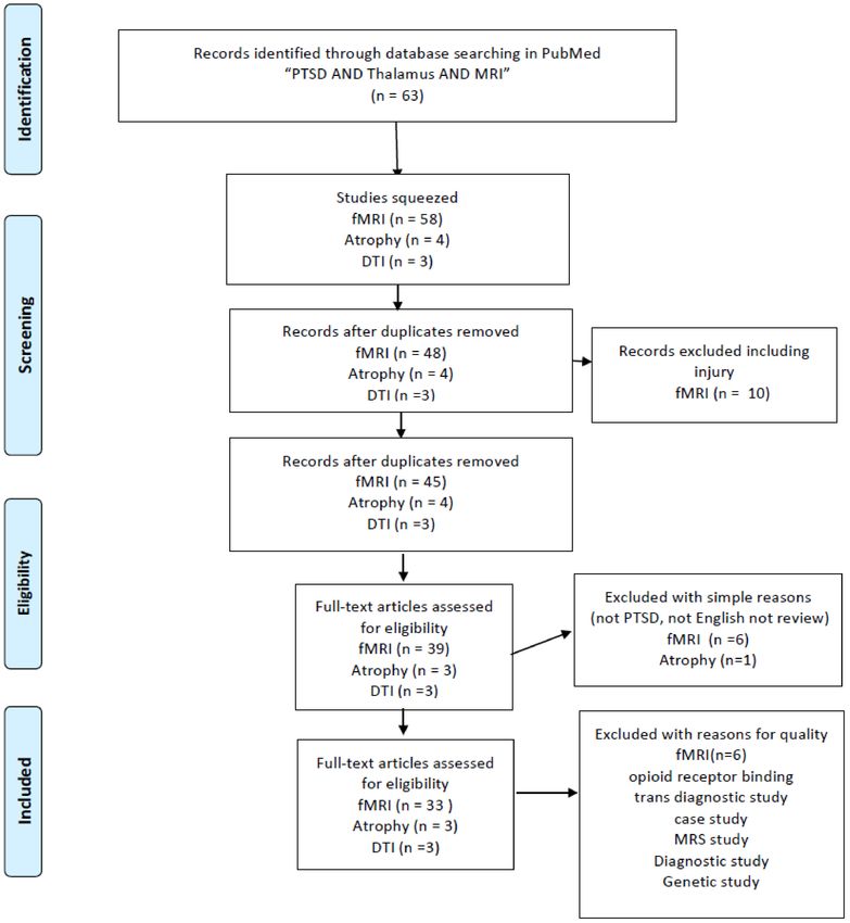
Int. J. Mol. Sci. 2021, 22, 1730                                                                                         2 of 18

                                        The thalamus was originally thought to act as a hub for relaying sensory information
                                   to the cortices and occasionally playing a role in processing this information. However,
                                   this view has gradually changed, and the thalamus is now considered to have many
                                   functions, acting as a sensory hub between other subcortical nuclei and the cortices and
                                   contributing to sleep and wake awareness, motor control, and cognition. Thalamic func-
                                   tional abnormalities are thought to contribute to the dysregulation of sensory filtering,
                                   circadian rhythms, levels of alertness, and consciousness [19]. It has been mentioned that
                                   fearful stimulation activates the thalamus [20]. In the context of fear-related learning,
                                   sensory processing, including visual processing in the thalamus, has been investigated
                                   with respect to its impact on amygdala function and output, rather than as an important
                                   psychological or pathophysiological process that shapes the development of PTSD [21].
                                   However, our animal voxel-based morphometry (VBM) study revealed the induction of
                                   brain atrophy in the thalamus by severe stress [18]. In addition, recent research work
                                   revealed the efficacy of interventions via visual technique: visual neurofeedback using
                                   implicit fear exposure [22,23] and visual game task [24]. Therefore, I consider the thalamus
                                   to be a promising area for stress research and thus conducted a mini literature review of
                                   the contribution of the thalamus to the pathogenesis of PTSD.

                                   2. Results
                                         This paper was initially designed as a systematic review. According to a result of
                                   literature research, the importance of the retinotectal pathway was recognized. At first,
                                   according to my bibliographic literature research, I found 132 studies mentioning PTSD and
                                   the thalamus. This total was reduced when I used narrower search terms: the number of
                                   studies mentioning “PTSD AND thalamus AND MRI” was 63. Unfortunately, I was not able
                                   to find sufficient numbers of volumetric studies (two studies with “PTSD AND thalamus
                                   AND MRI AND atrophy”, three with “PTSD AND thalamus AND DTI”) (Figure 1) and I
                                   also researched nine with “Childhood maltreatment AND MRI” squeezed to four papers
                                   volumetric study (Figure 2). I also read through 33 studies mentioning “PTSD AND
                                   thalamus AND MRI AND fMRI” and three studies with “PTSD AND thalamus AND
                                   MRI AND DTI” (Figure 1). Throughout the course of this study, I noticed the importance
                                   of retinotectal pathways and researched 12 including “PTSD AND colliculus” and two
                                   including “PTSD AND pulvinar”. I read through nine articles of them (Figure 3). I also
                                   aimed this research to bridge therapeutic interventions and researched 14 studies including
                                   “PTSD AND thalamus AND therapy” and I read through four reports of them (Figure 4).
                                   Tables 1–4 summarize the articles I included and provide a brief interpretation of each.
                                   The results of the review, separated according to subject area, are described below.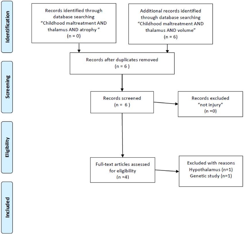
Int. J. Mol. Sci. 2021, 22, 1730                                                                                               3 of 18

        Figure 1. PRISMA flow diagrams [25] of literature research for PTSD neuroimaging research targeting on the thalamus.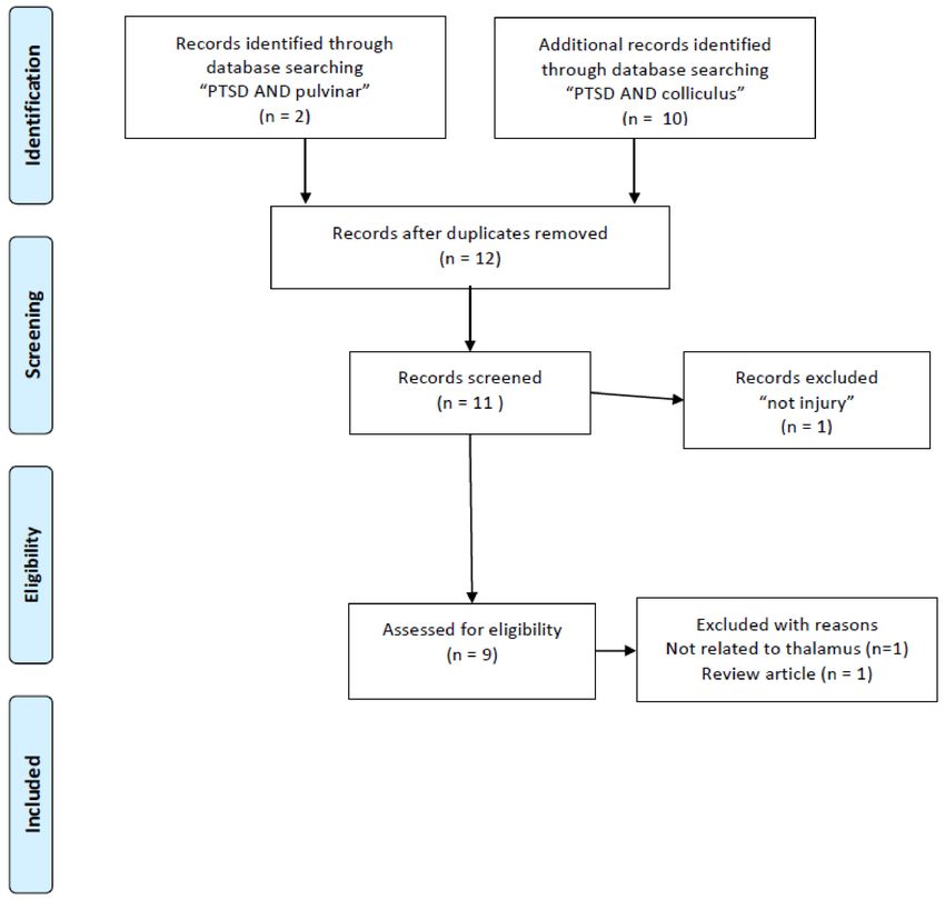
Int. J. Mol. Sci. 2021, 22, 1730                                                                                                4 of 18

      Figure 2. PRISMA flow diagrams [25] of literature research for brain structure research of childhood maltreatment targeting
      on the thalamus.

                                   3. Stress-Related Structural Change in the Thalamus
                                   3.1. Volume Change in Thalamus
                                         There are few reports that discuss stress-induced brain atrophy in the thalamus,
                                   and there is a report mentioning the trait of volume reduction in the thalamus which failed
                                   to reach significance [26,27]. Meta-analysis study of VBM via large-scale neuroimaging
                                   consortium study on PTSD did not reach significance in the thalamus volume [28]. On the
                                   other hand, volume loss in the right thalamus was detected in PTSD malingerers [29].
                                         However, there is a negative correlation between thalamic volume and re-experiencing
                                   in PTSD [30]. Although numbers of literature targeting PTSD have been small, a meta-
                                   analysis study mentioned brain atrophy in the bilateral thalamus [16]. It is difficult to
                                   separate mental stress from physical pain because chronic pain is sometimes contingent
                                   on the psychological toll of torture [17]. I consider that pain is probably one of the most
                                   important sources of physical and even psychological stress. The volume of the thalamus in
                                   the researches targeting childhood maltreatment has been studied; however, the volumetric
                                   changes in childhood maltreatment may be inconsistent. There is a negative correlation
                                   between childhood maltreatment score and the volume of the right thalamus [31], and there
                                   is a positive correlation with the volume of the left thalamus [32]. There is also a study on
                                   the volume reduction of the bilateral thalamus in physical childhood maltreatment [33],
                                   which may be a match to the result of pain studies, and it has been discussed stress
                                   paradigms may influence the volume variation in the thalamus [32]. However, no asso-
                                   ciations between age of exposure and volume of the thalamus are mentioned in other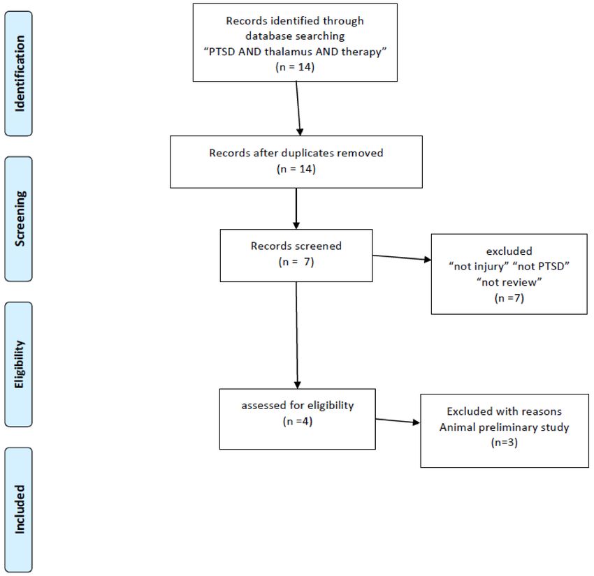
Int. J. Mol. Sci. 2021, 22, 1730                                                                                             5 of 18

                                   studies [34]. In this bibliographic literature research, meta-analysis study indicating vol-
                                   ume reduction of thalamus both in PTSD and childhood maltreatment was not detected.
                                   I elucidate the possibility that the variation of volume-change from stress paradigm may
                                   dampen the statistical power for meta-analysis.
                                         In an animal model, chronic stress induced dendritic atrophy in the medial thalamic
                                   nuclei, whose relationship to fear conditioning has been investigated [35]. It has been
                                   reported that a thinner right prefrontal cortex and larger right thalamus have been found
                                   to be related to denial and response prevention [36]. An animal VBM study of the response
                                   to a single prolonged stress factor indicated significant atrophy in the ventral lateral nuclei
                                   in the thalamus [18]. Generally, atrophic effect size of stress must be smaller than that of
                                   endogenous disease. Therefore, I consider that it is not sufficient to exclude participants
                                   with endogenous disease as a confounding factor for brain atrophy in clinical research.
                                   The summary of this literature study is described in Table 1.

                                   3.2. Diffusion Tensor Imaging (DTI)
                                         There are only a small number of studies that mention the abnormality of the thalamus
                                   in PTSD patients, and further research is required to determine whether the abnormality
                                   is in the white matter or the anatomical connectivity within the thalamus, or both. A DTI
                                   study reported a reduction in mean diffusivity (MD) and axial diffusivity (AD) in the
                                   right thalamus in PTSD patients [37]. The bilateral dorsal cingulum and right anterior
                                   corona radiation display low fractional anisotropy (FA) in PTSD patients, and low FA in
                                   the anterior corona radiation is correlated with PTSD severity, suggesting the relevance
                                   of the integrity of the limbic-thalamo-cortical tracts to PTSD [38]. It was reported that
                                   FA value improvement was detected in the anterior corona radiation and right thalamus
                                   of recovered fibromyalgia with PTSD symptom [39]. I was not able to detect any meta-
                                   analysis study detecting abnormality white matter abnormality around thalamus in PTSD;
                                   however, there is a meta-analysis study which reports the reduction of FA in the left
                                   anterior thalamic radiation and bilateral fornix in survivors in childhood maltreatment [40].
                                   A recent research study mentions that thalamic volume is related to increased anterior
                                   thalamic radiations in children with childhood maltreatment [41]. The discussion is similar
                                   to the previous paragraph, but I reason that the meta-analysis may not reach the level of
                                   statistical significance because of the influence of stress content. See Table 1 as the summary
                                   of literature research.

                                   3.3. Depleted Regional Activity in the Right Thalamus
                                         It has been mentioned that PTSD patients display depleted functional activity in
                                   the thalamus [42–44], and the right side of the thalamus is thought to have pathological
                                   significance [45], with psychotherapy increasing activity in the thalamus during memory re-
                                   trieval [46]. PTSD patients showed enhanced activity of the thalamus by seeing neutral (not
                                   traumatic) pictures [47]. In addition, resting-state functional MRI scans of PTSD patients
                                   have revealed enhanced regional connectivity in the thalamus [45,48,49]. Such patients
                                   exhibited depleted cluster coefficients within the bilateral thalamus, representing a fraction
                                   of all possible connections that connect the neighbors of a given node, in an analysis of
                                   gray matter structural connectivity [50]. PTSD patients showed strong positive correlations
                                   between the blood oxygen level–dependent (BOLD) signal and symptom severity in both
                                   the thalamus and the head of the caudate nucleus [51]. It has also been reported that the
                                   severity of early life stress is positively correlated to global-based connectivity in the thala-
                                   mus [52], and childhood maltreatment is thought to affect stress resilience. Resilience scores
                                   were positively correlated with BOLD signal strength in the right thalamus [53]. In addition,
                                   PTSD with dissociative symptoms (severe PTSD) is associated with higher activation of the
                                   left thalamus for nonconscious fear than nondissociative PTSD [54]. The summary of this
                                   literature research described in Table 1.
                                         In conclusion, I consider that there is laterality in the pathological significance of the
                                   thalamus for PTSD, and this may explain the discrepancies between reports with respect to
Int. J. Mol. Sci. 2021, 22, 1730 6 of 18
thalamic activity. Although the mechanism of pathological laterality in PTSD is not clear,
it has been known that visual perception and spatial awareness are right-side-dominant,
and this laterality may therefore occur because the pathogenesis of PTSD is dependent on
visual experience. With the progression of the disease, however, abnormalities may also
appear in the left thalamus.
3.4. Connectivity Research
Functional connections between the thalamus and amygdala, and between the thala-
mus and the anterior cingulate cortex (ACC), have been emphasized in PTSD researches.
Increased co-activity ACC, posterior cingulate cortex, and thalamus is detected in PTSD
patients [55]. PTSD can cause widespread increases in the activation of effective connectiv-
ity between the thalamus and the amygdala, striatum, rostral ACC, and ventral occipital
cortex, all of which encode scenes and could cause flashbacks [56]. Both resting-state [57]
and in-task [58,59] functional connectivity between the amygdala and the thalamus have
been reported, and in-task amygdala–thalamus connectivity is correlated with PTSD sever-
ity [58]. Some studies have shown that functional connectivity between the ACC and the
thalamus is depleted in PTSD patients [19,52,60,61], and emotional processing is particu-
larly strongly associated with the coactivity of the ACC and the posterior cingulate cortex
(PCC), as mediated by the thalamus [55].
The severity of PTSD may increase functional connectivity between the thalamus and
other sensory areas. Dissociative symptoms have been considered to indicate severe PTSD,
and dissociated PTSD patients show depleted connectivity between the left ventrolateral
thalamus (VLT) and the left superior frontal gyrus, right parahippocampal gyrus, and right
superior occipital gyrus. In contrast, they exhibit enhanced connectivity between the VLT
and the right insula, right middle frontal gyrus, superior temporal gyrus, right cuneus,
and left parietal lobe [62]. The connectivity between the pedunculopontine nuclei, as part of
the reticular activation system, and the anterior nucleus of the right thalamus is negatively
correlated with dissociative symptoms (derealization and depersonalization) [48].
The progression of PTSD may have brought enhancements in connections between
thalamus and autonomic systems. Enhanced functional connectivity between the thalamus
and the locus coeruleus has also been reported in PTSD [63]. Simultaneous enhancement
of neural activity in the periaqueductal nuclei and in the midline thalamic nuclei has
been reported in an animal PTSD model [64]. The thalamus is involved in the central
autonomic network (CAN), and low in-task heart rate variability (HRV) has been proposed
as a biomarker of PTSD; however, the covariation between brain connectivity related to the
CAN and HRV is diminished in PTSD patients [65]. High responders to stress exhibit local
brain atrophy in the ventral tegmental area (VTA) and enhanced connectivity between
the VTA and the thalamus [66]. The alexithymia scores (Toronto Alexithymia Scale 20)
of PTSD patients were positively correlated with suicide ideation [67] and there is the
positive correlation between the alexithymia scores and the response in the thalamus in
PTSD patients [68]. The summary of this literature research is described in Table 2.
Table 1. The summary of literature study for structure and regional activity in the thalamus.
Volume Change in Thalamus Reference Number
Volume reduction (not significant) [26–28]
Volume loss in the right thalamus was detected in PTSD malingerers [29]
Negative correlation between volume of right thalamus and re-experiencing [30]
Volume loss in the bilateral thalamus in pain meta-analysis [16]
Negative correlation between right thalamic volume and childhood maltreatment [31]
Positive correlation between left thalamic volume and childhood maltreatment [32]
Volume reduction in bilateral thalamus in cases of childhood physical maltreatment [33]
No associations between trauma exposure age and volume of thalamus [34]
Volume loss in the bilateral thalamus (ventrolateral nuclei) in animals under severe stress [18]
Thinner right prefrontal cortex and larger right thalamus are related to denial and response
[36]
prevention in PTSDInt. J. Mol. Sci. 2021, 22, 1730 7 of 18
Table 1. Cont.
Volume Change in Thalamus Reference Number
Diffusion Tensor Imaging (DTI)
Loss of MD and AD in right thalamus [37]
Low FA in bilateral dorsal cingulum and anterior corona radiate [38]
FA value improvement in anterior corona radiation and right thalamus in recovered
[39]
PTSD patients
Increased anterior thalamic radiation via childhood maltreatment correlated to
[40]
thalamic volume
Depleted Regional Activity in the Right Thalamus
Depleted regional activity [42–45]
Enhanced regional connectivity within thalamus [45,48,49]
Psychotherapy increased activity in thalamus during retrieval [46]
Enhanced activity of thalamus from showing PTSD patients a neutral picture [47]
Depleted cluster coefficients within bilateral thalamus [50]
BOLD signal positively correlated with symptoms [51]
Early life stress severity positively correlated with connectivity in thalamus [52]
Resilience score is positively correlated with BOLD signal in right thalamus in cases of
[53]
childhood maltreatment
Laterality of activation (pathological significance of right side) [46]
Enhanced activity in left thalamus during dissociation [54]
3.5. The Retinotectal Pathway in Fear-Related Learning in the Thalamus
I believe that the retinotectal pathway plays an important role in the progression
of PTSD pathogenesis. Visual information bypassed from geniculostriate system via the
superior colliculus (SC) projecting to the pulvinar as the retinotectal pathway. There are
some reports that SC itself directly plays a role combined with the periaqueductal gray as
an innate alarm system modulating defensive behavior [69] and activating in the social eye-
contact situation in PTSD patients [70,71]. Subliminal threat induces neural activity in SC
and periaqueductal gray in PTSD patients [72]. PTSD with dissociative subtype, compared
to PTSD without dissociation, increased resting state connectivity between SC and the
right dorsal lateral prefrontal cortex [73]. The pulvinar is a posterior part of the thalamus
and constitutes the retinotectal pathway, which is separate from the geniculostriate system.
A recent study demonstrated that the connectivity between pulvinar and V1 contributed to
fear anticipation [74]. Patients with pulvinar lesions exhibit disrupted implicit fear-related
visual processing [75]. The geniculostriate system contributes to form (ventral stream) and
space (dorsal stream) perception in visual processing. Both the geniculostriate and the
retinotectal pathways contribute to visual processing, and the latter in particular mediates
implicit processing of fearful stimuli [76,77]. Further research is needed to clarify the
function of the retinotectal pathway; however, it has been demonstrated that this circuit
contributes to fear learning [78].
This circuit may be necessary for the development of PTSD. It has been reported that
trauma survivors with a smaller pulvinar exhibit lower morbidity rates for PTSD [79],
and fMRI studies indicated enhanced connectivity between the SC and the ACC in PTSD
patients [63]. In addition, depletion of left pulvinar seed functional connectivity to sensory
regions (left superior parietal lobule, left middle temporal gyrus, and right postcentral
gyrus) in PTSD patients [80] and depletion of right pulvinar seed connectivity to primary
visual and higher sensory regions (left superior frontal gyrus, left superior parietal lobule,
bilateral precuneus, right inferior parietal lobule, right precentral gyrus medial segment)
in dissociative PTSD patients was reported [80]. Therefore, I consider that the retinotectal
pathway is likely to play a key role in fear conditioning, and disruption for pulvinar
function may have a preventative effect toward PTSD morbidity. The summary of this
literature research is presented in Table 3.Int. J. Mol. Sci. 2021, 22, 1730 8 of 18
Table 2. The summary of literature study for connectivity in the thalamus.
Connectivity Research Reference Number
Increased coactivity with ACC, posterior cingulate cortex,
[55]
and thalamus
Increase in effective connectivity from thalamus to amygdala [56–59]
Increase in effective connectivity from thalamus to ACC, striatum,
[56]
and occipital cortex
Positive correlation between thalamus−amygdala and PTSD severity [58]
Depletion of connectivity between thalamus and ACC [19,52,60,61]
Emotional processing correlation between thalamus and ACC/PCC [55]
Alteration of connectivity from VLT to other sensory areas in
[62]
dissociative PTSD patients
Enhanced connectivity of pedunculopontine nuclei (reticular
[48]
activation system) and anterior thalamic nucleus in dissociative PTSD
Enhanced connectivity between thalamus and locus coeruleus [63]
Simultaneous enhancement of activity in midline thalamus and
[64]
periaqueductal nuclei in animal PTSD model
Diminished correlation between CAN and HRV [65]
Enhanced connectivity between thalamus and VTA [66]
Positive correlation between thalamus activity and alexithymia [68]
Table 3. The summary of literature study for the retinotectal pathway.
The Retinotectal Pathway in Fear-Related Learning in the
Reference Number
Thalamus
SC directly modulates defense behavior [69]
SC is activated in social eye contact situations in PTSD patients [70,71]
Subliminal threat activates SC and periaqueductal gray [72]
Enhanced connectivity between SC and dorsal lateral prefrontal
[73]
cortex in dissociative PTSD
Pulvinar lesions disrupt implicit fear-related visual processing [75]
Pulvinar and V1 cortex contribute to fear anticipation [74]
Contribution of retinotectal pathway to implicit fear processing [76,77]
Contribution of retinotectal pathway to fear learning [78]
Smaller pulvinar in traumatized control [79]
Depletion of right pulvinar seed connectivity to sensory area in
[80]
dissociative PTSDInt. J. Mol. Sci. 2021, 22, 1730 9 of 18
Figure 3. PRISMA flow diagrams [25] for literature research for PTSD targeting retinotectal pathways.
4. Discussion on Therapeutic Implications Targeting the Thalamus
Based on this literature review, I believe that the retinotectal–pulvinar pathway and
thalamosensory connectivity contribute to the development of PTSD, and that depletion
in these regions before stress exposure may have preventative effects against worsening
PTSD. In fact, a recent clinical study reported that trauma-exposed control patients with-
out worsening PTSD exhibited depleted resting-state functional connectivity between
the thalamus and the postcentral gyrus, while both healthy controls and PTSD patients
did not exhibit such depletion [81]. A smaller pulvinar in similar traumatized control
patients has been also reported [79]. Defective pulvinar functioning may thus result in a
failure of fear processing [75], and I believe that reduced pulvinar function may contribute
to prevent the development of PTSD. On the other hand, the functional amygdala seed
connectivity study in PTSD indicates the enhancement of connectivity between centro-
medial amygdala and pulvinar, although the depletion between basolateral amygdala
and SC was detected [82]. In addition, recovered PTSD patients showed that the lower
tract strength of the amygdala–thalamus connection was normalized during recovery,
while that of amygdala–hippocampus connection remained low [83]. PTSD patients also
display disrupted regional activity of the thalamus, including the dorsal medial thalamus,
and future research should investigate whether this may hinder overwriting traumatic
memories with memories of ordinary daily life. The summary of literature research for
therapeutic implications is described in Table 4.
Taken together, I assume promising research: first, suppressing this pathway as a
direction of preventing the progression of PTSD; second, activating retinotectal pathwayInt. J. Mol. Sci. 2021, 22, 1730 10 of 18
followed with minimal stress exposure paradigm for desensitization of thalamus amygdala
connectivity; and third, activating geniculovisual cortex pathways to reduce contribution
of retinotectal pathway in visual fear processing.
In addition, I also would like to bridge the physiology of the thalamus to therapeutic
implications and consider the contribution of the physiology of thalamus to proposed
therapeutic interventions.
Figure 4. PRISMA flow diagrams [25] of literature research for contribution of thalamus physiology to therapy in PTSD.
4.1. EMDR
EMDR can be described as a methodology that extends PE. It is thought to take
advantage of the fact that eye movements facilitate learning, but the mechanism has long
been unclear, and many EMDR researchers themselves have relied on cognitive behavioral
therapy as the basis for its effectiveness. However, a recent animal study revealed that
projection from the SC to the mediodorsal thalamus may contribute to the processing of
conditioned fear [78], and especially mediodorsal thalamus may contribute in reactions
to fear memory [64,84–86]. Saccade eye movement is used for the desensitization of
traumatic memory in EMDR therapy, and a recent animal study demonstrated that visual
bilaterally alternating sensory stimulation, such as EMDR stimulation, provided a fear-
reducing effect, with sustained activation from the SC to the mediodorsal thalamus [87].
This result may partly explain the therapeutic mechanism of EMDR. Despite the finding that
limited pulvinar function may exert a preventative effect toward PTSD, the EMDR animal
model exhibited enhanced functioning of the SC and mediodorsal thalamus. The SC andInt. J. Mol. Sci. 2021, 22, 1730 11 of 18
mediodorsal thalamus activation may have a therapeutic effect by promoting the exposure
and desensitization of the thalamus–amygdala complex. Following EMDR treatment,
patients also showed a significant reduction in gray matter volume in the left thalamus
region [88], suggesting that the treatment may have modified the laterality that was induced
by PTSD. I consider that the efficacy of EMDR for treating PTSD supports the idea that the
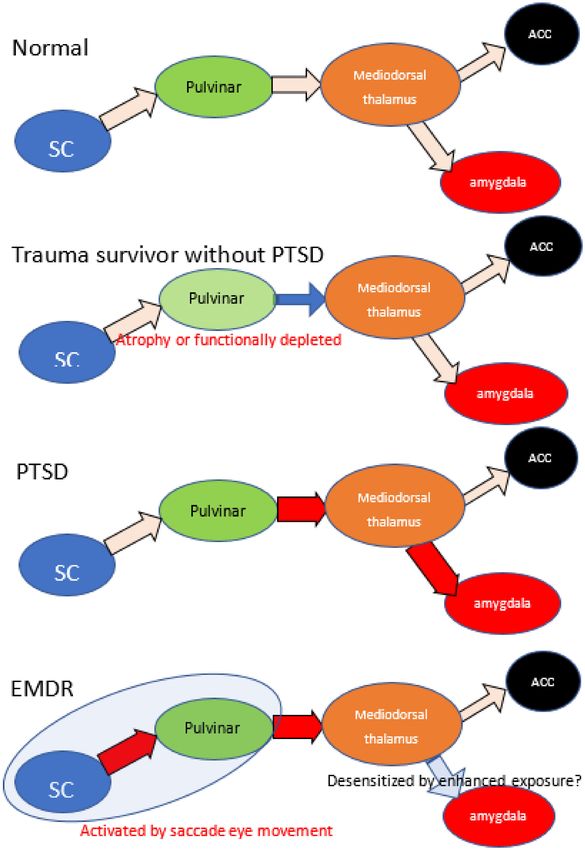
thalamus may be the key site of PTSD pathogenesis. See Figure 5 as a schematic summary.
Figure 5. The schematic summary of retinotectal pathway and hypothetic mechanism of EMDR. Because of reports low
morbidity rate of trauma survivors with smaller pulvinar, atrophy or dysfunction of pulvinar may have brought prevention
of progression in PTSD. PTSD patients indicate enhanced connectivity between thalamus and amygdala, depleted between
thalamus and ACC. EMDR intervention enhances the activity of retinotectal pathways and promotes the effects of exposure.
Red arrow indicates enhanced connectivity and blue arrow indicate reduced. SC: superior colliculus, ACC: anterior
cingulate cortex.Int. J. Mol. Sci. 2021, 22, 1730 12 of 18
Table 4. The summary of literature research for therapy for PTSD related to thalamus.
Discussion Reference Number
Trauma-exposed control showed depleted connectivity between
[81]
thalamus and postcentral gyrus
Enhanced connectivity between the centromedial amygdala and
[82]
pulvinar, and depletion between the basolateral amygdala and SC
Amygdala−thalamus connection enhanced during recovery process [83]
EMDR
Contribution of mediodorsal thalamus to fear processing [64,84–86]
Animal EMDR model provides sustained activation between SC and
[87]
mediodorsal thalamus with fear reducing effects
EMDR reduced gray matter volume in the left thalamus [88]
Hyperbaric Oxygen Therapy (HBOT)
HBOT enhances FA in the thalamic radiation, left thalamus,
[39]
and insula, with improved PTSD scores
Oxytocin Administration
Enhanced activity in the left thalamus during tasks both in PTSD
[89]
and controls
Oxytocin administration decreased connectivity between left
thalamus and amygdala in men with PTSD and traumatized controls, [89]
but increased connectivity in woman with PTSD
4.2. Functional MRI Neurofeedback Technique
A therapeutic approach targeting unconsciousness has been proposed [7], and one
that attempts to reprogram the unconscious using a fMRI-based technique called decoded
neurofeedback (DecNef) has been tried [22,23]. In conclusion, I could not find any literature
which indicates the contribution of thalamic change in this method. Tasks involving the
unconscious are complicated: they require computational calculation of brain activity in the
higher visual cortex during fear and control stimuli that attempt to stimulate brain activity
in the higher visual cortex to approximate that observed during fear stimuli without using
direct visual stimulation as non-explicit fear stimulation. This approach may have been
inspired by prolonged exposure therapy, but the activity of the amygdala does not appear
to be significantly altered in the learning phase. Only an acute BOLD signal response in
the amygdala under a fear-target confrontation was demonstrated [22]. Based on our belief
that the retinotectal pathway is more important than the geniculovisual cortex pathway in
the processing of visual fears, I assume that this intervention achieves therapeutic benefits
by reducing the contribution of the retinotectal pathway.
4.3. Visual Task Games
The visiospatial game task followed with minimal exposure has been applied for
prevention of PTSD via proof randomized control trial [24]. There is a debate that the
therapeutic mechanisms via competition for limited working memory resources for the
task [90] and I also could not find any literature describing the contribution of thalamic
change for this intervention. Although these studies were not designed for action on the
thalamus, it seems difficult to rule out that it is undergoing some action on the thalamus
via visual task. At present, there is no evidence that the thalamus is affected by this task;
however, it is certainly a task including discrimination and spatial cognition, and it can
be reasonably speculated that the geniculovisual pathway would be used. It is interesting
whether this intervention might produce preventative effect via reducing the contribution
of retinotectal pathway in the phase of PTSD progression.Int. J. Mol. Sci. 2021, 22, 1730 13 of 18
4.4. Hyperbaric Oxygen Therapy (HBOT)
I would like to introduce an intervention method that is neither medications nor
exposure. It has been shown in a randomized control trial that treating patients with
a history of childhood sexual abuse with HBOT results in an improvement of PTSD
symptoms and psychological distress, as well as an improved FA value (based on MRI-DTI)
in the anterior thalamic radiation, left thalamus, and left insula [39]. This treatment was
originally designed to treat fibromyalgia; however, it was subsequently suspected to have
activated the entire thalamus and may thus have additional therapeutic effects for PTSD.
HBOT may also promote the exposure and desensitization of thalamus–amygdala complex.
This treatment is expected to have few serious side effects, and I consider that HBOT is
thus a promising intervention for PTSD, which requires further research.
4.5. Oxytocin Administration
Oxytocin is a neurochemical agent that is thought to improve human interaction and
has been experimentally applied to PTSD therapy. Oxytocin administration enhanced
the activity of the left thalamus in both patients and controls during a distraction task
(resetting negative feelings with a working-memory task) and was negatively correlated
with error rates in this task in PTSD patients [89]. Oxytocin administration also decreased
the functional connectivity between the left thalamus and the amygdala in male PTSD
patients and trauma-exposed controls, although female patients indicated enhanced con-
nectivity between the left thalamus and the amygdala [89]. The mechanism of influencing
on thalamus amygdala connectivity with sexual variation has been unknown and the
potential efficacy of oxytocin administration should be further investigated.
4.6. Other Medications
Conventionally used medications may have some effect on the thalamus, but these in-
terventions do not yield satisfactory therapeutic effects. Several studies have thus indicated
a relationship between the thalamus and PTSD. These findings provide only a seed for
future research and are not sufficient to establish any new interventions. Selective serotonin
reuptake inhibitors (SSRIs) have been established as a treatment option for PTSD, and the
mediodorsal thalamus is a serotonin-rich area. Although one animal study showed that
chronic predator-scent exposure altered serotonin and dopamine levels in the thalamus [91],
another did not show a noticeable effect in the thalamus [92]. Prazosin: α -1 adrenergic
antagonist treatment has been already established as intervention to hyper arousal and
nightmares in PTSD [93]. Oral propranolol administration (1 mg/kg) improved PTSD
symptoms following the enhancement of activation of the thalamus and amygdala [94].
Noradrenergic system is intimately connected to the thalamus, and it is an interesting and
promising area for future research seeking the relationship between the noradrenergic
modulation and alteration of the thalamus in PTSD patients. A significant correlation
between re-experiencing and thalamic β2 nicotinic acetylcholine receptor binding using
single-photon emission computed tomography (SPECT) has already been described [95],
The micro-opioid system in the dorsomedial thalamus has also been found to contribute to
fear extinction [96]. However, these have not yet been targeted in medication research.
5. Methods
I conducted a bibliographic search in PubMed using the search terms “PTSD AND
thalamus AND MRI AND atrophy”, “PTSD AND thalamus AND MRI AND DTI”, “PTSD
AND thalamus AND MRI AND fMRI”. To include research other than MRI studies.
There are a few numbers for structural studies of abnormality in the thalamus and I added
“childhood maltreatment and MRI and atrophy” and “childhood maltreatment and MRI
and volume”. In addition, because the purpose of this review was to investigate established
treatment methods (especially EMDR) and explore new treatment approaches, I used the
search terms “PTSD AND thalamus AND therapy”. Finally, I applied the term “Not injury”
to the selection, and I excluded conference papers and studies not published in EnglishInt. J. Mol. Sci. 2021, 22, 1730 14 of 18
(Figure 1). After this research, we noticed the importance of the retinotectal pathway on
PTSD additional bibliographic search was carried out, “PTSD AND colliculus” and “PTSD
AND pulvinar”. These literature searches conform to the PRISMA guidelines [25].
Limitations
This article was initially planned as a systematic review for the relationships between
thalamus and PTSD and the contribution of retinotectal pathways for fear processing
was noticed. I aimed to bridge these findings to therapeutic mechanism; however, a few
articles were detected for the therapeutic contribution of thalamus in PTSD, especially for
neurofeedback techniques and visual task game. Although this may indicate a vanguard of
the therapeutic research targeting thalamus, I had to seek additional literature depending
upon the author’s experience in order to deepen the section of discussion. Therefore,
this may violate the validity and reproducibility of the discussion in this article.
6. Conclusions
Structural and volumetric researches detecting abnormalities in the thalamus have
not been sufficiently accumulated, and there may be problems categorizing stress contents
which might have caused failure to detect structural abnormalities in thalamus. How-
ever, animal studies demonstrated that the SC–pulvinar–mediodorsal thalamus–amygdala
pathways, contribute to visual fear processing. Based on this literature review, I pro-
pose that the activation of this system promotes the exposure and desensitization of the
thalamus–amygdala complex. It seems paradoxical that PTSD patients often require expo-
sure techniques for recovery, although many patients with acute stress disorder recover
spontaneously. A functional defect in an atrophy of the thalamus may be related to the
failure of desensitization of the thalamus–amygdala complex by exposure experienced in
everyday life. In addition, with the spread of COVID-19, the treatment of PTSD needs to
address the ongoing disaster. We believe that it would be promising to study interventions
for this site as preventive approaches for COVID-19 induce PTSD.
Author Contributions: The experimental design, implementation, analysis, and write-up were
conducted by T.Y. The author has read and agreed to the published version of the manuscript.
Funding: This study was supported by a Grant-in-Aid for Challenging Exploratory Research
(17K18711) from the Ministry of Education, Culture, Sports, Science and Technology (T.Y.) and
by a research grant from MSD K.K. (Tokyo, Japan) (T.Y.).
Institutional Review Board Statement: Not applicable.
Informed Consent Statement: Not applicable.
Data Availability Statement: No new data were created or analyzed in this study and data sharing
is not applicable to this article.
Conflicts of Interest: The other authors declare no conflict of interest.
References
1. Goldstein, R.B.; Smith, S.M.; Chou, S.P.; Saha, T.D.; Jung, J.; Zhang, H.; Pickering, R.P.; Ruan, W.J.; Huang, B.; Grant, B.F.
The epidemiology of DSM-5 posttraumatic stress disorder in the United States: Results from the National Epidemiologic Survey
on Alcohol and Related Conditions-III. Soc. Psychiatry Psychiatr. Epidemiol. 2016. [CrossRef]
2. Tran, K.; Moulton, K.; Santesso, N.; Rabb, D. Cognitive Processing Therapy for Post-Traumatic Stress Disorder: A Systematic Review and
Meta-Analysis; Canadian Agency for Drugs and Technologies in Health: Ottawa, ON, Canada, 2016.
3. Matsumoto, K.; Sakuma, A.; Ueda, I.; Nagao, A.; Takahashi, Y. Psychological Trauma after the Great East Japan Earthquake.
Psychiatry Clin. Neurosci. 2016. [CrossRef]
4. American Psychiatric Association. Diagnostic and Statistical Manual of Mental Disorder, 5th ed.; American Psychiatric Publishing:
Arlington, VA, USA, 2013.
5. Foa, E.B.; Gillihan, S.J.; Bryant, R.A. Challenges and Successes in Dissemination of Evidence-Based Treatments for Posttraumatic
Stress: Lessons Learned From Prolonged Exposure Therapy for PTSD. Psychol. Sci. Public Interest 2013, 14, 65–111. [CrossRef]Int. J. Mol. Sci. 2021, 22, 1730 15 of 18
6. Zhu, X.; Suarez-Jimenez, B.; Lazarov, A.; Helpman, L.; Papini, S.; Lowell, A.; Durosky, A.; Lindquist, M.A.; Markowitz, J.C.;
Schneier, F.; et al. Exposure-based therapy changes amygdala and hippocampus resting-state functional connectivity in patients
with posttraumatic stress disorder. Depress. Anxiety 2018, 35, 974–984. [CrossRef]
7. Sohn, E. Decoding the neuroscience of consciousness. Nature 2019, 571, S2–S5. [CrossRef]
8. Society of Clinical Psychology Division 12 of the APA. Eye Movement Desensitization and Reprocessing for Post-Traumatic Stress
Disorder. Available online: www.div12.org (accessed on 3 November 2020).
9. Nardo, D.; Hogberg, G.; Looi, J.C.; Larsson, S.; Hallstrom, T.; Pagani, M. Gray matter density in limbic and paralimbic cortices is
associated with trauma load and EMDR outcome in PTSD patients. J. Psychiatr. Res. 2010, 44, 477–485. [CrossRef]
10. Chen, S.; Xia, W.; Li, L.; Liu, J.; He, Z.; Zhang, Z.; Yan, L.; Zhang, J.; Hu, D. Gray matter density reduction in the insula in fire
survivors with posttraumatic stress disorder: A voxel-based morphometric study. Psychiatry Res. 2006, 146, 65–72. [CrossRef]
11. Hakamata, Y.; Matsuoka, Y.; Inagaki, M.; Nagamine, M.; Hara, E.; Imoto, S.; Murakami, K.; Kim, Y.; Uchitomi, Y. Structure of
orbitofrontal cortex and its longitudinal course in cancer-related post-traumatic stress disorder. Neurosci. Res. 2007, 59, 383–389.
[CrossRef]
12. Carrion, V.G.; Weems, C.F.; Watson, C.; Eliez, S.; Menon, V.; Reiss, A.L. Converging evidence for abnormalities of the prefrontal
cortex and evaluation of midsagittal structures in pediatric posttraumatic stress disorder: An MRI study. Psychiatry Res. 2009,
172, 226–234. [CrossRef]
13. Tavanti, M.; Battaglini, M.; Borgogni, F.; Bossini, L.; Calossi, S.; Marino, D.; Vatti, G.; Pieraccini, F.; Federico, A.;
Castrogiovanni, P.; et al. Evidence of diffuse damage in frontal and occipital cortex in the brain of patients with post-traumatic
stress disorder. Neurol. Sci. 2012, 33, 59–68. [CrossRef]
14. Zhang, J.; Tan, Q.; Yin, H.; Zhang, X.; Huan, Y.; Tang, L.; Wang, H.; Xu, J.; Li, L. Decreased gray matter volume in the left
hippocampus and bilateral calcarine cortex in coal mine flood disaster survivors with recent onset PTSD. Psychiatry Res. 2011,
192, 84–90. [CrossRef]
15. Rogers, M.A.; Yamasue, H.; Abe, O.; Yamada, H.; Ohtani, T.; Iwanami, A.; Aoki, S.; Kato, N.; Kasai, K. Smaller amygdala volume
and reduced anterior cingulate gray matter density associated with history of post-traumatic stress disorder. Psychiatry Res. 2009,
174, 210–216. [CrossRef]
16. Pan, P.L.; Zhong, J.G.; Shang, H.F.; Zhu, Y.L.; Xiao, P.R.; Dai, Z.Y.; Shi, H.C. Quantitative meta-analysis of grey matter anomalies
in neuropathic pain. Eur. J. Pain 2015, 19, 1224–1231. [CrossRef]
17. Tsur, N.; Defrin, R.; Ginzburg, K. PTSD, Orientation to Pain, and Pain Perception in Ex-prisoners of War who Underwent Torture.
Psychosom. Med. 2017. [CrossRef]
18. Yoshii, T.; Oishi, N.; Ikoma, K.; Nishimura, I.; Sakai, Y.; Matsuda, K.; Yamada, S.; Tanaka, M.; Kawata, M.; Narumoto, J.; et al.
Brain atrophy in the visual cortex and thalamus induced by severe stress in animal model. Sci. Rep. 2017, 7, 12731. [CrossRef]
19. Yin, Y.; Jin, C.; Hu, X.; Duan, L.; Li, Z.; Song, M.; Chen, H.; Feng, B.; Jiang, T.; Jin, H.; et al. Altered resting-state functional
connectivity of thalamus in earthquake-induced posttraumatic stress disorder: A functional magnetic resonance imaging study.
Brain Res 2011, 1411, 98–107. [CrossRef]
20. Burra, N.; Hervais-Adelman, A.; Celeghin, A.; de Gelder, B.; Pegna, A.J. Affective blindsight relies on low spatial frequencies.
Neuropsychologia 2019, 128, 44–49. [CrossRef]
21. Liberzon, I.; Abelson, J.L. Context Processing and the Neurobiology of Post-Traumatic Stress Disorder. Neuron 2016, 92, 14–30.
[CrossRef]
22. Koizumi, A.; Amano, K.; Cortese, A.; Shibata, K.; Yoshida, W.; Seymour, B.; Kawato, M.; Lau, H. Fear reduction without fear
through reinforcement of neural activity that bypasses conscious exposure. Nat. Hum. Behav. 2016, 1, 6. [CrossRef]
23. Taschereau-Dumouchel, V.; Cortese, A.; Chiba, T.; Knotts, J.D.; Kawato, M.; Lau, H. Towards an unconscious neural reinforcement
intervention for common fears. Proc. Natl. Acad. Sci. USA 2018, 115, 3470–3475. [CrossRef]
24. Iyadurai, L.; Blackwell, S.E.; Meiser-Stedman, R.; Watson, P.C.; Bonsall, M.B.; Geddes, J.R.; Nobre, A.C.; Holmes, E.A. Preventing
intrusive memories after trauma via a brief intervention involving Tetris computer game play in the emergency department:
A proof-of-concept randomized controlled trial. Mol. Psychiatry 2017. [CrossRef]
25. Moher, D.; Liberati, A.; Tetzlaff, J.; Altman, D.G. Preferred reporting items for systematic reviews and meta-analyses: The PRISMA
statement. PLoS Med. 2009, 6, e1000097. [CrossRef]
26. Sussman, D.; Pang, E.W.; Jetly, R.; Dunkley, B.T.; Taylor, M.J. Neuroanatomical features in soldiers with post-traumatic stress
disorder. BMC Neurosci. 2016, 17, 13. [CrossRef]
27. Filipovic, B.R.; Djurovic, B.; Marinkovic, S.; Stijak, L.; Aksic, M.; Nikolic, V.; Starcevic, A.; Radonjic, V. Volume changes of corpus
striatum, thalamus, hippocampus and lateral ventricles in posttraumatic stress disorder (PTSD) patients suffering from headaches
and without therapy. Cent. Eur. Neurosurg. 2011, 72, 133–137. [CrossRef]
28. Logue, M.W.; van Rooij, S.J.H.; Dennis, E.L.; Davis, S.L.; Hayes, J.P.; Stevens, J.S.; Densmore, M.; Haswell, C.C.; Ipser, J.;
Koch, S.B.J.; et al. Smaller Hippocampal Volume in Posttraumatic Stress Disorder: A Multisite ENIGMA-PGC Study: Subcortical
Volumetry Results From Posttraumatic Stress Disorder Consortia. Biol. Psychiatry 2018, 83, 244–253. [CrossRef]
29. Butler, O.; Herr, K.; Willmund, G.; Gallinat, J.; Zimmermann, P.; Kuhn, S. Neural correlates of response bias: Larger hippocampal
volume correlates with symptom aggravation in combat-related posttraumatic stress disorder. Psychiatry Res. Neuroimag. 2018,
279, 1–7. [CrossRef]Int. J. Mol. Sci. 2021, 22, 1730 16 of 18
30. Shucard, J.L.; Cox, J.; Shucard, D.W.; Fetter, H.; Chung, C.; Ramasamy, D.; Violanti, J. Symptoms of posttraumatic stress disorder
and exposure to traumatic stressors are related to brain structural volumes and behavioral measures of affective stimulus
processing in police officers. Psychiatry Res. 2012, 204, 25–31. [CrossRef]
31. Duarte, D.G.; Neves Mde, C.; Albuquerque, M.R.; de Souza-Duran, F.L.; Busatto, G.; Corrêa, H. Gray matter brain volumes in
childhood-maltreated patients with bipolar disorder type I: A voxel-based morphometric study. J. Affect. Disord. 2016, 197, 74–80.
[CrossRef]
32. Liao, M.; Yang, F.; Zhang, Y.; He, Z.; Song, M.; Jiang, T.; Li, Z.; Lu, S.; Wu, W.; Su, L.; et al. Childhood maltreatment is associated
with larger left thalamic gray matter volume in adolescents with generalized anxiety disorder. PLoS ONE 2013, 8, e71898.
[CrossRef]
33. Hanson, J.L.; Chung, M.K.; Avants, B.B.; Shirtcliff, E.A.; Gee, J.C.; Davidson, R.J.; Pollak, S.D. Early stress is associated
with alterations in the orbitofrontal cortex: A tensor-based morphometry investigation of brain structure and behavioral risk.
J. Neurosci. 2010, 30, 7466–7472. [CrossRef]
34. Pechtel, P.; Lyons-Ruth, K.; Anderson, C.M.; Teicher, M.H. Sensitive periods of amygdala development: The role of maltreatment
in preadolescence. NeuroImage 2014, 97, 236–244. [CrossRef]
35. Dagnino-Subiabre, A.; Munoz-Llancao, P.; Terreros, G.; Wyneken, U.; Diaz-Veliz, G.; Porter, B.; Kilgard, M.P.; Atzori, M.; Aboitiz, F.
Chronic stress induces dendritic atrophy in the rat medial geniculate nucleus: Effects on auditory conditioning. Behav. Brain Res.
2009, 203, 88–96. [CrossRef]
36. Mutluer, T.; Şar, V.; Kose-Demiray, Ç.; Arslan, H.; Tamer, S.; Inal, S.; Kaçar, A. Lateralization of Neurobiological Response in
Adolescents with Post-Traumatic Stress Disorder Related to Severe Childhood Sexual Abuse: The Tri-Modal Reaction (T-MR)
Model of Protection. J. Trauma Dissoc. 2018, 19, 108–125. [CrossRef]
37. Lei, D.; Li, L.; Li, L.; Suo, X.; Huang, X.; Lui, S.; Li, J.; Bi, F.; Kemp, G.J.; Gong, Q. Microstructural abnormalities in children with
post-traumatic stress disorder: A diffusion tensor imaging study at 3.0T. Sci. Rep. 2015, 5, 8933. [CrossRef]
38. Sanjuan, P.M.; Thoma, R.; Claus, E.D.; Mays, N.; Caprihan, A. Reduced white matter integrity in the cingulum and anterior
corona radiata in posttraumatic stress disorder in male combat veterans: A diffusion tensor imaging study. Psychiatry Res. 2013,
214, 260–268. [CrossRef]
39. Hadanny, A.; Bechor, Y.; Catalogna, M.; Daphna-Tekoah, S.; Sigal, T.; Cohenpour, M.; Lev-Wiesel, R.; Efrati, S. Hyperbaric Oxygen
Therapy Can Induce Neuroplasticity and Significant Clinical Improvement in Patients Suffering From Fibromyalgia With a
History of Childhood Sexual Abuse-Randomized Controlled Trial. Front. Psychol. 2018, 9, 2495. [CrossRef]
40. Lim, L.; Howells, H.; Radua, J.; Rubia, K. Aberrant structural connectivity in childhood maltreatment: A meta-analysis. Neurosci.
Biobehav. Rev. 2020, 116, 406–414. [CrossRef]
41. Jung, M.; Takiguchi, S.; Hamamura, S.; Mizuno, Y.; Kosaka, H.; Tomoda, A. Thalamic Volume Is Related to Increased Anterior
Thalamic Radiations in Children with Reactive Attachment Disorder. Cereb. Cortex 2020, 30, 4238–4245. [CrossRef]
42. Lanius, R.A.; Williamson, P.C.; Densmore, M.; Boksman, K.; Gupta, M.A.; Neufeld, R.W.; Gati, J.S.; Menon, R.S. Neural correlates
of traumatic memories in posttraumatic stress disorder: A functional MRI investigation. Am. J. Psychiatry 2001, 158, 1920–1922.
[CrossRef]
43. Yan, X.; Brown, A.D.; Lazar, M.; Cressman, V.L.; Henn-Haase, C.; Neylan, T.C.; Shalev, A.; Wolkowitz, O.M.; Hamilton, S.P.;
Yehuda, R.; et al. Spontaneous brain activity in combat related PTSD. Neurosci. Lett. 2013, 547, 1–5. [CrossRef]
44. Suarez-Jimenez, B.; Albajes-Eizagirre, A.; Lazarov, A.; Zhu, X.; Harrison, B.J.; Radua, J.; Neria, Y.; Fullana, M.A. Neural signatures
of conditioning, extinction learning, and extinction recall in posttraumatic stress disorder: A meta-analysis of functional magnetic
resonance imaging studies. Psychol. Med. 2020, 50, 1442–1451. [CrossRef]
45. Roos, A.; Fouche, J.P.; Stein, D.J. Brain network connectivity in women exposed to intimate partner violence: A graph theory
analysis study. Brain Imag. Behav. 2017, 11, 1629–1639. [CrossRef]
46. Peres, J.F.; Newberg, A.B.; Mercante, J.P.; Simão, M.; Albuquerque, V.E.; Peres, M.J.; Nasello, A.G. Cerebral blood flow changes
during retrieval of traumatic memories before and after psychotherapy: A SPECT study. Psychol. Med. 2007, 37, 1481–1491.
[CrossRef]
47. Neumeister, P.; Feldker, K.; Heitmann, C.Y.; Helmich, R.; Gathmann, B.; Becker, M.P.I.; Straube, T. Interpersonal violence in
posttraumatic women: Brain networks triggered by trauma-related pictures. Soc. Cogn. Affect. Neurosci. 2017, 12, 555–568.
[CrossRef]
48. Thome, J.; Densmore, M.; Koppe, G.; Terpou, B.; Théberge, J.; McKinnon, M.C.; Lanius, R.A. Back to the Basics: Resting State
Functional Connectivity of the Reticular Activation System in PTSD and its Dissociative Subtype. Chronic Stress 2019, 3. [CrossRef]
49. Zhong, Y.; Zhang, R.; Li, K.; Qi, R.; Zhang, Z.; Huang, Q.; Lu, G. Altered cortical and subcortical local coherence in PTSD:
Evidence from resting-state fMRI. Acta Radiol. 2015, 56, 746–753. [CrossRef]
50. Mueller, S.G.; Ng, P.; Neylan, T.; Mackin, S.; Wolkowitz, O.; Mellon, S.; Yan, X.; Flory, J.; Yehuda, R.; Marmar, C.R.; et al. Evidence
for disrupted gray matter structural connectivity in posttraumatic stress disorder. Psychiatry Res. 2015, 234, 194–201. [CrossRef]
51. Mickleborough, M.J.; Daniels, J.K.; Coupland, N.J.; Kao, R.; Williamson, P.C.; Lanius, U.F.; Hegadoren, K.; Schore, A.; Densmore,
M.; Stevens, T.; et al. Effects of trauma-related cues on pain processing in posttraumatic stress disorder: An fMRI investigation.
J. Psychiatry Neurosci. 2011, 36, 6–14. [CrossRef]
52. Philip, N.S.; Tyrka, A.R.; Albright, S.E.; Sweet, L.H.; Almeida, J.; Price, L.H.; Carpenter, L.L. Early life stress predicts thalamic
hyperconnectivity: A transdiagnostic study of global connectivity. J. Psychiatr. Res. 2016, 79, 93–100. [CrossRef]Int. J. Mol. Sci. 2021, 22, 1730 17 of 18
53. Daniels, J.K.; Hegadoren, K.M.; Coupland, N.J.; Rowe, B.H.; Densmore, M.; Neufeld, R.W.; Lanius, R.A. Neural correlates and
predictive power of trait resilience in an acutely traumatized sample: A pilot investigation. J. Clin. Psychiatry 2012, 73, 327–332.
[CrossRef]
54. Felmingham, K.; Kemp, A.H.; Williams, L.; Falconer, E.; Olivieri, G.; Peduto, A.; Bryant, R. Dissociative responses to conscious
and non-conscious fear impact underlying brain function in post-traumatic stress disorder. Psychol. Med. 2008, 38, 1771–1780.
[CrossRef]
55. Ramage, A.E.; Laird, A.R.; Eickhoff, S.B.; Acheson, A.; Peterson, A.L.; Williamson, D.E.; Telch, M.J.; Fox, P.T. A coordinate-based
meta-analytic model of trauma processing in posttraumatic stress disorder. Hum. Brain Mapp. 2013, 34, 3392–3399. [CrossRef]
56. Bourne, C.; Mackay, C.E.; Holmes, E.A. The neural basis of flashback formation: The impact of viewing trauma. Psychol. Med.
2013, 43, 1521–1532. [CrossRef]
57. Zhu, X.; Helpman, L.; Papini, S.; Schneier, F.; Markowitz, J.C.; Van Meter, P.E.; Lindquist, M.A.; Wager, T.D.; Neria, Y. Altered
resting state functional connectivity of fear and reward circuitry in comorbid PTSD and major depression. Depress. Anxiety 2017,
34, 641–650. [CrossRef]
58. Ben-Zion, Z.; Zeevi, Y.; Keynan, N.J.; Admon, R.; Kozlovski, T.; Sharon, H.; Halpern, P.; Liberzon, I.; Shalev, A.Y.;
Benjamini, Y.; et al. Multi-domain potential biomarkers for post-traumatic stress disorder (PTSD) severity in recent trauma
survivors. Transl. Psychiatry 2020, 10, 208. [CrossRef]
59. Morey, R.A.; Dunsmoor, J.E.; Haswell, C.C.; Brown, V.M.; Vora, A.; Weiner, J.; Stjepanovic, D.; Wagner, H.R., 3rd; LaBar, K.S.
Fear learning circuitry is biased toward generalization of fear associations in posttraumatic stress disorder. Transl. Psychiatry 2015,
5, e700. [CrossRef]
60. Lanius, R.A.; Williamson, P.C.; Hopper, J.; Densmore, M.; Boksman, K.; Gupta, M.A.; Neufeld, R.W.; Gati, J.S.; Menon, R.S. Recall
of emotional states in posttraumatic stress disorder: An fMRI investigation. Biol. Psychiatry 2003, 53, 204–210. [CrossRef]
61. Kennis, M.; Rademaker, A.R.; van Rooij, S.J.; Kahn, R.S.; Geuze, E. Altered functional connectivity in posttraumatic stress disorder
with versus without comorbid major depressive disorder: A resting state fMRI study. F1000Res 2013, 2, 289. [CrossRef]
62. Lanius, R.A.; Williamson, P.C.; Bluhm, R.L.; Densmore, M.; Boksman, K.; Neufeld, R.W.; Gati, J.S.; Menon, R.S. Functional
connectivity of dissociative responses in posttraumatic stress disorder: A functional magnetic resonance imaging investigation.
Biol. Psychiatry 2005, 57, 873–884. [CrossRef]
63. Steuwe, C.; Daniels, J.K.; Frewen, P.A.; Densmore, M.; Theberge, J.; Lanius, R.A. Effect of direct eye contact in women with
PTSD related to interpersonal trauma: Psychophysiological interaction analysis of connectivity of an innate alarm system.
Psychiatry Res. 2015, 232, 162–167. [CrossRef]
64. Della Valle, R.; Mohammadmirzaei, N.; Knox, D. Single prolonged stress alters neural activation in the periacqueductal gray and
midline thalamic nuclei during emotional learning and memory. Learn Mem. 2019, 26, 1–9. [CrossRef]
65. Thome, J.; Densmore, M.; Frewen, P.A.; McKinnon, M.C.; Théberge, J.; Nicholson, A.A.; Koenig, J.; Thayer, J.F.; Lanius, R.A.
Desynchronization of autonomic response and central autonomic network connectivity in posttraumatic stress disorder.
Hum. Brain Mapp. 2017, 38, 27–40. [CrossRef]
66. Magalhaes, R.; Barriere, D.A.; Novais, A.; Marques, F.; Marques, P.; Cerqueira, J.; Sousa, J.C.; Cachia, A.; Boumezbeur, F.;
Bottlaender, M.; et al. The dynamics of stress: A longitudinal MRI study of rat brain structure and connectome. Mol. Psychiatry
2018, 23, 1998–2006. [CrossRef]
67. De Berardis, D.; Vellante, F.; Fornaro, M.; Anastasia, A.; Olivieri, L.; Rapini, G.; Serroni, N.; Orsolini, L.; Valchera, A.;
Carano, A.; et al. Alexithymia, suicide ideation, affective temperaments and homocysteine levels in drug naïve patients with
post-traumatic stress disorder: An exploratory study in the everyday ‘real world’ clinical practice. Int. J. Psychiatry Clin. Pract.
2020, 24, 83–87. [CrossRef]
68. Frewen, P.A.; Pain, C.; Dozois, D.J.; Lanius, R.A. Alexithymia in PTSD: Psychometric and FMRI studies. Ann. N. Y. Acad. Sci.
2006, 1071, 397–400. [CrossRef]
69. Forcelli, P.A.; Waguespack, H.F.; Malkova, L. Defensive Vocalizations and Motor Asymmetry Triggered by Disinhibition of the
Periaqueductal Gray in Non-human Primates. Front. Neurosci. 2017, 11, 163. [CrossRef]
70. Thome, J.; Frewen, P.; Daniels, J.K.; Densmore, M.; Lanius, R.A. Altered connectivity within the salience network during direct
eye gaze in PTSD. Borderline Pers. Disord. Emot. Dysregul. 2014, 1, 17. [CrossRef]
71. Steuwe, C.; Daniels, J.K.; Frewen, P.A.; Densmore, M.; Pannasch, S.; Beblo, T.; Reiss, J.; Lanius, R.A. Effect of direct eye contact in
PTSD related to interpersonal trauma: An fMRI study of activation of an innate alarm system. Soc. Cogn. Affect. Neurosci. 2014,
9, 88–97. [CrossRef]
72. Terpou, B.A.; Densmore, M.; Thome, J.; Frewen, P.; McKinnon, M.C.; Lanius, R.A. The Innate Alarm System and Subliminal
Threat Presentation in Posttraumatic Stress Disorder: Neuroimaging of the Midbrain and Cerebellum. Chronic Stress 2019, 3.
[CrossRef]
73. Olivé, I.; Densmore, M.; Harricharan, S.; Théberge, J.; McKinnon, M.C.; Lanius, R. Superior colliculus resting state networks in
post-traumatic stress disorder and its dissociative subtype. Hum. Brain Mapp. 2018, 39, 563–574. [CrossRef]
74. Koizumi, A.; Zhan, M.; Ban, H.; Kida, I.; De Martino, F.; Vaessen, M.J.; de Gelder, B.; Amano, K. Threat Anticipation in Pulvinar
and in Superficial Layers of Primary Visual Cortex (V1). Evidence from Layer-Specific Ultra-High Field 7T fMRI. eNeuro 2019, 6.
[CrossRef]You can also read