Discovery and Validation of a Urinary Exosome mRNA Signature for the Diagnosis of Human Kidney Transplant Rejection - JASN
←
→
Page content transcription
If your browser does not render page correctly, please read the page content below
CLINICAL RESEARCH             www.jasn.org

Discovery and Validation of a Urinary Exosome mRNA
Signature for the Diagnosis of Human Kidney
Transplant Rejection
Rania El Fekih,1 James Hurley,2 Vasisht Tadigotla,2 Areej Alghamdi,1 Anand Srivastava,1
Christine Coticchia,2 John Choi,1 Hazim Allos,1 Karim Yatim,1 Juliano Alhaddad ,1
Siawosh Eskandari,1 Philip Chu,1 Albana B. Mihali ,1 Isadora T. Lape,1 Mauricio P. Lima Filho,1
Bruno T. Aoyama,1 Anil Chandraker,1 Kassem Safa,3 James F. Markmann,3
Leonardo V. Riella ,1 Richard N. Formica ,4 Johan Skog,2 and Jamil R. Azzi1
Due to the number of contributing authors, the affiliations are listed at the end of this article.

ABSTRACT
Background Developing a noninvasive clinical test to accurately diagnose kidney allograft rejection is
critical to improve allograft outcomes. Urinary exosomes, tiny vesicles released into the urine that carry
parent cells’ proteins and nucleic acids, reflect the biologic function of the parent cells within the kidney,
including immune cells. Their stability in urine makes them a potentially powerful tool for liquid biopsy and
a noninvasive diagnostic biomarker for kidney-transplant rejection.
Methods Using 192 of 220 urine samples with matched biopsy samples from 175 patients who underwent a
clinically indicated kidney-transplant biopsy, we isolated urinary exosomal mRNAs and developed rejec-
tion signatures on the basis of differential gene expression. We used crossvalidation to assess the perfor-
mance of the signatures on multiple data subsets.
Results An exosomal mRNA signature discriminated between biopsy samples from patients with all-cause
rejection and those with no rejection, yielding an area under the curve (AUC) of 0.93 (95% CI, 0.87 to 0.98),
which is significantly better than the current standard of care (increase in eGFR AUC of 0.57; 95% CI, 0.49 to 0.65).
The exosome-based signature’s negative predictive value was 93.3% and its positive predictive value was 86.2%.
Using the same approach, we identified an additional gene signature that discriminated patients with T cell–
mediated rejection from those with antibody-mediated rejection (with an AUC of 0.87; 95% CI, 0.76 to 0.97). This
signature’s negative predictive value was 90.6% and its positive predictive value was 77.8%.
Conclusions Our findings show that mRNA signatures derived from urinary exosomes represent a power-
ful and noninvasive tool to screen for kidney allograft rejection. This finding has the potential to assist
clinicians in therapeutic decision making.

JASN 32: ccc–ccc, 2021. doi: https://doi.org/10.1681/ASN.2020060850

                                                                Received June 17, 2020. Accepted December 26, 2020.
CKD is a major health concern in the Unites States
and worldwide.1 Although patients with ESKD re-                 Published online ahead of print. Publication date available at
quire either dialysis or transplantation to sustain             www.jasn.org.

their life, the latter remains the treatment of                 Present address: Dr. Anand Srivastava, Division of Nephrology
choice.2–4 However, long-term graft survival re-                and Hypertension, Center for Translational Metabolism and
                                                                Health, Institute for Public Health and Medicine, Northwestern
mains a major challenge, due mostly to acute and                University Feinberg School of Medicine, Chicago, Illinois.
chronic rejection. Although the rate of acute rejec-
                                                                Correspondence: Dr. Johan Skog, Exosome Diagnostics, a Bio-
tion has decreased in the modern era of potent im-
                                                                Techne brand, 266 2nd Avenue, Waltham, MA 02451, or Dr. Jamil
munosuppression,5 recent reported incidence of                  Azzi, Transplant Research Center, Brigham and Women’s Hospital,
acute rejections in literature ranges from 11% to               Harvard Medical School, 221 Longwood Avenue, Boston, MA 02115.
26%.6–9 During the first year after transplantation,             E-mail: johan.skog@bio-techne.com or jazzi@bwh.harvard.edu

the incidence of acute rejection is around 7.9%.10              Copyright © 2021 by the American Society of Nephrology

JASN 32: ccc–ccc, 2021                                                                                             ISSN : 1046-6673/3204-ccc   1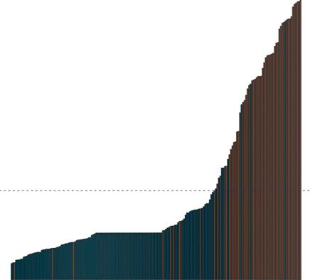
CLINICAL RESEARCH    www.jasn.org

This has been associated with poor long-term allograft sur-
                                                                     Significance Statement
vival.11 The implementation of the Banff classification in 1991
provided a valuable tool for histopathologic diagnosis of            The traditional biomarkers currently used to monitor a kidney al-
kidney-transplant injury and allowed for standardization             lograft for rejection are late markers of injury and they lack sensi-
                                                                     tivity and specificity. Allograft biopsies on the other hand, are in-
when comparing biopsy sample results between different
                                                                     vasive and costly. The authors describe the discovery and validation
studies.12 Serum creatinine, eGFR and its increase (expressed        of two urinary exosomal mRNA multigene signatures for the di-
as D eGFR), and urinary protein excretion are traditional bio-       agnosis of acute T cell–mediated and antibody-mediated rejection
markers currently used to monitor the kidney allograft, but          and chronic, active antibody-mediated rejection in recipients of
they lack desired sensitivity, specificity, and predictive abil-      kidney transplant. Using a clinically validated platform for exosome
                                                                     isolation and analysis, they demonstrated the high stability of uri-
ity.13 Kidney allograft biopsy specimens, with histopathologic
                                                                     nary exosomes and the reliability of this approach in monitoring
evaluation, remain the gold standard in diagnosing acute re-         patients for allograft rejection. One gene signature for all-cause
jection. However, there are limitations to their use because         rejection and another for discriminating T cell–mediated rejection
biopsies are invasive, costly, and can be associated with signif-    from antibody-mediated rejection showed high predictive perfor-
icant morbidity.14,15 Several biomarkers have been identified         mances and offer clinicians the possibility of new tools for moni-
                                                                     toring emergence of rejection in kidney allografts.
as potential noninvasive tools to diagnose early graft rejection,
including CD3ɛ mRNA, IP-10 (CXCL10) mRNA, and 18S ri-
bosomal RNA isolated from urine pellet, described in the            within membrane-bound vesicles.25 With recent techniques
CTOT04 study.8 Urinary CXCL9 mRNA was highly expressed              incorporating nanofiltration, affinity, microfluidics, and
in patients with acute rejection compared with patients with-       tangential flow fractionation, along with many others, it is
out rejection in multicenter study which included 280 recip-        possible to isolate exosome-enriched fractions.26 The RNA
ients of kidney transplants.16 Recently, donor-derived cellfree     transcriptome can be efficiently profiled in urine exosomes,
DNA (dd-cfDNA) has been introduced to the clinical practice         and this exosomal RNA has been shown to be a valuable source
as a novel biomarker for graft rejection after solid organ trans-   for biomarker discovery and integration of these gene signa-
plantation. Despite results showing good performances in            tures into clinical applications.27–29 Urinary exosome RNA
discriminating active rejection from no-rejection status, dd-       diagnostic assays are being used today, and are even included
cfDNA using the currently defined threshold of 1% did not            in the National Comprehensive Cancer Network guidelines
discriminate well between no rejection and lower grades of          for early detection of prostate cancer.29–32
cellular rejection, such as acute cellular rejection (ACR) 1A.7        Our hypothesis was that the use of the urinary exosome
    Exosomes are nanometer-sized vesicles (between 50 and           mRNA gene signature could represent a rapid, noninvasive
200 nm) released by cells to mediate cell-to-cell communica-        assay to diagnose acute rejection in kidney allografts. We in-
tion by delivering proteins and nucleic acids, such as mRNAs        cluded 192 urine samples collected from patients who received
and microRNAs.17 Exosomes are released into the urine dur-          a renal transplant and were undergoing clinically indicated
ing fusion of the multivesicular body with the apical plasma        biopsies at three centers across the Unites States. We measured
cell membrane, or by direct budding of the plasma mem-              mRNA directly from urinary exosomes to identify a specific
brane.18 They carry the parent cells’ surface proteins and nu-      exosome RNA signature for kidney rejection. Whereas previ-
cleic acids and, thus, reflect the biologic function of the parent   ous data identified a urinary cell mRNA signature,8,24 we re-
cell. In the transplanted kidney, exosomes originate from           port for the first time the development of a urinary exosome
glomerular podocytes, renal tubular cells, and from the uroe-       mRNA signature in recipients of kidney transplants undergo-
pithelium.19 Exosomes shed into the urine and, therefore, rep-      ing T cell–mediated rejection (TCMR) and/or antibody-
resent an easily accessible, noninvasive window into ongoing        mediated rejection (ABMR).
pathologic processes within the kidney. We, and others, have
recently shown that urinary exosomes are enriched with pro-
teins derived from immune cells within the kidney transplant
during rejection.20–22 Thus, urinary exosomes can provide           METHODS
investigators with a unique, concentrated sampling of mem-
brane and cytosolic proteins during allograft rejection, and        Patient and Sample Information
can further provide information regarding RNA derived               The study was approved by the institutional review board at
from cells residing within the kidneys, including infiltrating       each site, and the patients provided written informed consent
lymphocytes.                                                        in accord with the Declaration of Helsinki. We enrolled 175
    Although there are no data on the association between uri-      recipients of kidney transplants at the time of a clinically in-
nary exosome–derived mRNA signatures and kidney allograft           dicated renal biopsy from three renal centers. A total of 220
rejection, mRNA signatures from urinary cell pellets have been      urine samples were collected from patients with matched bi-
associated with active rejection.23,24 Compared with urinary        opsy specimens for urinary exosomal mRNA profiling.
mRNA isolated from whole urinary cells, urinary exosomal            Among the 175 patients, 44 had repeat biopsies, with 30 pa-
mRNA has shown a greater stability due to the encapsulation         tients having two biopsies and seven patients having more

2        JASN                                                                                                       JASN 32: ccc–ccc, 2021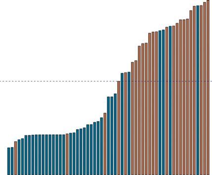
www.jasn.org      CLINICAL RESEARCH

than two biopsies. Demographic and clinical characteristics,           On the basis of the initial analysis, a subset of assays was
and information on the donors, were collected from the med-         identified and plated onto a custom TaqMan OpenArray
ical chart. eGFR was calculated using the Modification of Diet       panel. This panel consisted of 112 TaqMan assays. For this
in Renal Disease equation.33 We used the on-site pathologist’s      panel, 5 ml cDNA was preamplified with a pool of the 112
renal transplant biopsy specimen report to define active re-         assays, using the manufacturer’s directions. The preamplifica-
jection, in accordance with the Banff Working Groups’ crite-        tion reactions were diluted before mixing samples with the
ria.34 We excluded from our primary analysis 23 samples that        TaqMan OpenArray Real-Time PCR Master Mix. Reaction
were diagnosed as borderline cell-mediated rejection, and five       mixes were loaded onto the OpenArray plates and the plates
samples that were diagnosed with BK virus nephropathy. For          were run on the QuantStudio 12K Flex Real-Time PCR system
our analyses, we integrated TCMR and acute and chronic ac-          (Thermo Fisher), using the preset protocol for this panel.
tive ABMR to form the rejection group, and we distinguished         Analysis of samples described here used the 112 TaqMan as-
them from samples that were classified as having no rejection        says common to all samples.
on the basis of biopsy specimen reports. Biopsy specimen
reports with a diagnosis of mixed ABMR and TCMR were                Statistical Analyses
grouped with the TCMR subgroup, and those with mixed                Genes with data missing from .20% of the samples were
borderline TCMR and ABMR were grouped with the ABMR                 excluded from the analysis. Missing data were imputed using
subgroup.                                                           a nonparametric missing-value imputation.35 The Ct values
                                                                    from the OpenArray were normalized to PGK1. The Boruta36
Detection of Donor-Specific Antibodies                               algorithm was used to select genes that were most relevant for
The presence of anti-HLA antibodies was assessed by LAB-            prediction.. A support vector machine (SVM) with a radial
Screen Mixed (One Lambda Inc., Canoga Park, CA), analyzed           kernel was fit to the relevant features using a stratified repeated
on a Luminex platform. In the event of a positive assay, this       K-fold crossvalidation (K510, repeats510) to generate the
was followed by LABScreen Single Antigen Class I/Class II           rejection probabilities using the caret package.37 This ap-
(One Lambda Inc.). A normalized mean fluorescence intensity          proach gives a better indication of how well the model will
$3000 for class I or $1000 for class II is considered positive at   perform on unseen data compared with just one train-test split
our center.                                                         in a hold-out method, which makes it highly dependent on how
                                                                    the data are split in test and train datasets. The pROC package
Urinary Exosome Isolation, mRNA Extraction, and                     was used to generate the receiver-operating-characteristic
Gene-Expression Analysis                                            curves.38 Associations between clinical and demographic factors
The second voided urine sample was collected on the morning         were computed using the t test for continuous variables and the
of the biopsy, and whole urine samples were stored at 280°C.        Pearson chi-squared test for categoric variables. Area under the
Three in-house controls were used, consisting of one-pooled         curve (AUC) comparison was performed using the DeLong test.
male sample, one-pooled female sample, and one-pooled male          Data reporting and analyses were conducted using R version 3.3.
and female sample. Samples were thawed and up to 20 ml              Two-tailed P values #0.05 were considered statistically signifi-
urine was centrifuged to pellet cells and cellular debris at        cant. Sample size was calculated for a negative predictive value
2000 3 g for 20 minutes before the extraction. Exosomes             (NPV) and specificity of 90%, with a 10% width for the 95% CI
were isolated using a urine-exosome isolation kit, as described     at a prevalence of 20%.39 On the basis of this calculation, the
previously (Figure 1).29–31 RNA was eluted in 16 ml nuclease-       required sample size was estimated to be 173 samples.
free water, 14 ml of which was used in a 20 ml reverse-
transcription reaction using the VILO cDNA synthesis kit
(Thermo Fisher).                                                    RESULTS
    The first round of samples was analyzed using the TaqMan
OpenArray Human Inflammation Panel (Thermo Fisher).                  Patients’ Characteristics and Biopsy Specimens
This panel consists of 586 TaqMan assays for genes that have        A total of 192 urine samples that have matched biopsy spec-
been studied as targets for a range of inflammatory diseases,        imens were included. Exosomal mRNA showed excellent sta-
and it includes 21 endogenous control assays. To prepare the        bility in urine stored at 4°C for 2 weeks. Figure 2 shows the
samples for quantitative PCR (qPCR), 10 ml cDNA was split           expression of three targets from eight samples. The stability of
into two equal portions and preamplified with two pools of           mRNA is critical for developing clinically useful diagnostic
mixed primers, following the manufacturer’s directions. The         tests because the samples can be safely cold-pack shipped
preamplification reactions were mixed and diluted before             from the patient’s residence to a central laboratory for analysis,
mixing with TaqMan OpenArray Real-Time PCR Master                   where they can be either processed immediately or stored at
Mix. Reaction mixes were loaded onto the OpenArray plates           14°C for up to 2 weeks. Our study included matched urine
and the plates were run on the QuantStudio 12K Flex Real-           samples for biopsy specimens showing TCMR (grades IA, IB,
Time PCR system (Thermo Fisher) using the preset protocol           IIA, IIB) and acute active and chronic active ABMR subgroups
for this panel.                                                     of rejection, on the basis of the Banff classification, and we used

JASN 32: ccc–ccc, 2021                                                                        Urinary Exosomes in Transplantation    3
CLINICAL RESEARCH www.jasn.org
Standard Exosomal RNA OpenArray real time PCR
Analysis
Urine sample extraction RNA profiling
3000
2000
Delta R
Multivariate
Analysis
1000
0
0 10 20 30 40
Cycle
Figure 1. Simple collection protocol enables ease of use. Urine samples are collected in a standard urine cup at any time during the
day. The exosomes are isolated from the urine sample, followed by an RNA extraction, and RT-qPCR analysis of the target genes. The
relative quantities of each target gene are inputted into an algorithm to generate a single score from zero to one.
the term “active rejection” to distinguish these samples from any-cause rejection group included a higher proportion of pa-
other biopsy specimens without rejection. There were 59 bi- tients with previous rejection episodes (P,0.001) and longer
opsy specimens with rejection, and 133 biopsy specimens with- time since biopsy when compared with the group without re-
out rejection (30.7% prevalence). Figure 3 shows the results for jection (P50.02). The difference in the proportion of Black
the 192 biopsy specimens that had matched urine samples. patients between the groups was NS (P50.47). Among the
Table 1 shows the baseline characteristics of the study cohorts. any-cause rejection group, 59.3% of cases of rejection were
The mean (interquartile range) age of patients with any-cause due to active TCMR, and 40.7% were attributed to ABMR.
rejection was 51.0 (38.0–64.5) years, and 51.6 (40.8–65) years
in patients without rejection. Median (interquartile range) Identifying Any-Cause Rejection Signature from
eGFR levels were 32.85 (22.13–44.56) ml/min per 1.73 m2 Urinary Exosomes
in patients with any-cause rejection, and 37.89 (25.95–50.89) We compared mRNA from urinary exosomes collected from
ml/min per 1.73 m 2 in patients with no rejection. The patients with biopsy sample–proven, any-cause rejection with
105
100
95
% ExoRNA Yield
90
85
80
75
70
DAY1 DAY2 DAY3 DAY7 DAY8 DAY9 DAY14 DAY15
Number of Days
Figure 2. Urinary exosomal RNA is stable over 2 weeks at 4°C. The urine samples were collected and stored at 4°C for up to 2 weeks.
Exosomes were extracted at different time points, followed by RT-qPCR to analyze the yield and integrity of the RNA. The urinary
exosome RNA (exoRNA) was stable over 2 weeks (average yield from three separate genes). The error bars represent the SD of the
percentage of exosomal RNA yield across three different genes.
4 JASN JASN 32: ccc–ccc, 2021www.jasn.org CLINICAL RESEARCH
220 Biopsy Samples
175 Patients
28 excluded:
Analysis Cohort:
5 BKV
192 biopsies
23 Borderline TCMR
59 Any Cause Rejection 133 No Rejection
35 TCMR
17 IA
9 IB
8 Acute ABMR 16 chronic, active ABMR
4 IIA
2 IIB
3 chronic TCMR
Figure 3. CONSORT flow diagram and histologic diagnosis of enrolled patients. Of the 220 initially collected samples, 192 were
further analyzed in this cohort.
urine samples from patients without rejection. To identify rel- of investigated genes, we performed feature selection using
evant genes in urinary exosomes that could predict any-cause Boruta to identify the relevant features. A repeated, stratified,
rejection, we first analyzed the samples using the TaqMan K-fold classification model (K510, repeats510) with an SVM
OpenArray Human Inflammation Panel. This panel consists using a radial-basis-function kernel was used for classification.
of 586 TaqMan assays for genes that have been studied as The stratification ensures there is a similar percentage of sam-
targets for a range of inflammatory diseases, and it includes ples with rejection in each of the folds. This process is repeated
21 endogenous control assays. For subsequent analyses, a sub- ten times, with a different randomization in each repeat, to
set of 112 TaqMan assays was identified and plated onto a generate the final classification model. We used crossvalida-
custom TaqMan OpenArray panel. Given the large number tion instead of a hold-out method because crossvalidation
Table 1. Baseline characteristics of patients
Clinical Cohort (n5192)
Characteristic P Value
No Rejection (n5133) Any-Cause Rejection (n559)
Age, yr 51.6615.1 51.0616.2 0.80a
Female, % 32.3 45.8
Race, %
White 83.6 88.0 0.47b
Black 16.4 22.0 0.47b
SCr at biopsy, mg/dl 1.8 (1.5–2.6) 2.2 (1.7–2.8) 0.39a
eGFR ml/min per 1.73 m2 37.9 (25.6–50.9) 32.9 (22.1–44.6) 0.02a
Previous rejection, % 15.2 42.4 8.23310207b
Deceased donor, % 43.0 51.9 0.65b
Time to biopsy, d 215 (46–1751) 1250 (295–3063) 0.02a
Thymoglobulin, % 60.5 69.4 0.36b
UPCR 0 (0–0.28) 0 (0–1.51) 0.27a
DSA, % 18.5 44.2 0.002a
PRA, % 2.0 (2.0–41.25) 25.0 (2.0–61.25) 0.02a
Data presented as frequencies, mean6SD, and median (interquartile range). All demographic and clinical data are based on the day of biopsy, timed with the urine
collection. SCr, serum creatinine; UPCR, urinary protein-creatinine ratio; PRA, panel-reactive antibody.
a
P values from t test.
b
P values from chi-squared test.
JASN 32: ccc–ccc, 2021 Urinary Exosomes in Transplantation 5CLINICAL RESEARCH www.jasn.org
improves the generalizability of the gene signature by validat- significantly worse than our any-case rejection signature
ing the performance on multiple train-test subsets of the data, (P50.03 and P52.2 x 10-9 respectively). Combining D eGFR
and this results in a much more stable estimate of the perfor- with DSA and urinary protein-creatinine ratio did not further
mance. This allowed us to identify a multigene signature improve the AUC (Supplemental Figure 1).
(CXCL11, CD74, IL32, STAT1, CXCL14, SERPINA1, B2M,
C3, PYCARD, BMP7, TBP, NAMPT, IFNGR1, IRAK2, and Discriminating TCMR from ABMR
IL18BP) that discriminated biopsy samples with any-cause We also compared the TCMR samples with the ABMR samples
rejection from those with no rejection. The AUC was 0.93 to derive an additional signature to discriminate between these
(95% CI, 0.87 to 0.98) (Figure 4). To compare the perfor- two forms of rejection. Applying the same optimization and
mance of this signature against current clinical practice, we classification approach used for any-cause rejection, we iden-
also generated an AUC for the change (D) in eGFR (D eGFR) tified a multigene signature (CD74, C3, CXCL11, CD44, and
(Figure 4). The AUC for D eGFR for this set of patients was IFNAR2) that could distinguish TCMR from ABMR. The AUC
0.57 (95% CI, 0.49 to 0.65), which was significantly inferior for this signature was 0.87 (95% CI, 0.76 to 0.97) (Figure 6). A
(P,0.001) to the performance of the multigene signature. We cut point was derived to maximize the NPV and sensitivity to
also derived a cut point to rule out any-cause rejection by rule out ABMR (Figure 5B). Samples with a positive signature
optimizing Youden J (Figure 5A). This resulted in an NPV of for all-cause rejection were analyzed for the second signature.
93.3% (95% CI, 87.7% to 96.4%) and a sensitivity of 84.7% If the second signature is negative, the patient has TCMR and
(95% CI, 73.5% to 91.8%). The positive predictive value ABMR is ruled out with an NPV of 90.6% (95% CI, 75.8% to
(PPV) for discriminating active rejection was 86.2% (95% 96.8%) and a PPV of 77.8% (95% CI, 59.2% to 89.4%). The
CI, 75.1% to 92.8%) (Table 2). sensitivity to discriminate TCMR from ABMR was 87.5%
We also analyzed the data on donor specific antibodies (95% CI, 69.0% to 95.7%) and the specificity was 82.9%
(DSAs) to assess whether the presence of DSAs against HLA (95% CI, 67.3% to 92.0%) (Table 3).
class I or II was associated with an increased risk of ABMR and
of any-cause rejection. As shown in Supplemental Figure 1, the Borderline-Rejection Samples
presence of DSAs has an AUC (95% CI) of 0.72 (0.61–0.83) for We then applied our all-cause rejection signature to the urine
ABMR and 0.64 (0.56–0.72) for any-cause rejection, which are samples in our cohort from patients diagnosed with border-
line rejection. Follow-up information was available for 18 out
of the 23 samples. One patient was excluded from the analysis
1.0 for having recurrent glomerulopathy in addition to the histo-
logic diagnosis of borderline rejection. Among the eight sam-
ples from patients with borderline rejection who showed a
0.8 predicted negative signature of all-cause rejection, only two
patients showed a decrease in eGFR of .30%, 12 months after
biopsy. One of those two patients developed TCMR 1B and
0.6 BK-virus nephritis less than a year later, and the other patient
Sensitivity
developed TCMR 1A on two subsequent biopsies, 1 and
3 months later. The other six patients remained stable with
0.4 no change in eGFR (,5% change) or development of protein-
uria, and they did not need any further intervention. Among
the nine samples from patients with borderline rejection who
0.2 showed a predicted positive signature, only three patients re-
mained with stable eGFR (,5% change) and no proteinuria at
Exosome [AUC:0.93 (95% CI: 0.87-0.98)] 12 months postbiopsy. The other six patients had either a de-
0.0 deltaGFR [AUC:0.57 (95% CI: 0.49-0.65)] crease in eGFR of .30% (two patients) and/or persistent pro-
0.0 0.2 0.4 0.6 0.8 1.0 teinuria (five patients, urinary protein-creatinine ratio .0.9).
1 - Specificity It is important to note that borderline-rejection samples were
not used in the development of the all-cause rejection signa-
Figure 4. Exosome RNA signature significantly outperforms D ture, and the signature is only meant to diagnose ongoing re-
eGFR in discriminating any-cause rejection. The receiver-oper-
jection and not act as a predictor of rejection.
ating-characteristic analysis and AUC is shown for the exosome
RNA signature and compared with D eGFR. The fraction of true
positive results (sensitivity) and the fraction of false positive re-
sults (1–specificity) for diagnosis of any-cause acute rejection are DISCUSSION
displayed on the y and x axis, respectively. The AUC for the RNA
signature is 0.93 (95% CI, 0.87 to 0.98) and the AUC for D eGFR is In this study, we report a noninvasive test to detect active
0.57 (95% CI, 0.49 to 0.65). kidney-transplant rejection from urine samples of patients
6 JASN JASN 32: ccc–ccc, 2021www.jasn.org CLINICAL RESEARCH
A B
1.0 1.0
0.8 0.8
0.6 0.6
0.4 0.4
0.2 0.2
0.0 0.0
Figure 5. Waterfall plot of the urinary exosome gene scores demonstrates the high NPV of the exosomal RNA signature. The blue
dotted line represents the score cutoff for each of the gene signatures. (A) Discriminating any-cause rejection from no rejection (red
bars denote samples that have clinical rejection). (B) Discriminating ABMR from TCMR (red bars denote samples with ABMR).
undergoing a for-cause biopsy, with diverse pathologic diag- suited for diagnostic assays. We analyzed 112 target genes by
nosis including both ABMR and cellular-mediated rejection. TaqMan qPCR across all samples, using the OpenArray plat-
We decided to exclude the borderline cellular-rejection sam- form, and identified an exosomal mRNA signature that
ples because many studies have shown that borderline cellular significantly correlates with active rejection. The mRNA sig-
rejection represents a heterogeneous group that ranges from nature was developed using an SVM classifier with a radial-
insignificant inflammation to a clinically significant cellular basis-function kernel. An SVM classifier was chosen due to
rejection resulting in histologic consequences.40 Furthermore, underlying nonlinearity of the data and its ability to better
the immunologic and clinical significance of borderline rejec- handle outliers. Stratification was chosen to ensure that all
tion is still a subject of debate in the transplant community.41 of the different folds had the same proportion of rejected sam-
We used a clinically validated exosome isolation platform, ples to minimize any bias due to the underlying prevalence of
which has been used for the ExoDx Prostate (Intelliscore) rejection in the dataset. We also chose crossvalidation instead
test and has been performed on .50,000 patients to date.29–31
This platform also enabled isolation and analysis of mRNA
1.0
signatures in urinary exosomes to predict any-cause active re-
jection in patients with kidney transplant at the time of allo-
graft biopsy. Compared with exosomal proteins, RNA is well
0.8
protected inside the vesicle and can be stably assessed from
urine samples even after prolonged storage, whereas urine
samples for exosomal protein study can only be safely stored
0.6
for about 12 hours,20 because proteins that are associated with
Sensitivity
the outer membrane are exposed to protease activity. Also, it is
sometimes more challenging to robustly assess and discrimi-
0.4
nate exosome proteins from free proteins (or those sticking
nonspecifically to the outside of the vesicle). RNA detection
methods (such as RT-qPCR used here) is highly sensitive
0.2
down to single-copy levels, and RT-PCR is a well-known
method for clinical laboratories and is exceptionally well
0.0 AUC: 0.865 (0.761–0.970
Table 2. Gene signature discrimination performance 0.0 0.2 0.4 0.6 0.8 1.0
characteristics for overall rejection 1 - Specificity
Metric Performance (95% CI), % Figure 6. Exosome RNA signature can accurately discriminate
NPV 93.3 (87.7 to 96.4) TCMR from ABMR. Receiver-operating-characteristic curve
Sensitivity 84.7 (73.5 to 91.8) showing the fraction of true positive results (sensitivity) and the
Specificity 94.0 (88.6 to 96.9) fraction of false positive results (1–specificity) for discriminating
PPV 86.2 (75.1 to 92.8) TCMR from ABMR (AUC, 0.87; 95% CI, 0.76 to 0.97).
JASN 32: ccc–ccc, 2021 Urinary Exosomes in Transplantation 7CLINICAL RESEARCH www.jasn.org
Table 3. Gene signature discrimination performance allograft rejection in multiple studies.43–46 Although some of
characteristics discriminating TCMR from ABMR the genes identified are not known to drive allograft rejection,
Metric Performance (95% CI), % such as NAMPT, they have been shown to play a role in in-
NPV 90.6 (75.8 to 96.8) flammation and immune-cell activation.47 The PYCARD gene
Sensitivity 87.5 (69.0 to 95.7) codes for the ASC protein, which is widely expressed in dif-
Specificity 82.9 (67.3 to 92.0) ferent cells, including B cells, monocytes, and mature T cells. It
PPV 77.8 (59.2 to 89.4) has been described as an adaptor protein that participates in
the inflammasome assembly and has been reported to play a
part in different autoimmune processes and in viral and bac-
of a single training/test split to obtain a more accurate estimate terial infections.48,49 Interestingly, our signature that discrim-
of the performance of the model, which is not dependent on inates TCMR from ABMR showed some overlapping with the
how the initial training/test split was generated. This poten- any-cause rejection signature. We note that three of these
tially leads to lower variance in the performance of the classi- genes (CXCL11, CD74, and C3) are present in the all-cause
fier, leading to a better estimate of future performance on rejection signature, and that two additional genes (IFNAR2
unseen data. and CD44) can help distinguish ABMR from TCMR. All five of
Both the any-cause rejection signature and the signature to these genes which are part of the ABMR/TCMR signature are
distinguish between TCMR and ABMR demonstrated excel- significantly overexpressed (P,0.05) in ABMR compared
lent correlation with histopathologic diagnosis. The any-cause with TCMR.
rejection signature also demonstrated a higher performance CD74, the invariant chain of MHC class II, is strongly ex-
over the current indicators of allograft function, such as eGFR, pressed in cells involved in the presentation of antigens like
which showed an AUC of 0.57 discriminating active rejection dendritic cells, B cells, and macrophages. It also plays a role in
from nonrejection status. All kidney transplant biopsies in our regulating protein trafficking; dendritic-cell migration; and
cohort were clinically indicated, and 30.7% of these biopsy T-lymphocyte homing, proliferation, and cytokine secre-
specimens revealed active rejection. Considering that real- tion.50 CD44, a cell surface glycoprotein involved in cell ad-
life active-rejection prevalence is consistently lower than it is hesion, migration, and homing,51,52 acts as a coreceptor to
in our study population, 10 and that NPV and PPV are CD74. 53 The IFNAR2 is a subunit of the INFA receptor
prevalence-dependent metrics (because NPV increases and
PPV decreases when prevalence decreases), our signature is
expected to show an even higher NPV if it holds up in a stable Patient being tested for
disease population. When we adjust the prevalence to 20%, the any-cause Rejection
NPV will be 96.1%, and, when adjusted to the 7.9% first year
incidence for any-cause rejection, NPV will be 98.6% and PPV
will be 54.7%.
Patient below cut-point
Therefore, the urinary exosome RNA assay for any-cause
rejection can potentially be used to avoid unnecessary biopsies
in patients with clinical suspicion of rejection. Whereas the Yes No
high NPV suggests that only one in 15 patients would poten- NPV=93.3% PPV=86.2%
tially miss a clinically indicated biopsy even at the very high
(30.7%) prevalence in this cohort, the high PPV (86.2%) sup- Rule out rejection
TCMR vs ABMR
ports the possibility for patients with active rejection to be and continue monitoring
treated on time. The strong performances of urinary exosome
gene signature in discriminating TCMR from ABMR status
(AUC, 0.87; NPV, 90.6%) can help to refine the diagnosis by
Patient below cut-point
ruling out an ABMR and provide the clinicians with useful
information for clinical management decisions (Figure 7).
Most of the genes identified in our signatures have an es- Yes No
NPV=90.6% PPV=77.8%
tablished role in immune activation that can explain the re-
jection process. BMP7 (a protein of the TGFb superfamily)
and the proinflammatory cytokines CXCL14, B2M, and IL32 TCMR ABMR
identified in our all-cause rejection signature have recently
been shown to be significantly induced in a kidney transplant Figure 7. Schematic representation of the evaluation chart for
undergoing mixed cellular-mediated rejection and ABMR, as recipients of kidney transplants. Serial monitoring of urinary
analyzed by single-cell analysis.42 Similarly, C3 and CXCL11, exosome could be performed, and clinical management deci-
identified in both any-cause rejection and TCMR, have been sions made, according to the positivity or negativity of the
found to be upregulated in gene-expression profiling in acute signature.
8 JASN JASN 32: ccc–ccc, 2021www.jasn.org CLINICAL RESEARCH
coupled to the tyrosine protein kinase JAK1, and that binds to IntelliScore test has been validated in two independent, pro-
the latent form of the transcription factors STAT1 or STAT2, spective, multicenter studies and in a large utility study, with a
participating in amplifying the production of chemokines, blinded control arm, showing the value of the exosome
such as CXCL10.54 platform.29–31 This is the first study describing an exosome
Previous studies by Suthanthiran et al.8 have successfully mRNA signature for any-cause kidney rejection, and we ap-
used mRNA derived from urinary cell pellets to identify po- plied a similar approach as the prostate cancer signature on
tential markers of acute rejection.24 Although cells in urine are urinary exosomes.
often dead or dying, and RNA within dead cells is quickly There are a few limitations to this study. First, we have
subjected to degradation, the extreme stability of exosome conducted a cross-sectional study and have not collected serial
RNA (stable in urine for 2 weeks) can enable a more robust urine samples, which prevented us from testing how early the
diagnostic platform. A more recent biomarker that is currently signature can predict rejection before the clinically indicated
used in the clinic is dd-cfDNA. Bloom et al.7 measured the biopsy. Second, our study has not included an independent
plasma level of dd-cfDNA and used a threshold of 1% to dis- validation cohort. However, the crossvalidation technique we
criminate patients with allograft rejection status from those used has been shown to improve the variability and
without. Although they were able to show that dd-cfDNA selection bias.
discriminated an active rejection status with an AUC of 0.74 In this report, we show the high-performance characteris-
and an NPV of 84%, samples with grade-IA TCMR did not tics of urinary exosome RNA to discriminate active rejection
reach the 1% threshold.7 In a subsequent study, Huang et al.55 from no rejection and ABMR from TCMR. We did not directly
found that the same dd-cfDNA test has not been able to dis- compare how the exosome signature performs compared with
criminate TCMR from nonrejection status, despite strong per- a cell pellet RNA assay; however, the high stability of urinary
formance in ABMR. In contrast to these studies, our any-cause exosomes supports the possibility of identifying the nucleic
rejection signature is clearly able to discriminate TCMR from acid signature more accurately, even without complicated
nonrejection, despite 42.9% of the cases being Banff IA. How- RNA preservatives in the urine-collection device. Our gene
ever, given the complexity of the molecular perturbations dur- signatures offer the possibility to provide the clinician with a
ing kidney allograft rejection, it is becoming increasingly ev- new tool to help differentiate causes of graft dysfunction in the
ident that the combination of multiple biomarkers can kidney allograft. Further validating studies, with prospectively
improve the performance of any individual test. In a recent collected samples, would help confirm our results in larger
publication, Sarwal et al.56 used a combination of genomic, patient cohorts, and evaluate the potentials of this assay for
metabolic, and proteomic urinary biomarkers in recipients of early detection of kidney allograft rejection.
kidney transplants. Similarly, we envision that combining uri-
nary exosomal transcriptomic and proteomic profiling can
improve the performance of our current signature. DISCLOSURES
The strengths of this urinary exosome mRNA study is that
we have (1) been able to unveil a combination of biomarkers J.R. Azzi, C. Coticchia, J. Hurley, J. Skog, and V. Tadigotla have intellectual
involved in active allograft kidney rejection in a large number properties related to this work. J.R. Azzi reports having intellectual properties
of patients and urine samples, (2) developed a signature that and receiving royalties from Accrue Health Inc.; receiving research funding
distinguishes TCMR from ABMR, and (3) showed high pre- from the American Diabetes Association, American Heart Association, and
Qatar Research Fund; being a scientific advisor for CareDx; and having in-
dictive performances in our results that will allow the clinical tellectual properties in ExosomeDx. C. Coticchia, J. Hurley, J. Skog, and
use of this test to rule out rejection in patients. Assuming a V. Tadigotla are employees of Exosome Diagnostics, a Bio-Techne brand.
high prevalence of rejection in a cohort of patients with clin- R.N. Formica reports being the president of the American Society of Trans-
ically indicated biopsies, and considering that urine samples plantation; having consultancy agreements with Genentech Pharmaceutical
are easy to collect and urinary exosomal mRNA is highly sta- and Veloxis Pharmaceuticals; being on a speakers bureau for Novartis Phar-
maceuticals; being on the visiting committee for the Scientific Registry of
ble, with a good positive predictive value increasing with Transplant Recipients (January 1, 2018); and being a member of the United
higher prevalence, our test can prove useful to also rule in Network for Organ Sharing/Organ Procurement and Transplantation Net-
rejection. This is particularly true in patients with high-risk work Membership and Professional Standards Committee. J.F. Markmann
biopsies, patients who live far from transplant centers, and in reports having consultancy agreements with, ownership interest in, and being
a scientific advisor for (or member of) eGenesis and QihanBio. L.V. Riella
patients living through pandemics such as coronavirus disease
reports receiving research funding from Bristol Myers Squibb and Visterra,
2019. This is important because we have shown that urine and being a scientific advisor for or member of CareDx. J. Skog reports having
stored at 4°C maintains stable exosomal mRNA for up to 2 ownership interest in Bio-Techne and patents and inventions with Mas-
weeks, similar to what has been shown before.29 sachusetts General Hospital. A. Srivastava reports being on a speakers bureau
Integration of urinary exosomes into clinical practice has for AstraZeneca; receiving honoraria from AstraZeneca and Horizon Thera-
emerged in fields outside kidney transplantation. The world’s peutics PLC; and having consultancy agreements with CVS Caremark and Tate
& Latham (medicolegal consulting). A. Chandraker reports consultancy
first exosome-based diagnostic assay (ExoDx Prostate Intelli- agreements with Mitobridge, Shire, Amgen, Immucor, Natera, and Allovir;
Score test) was launched in 2016 and is a prostate cancer test receiving research funding from ReNu, CSL, Shire, Amgen, and Allovir; re-
which uses urinary exosome RNA. The ExoDx Prostate ceiving honoraria from Hansa, Natera, and eGenesis; reports being a scientific
JASN 32: ccc–ccc, 2021 Urinary Exosomes in Transplantation 9CLINICAL RESEARCH www.jasn.org
advisor or membership with American Society of Transplantation as Devel- donor-derived cell-free DNA via massively multiplex PCR. J Clin Med 8:
opment Chair, Transplant Therapeutics Consortium as Governance Commit- 19, 2018
tee and Past Chair, CEoT as Co-Chair, Transplant Metrics as Chair, and Sci- 7. Bloom RD, Bromberg JS, Poggio ED, Bunnapradist S, Langone AJ,
entific American as Editor for Nephrology. All remaining authors have Sood P, et al.; Circulating Donor-Derived Cell-Free DNA in Blood for
nothing to disclose. Diagnosing Active Rejection in Kidney Transplant Recipients (DART)
Study Investigators: Cell-free DNA and active rejection in kidney allo-
grafts. J Am Soc Nephrol 28: 2221–2232, 2017
8. Suthanthiran M, Schwartz JE, Ding R, Abecassis M, Dadhania D,
FUNDING Samstein B, et al.; Clinical Trials in Organ Transplantation 04 (CTOT-04)
Study Investigators: Urinary-cell mRNA profile and acute cellular re-
This project was supported by American Heart Association award jection in kidney allografts. N Engl J Med 369: 20–31, 2013
13FTF17000018 (to J.R. Azzi) and National Institutes of Health (NIH) grant 9. Christakoudi S, Runglall M, Mobillo P, Tsui T-L, Duff C, Domingo-Vila C,
RO1 AI134842 (to J.R. Azzi). This project was also partially supported by et al.: Development of a multivariable gene-expression signature tar-
Exosome Diagnostics, a Bio-Techne brand. A. Srivastava was supported by geting T-cell-mediated rejection in peripheral blood of kidney trans-
NIH Clinical Center grant F32DK11106. plant recipients validated in cross-sectional and longitudinal samples.
EBioMedicine 41: 571–583, 2019
10. Hart A, Smith JM, Skeans MA, Gustafson SK, Stewart DE, Cherikh WS,
et al.: OPTN/SRTR 2015 annual data report: Kidney. Am J Transplant 17
ACKNOWLEDGMENTS [Suppl 1]: 21–116, 2017
11. Opelz G, Döhler B; Collaborative Transplant Study Report: Influence of
The authors would like to thank the physician assistants at the Renal Trans- time of rejection on long-term graft survival in renal transplantation.
plant Division, Brigham and Women’s Hospital and the transplant coordi- Transplantation 85: 661–666, 2008
nators at Yale School of Medicine and Massachusetts General Hospital for their 12. Solez K, Axelsen RA, Benediktsson H, Burdick JF, Cohen AH, Colvin RB,
logistic help in enrolling patients and collecting samples. In particular, the et al.: International standardization of criteria for the histologic di-
authors thank Michelle Kopp, Jill Lynch, Kaitlyn McGowan, Keri Foley, Jon- agnosis of renal allograft rejection: The Banff working classification of
athan Andrade, and Sheri Talbott. The authors also thank Ricarda Tomlin kidney transplant pathology. Kidney Int 44: 411–422, 1993
from Yale for the help. 13. Waikar SS, Betensky RA, Bonventre JV: Creatinine as the gold standard
Dr. Johan Skog and Dr. Jamil R. Azzi contributed to the conception and for kidney injury biomarker studies? Nephrol Dial Transplant 24:
design; Dr. Anand Srivastava, Siawosh Eskandari, Dr. Albana B. Mihali, and 3263–3265, 2009
Dr. Juliano Alhaddad contributed to data acquisition; Dr. James Hurley, Dr.
14. Ferguson C, Winters S, Jackson S, McToal M, Low G: A retrospective
Christine Coticchia, Dr. Vasisht Tadigotla, and Dr. Johan Skog performed
analysis of complication and adequacy rates of ultrasound-guided na-
assays and data analysis; Dr. Rania El Fekih and Dr. Jamil R. Azzi drafted the
tive and transplant non-focal renal biopsies. Abdom Radiol (NY) 43:
manuscript; all authors contributed to data interpretation and manuscript
2183–2189, 2018
revisions, and all authors approved the final version of the manuscript.
15. Furness PN, Taub N; Convergence of European Renal Transplant Pa-
thology Assessment Procedures (CERTPAP) Project: International var-
iation in the interpretation of renal transplant biopsies: Report of the
SUPPLEMENTAL MATERIAL CERTPAP Project [published correction appears in Kidney Int 60: 2429,
2001]. Kidney Int 60: 1998–2012, 2001
16. Hricik DE, Nickerson P, Formica RN, Poggio ED, Rush D, Newell KA,
This article contains the following supplemental material online at http://
et al.; CTOT-01 consortium: Multicenter validation of urinary CXCL9 as
jasn.asnjournals.org/lookup/suppl/doi:10.1681/ASN.2020060850/-/
a risk-stratifying biomarker for kidney transplant injury. Am J Transplant
DCSupplemental.
13: 2634–2644, 2013
Supplemental Figure 1. Receiver-operating-characteristic (ROC) curves
comparing performance of various clinical covariates. 17. Lai CP-K, Breakefield XO: Role of exosomes/microvesicles in the ner-
vous system and use in emerging therapies. Front Physiol 3: 228, 2012
18. Gonzales PA, Pisitkun T, Hoffert JD, Tchapyjnikov D, Star RA, Kleta R,
et al.: Large-scale proteomics and phosphoproteomics of urinary exo-
REFERENCES somes. J Am Soc Nephrol 20: 363–379, 2009
19. Pisitkun T, Shen R-F, Knepper MA: Identification and proteomic pro-
1. Hill NR, Fatoba ST, Oke JL, Hirst JA, O’Callaghan CA, Lasserson DS, filing of exosomes in human urine. Proc Natl Acad Sci U S A 101:
et al.: Global prevalence of chronic kidney disease – a systematic review 13368–13373, 2004
and meta-analysis. PLoS One 11: e0158765, 2016 20. Sigdel TK, Ng YW, Lee S, Nicora CD, Qian W-J, Smith RD, et al.: Per-
2. Mange KC, Joffe MM, Feldman HI: Effect of the use or nonuse of long- turbations in the urinary exosome in transplant rejection. Front Med
term dialysis on the subsequent survival of renal transplants from living (Lausanne) 1: 57, 2015
donors. N Engl J Med 344: 726–731, 2001 21. Lim J-H, Lee C-H, Kim KY, Jung H-Y, Choi J-Y, Cho J-H, et al.: Novel urinary
3. Meier-Kriesche H-U, Kaplan B: Waiting time on dialysis as the strongest exosomal biomarkers of acute T cell-mediated rejection in kidney transplant
modifiable risk factor for renal transplant outcomes: A paired donor recipients: A cross-sectional study. PLoS One 13: e0204204, 2018
kidney analysis. Transplantation 74: 1377–1381, 2002 22. Park J, Lin H-Y, Assaker JP, Jeong S, Huang C-H, Kurdi T, et al.: In-
4. Gill JS, Tonelli M, Johnson N, Pereira BJG: Why do preemptive kidney tegrated kidney exosome analysis for the detection of kidney transplant
transplant recipients have an allograft survival advantage? Trans- rejection. ACS Nano 11: 11041–11046, 2017
plantation 78: 873–879, 2004 23. Afaneh C, Muthukumar T, Lubetzky M, Ding R, Snopkowski C, Sharma VK,
5. Kaplan B, Meier-Kriesche HU: Renal transplantation: A half century of et al.: Urinary cell levels of mRNA for OX40, OX40L, PD-1, PD-L1, or PD-L2 and
success and the long road ahead. J Am Soc Nephrol 15: 3270–3271, acute rejection of human renal allografts. Transplantation 90: 1381–1387, 2010
2004 24. Muthukumar T, Dadhania D, Ding R, Snopkowski C, Naqvi R, Lee JB,
6. Sigdel TK, Archila FA, Constantin T, Prins SA, Liberto J, Damm I, et al.: et al.: Messenger RNA for FOXP3 in the urine of renal-allograft recipi-
Optimizing detection of kidney transplant injury by assessment of ents. N Engl J Med 353: 2342–2351, 2005
10 JASN JASN 32: ccc–ccc, 2021www.jasn.org CLINICAL RESEARCH
25. Miranda KC, Bond DT, McKee M, Skog J, Păunescu TG, Da Silva N, 41. Becker JU, Chang A, Nickeleit V, Randhawa P, Roufosse C: Banff bor-
et al.: Nucleic acids within urinary exosomes/microvesicles are poten- derline changes suspicious for acute T cell-mediated rejection: Where
tial biomarkers for renal disease. Kidney Int 78: 191–199, 2010 do we stand? Am J Transplant 16: 2654–2660, 2016
26. Jia S, Zocco D, Samuels ML, Chou MF, Chammas R, Skog J, et al.: 42. Wu H, Malone AF, Donnelly EL, Kirita Y, Uchimura K, Ramakrishnan SM,
Emerging technologies in extracellular vesicle-based molecular diag- et al.: Single-cell transcriptomics of a human kidney allograft biopsy
nostics. Expert Rev Mol Diagn 14: 307–321, 2014 specimen defines a diverse inflammatory response. J Am Soc Nephrol
27. Yang F, Liao X, Tian Y, Li G: Exosome separation using microfluidic 29: 2069–2080, 2018
systems: Size-based, immunoaffinity-based and dynamic methodolo- 43. Sreekumar R, Rasmussen DL, Wiesner RH, Charlton MR: Differential
gies. Biotechnol J 12: 1600699, 2017 allograft gene expression in acute cellular rejection and recurrence of
28. He L, Zhu D, Wang J, Wu X: A highly efficient method for isolating hepatitis C after liver transplantation. Liver Transpl 8: 814–821, 2002
urinary exosomes. Int J Mol Med 43: 83–90, 2019 44. Chen R, Sigdel TK, Li L, Kambham N, Dudley JT, Hsieh S-C, et al.:
29. McKiernan J, Donovan MJ, O’Neill V, Bentink S, Noerholm M, Belzer S, Differentially expressed RNA from public microarray data identifies
et al.: A novel urine exosome gene expression assay to predict high- serum protein biomarkers for cross-organ transplant rejection and
grade prostate cancer at initial biopsy. JAMA Oncol 2: 882–889, 2016 other conditions. PLOS Comput Biol 6: e1000940, 2010
30. McKiernan J, Donovan MJ, Margolis E, Partin A, Carter B, Brown G, 45. Spivey TL, Uccellini L, Ascierto ML, Zoppoli G, De Giorgi V, Delogu LG,
et al.: A prospective adaptive utility trial to validate performance of a et al.: Gene expression profiling in acute allograft rejection: Chal-
novel urine exosome gene expression assay to predict high-grade lenging the immunologic constant of rejection hypothesis. J Transl
prostate cancer in patients with prostate-specific antigen 2-10ng/ml Med 9: 174, 2011
at initial biopsy. Eur Urol 74: 731–738, 2018 46. Akalin E, Hendrix RC, Polavarapu RG, Pearson TC, Neylan JF, Larsen
31. Tutrone R, Donovan MJ, Torkler P, Tadigotla V, McLain T, Noerholm M, CP, et al.: Gene expression analysis in human renal allograft biopsy
et al.: Clinical utility of the exosome based ExoDx Prostate(IntelliScore) samples using high-density oligoarray technology. Transplantation 72:
EPI test in men presenting for initial biopsy with a PSA 2-10 ng/mL. 948–953, 2001
Prostate Cancer Prostatic Dis 23: 607–614, 2020 47. Halvorsen B, Espeland MZ, Andersen GØ, Yndestad A, Sagen EL,
32. Carroll PR, Kellogg Parsons J, Andriole G, Bahnson RR, Castle EP, Rashidi A, et al.: Increased expression of NAMPT in PBMC from patients
Catalona WJ, et al.: NCCN guidelines insights: Prostate cancer early with acute coronary syndrome and in inflammatory M1 macrophages.
detection, Version 2.2016. J Natl Compr Canc Netw 14: 509–519, 2016 Atherosclerosis 243: 204–210, 2015
33. Levey AS, Stevens LA, Schmid CH, Zhang YL, Castro AF 3rd, Feldman 48. Martin BN, Wang C, Zhang CJ, Kang Z, Gulen MF, Zepp JA, et al.: T cell-
HI, et al.: A new equation to estimate glomerular filtration rate [pub- intrinsic ASC critically promotes T(H)17-mediated experimental auto-
lished correction appears in Ann Intern Med 155: 408, 2011]. Ann Intern immune encephalomyelitis. Nat Immunol 17: 583–592, 2016
Med 150: 604–612, 2009 49. Ichinohe T, Lee HK, Ogura Y, Flavell R, Iwasaki A: Inflammasome rec-
34. Haas M, Loupy A, Lefaucheur C, Roufosse C, Glotz D, Nankivell BJ, ognition of influenza virus is essential for adaptive immune responses.
et al.: The Banff 2017 Kidney Meeting Report: Revised diagnostic cri- J Exp Med 206: 79–87, 2009
teria for chronic active T cell-mediated rejection, antibody-mediated 50. Su H, Na N, Zhang X, Zhao Y: The biological function and significance of
rejection, and prospects for integrative endpoints for next-generation CD74 in immune diseases. Inflamm Res 66: 209–216, 2017
clinical trials. Am J Transplant 18: 293–307, 2018 51. DeGrendele HC, Estess P, Picker LJ, Siegelman MH: CD44 and its li-
35. Stekhoven DJ, Bühlmann P: MissForest--non-parametric missing value gand hyaluronate mediate rolling under physiologic flow: A novel
imputation for mixed-type data. Bioinformatics 28: 112–118, 2012 lymphocyte-endothelial cell primary adhesion pathway. J Exp Med
36. Kursa MB, Rudnicki WR: Feature selection with the Boruta package. 183: 1119–1130, 1996
J Stat Softw 36: 1–13, 2010 52. Ponta H, Sherman L, Herrlich PA: CD44: From adhesion molecules to
37. Kuhn M: Building predictive models in R using the caret package. J Stat signalling regulators. Nat Rev Mol Cell Biol 4: 33–45, 2003
Softw 28: 1–26, 2008 53. Schröder B: The multifaceted roles of the invariant chain CD74--More than
38. Robin X, Turck N, Hainard A, Tiberti N, Lisacek F, Sanchez J-C, et al.: just a chaperone. Biochim Biophys Acta 1863[6 Pt A]: 1269–1281, 2016
pROC: an open-source package for R and S1 to analyze and compare 54. Takaoka A, Yanai H: Interferon signalling network in innate defence.
ROC curves. BMC Bioinformatics 12: 77, 2011 Cell Microbiol 8: 907–922, 2006
39. Buderer NMF: Statistical methodology: I. Incorporating the prevalence 55. Huang E, Sethi S, Peng A, Najjar R, Mirocha J, Haas M, et al.: Early clinical
of disease into the sample size calculation for sensitivity and specificity. experience using donor-derived cell-free DNA to detect rejection in
Acad Emerg Med 3: 895–900, 1996 kidney transplant recipients. Am J Transplant 19: 1663–1670, 2019
40. Nankivell BJ, Agrawal N, Sharma A, Taverniti A, P’Ng CH, Shingde M, 56. Yang JYC, Sarwal RD, Sigdel TK, Damm I, Rosenbaum B, Liberto JM,
et al.: The clinical and pathological significance of borderline T cell- et al.: A urine score for noninvasive accurate diagnosis and prediction of
mediated rejection. Am J Transplant 19: 1452–1463, 2019 kidney transplant rejection. Sci Transl Med 12: eaba2501, 2020
AFFILIATIONS
1
Renal Division, Transplantation Research Center, Brigham and Women’s Hospital and Children’s Hospital, Harvard Medical School, Boston,
Massachusetts
2
Exosome Diagnostics, a Bio-Techne brand, Waltham, Massachusetts
3
Transplant Center, Massachusetts General Hospital, Harvard Medical School, Boston, Massachusetts
4
Section of Nephrology, Yale School of Medicine, New Haven, Connecticut
JASN 32: ccc–ccc, 2021 Urinary Exosomes in Transplantation 11Supplemental Table of Content 1- Supplemental Figure 1. Receiver-Operating-Characteristic (ROC) curve for any cause rejection and ABMR
Supplemental Figure 1. Receiver-Operating-Characteristic (ROC) curve for any cause rejection and
ABMR. The ROC analysis and area under the curve (AUC) is shown for the DSA in ABMR and in any-
cause rejection and compared to delta eGFR_DSA_UPCR. The fraction of true positive results
(sensitivity) and the fraction of false positive results (1 – specificity) for diagnosis of any-cause rejection
are displayed on the y-and x-axis, respectively. The AUC for the DSA in ABMR is 0.72 (95% CI 0.61-
0.83), the AUC for DSA in any cause rejection is 0.64 (95%CI 0.56-0.72), and the AUC for delta eGFR
combined with DSA and UPCR is 0.57 (95%CI 0.46-0.69).
You can also read