Review Serological assessment of type I collagen burden in scleroderma spectrum disorders: A systematic review
←
→
Page content transcription
If your browser does not render page correctly, please read the page content below
Review
             Serological assessment of type I collagen burden in
            scleroderma spectrum disorders: A systematic review
             M. Dziadzio1,2, R.E. Smith1, D.J. Abraham1, R.J. Stratton1, A. Gabrielli2,
                                   C.M. Black1, C.P. Denton1

1
 Centre for Rheumatology and Connective      ABSTRACT                                     severity, in view of their low specificity
Tissue Diseases, Royal Free and University   Rationale: The aim of the study was to       and the heterogeneity of the results of
College Medical School, University Col-      evaluate the validity of collagen type I     various studies. Lack of standardized
lege, London, UK; 2Istituto di Clinica       metabolites as markers of disease acti -     routine evaluation of SSc patients in
Medica, Ematologia ed Immunologia
Clinica, Università di Ancona, Italy         vity in scleroderma (SSc), through a sys -   clinical studies might have accounted
                                             tematic review of the literature and by      for the variability of the findings. How -
Magdalena Dziadzio, MD, PhD; Roy E.
Smith, PhD; David J. Abraham, PhD;           validating the results by measuring col -    ever, due to the small sizes of most pub -
Richard J. Stratton, MD, FRCP; Armando       lagen type I metabolites in well charac -    lished studies, demonstration of no ef -
Gabrielli, MD, Carol M. Black, MD,           terized patients with scleroderma spec -     fect should come from large-scale ran -
PRCP; and Christopher P. Denton,             trum disorders and in Raynaud’s pheno -      domised trials. Longitudinal serial an -
PhD, FRCP.                                   menon.                                       alysis of these molecules in individual
This work was in part supported by the       Methods: A systematic review was per -       patients may play a future role in the
grant from the Scleroderma Society, UK.      formed of studies of collagen type I me -    evaluation of the response to fibro -
We would also like to acknowledge gener-     tabolites in scleroderma spectrum dis -      blast-targeting therapeutic strategies
ous support from the Raynaud’s and Scler-
                                             orders published from 1980 to 2003.          in scleroderma patients.
oderma Society, UK and the Arthritis
Research Campaign.                           The collected results from the literature
                                             were compared with our own measure -         Introduction
Please address correspondence and reprint
requests to: Dr Christopher P. Denton,       ments of collagen type I metabolites         Type I collagen excess is a hallmark
Centre for Rheumatology, Royal Free          (PINP and ICTP) in a small number of         pathological feature of established scle-
and University College Medical School,       well characterized patients within the       roderma (systemic sclerosis, SSc) (1).
University College London, Rowland Hill      scleroderma spectrum and in patients         Collagens are the major fibrillar proteins
Street, London NW3 2PF, U.K.                 with primary and “autoimmune” Ray -          in extracellular matrix (ECM) and colla-
E-mail: c.denton@rfc.ucl.ac.uk               naud’s phenomenon. Peptide concen -          gen type I (Col I) is the main component
Received on August 1, 2003; accepted         trations from all sources, including the     of ECM in the skin, bones and ligaments
in revised form on March 4, 2004.            present study, were compared. Report -       (2, 3). Increase in Col I synthesis by le-
Clin Exp Rheumatol 2004; 22: 356-367.        ed correlations between peptide con -        sional dermal and lung fibroblasts is
© Copyright CLINICAL AND EXPERIMEN-          centrations and clinical variables were      characteristic for SSc (4-5). Several ef-
TAL RHEUMATOLOGY 2004.                       also analysed.                               forts have been made to identify the
                                             Results: Of 19 papers identified by an       mechanisms of upregulation of Col I in
Key words: Scleroderma,systemic;             extensive Medline search, 12 were eli -      SSc in vitro and many signal transduc-
Raynaud’s disease; collagen type I;          gible for systematic analysis. There was     tion factors involved in this process have
biological markers; radioimmuno-             a considerable heterogeneity in the re -     been identified (6-7). Increased rates of
assay; review literature; confounding        sults with a wide range of metabolite        collagen gene transcription (6-7) and in-
factors (epidemiology); severity of          concentrations. Values from disease          creased levels of tissue inhibitors of
illness index; sensitivity and               groups and healthy controls overlap -        matrix metalloproteinase-1 (TIMP-1)
specificity.                                 ped. These findings were confirmed by        (8) seem also to account for elevated
                                             our study where, similarly, there was a      levels of Col I in SSc.
                                             large range of values in all groups, but     Excessive collagen synthesis and depo-
                                             particularly in the diffuse SSc subset.      sition in SSc was a rationale for mea-
                                             When the correlation between peptide         surements of Col I turnover in the as-
                                             levels and clinical variables was asses -    sessment of SSc activity and severity in
                                             sed, large discordance between the stu -     vivo. Collagen metabolites reflect the
                                             dies was observed.                           dynamic nature of the ECM (2-3). Pro-
                                             Conclusions: We have not found suffi -       collagens are secreted by the fibro-
                                             cient evidence to support the use of ser -   blasts in the ECM space where the N-
                                             um markers of collagen turnover in the       terminal [PINP] and C-terminal [PICP]
                                             assessment of scleroderma activity and       propeptides are cleaved by specific pro-

                                                               356
Type I collagen burden in scleroderma / M. Dziadzio et al. REVIEW
teases; N- and C-terminal telopeptides ria: direct assessments expressed as con- mented our systematic review with an
are cross-linked products of collagen centration of Col I peptides (PINP, investigation of the differences between
degradation, released to the circulation PICP and/or ICTP) in body fluids of pa- the collagen peptide concentrations:
(2,3,9-11). Specific methods have been tients with a diagnosis of systemic scle- mean and/or median concentrations
developed to measure these peptides in rosis, localised scleroderma or Ray- (with range and/or standard deviations,
body fluids (9-10, 12). Their concentra- naud’s phenomenon; research involv- where available) were compared be-
tions have been measured in the serum, ing only humans (in vitro studies were tween the sources for PINP, PICP and
plasma, urine, blister fluid or broncho- not included); only primary studies (and ICTP and the data expressed in a graph-
alveolar lavage of patients with SSc not reviews of studies) were analysed. ical form. Correlations between the
and found elevated by some authors, Both prospective and retrospective stu- variables (serological versus clinical)
who suggested their utility as surrogate dies were eligible. The presence of a were analysed and tabulated for single
markers of disease activity (13-25). healthy control group was not part of the studies (Table I).
Discordant results from earlier studies inclusion criteria. There was no mini-
meant that measurements of collagen mum sample size, since most available Method
metabolites in SSc patient sera have studies were very small. The main ob- Study of type I collagen peptides
not been recommended for routine eva- jective of the analysis was to evaluate Patients
luation of SSc severity and activity the validity of Col I peptides as mark- Patients were recruited from the outpa-
(26-29). In fact, Col I metabolites have ers of disease process in SSc as well as tient clinic at the Rheumatology De-
not been included in the proposed non- the assessment of the variability and/or partment, Royal Free Hospital over a
organ based laboratory markers in SSc repeatability of the assay results be- 24-week period. Ten patients for each
(26) for clinical investigation studies and tween different studies. The eligible disease group (primary Raynaud’s phe-
clinical practice recommended by the papers were analysed; key data ele- nomenon, “autoimmune” Raynaud’s
European Scleroderma Study Group ments included: patient group charac- phenomenon, limited cutaneous SSc and
(28) and recent Consensus Conference teristics, material studied, peptide type, diffuse cutaneous SSc) as well as for
on scleroderma (27). measurement technique used, mean and/ healthy control (HC) group, were enrol-
The purpose of this work was a system- or median peptide concentrations, re- led into the study (Tables IIa and IIb).
atic review of major published studies ported clinical and biochemical corre- Ethical committee approval was obtain-
of collagen type I metabolites in pa- lations and conclusions. As there were ed and patients gave written consent to
tients with scleroderma spectrum disor- a limited number of studies with clear- participate.
ders, in order to gain more insight into ly reported peptide concentrations, re- Systemic sclerosis: Patients were diag-
the significance of Col I measurements sults that required extrapolations from nosed with SSc, according to the Amer-
in vivo. We validated our findings by graphs or derivation from figures or ican College of Rheumatology (for-
measuring Col I peptides in a small tables were also captured; results that merly, the American Rheumatism As-
number of well characterized patients were only given for a single patient sociation) preliminary criteria for the
with scleroderma spectrum disorders, were not analysed. classification of SSc (30). According to
stratified by disease subsets, as well as Meta-analysis or systematic literature the classification system proposed by
in healthy controls, and rationalized review are used to examine the relevant LeRoy et al. (31), 10 patients (8 females,
our data in the context of published stu- literature for general trends and pat- 2 male) had diffuse cutaneous systemic
dies which used high sensitivity radio- terns. Meta-analysis refers to the statis- sclerosis and 10 (9 female, 1 male) had
immunoassay.We also attempted to va- tical analysis of a large collection of re- limited cutaneous systemic sclerosis.
lidate our measurements against the re- sults from preferably homogeneous in- Epidemiological and clinical variables
cent consensus descriptive disease var- dividual studies for the purpose of inte- of each SSc patient have been investi-
iables (disease activity and severity in- grating the findings. In our case, the gated, following guidelines formulated
dices) (27, 29). heterogeneity of study designs, very recently (29). In particular, the presence
small samples, insufficient clinical of organ involvement, disease activity
Systematic review strategy characterisation of patient groups, lack and severity has been assessed follow-
The published literature from January of control groups in some studies and ing the recent guidelines (27, 29).
1980 to January 2003 was searched. different materials analysed by various Duration of SSc was defined as time
The search started with a broad Med- methods meant that meta-analysis was from diagnosis (years).
line search using terms “systemic scle- not an adequate tool for our purposes. Raynaud’s phenomenon: Raynaud’s
roderma” or “localised scleroderma” Systematic literature review was, there- phenomenon (RP) was diagnosed on
and “PINP”, “PICP”, “ICTP” and “hu- fore, chosen as a more appropriate ana- the basis of a history of episodic digital
man”. The electronic searches were sup- lytical method; it uses a method of vasospasm with triphasic colour chan-
plemented by a thorough search of the qualitative rather than quantitative mani- ges and confirmed by thermography.
reference lists of all eligible studies. pulation of the published information Capillaroscopic abnormalities were
The review process was based on the with subjective rather than statistical scored using the commonly accepted
following pre-defined inclusion crite- analysis of the results. We have supple- capillaroscopy scale (32). Patients were
357REVIEW Type I collagen burden in scleroderma / M. Dziadzio et al.
investigated for the presence of antinu- Table I. Analysis of the published literature.
clear antibodies, anti-centromere anti-
Reference Patients Peptide Method Results
bodies, anti-dsDNA antibodies, anti-
ENA antibodies including anti-Scl-70,
anti-PM-Scl, anti-nRNP, anti-Jo-1, anti-
Ro and anti-La, anti-Sm, and anti-poly-
merase I and III antibodies as well as
for rheumatoid factor. Patients with RP,
no significant nailfold capillaroscopic
abnormalities (32) and absent autoanti-
Scheja et al. (14) 54 30 24 S ✓ SSc = HC
bodies were defined as having primary dcSSc > lcSSc
Raynaud’s phenomenon (PRP). Patients
with capillaroscopic abnormalities, pos-
itive for one or more autoantibodies stu-
Zachariae et al. (15) No 4 3 S ✓ SSc = NR
died and no clinical signs or symptoms S ✓ SSc = NR
of underlying connective tissue disease
were defined as “autoimmune Ray- Kikuchi et al. (16) 30 39 S ✓ Localised > HC
naud’s phenomenon” (ARP). Ten pa- GM > LM
tients were included in each group, their
characteristics are shown in Table IIa.
Kikuchi et al. (17) 21 28 33 S ✓ SSc > HC
Healthy controls: Control samples were dcSSc > lcSSc
obtained from ten healthy age and sex
matched volunteers (Table IIa). Kikuchi et al. (18) 10 3 8 S ✓ SSc> HC
Fb dcSSc > lcSSc
Serum markers (samples)
Blood samples were taken from each
subject in the morning. Serum was ob- Heickendorffe ta l.(19) 40 25 S ✓ SSc = HC
tained by centrifugation of whole blood
at 3000 G for 10 minutes and aliquots SScuninvolvedskin=
were stored at –20ºC until assayed. 11 11 BF ✓ HC; SSc involved
Levels of type I aminoterminal propep- skin>SSc uninvolved
tide of collagen type I [PINP] and car- 36 25 S ✓ SSc = HC
boxyterminal telopeptide of collagen
type I [ICTP] were evaluated using Sondergaardetal .(20) 11 10 3 P ✓ SSc = HC
commercially available radioimmuno- BF ✓ SSc > HC
assay (RIA) (Orion Diagnostica, Es-
poo, Finland). There is no documented
cross-reaction with propeptide of type Hunzelmann etal.(21) No 10 22 6 S ✓ SSc = NR = localised
III procollagen in these assays (Orion S ✓ SSc > NR
Diagnostica). Valat et al. (22) No 22 SSc
In brief, 50 µl of appropriate standards, no ILD; S ✓ SSc = NR
controls and serum samples for PINP 23 SSc ILD +ve = ILD -ve
assay were mixed in 2-ml Eppendorf with ILD
tubes with 200 µl of PINP antiserum
Dziadzio et al. (23) No 25 18 9 S ✓ SSc = PRP
IgG (rabbit) and 200 µl of 125I-labelled
PINP. For the ICTPassay, the appropri-
ate samples were of 100 µl, the rest of
the procedure was identical. After a 2- Scheja et al. (24) 31 12 23 13 S ✓ SSc = HC
S ✓ SSc = HC
hour incubation at 37ºC 500 µl of a sec- S ✓ SSc > HC
ond antibody (goat anti-rabbit IgG dcSSc > lcSSc
bound to solid particles in order to pre-
cipitate the antigen-antibody complex) S ✓ SSc = HC
Allanore et al. (25) 25 14 19 S ✓ SSc > HC
was added and tubes were thoroughly CTX-I dcSSc > lcSSc
mixed and then left to stand for 30 min-
utes at room temperature. Tubes were HC: healthy controls; NR: normal range; GM: generalised morphoea; LM: linear morphoea;
subsequently centrifuged for 15 min- ILD: interstitial lung disease; P: plasma; BF: blister fluid; S: Serum; Fb: Fibroblast culture medium;
utes at 2000 G at 4ºC. The supernatant CTX-1: Carboxyterminal telopeptide region of Col I.
358Type I collagen burden in scleroderma / M. Dziadzio et al. REVIEW
was then removed and the sediment
containing the precipitated antibody-
Correlations Conclusions and authors’comments
antigen complex was counted for 1 min-
ute per tube in a gamma counter (Wal -
lac). Standard curves were produced
by calculating the binding of six stan-
dards as a percentage of the maximum
possible binding and by plotting these
values. The sensitivity of this method
was 2 µgl-1 for PINP and 0.5 µgl-1 for
✓ In HC [PICP] was significantly higher
in men than women (p < 0.001) and ICTP; the inter- and intra-assay coeffi-
was related to height (p < 0.001) and cient of variation was about 5% for
weight (p < 0.02). both assays (Orion Diagnostica).
✓ PUVAtreatment reduces [PICP].
✓ PUVAtreatment has no effect on [ICTP].
Statistical analysis
Statistical analysis was carried out on
Y Y Y Y ✓ Positive correlation between the non-transformed data using the Mood
presence of autoantibodies and [PICP] median test (non-parametric equivalent
in localized scleroderma.
of one-way analysis of variance) and
pair-wise comparison using Mann-
Y Y ✓
Whitney. Logarithmic transformation
was subsequently applied in order to
✓ Positive correlation between serum normalize the data. One-way analysis
[PICP] and [PICP] in the fibroblast
culture media (Fb) from the same
of variance (ANOVA) was carried out
patient with SSc. and individual group comparisons were
made using unpaired t-test assuming
✓ Treatment with D-penicillamine, unequal variance. Significance was set
steroids and cyclophosphamide reduces at 5%. Analysis was performed for HC,
[PICP], suggesting their use for SSc
treatment. RP and SSc as well as for patient sub-
✓ groups (HC, PRP, ARP, lcSSc and
dcSSc), Multiple regression on log-
✓ transformed peptide concentration data
was used to investigate the correlations
✓ Fibrogenetic process takes place in the between patient demographics (age,
✓ transitional zone of SSc skin. Blister duration of Raynaud’s phenomenon,
fluid (BF) can be used to monitor the duration of disease and patient sub-
progression of SSc skin lesions in vivo.
group) and peptide concentrations. Li-
N N N N ✓ near regression was also carried out for
Y Y N Y ✓ clinical variables (defined as disease
activity and severity scores (27, 29))
and log-transformed [PINP] and
N N ✓ [ICTP]; again, correlations were per-
formed for the scleroderma subsets (lc-
12-week treatment with losartan led SSc and dcSSc). Regression was also
✓
to a significant reduction in [PINP], used to investigate any correlation be-
suggesting this drug’s disease
tween [PINP] and [ICTP]; in this ana-
modifying properties.
lysis all subgroups were pooled togeth-
N N N N N ✓ er. The Minitab statistics package
N N N N N ✓
(Minitab Inc, PA) was used.
N N Y Y N ✓
Results
N N N ✓ No difference in [PICP] and [CTX-I] Literature review
Y Y Y ✓ between patients receiving and those
not receiving steroids and/or The search identified nineteen reports.
D-penicillamine. Twelve papers were considered eligi-
Correlation between clinical variables and peptide concentration:Y(yes) if found
ble for systematic analysis (14-25).
and N(n) if no correlation. The results are shown in Table I. Stud-
ies selected were heterogeneous for pa-
359REVIEW Type I collagen burden in scleroderma / M. Dziadzio et al.
Table II (a). Characteristics of scleroderma patients, Raynaud’s phenomenon patients, and healthy controls.
Healthy Primary Autoimmune Raynaud’s Limited Diffuse SSc
Controls Raynaud’s Raynaud’s phenomenon cutaneous cutaneous (lcSSc +
Phenomenon phenomenon (PRP+ ARP) SSc SSc dcSSc)
Number of subjects 10 10 10 20 10 10 20
Sex (Female: Male) 8:2 8:2 9:1 17:3 9:1 8:2 17:3
Age (years)
Median (Range) 42.9 (28-64) 40.3 (24-59) 38.5 (16-58) 39.5 (16-59) 46.4 (22-72) 47.1 (18-73) 45.2 (18-73)
Mean ± SD 41.0 ± 11.8 40.3 ± 12.2 36.1 ± 14.8 39.1 ±13.1 44.6 ± 10.5 48.1 ± 15.6 46.2 ± 12.7
RP Duration (years)
Median (Range) NA 11 (1-30) 6 (1-43) 6.5 (1-43) 17.5 (2-50) 3.5 (1-35) 9 (1-50)
Mean ± SD 13.6 ± 10.4 11.3 ± 14.0 12.4 ± 12.1 20.8 ± 14.3 7.9 ± 9.5 14.3 ± 13.6
SSc Duration
Median (Range) NA NA NA NA 10 (2-24) 3 (1-24) 6 (1-24)
Mean ± SD 11.9 ± 7.6 6.6 ± 8.7 9.3 ± 8.4
Autoantibody profile
ANApositive 0 0 10 10 10 10 20
ACApositive 0 0 0 0 8 0 8
Anti-Scl70 positive 0 0 0 0 1 6 7
ANA: antinuclear antibodies; ACA: anti-centromere antibodies; NA: not applicable.
Table II (b). Scleroderma activity and severity scores (29).
Scleroderma activity Limited cutaneous SSc Diffuse cutaneous SSc SSc (lcSSc + dcSSc)
Active: inactive 3:7 4:6 7:13
Organ involvement Y/N Severity Y/N Severity Y/N Severity
median (range) Mean ± SD median (range) Mean ± SD median (range) Mean ± SD
General 7/3 1 (0-3) 1.20 ± 1.14 6/4 1 (0-2) 0.70 ± 0.67 13/7 1 (0-3) 0.95 ± 0.94
Peripheral vascular 9/1 2 (0-3) 1.80 ± 0.79 10/0 1 (1-3) 1.50 ± 0.71 19/1 2 (0-3) 1.65 ± 0.75
Skin 9/1 1 (0-2) 1.10 ± 0.57 10/0 2 (1-4) 2.00 ± 0.94 19/1 1 (0-4) 1.55 ± 0.89
Joint/tendon 3/7 0 (0-2) 0.40 ± 0.70 3/7 0 (0-3) 0.60 ± 1.07 6/14 0 (0-3) 0.50 ± 0.89
Muscle 3/7 0 (0-1) 0.30 ± 0.48 1/9 0 (0-1) 0.10 ± 0.32 4/16 0 (0-1) 0.20 ± 0.41
GI tract 9/1 1 (0-3) 1.10 ± 0.74 8/2 1 (0-1) 0.80 ± 0.42 17/3 1 (0-3) 0.95 ± 0.60
Lung 5/5 0.5 (1-2) 0.70 ± 0.82 7/3 1 (0-4) 1.30 ± 1.25 12/8 1 (0-4) 1.00 ± 1.08
Heart 2/8 0 (0-1) 0.20 ± 0.42 1/9 0 (0-1) 0.10 ± 0.32 3/17 0 (0-1) 0.15 ± 0.37
Kidney 1/9 0 (0-3) 0.30 ± 0.95 1/9 0 (0-2) 0.20 ± 0.63 2/18 0 (0-3) 0.25 ± 0.79
Severity score 6 (3-12) 6.80 ± 3.05 7 (4-15) 7.40 ± 3.10 6.5 (3-15) 7.10 ± 3.01
Scleroderma activity is scored between 0-11: active disease is denoted by a score ≥ 3 (27). The severity of organ involvement is scored between 0 and 4,
with 0 denoting no involvement. The median and range shown is for all patients in a group. Scleroderma severity score ranges from 0 to 36 (27).
tient characteristics, study design, ma- ments) or enzyme-linked immunoab- 87% increase in [PICP] in dermal blis-
terials studied (serum, plasma, blister sorbent assay (ELISA) (6 out of 19 mea- ter fluid from SSc patients as compared
fluid, supernatants), methods used (RIA, surements). There was discordance re- to healthy controls; this was not observ-
ELISA) and the presence of healthy garding the correlation(s) between pep- ed for plasma. Carboxyterminal telopep-
control group as opposed to reference tide concentrations and different clini- tide region of Col I (CTX-I), biologi-
normal values. Healthy control groups cal parameters between the studies (Ta- cally similar to ICTP, was studied by
were not included in 4 out of 12 studies; ble I). [ICTP], [PINP] and [PICP] in Allanore et al. (25) and its levels were
in these papers, the reference range pro- SSc were raised in 2 out of 5, 1 out of 3 found increased in SSc patients.
vided by the manufacturer of the kit was and 3 of 9 papers, respectively. For Comparative analysis of the mean and/
used as a normal control range. Peptide [PICP], the same research group per- or median [PICP], [PINP] and [ICTP]
concentrations have been measured usi- formed all three positive studies (16 - values was performed and is shown in
ng either RIA (13 out of 19 measure- 18). Sondergaard et al. (20) found an Figures 1 - 3. It was carried out on 11
360Type I collagen burden in scleroderma / M. Dziadzio et al. REVIEW
out of 12 papers; no numeric values were
provided for either [PICP] or [ICTP] in
one paper (15). Data were presented as
median (with range) by most authors
due to a non-normal distribution of the
values. In fact, very large ranges can be
seen (Figs. 1- 3), in particular in the dc-
SSc groups. In the graphical represen-
tation each symbol represents either the
mean or the median and the horizontal
lines are the group standard deviations
(SD) or range; for the “manufacturer’s”
reference ranges a thick horizontal line
is used.
Study of type I collagen peptides
Patients
Fig. 1. Comparison of the concentrations of PICPreported by different authors.
Characteristics of healthy controls and
patients are summarized in Tables IIa
and IIb. Groups were matched by age
with no significant differences between
them (Table IIa). Sex differences were
ignored in all analyses as there was a
42:8 F:M ratio, reflecting naturally
occurring female preponderance for RP
and SSc. All ARP patients had abnor-
mal capillaries on capillaroscopy and
had positive autoantibodies (ANA pre-
sent in a titre ≥ 1:1000 in 8 patients,
with a homogeneous pattern in 1 pa-
tient, and fine speckled and cytoplasmic
pattern in 7 patients, and ≥ 1:100 in 2
patients, with a fine speckled pattern).
PINP and ICTP concentrations
Mean (±SD) and median (and range)
serum PINP and ICTP concentrations Fig. 2. Comparison of the concentrations of PINPreported by different authors.
(µgl -1) are shown in Table III. Single
patient measurements are shown in
Figure 4 [PINP] and Figure 5 [ICTP].
In Figures 4 and 5 we have shown non-
transformed data to allow ease of com-
parison with other published results; p-
values on these graphs derived from the
analysis of log-transformed data.
A large range and standard deviations
were observed for both [PINP] and
[ICTP]. The largest scatter was seen for
[ICTP] in the dcSSc group. One-way
ANOVAon log-transformed data (in or-
der to normalise the data) was signifi-
cant for PINP (pREVIEW Type I collagen burden in scleroderma / M. Dziadzio et al.
Table III. PINPand ICTPconcentrations in serum (µg/l).
Healthy Primary Autoimmune Raynaud’s Limited Diffuse SSc
Controls Raynaud’s Raynaud’s Phenomenon cutaneous cutaneous (lcSSc + dcSSc)
Phenomenon Phenomenon (PRP+ ARP) SSc SSc
(HC) (PRP) (APR) (RP) (lcSSc) (dcSSc)
PINP
Mean ± SD 52.7 ± 6.6 39.8 ± 4.3 50.7 ± 15.2 45.2 ± 12.2 70.1 ± 26.2 72.7 ± 35.2 71.4 ± 30.2
Median 51.8 40.3 49.3 42.0 66.1 60.4 62.9
range 42.2 - 61.5 34.2 - 47.7 31.1 - 85.6 31.1 – 85.6 47.4 - 137.8 39.9 - 156.5 39.9 – 156.5
p < 0.0002 p < 0.02 p < 0.0003 p < 0.01 versus p < 0.02 versus HC;
versus HC versus HC versus PRP; PRP p < 0.002 versus
p < 0.03 versus RP
ARP
ICTP
Mean ± SD 5.4 ± 3.6 3.4 ± 2.8 5.2 ± 5.0 4.2 ± 4.0 5.1 ± 3.5 13.4 ± 12.9 9.5 ± 10.1
Median 3.5 2.4 3.3 2.5 4.8 5.2 4.8
range 2.3 - 12.4 0.9 - 8.2 0.6 - 14.7 0.6 – 14.7 1.3 - 13.9 2.9 - 33 1.3 - 33
p < 0.05 versus p < 0.01 versus
ARP; RP
p < 0.02 versus
PRP
Note: all group t-tests performed on log-transformed data.
for PRP (pType I collagen burden in scleroderma / M. Dziadzio et al. REVIEW
abundant components of the extracellu-
lar matrix (ECM). Col I is a heterodi-
mer of two alpha1(I) and one alpha2(I)
collagen polypeptides (chains). Each of
these chains is coiled into a left-handed
helix; the three helical chains are then
twisted around each other into a right-
handed super-helix. The collagen mole-
cule is rich in proline and hydroxypro-
line, which are rigid, cyclic aminoacids
and contribute to the stability of the
triple helix. The synthesis of both
chains is highly regulated by different
cytokines at the transcriptional level.
Col I is first synthesized intracellularly
as a longer molecule, procollagen, which
contains additional “propeptides” (2-3)
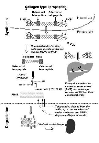
Fig. 5. ICTPlevels of individual subjects. (Fig. 7). The procollagen polypeptide
Key: ✧ = individual subjects; horizontal bar = median; long vertical bar = interquartile range. chains are produced by the fibroblasts
Ten subjects in each group: HC = healthy controls, PRP= primary Raynauds’s phenomenon; ARP =
and then secreted in the extracellular
autoimmune Raynaud’s phenomenon; lcSSc = limited cutaneous systemic sclerosis; dcSSc = diffuse
cutaneous systemic sclerosis. For explanation of p-values, see text. space where the N-terminal [PINP] and
C-terminal [PICP] propeptides are
cleaved by specific amino- and car-
boxy- proteases (Figure 7)(11). Mature
collagen molecules aggregate into sta-
ble, cross-linked collagen fibrils and
form the ECM. Col I is degraded in nor-
mal remodelling associated with physi-
ological processes such as morphogene-
sis, growth, wound healing and physio-
logical bone turnover (2,3). Also, cell –
cell and cell–matrix interactions in-
clude ECM proteolysis. There are four
major enzyme classes involved in Col I
degradation: aspartate, cysteine, serine
proteases and matrix metalloproteinases
(MMPs). Proteolytic activity of these
enzymes leads to the formation of vari-
ous degradation fragments including
several triple-helical peptides as well
Fig. 6. Correlation between ICTPconcentration ([ICTP]) and PINPconcentration ([PINP]) in the cur- as telopeptides, which are small amino-
rent study, shown as a log-log plot.
Notes: Pre-transform units: µg/l. Best fit straight line shown; correlation r2 = 0.175, p < 0.003. acid sequences originating from the
non-helical ends of collagen molecules.
Normal Col I production, deposition in
roderma sera. However, due to large ously. For educational purposes, we ECM and its degradation is regulated at
variability in the data from previous stu- discuss briefly the principles of colla- many levels. Abnormalities at any level
dies looking at collagen metabolites in gen metabolism, its alterations in SSc, may cause defective synthesis, accumu-
SSc, these molecules are not currently the significance and clinical application lation or degradation leading to fibrosis
recommended as standard laboratory of collagen pro- and telopeptide mea- (3,6,7).
markers of disease activity (26), des- surements. Subsequently we discuss
pite their obvious relevance to the bur- the results from the published clinical Collagen type I in scleroderma
den of fibrosis. With clear criteria for studies and compare them to our find- In fibrotic diseases there is a disturbed
descriptive studies in SSc we felt confi- ings. balance between the process of synthe-
dent in adding our own dataset using sis and degradation, with net gain in
optimal assays in very well defined dis- Type I collagen biosynthesis and collagen and fibrotic tissue (6-7). Scle-
tinctive disease groups, including “auto- metabolism roderma fibroblasts in vitro continue to
immune” Raynaud’s, not studied previ- Collagen type I (Col I) is one of the most synthesize increased amounts of colla-
363REVIEW Type I collagen burden in scleroderma / M. Dziadzio et al.
gen for several passages (4). Elevated
levels of Col I in scleroderma skin (5)
is primarily due to the increased rate of
collagen gene transcription, regulated by
various factors including PDGF, TGF-
beta and CTGF to name but a few (4, 6-
7). The transcriptional rate of genes en-
coding pro-alpha2(I) collagen is in-
creased in SSc fibroblasts, suggesting
alterations in transcription factors. Data
regarding the catabolism of collagen in
SSc are heterogeneous. Tissue inhibi-
tors of metalloproteinase 1 (TIMP-1)
may contribute to increased collagen
burden in SSc; in fact serum concentra-
tions of TIMP-1 were found raised in
SSc (8).
Type I collagen propeptides and telo-
peptides: assessment of the rate of
Col I synthesis and degradation
Procollagen type I contains N-terminal
and C-terminal propeptides (PINP and
PICP) which are cleaved off by specif-
ic proteases belonging to a family of
zinc-dependent metalloproteinases in a
stechiometric relationship with colla-
gen biosynthesis (2,11). PINP has a
molecular weight of 35 kDa and con-
tains three distinct structural domains:
a globular amino-terminal domain, a
central collagen-like domain and an-
other short-globular domain. PICP has
a molecular weight of about 100 kDa
and globular conformations without
any collagen-like domains. Both pro-
peptides contain cysteine and sugars, not
Fig. 7. Schematic representation of type I collagen synthesis and degradation.
found in type I collagen. The propep-
Key: PINP: N-terminal propeptide of type I collagen; PICP: C-terminal propeptide of type I collagen;
tides account for one-third of the bulk MMPs: matrix metalloproteinases; PYD: pyridinoline; DPD: deoxypyridinoline.
of the procollagen molecule. They pre-
vent premature fibril formation and help
to direct assembly of the protein into stant temporal or disease-specific cor- They are the most immunogenic parts
fibrils. After they are cleaved from the relation exists between these molecules of the type I collagen and play a crucial
molecule they also seem to play a role (3). Recently, extremely high PICPlev- role in fibrillogenesis. They are prima-
in the control of the amount of procol- els have been described as a familial ry sites of covalent cross-linking which
lagen synthesized by the cells (33). case and determined genetically as an stabilises the fibrils. N-terminal telo-
PINP is degraded via the scavenger re- apparent autosomal dominant condi- peptide is cleaved by cathepsin K (a
ceptors of liver endothelial cells, which tion, with no effect on survival and nor- cysteine protease) that is also involved
are not hormone–sensitive and there- mal concentrations of PINP and ICTP in generating a C-terminal telopeptide
fore the regulation of the clearance of (34). The assays (RIA or ELISA) use [ICTP]. MMPs are not implicated in
PINPis less dependent on different fac- polyclonal antibodies against the pro- the release of either N-terminal telo-
tors including sex, age and treatment(s) peptides for PINP and PICP and have peptide or ICTP (35). One molecule of
(33). In contrast, PICP is degraded in been widely used to assess the rate of telopeptide is composed, of either an
the liver through the mannose-6-phos- collagen synthesis (9-10). N- or a C-terminal fragment from the
phate receptors on endothelial cells. Telopeptides are small aminoacid se- alpha1 (I) chain, one helical segment
Despite the fact that PICPand PINPare quences originating from the nonheli- from the alpha1 or alpha2 chain of col-
derived from the same molecule, no con- cal ends of collagen molecules (10, 34). lagen and a cross-linker (pyridinoline
364Type I collagen burden in scleroderma / M. Dziadzio et al. REVIEW
(PYD) or deoxypyridinoline (DPD)). corticosteroids, commonly used in scler- The evidence for the connective tissue
N-terminal and C-terminal telopeptides oderma, decrease the rate of collagen constantly undergoing remodelling is
are small molecules (about 10 kDa) and synthesis (2,3,37-39). Also, collagen clearly demonstrated by the heteroge-
are released into the circulation after telopeptides are cleared by the kidneys neity of the findings in the literature.
the degradation of several mature col- (10, 36) but the presence or absence of Collagen turnover seems to be diverse
lagen molecules. They are eliminated SSc-renal involvement was not dis- among patients with scleroderma spec-
by the kidneys and tend to accumulate cussed in the series of patients in most trum disorders and is related to disease
in patients with renal failure (36). studies (Table I). Renal impairment is course, severity and many phenotypical
Old methods for evaluating collagen de- not rare in SSc: glomerular filtration features and possibly genetic factors
gradation include measurements of ur- rate was found reduced in about 60% of yet to be well characterised. The posi-
ine hydroxyproline and urine and ser- SSc patients and the prevalence of re- tive correlation between lung fibrosis
um cross-link analyses (PYD and DPD). nal crisis is about 10% in patients with and raised levels of collagen metabolite
Hydroxyproline estimation is limited SSc (40). Correlation patterns between concentrations has been suggested as a
by the fact that it is derived from any collagen metabolite levels and various reflection of lung involvement; similar-
collagen type; only 10% of hydroxypro- clinical parameters were very heteroge- ly, extent of skin involvement and
line is excreted in the urine and a pro- neous between the studies. Diffuse SSc raised collagen concentrations were sug-
portion of it derives from PINP, which was most commonly associated with gested as a reflection of disease activi-
contains a collagenous domain. Simi- high [PINP], [PICP] or [ICTP]. Discor- ty. In our view this interpretation is not
larly, assays of PYD and DPD cross- dant results were found for the correla- that straightforward: assessment of dis-
links are not specific for Col I and re- tion between peptide levels and disease ease activity is complex and should be
quire tedious HPLC analysis (10). About activity, organ involvement and the ex- based on more than a few variables, as
ten years ago new immunological as- tension of skin involvement (Table I). recently indicated (27).
says have been developed for the mea- Substantial heterogeneity, both among
surements of ICTP antigen (10). These different patients at presentation and Pitfalls of the clinical studies of
assays are superior to the previous ones, among diverse phases of the disease collagen metabolites in SSc
as they are Col I specific and, because course in the single patient, is a basis SSc is a rare disease (with a prevalence
the ICTP antigen is cross-linked, it is for these inter-individual variabilities; of 12.4/100,000 in the UK (41)) and it
known to derive from collagen fibrils. however, lack of standardization in the is not surprising that most published re-
evaluation of these epidemiological sults derived from small or very small
Measurements of collagen type I and clinical features of SSc patients in studies. Also, the presence of “publica-
metabolites in clinical studies in SSc the studies analysed here makes the tion bias” indicates cautious interpreta-
Several groups have measured the con- interpretation of these results difficult. tion of the results. The small number of
centrations of collagen metabolites in Efforts by the scientific community to patients studied did not permit an ana-
the serum, plasma, bronchoalveolar lav- identify a core set of clinical and labo- lysis with stratification by potential
age or blister fluid from SSc patients ratory variables for organ assessment confounding factor(s). Various studies
with heterogeneous results; correlation and for their inclusion in research stud- showed skewed data: stratification by
with disease subset, disease activity or ies should promote future production disease category has not eliminated the
organ involvement also varied between of high-quality data (26-27). heterogeneity of the results. Also, sta-
the studies (Table I). There was a wide The results of our study had a similar tistical subgroup analysis (based on the
range of values, especially in the dcSSc trend to the studies reviewed in this disease subset) lacks in precision and
groups and for high concentrations (Figs. work. Although [PINP] and [ICTP] were can be misleading, producing spurious
1-3). elevated in SSc, there was a large inter- results, which can be at risk of both type I
Also, there was no clear separation of individual variation in the values, espe- or type II errors. However, demonstra-
the values between healthy controls and cially in the dcSSc group, which also tion of no effect should come from
disease groups, with overlapping values overlapped with healthy controls (Figs. large-scale randomised trials.
in all studies. Although it is known that 4, 5). Interestingly, we have found posi-
radioimmunoassay loses its sensitivity tive correlation between [PINP] and Type III collagen
with low levels of radioactivity, which, the age of onset and the duration of RP; Type III collagen (2, 3) is not a subject
in the case of collagen metabolite mea- these findings should be confirmed in a of our analysis. Briefly, unlike Col I
surements using competitive assays, larger study. Collagen peptide levels metabolites, N-terminal type III colla-
corresponds with high concentrations, were not increased in patients with gen peptide (PIIINP) has been propos-
other factors can also play a role. Clini- ARP; lower levels of both [PINP] and ed by the Consensus conference (26) as
cal parameters such as skin score, dis- [ICTP] were found in the PRP group. one of the candidate non-organ based
ease subset, disease stage, number of Therefore it does not appear that colla- laboratory markers in SSc. Although
organ-based complications or treat- gen peptides can be used as predictive collagen type III has been considered
ment(s) might have accounted for the markers of progression to CTD in pa- more specific than type I collagen for
variability of the results. For example, tients with ARP. connective tissue turnover due to its
365REVIEW Type I collagen burden in scleroderma / M. Dziadzio et al.
absence from bone, it is not a specific enrolled into the clinical studies pro- ONEN SL: The aminoterminal propeptide of
marker for scleroderma (2). Recently, posed recently (27) should improve the type I procollagen: evaluation of a commer-
cial radioimmunoassay kit and values in
PIIINP has been used as an alternative quality of the results and enable pub- healthy subjects. Clin Biochem 1997; 30: 35-
to liver biopsy in the longitudinal mon- lished results to be combined for the 40.
itoring of patients with psoriasis treated purpose of integrating the findings. 13. LAMMI L, RYHANEN L, LAKARI E et al.:
Type III and type I procollagen markers in fi-
with methotrexate and therefore at risk We propose that longitudinal analysis
brosing alveolitis. Am J Respir Crit Care
of developing hepatic fibrosis (42). of collagen metabolites in individual Med 1999; 159: 818-23.
patients may show if these molecules 14. SCHEJA A, HELLMER G, WOLLHEIM A,
Collagen metabolites as serial can be of clinical use in the long-term AKESSON A: Carboxyterminal type I procol-
lagen peptide concentrations in systemic
markers of disease? follow-up of single SSc patients. Per- sclerosis: higher levels in early diffuse dis-
It seems plausible that serum collagen haps the use of Col I peptides will be ease. Br J Rheumatol 1993; 32: 59-62.
metabolites should be best used in seri- justified in the evaluation of the re- 15. ZACHARIAE H, BJERRING P, HEICKEN-
al investigations of individual patient sponse to the new therapeutic strategies DORFF L, MOLLER B, WALLEVIK K, ANGE-
LO H: Photophoresis in systemic sclerosis:
progression or response to treatment ra- which target fibroblasts in SSc. clinical and serological studies using markers
ther than as a diagnostic marker in ser- of collagen metabolism. Acta Derm Venereol
ies of patients. In diseases such as bone References (Stockh) 1993; 73: 356-361.
1. BLACK CM, DENTON CP: Systemic sclerosis 16. KIKUCHI K, SATO S, KADONO T, IHN H,
metastases [ICTP] (43), rheumatoid
and related disorders in adults and children. TAKEHARA K: Serum concentration of pro-
arthritis [ICTP] (44), multiple myelo- In MADDISON PG, ISENBERG DA, WOO P collagen type I carboxyterminal propeptide
ma [ICTP] (45), Paget’s disease [PINP] and GLASS DN (Eds): Oxford Textbook of in localized scleroderma. Arch Dermatol
(46) or osteoporosis [PINP] [ICTP] Rheumatology, 2nd ed., Oxford University 1994; 130: 1269-72.
Press, Inc. 1998; Vol. 2: 1217-47. 17. KIKUCHI K, IHN H, SATO S et al.: Serum
(33,37), the efficacy of specific thera- 2. PROCKOP DJ, KIVIRIKKO KI, TUDERMAN concentration of procollagen type I carboxy-
pies have been monitored by measur- L, GUZMAN NA: The biosynthesis of colla- terminal propeptide in systemic sclerosis.
ing their serum levels in individual pa- gen and its disorders. First of two parts. N Arch Dermatol Res 1994; 286: 77-80.
tients. Zachariae et al. (15) used PICP, Engl J Med 1979; 301: 13-23. 18. KIKUCHI K, KADONO T, FUJIMOTO M, IHN
3. PROCKOP DJ, KIVIRIKKO KI, TUDERMAN H, SATO S, TAKEHARA K: Elevated procol-
ICTP and PIIINP to assess longitudi- L, GUZMAN NA : The biosynthesis of colla- lagen type I carboxyterminal propeptide pro-
nally the response of SSc patients to gen and its disorders. Second of two parts. N duction in cultured scleroderma fibroblasts.
photophoresis; readings obtained every Engl J Med 1979; 301: 77-85. Dermatol 1995; 190: 104-8.
4 weeks showed large variability. We 4. LEROY EC: Increased collagen synthesis by 19. HEICKENDORFF L, ZACHARIAE H, BJER-
scleroderma fibroblasts in vitro. J Clin Invest RING P, HALKIER-SORENSEN L, SONDER-
might hypothesize that the assessment 1974; 54: 880-9. GAARD K : The use of serologic markers for
of serial measurements for single pa- 5. SCHARFFETTER K, LANKAT-BUTTGEREIT collagen synthesis and degradation in sys-
tients, possibly integrated as “area un- B, KRIEG T: Localization of collagen mRNA temic sclerosis. J Am Ac Dermatol 1995; 32:
der the curve” could correlate with cli- in normal and scleroderma skin by in situ hy- 584-8.
bridization. J Invest Dermatol 1988; 90: 48- 20. SONDERGAARD K, HECKENDORFF L, RIS-
nical variables such as disease activity 50. TELI L et al.: Increased levels of type I and
or severity scores. 6. GHOSH AK: Factors involved in the regula- III collagen and hyaluronan in scleroderma
tion of type I collagen gene expression: im- skin. Br J Dermatol 1997; 136: 47-53.
plication in fibrosis. Exp Biol Med 2002; 21. HUNZELMANN N, RISTELI J, RISTELI L et
Conclusions
227: 301-14. al.: Circulating type I collagen degradation
In summary, we have not found suffi- 7. DENTON CP, ABRAHAM DJ: Transforming products: a new serum marker for clinical
cient evidence that collagen type I pep- growth factor-beta and connective tissue severity in patients with scleroderma ? Br J
tides are good serological markers for growth factor: cytokines in scleroderma Dermatol 1998; 139: 1020-5.
pathogenesis. Curr Opin Rheumatol 2001; 22. VALAT C, DIOT P, DIOT E: Serum III pro-
the assessment of SSc activity or sever- 13: 506-11. collagen, but not serum I procollagen, is pre-
ity. Our study confirmed a large range 8. YOUNG-MIN SA, BEETON C, LAUGHTON R dictive of lung involvement in systemic scle-
of values for PINP, PICP and ICTP con- et al. : Serum TIMP-1, TIMP-2 and MMP-1 rosis. Clin Exp Rheumatol 1998; 16: 517-8.
centrations in a small well-character- in patients with systemic sclerosis, primary 23. DZIADZIO M, DENTON CP, SMITH R et al.:
Raynaud’s phenomenon, and in normal con- Losartan therapy for Raynaud’s phenomenon
ised SSc population studied. trols. Ann Rheum Dis 2001; 60: 846-51. and scleroderma: clinical and biochemical
The available literature on Col I pep- 9. RISTELI L, RISTELI J: Biochemical markers findings in a fifteen-week, randomized, par-
tides in SSc is represented by few rela- of bone metabolism. Ann Med 1993; 25: 385- allel-group, controlled trial. Arthritis Rheum
tively small studies often lacking con- 93. 1999; 42: 2646-55.
10. RISTELI J, ELOMAA I, NIEMI S, NOVAMO A, 24. SCHEJA A, WILDT M, WOLLHEIM A, AK-
trol groups and stratification for dis- RISTELI L: Radioimmunoassay for the pyri- ESSON A, SAXNE T : Circulating collagen
ease subset and showing large hetero- dinoline cross-linked carboxyterminal telo- metabolites in systemic sclerosis. Differen-
geneity in the results and low specifici- peptide of type I collagen. A new serum mar- ces between limited and diffuse form and re-
ty for collagen type I metabolites in SSc. ker of bone collagen degradation. Clin Chem lationship with pulmonary involvement.
1993; 39: 635-40. Rheumatology 2000; 39: 1110-3.
Reliable demonstration of “no effect” 11. PROCKOP DJ, SIERON AL, LI SW: Procolla- 25. ALLANORE Y, BORDERIE D, LEMARECHAL
should come from meta-analysis of gen N-proteinase and procollagen C-protein - H, CHERRUAU B, EKINDJIAN OG, KAHAN
large-scale randomised trials which is ase. Two unusual metalloproteinases that are A: Correlation of serum collagen I carboxy-
essential for procollagen processing probably terminal telopeptide concentrations with cut-
plausibly difficult to achieve in the
have important role in development and cell aneous and pulmonary involvement in sys-
field of rare diseases such as SSc. Stan- signalling. Matrix Biol 1998; 16: 399-408. temic sclerosis. J Rheumatol 2003; 30: 68-
dardised evaluation of SSc patients 12. TAHTELAR, TURPEINEN M, SORVA R, KAR- 73.
366Type I collagen burden in scleroderma / M. Dziadzio et al. REVIEW
26. M CHUGH NJ, DISTLER O, GIACOMELLI R, SEN SB, CHRISTIANSEN C: Diurnal varia- adults. Scand J Clin Lab Invest 1997; 57:
RIEMEKASTEN G: Non-organ based labora- tion in type I collagen synthesis and degrada- 133-40.
tory markers in systemic sclerosis. Clin Exp tion in healthy premenopausal women. J 40. KINGDON EJ, KNIGHT CJ, DUSTAN K et
Rheumatol 2003; 21 (Suppl. 29): S32-S38. Bone Miner Res 1992; 7: 1307-11. al.: Calculated glomerular filtration rate is a
27. VALENTINI G, MEDSGER TAJ R, SILMAN AJ, 34. SORVA A, TAHTELA R, RISTELI J et al.: useful screening tool to identify scleroderma
BOMBARDIERI S: Conclusion and identifi- Familial high serum concentrations of the patients with renal impairment. Rheumatol -
cation of the core set of variables to be used carboxy-terminal propeptide of type I procol- ogy (Oxford) 2003; 42: 26-33.
in clinical investigations. Clin Exp Rheuma - lagen. Clin Chem 1994; 40: 1591-3. 41. HOPKINSON ND, BURNS AM: The frequen-
tol 2003; 21 (Suppl. 29): S47-56. 35. VITAGLIANO L, NEMETHY G, ZAGARI A, cy of progressive systemic sclerosis in a non-
28. VALENTINI G, DELLA ROSSA S, BOM- SCHERAGA HA: Structure of the type I colla- industrialised, semi-rural population in South-
BARDIERI S et al.: European multicentre gen molecule based on conformational ener- ern England. Rheumatology 2000; 39S: 253.
study to define disease activity criteria for gy computations: the triple-stranded helix 42. ZACHARIAE H, HEICKENDORFF L, SO-
systemic sclerosis. Identification of disease and the N-terminal telopeptide. J Mol Biol GAARD H: The value of amino-terminal pro-
activity variables and the development of 1995; 247: 69-80. peptide of type III procollagen in routine
preliminary activity indexes. Ann Rheum Dis 36. URENA P, D E VERNEJOUL M : Circulating screening for methotrexate-induced liver fi-
2001;60:592-8. biochemical markers of bone remodelling in brosis: a 10-year follow-up. Br J Dermatol
29. APPENDIX: Manual of signs, symptoms, meth- uremic patients. Kidney Int 1999; 55: 2141- 2001; 144: 100-3.
ods and procedures for the assessment of the 56. 43. KOIZUMI M, OGATAE : Bone metabolic mar-
patient with SSc. Clin Exp Rheumatol 2003; 37. HASLING C, ERIKSEN EF, MELKKO J, RIS- kers as gauges of metastasis to bone: a re-
21 (Suppl. 29): S49-56. TELI L, CHARLES P, MOSEKILDE L: Effects view. Ann Nucl Med 2002; 16: 161-8.
30. SUBCOMMITEE FOR S CLERODERMA CRITERIAOF of a combined estrogen-gestagen regimen on 44. HAKALAM, RISTELI L, MANELIUS J, NIEMI-
THE A MERICAN RHEUMATISM ASSOCIATION serum levels of carboxy-terminal propeptide NEN P, RISTELI J: Increased type I collagen
DIAGNOSTICAND THERAPEUTIC C RITERIA COM- of human type I procollagen in osteoporosis. degradation correlates with disease severity
MITTEE: Preliminary criteria for the classifi- J Bone Miner Res 1991; 6: 1295-300. in rheumatoid arthritis. Ann Rheum Dis 1993;
cation of systemic sclerosis (scleroderma). 38. AUTIO P, RISTELI J, KIISTALA U, RISTELI 52: 866-9.
Arthritis Rheum 1980; 23: 581-90. L, KARVONEN J, OIKARINEN A: Serum 45. ELOMAA I, VIRKKUNEN P, RISTELI L, RIS-
31. LEROY EC, BLACK CM, FLEISHMAJER R et markers of collagen synthesis and degrada- TELI J : Serum concentration of the cross-
al.: Scleroderma (systemic sclerosis): classi- tion in skin diseases. Altered levels in dis- linked carboxyterminal telopeptide of type I
fication, subsets and pathogenesis. J eases with systemic manifestation and during collagen [ICTP] is a useful prognostic indica-
Rheumatol 1988; 15: 202-5. systemic glucocorticoid treatment. Arch Der - tor in multiple myeloma. Br J Cancer 1992;
32. MARICQ HR : Wide-field capillary micro- matol Res 1993; 285: 322-7. 66: 337-41.
scopy. Technique and rating scale for abnor- 39. WOLTHERS OD, HEUCK C, HANSEN M, 46. PONS F, ALVAREZ L, PERIS P et al.: Quan-
malities seen in scleroderma and related dis- KOLLERUP G: Serum and urinary markers of titative evaluation of bone scintigraphy in the
orders. Arthritis Rheum 1981; 24: 1159-65. types I and III collagen turnover during assessment of Paget’s disease activity. Nucl
33. HASSAGER C, RISTELI J, RISTELI L, JEN- short-term prednisolone treatment in healthy Med Commun 1999; 20: 525-8.
367You can also read