Use of RNA sequencing to detect abnormal transcription of the collagen α 2 (VI) chain gene that can lead to Bethlem myopathy - Spandidos Publications
←
→
Page content transcription
If your browser does not render page correctly, please read the page content below
INTERNATIONAL JOURNAL OF MOlecular medicine 47: 28, 2021

                      Use of RNA‑sequencing to detect abnormal
                   transcription of the collagen α‑2 (VI) chain gene
                          that can lead to Bethlem myopathy
       JINGZI ZHONG1, YANSHU XIE1, YIWU DANG2, JIAPENG ZHANG1, YINGRU SONG3 and DAN LAN1

                                     Departments of 1Pediatrics, 2Pathology and 3Radiology,
               The First Affiliated Hospital of Guangxi Medical University, Nanning, Guangxi 530021, P.R. China

                                      Received March 15, 2020; Accepted November 27, 2020

                                                     DOI: 10.3892/ijmm.2021.4861

Abstract. Bethlem myopathy (BM) is an autosomal dominant               collagen protein within cultured fibroblasts was detected
or autosomal recessive disorder and is usually associated with         using immunocytochemistry. The mutation c.736‑1G>C in the
mutations in the collagen VI genes. In the present study, the          COL6A2 gene caused aberrant splicing and led to premature
pathogenicity of a novel splice‑site mutation was explored             termination of protein translation. In conclusion, these findings
using RNA‑sequencing in a family with suspected BM, and                may improve our knowledge of mutations of the COL6A2 gene
a myopathy panel was performed in the proband. The genetic             associated with BM and demonstrated that RNA‑sequencing
status of all family members was confirmed using Sanger                can be a powerful tool for finding the underlying mechanism
sequencing. Clinical data and magnetic resonance imaging               of a disease‑causing mutations at a splice site.
(MRI) features were also documented. In silico analysis
was performed to predict the effects of the splice mutation.           Introduction
RNA‑sequencing and reverse transcription (RT)‑PCR were
used to assess aberrant splicing. Immunocytochemistry was              Collagen VI is an important component of the extracellular
conducted to measure collagen VI protein levels within the             matrix (ECM). It forms a microfibrillar network that is usually
gastrocnemius and in cultured skin fibroblasts. The results            found to be in close association with the cell and surrounding
revealed that three patients in the family shared a similar            basement membrane. Collagen VI is also found in the inter‑
classic BM presentation. MRI revealed distinct patterns of fatty       stitial space of numerous tissues, including muscles, tendons,
infiltration in the lower extremities. A novel splicing mutation       skin, cartilage and intervertebral discs (1). Collagen VI is
c.736‑1G>C in the collagen α‑2 (VI) chain (COL6A2) gene                encoded by six different genes, namely collagen α‑1 (VI)
was found in all three patients. In silico analysis predicted          chain (COL6A1), COL6A2, COL6A3, COL6A4, COL6A5 and
that the mutation would destroy the normal splice acceptor             COL6A6. Collagen VI‑related myopathy (COL6‑RD) is caused
site. RNA‑sequencing detected two abnormal splicing vari‑              by pathogenic variants in three collagen VI genes: COL6A1,
ants adjacent to the mutation site, and RT‑PCR confirmed               COL6A2 and COL6A3. These mutations can result in two
the RNA‑sequencing findings. Furthermore, a defect in the              main types of muscle disorders that can range from severe
                                                                       forms of Ullrich congenital muscular dystrophy (UCMD) to
                                                                       milder forms of Bethlem myopathy (BM) (2).
                                                                            A child with typical UCMD cannot walk independently, or
Correspondence to: Professor Dan Lan, Department of Pediatrics,        can only do so for short periods of time. These patients also
The First Affiliated Hospital of Guangxi Medical University,           present with changes to their skin and joints, including vari‑
6 Shangyong Road, Nanning, Guangxi 530021, P.R. China                  able proximal contractures and distal hyperlaxity, congenital
E‑mail: land6785@163.com                                               hip dislocations, follicular hyperkeratosis over some of the
                                                                       extensor surfaces of the limbs and soft velvety skin on their
Abbreviations: BM, Bethlem myopathy; COL6‑RD, collagen                 palms and soles of their hands and feet. In addition, there
type VI‑related dystrophies; ECM, extracellular matrix; MRI,
                                                                       is usually abnormal wound healing, which can result in the
magnetic resonance imaging; NMD, non‑sense mediated decay;
PolyPhen‑2, Polymorphism phenotyping version 2; RT‑PCR,
                                                                       formation of keloid scars. BM is a relatively mild disorder,
reverse transcription‑polymerase chain reaction; THD, triple‑helical   and can be characterized by some proximal muscle weakness
domains; UCMD, Ullrich congenital muscular dystrophy                   and contractures, with the onset of these symptoms occur‑
                                                                       ring within the first two decades of life (2). These two major
Key words: Bethlem myopathy, collagen α‑2 (VI) chain, abnormal         phenotypes are not distinct and an intermediate phenotype,
transcription, splice‑site mutation, RNA‑sequencing                    described as mild UCMD with severe BM, has been defined
                                                                       by number of previous studies (2,3). BM is transmitted as
                                                                       an autosomal dominant or autosomal recessive inherited
                                                                       disorder and is usually associated with mutations in the COL6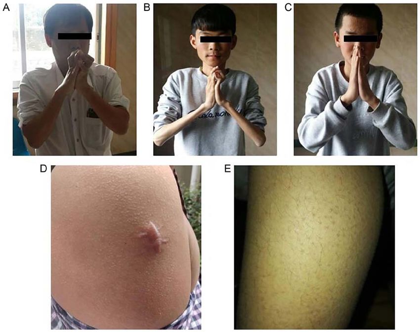
2                                  ZHONG et al: ABNORMAL COL6A2 GENE IN BETHLEM MYOPATHY

genes (4,5). The prevalence of BM in the northern region of            Routine periodic acid‑Schiff (PAS) staining was performed on
England in 2007 was 0.77/10,000 (6).                                   muscle cryostat sections to investigate the presence of normal
    The distribution of the mutations in the COL6 genes is             and abnormal glycogen levels.
somewhat uniform, and lacks mutation hot spots. The most
common mutations in COL6‑related myopathy are splicing                 Biological materials. All the biological materials, including
and missense mutations (3,7). As a consequence of this notable         peripheral blood samples obtained from the cubital vein,
heterogeneity at the clinical and molecular levels in the COL6         skin biopsies, muscle biopsies as well as fibroblasts for cell
genes, it appears to be difficult to establish a correlation between   culture, were collected after the appropriate informed consent
the phenotype and the genotype of this disease. In a previous          was obtained. The use of all appropriate human tissues was
study, dominant splice‑site mutations that cause exon skipping         approved by The Medical Ethics Committee of the First
were observed in cases classed as moderate to progressive (8).         Affiliated Hospital of Guangxi Medical University (Nanning,
    Two common splice sites, the canonical splice donor GT             China; approval no. 2017‑KY‑E‑154) and the experimental
and acceptor AG, are present in 98.71% of all mammalian                protocols were performed in accordance with the institutional
genes (9). Mutations in these canonical dinucleotides always           guidelines and regulations. All nine family members donated
result in splicing errors when pre‑mRNA transcripts are trans‑         venous blood samples Genomic DNA was extracted with
formed into mature mRNAs (10). It was previously reported              Qiagen FlexiGene DNA kit (cat. no. 51206; Qiagen China
that sequence variations surrounding certain canonical splice          Co., Ltd.) according to the manufacturer's protocol. A muscle
sites may not result in splicing errors because these exon and         biopsy was obtained from the proband and skin biopsies were
intron elements are only weakly conserved, and their altera‑           obtained from the inner side of the thigh in the proband and
tions do not always disrupt processes related to splicing (11).        a son. Normal skin and muscle tissues were collected from
Therefore, it is essential to determine the presence of aber‑          two male patients (median age 24 years) receiving skin or
rant mRNAs in order to predict the clinical phenotypes of              muscle resections for non‑muscular disease‑related reasons,
diseases. RNA‑sequencing uses high‑throughput sequencing               including circumcisions. Fibroblasts were cultured from the
technology to rapidly and comprehensively obtain the tran‑             skin biopsies and were used as primary cultures.
scriptional status of biological samples at a specific time (12).
In the present study, RNA‑sequencing was successfully used             DNA analysis. To systematically search for the disease‑causing
to analyze the influence of splice mutations.                          genes, a myopathy panel based on next‑generation sequencing
    The current study reported three patients from a family who        was performed on the proband. The myopathy panel (Table SI)
presented with typical clinical features of BM. A novel hetero‑        was a kit designed by the Zhongguancun Huakang Gene
zygous COL6A2 mutation (c.736‑1G>C) was identified in this             Institute (http://www.kangso.net/) and produced using the
family. The mechanism of this mutation has not been previ‑             Agilent SureSelect Target Enrichment technique (13).
ously reported in BM, to the best of our knowledge. Adiagnosis             High‑throughput single‑end sequencing was performed on
of BM was made by observation of the clinical characteristics          an Illumina NextSeq500 platform (Illumina, Inc.). In order to
and MRI features of the patients. Bioinformatics, in silico            construct the exome library, 4 µg genomic DNA from periph‑
analysis, RNA‑sequencing, reverse transcription (RT)‑PCR               eral blood samples of each patient was sheared into fragments
and immunocytochemistry were performed to confirm the                  of 150‑250 bp in length by sonication (30 sec on/30 sec off,
pathogenicity and mechanism of the mutation. The present               0˚C for 5 min) and subsequently hybridized for enrichment,
findings may help improve the accurate molecular diagnosis             using the manufacturer's standard protocol (SureSelect
of the disorder, and illustrated a previously unrecognized             Target Enrichment System Target Sequence Enrichment
category of BM.                                                        kit, cat. no. 5190‑5931; Agilent). Indexing primers with 8 bp
                                                                       index A01‑H12 were used to amplify the captured library,
Materials and methods                                                  and PCR was performed to amplify the SureSelect‑enriched
                                                                       DNA library. The amplified captured library was puri‑
Patients and their families. This study was from January 2015          fied using AMPure XP magnetic beads. To evaluate the
to August 2018. The clinical data of the affected father               DNA quality and quantity, it was necessary to use the 2100
(proband) and two sons were collected at The First Affiliated          Bioanalyzer instrument (Agilent) and a High Sensitivity DNA
Hospital of Guangxi Medical University (Nanning, China).               kit (Agilent) according to the manufacturer's protocols. The
Three male patients age ranged from 40 to 14 years. The                final concentration of the final library was >1.52x10‑6 mmol/l.
median age was 24 years. The family had nine members, the              After obtaining the human reference genome from the
parents, two sisters and a brother of the proband, his wife and        University of California, Santa Cruz database (build 37.1,
two sons. Information such as symptoms at onset and creatine           version hg19, http://genome.ucsc.edu/), sequence alignment
kinase levels were determined by means of a commercially               was performed using the Burrows‑Wheeler Alignment
available Creatine Kinase reagent kit (cat. no. 23‑666‑208;            tool (14). Picard (http://sourceforge.net/projects/picard/) was
Thermo Fisher Scientific, Inc.) via rate‑assay spectrophotom‑          then employed to mark duplicates resulting from PCR ampli‑
etry according to the manufacturer's instructions, as well as          fication. All candidate mutations were subsequently filtered
ultrasonic cardiogram results, were obtained. Muscle MRI was           against several programs, including the single nucleotide
performed using the thighs of the three patients and a normal          polymorphism database (http://www.ncbi.nlm.nih.gov/proj‑
control (the grandfather). The MRI results were assessed by            ects/SNP/snp_summary.cgi), the international HapMap Project
two independent and experienced radiologists and a neurolo‑            (http://hapmap.ncbi.nlm.nih.gov/) and the 1,000 genomes
gist. The right gastrocnemius was biopsied in the father.              project (2012 April release, http://www.1000genomes.org/) in
INTERNATIONAL JOURNAL OF MOlecular medicine 47: 28, 2021 3
order to remove loci polymorphisms. Polymorphism pheno‑ Skin fibroblast cultures. To extract total RNA for transcript
typing version 2 (PolyPhen‑2) was used for sorting intolerant analysis, skin biopsies from two patients (proband and his
from tolerant and mutation tasters to predict whether an amino elder son) and normal controls were immersed into Dulbecco's
acid substitution could affect the functions of the protein. Next, modified Eagle's medium (cat. no. 22390; Gibco; Thermo Fisher
Sanger sequencing was used to validate whether the identified Scientific, Inc.) supplemented with 20% heat‑inactivated fetal
potential disease‑causing variant occurred in the individual bovine serum (cat. no. A11‑043; PAA Laboratories GmbH; GE
family members (the proband's parents, brother, two sisters, Healthcare) and penicillin‑streptomycin (cat. no. 15070‑063;
two sons and his wife). Gibco; Thermo Fisher Scientific, Inc.). Fibroblasts from skin
biopsies were cultured in Dulbecco's modified Eagle's medium
In silico analysis. To evaluate the probability of splice site containing 20% fetal bovine serum (Sigma‑Aldrich; Merck
mutations being able to disrupt the normal splicing of COL6A2, KGaA) and penicillin‑streptomycin (Sigma‑Aldrich; Merck
two online bioinformatics tools were used for prediction KGaA) at 37˚C in an atmosphere of 95% air and 5% CO2 (15).
studies: Human Splicer Finder (http://www.umd.be/HSF/)
and Alternative Splice Site Predictor (http://wangcomputing. Transcript analysis. In order to validate the results of
com/assp/index.html). ExPASy (https://www.expasy.org/)was RNA‑sequencing analysis and the effect of the splice site muta‑
used to predict their impact on the protein that they would tion, transcript analysis was performed using RT‑PCR with
produce. RNA from fibroblasts derived from two patients (proband and
his elder son) and two healthy individuals as controls. TRIzol®
RNA‑sequencing analysis. The effect of the splicing mutation (0.5 ml; Invitrogen; Thermo Fisher Scientific, Inc.) was
on COL6A2 mRNA was determined by RNA‑sequencing used to extract total RNA using the standard manufacturer's
analysis. Tissue from the muscle of the proband and a instructions.
non‑muscle disease control were selected to extract total RNA In order to reveal the COL6A2 cryptic splice acceptor,
for RNA‑sequencing analysis. Construction of the sequencing RT‑PCR was performed with a forward primer within the 3' of
libraries followed the standard protocols of the TruSeq™ RNA exon 3 and a reverse primer within exon 5. The primer pairs were
Sample Preparation guide (Illumina, Inc.). TruSeq® Stranded as follows: Forward, 5'‑CTCTACCGCAACGACTACGC‑3' and
mRNA Library Prep kit (Illumina, Inc.) was used to prepare reverse, 5'‑TTCCAGGCAGCTCACCTT‑3'. RT‑PCR, revealing
the sequencing libraries. The poly‑A containing mRNA mole‑ COL6A2 exon skipping, was performed using a forward primer
cules are purified from total RNA using poly‑T oligo‑attached within exon 3 and a reverse primer within exon 6. The primer
magnetic beads. During the elution of the poly‑A RNA, the pairs were as follows: Forward, 5'‑CCCA ACCAGA ACCTG
RNA is also fragmented to 120‑200‑bp and primed for cDNA AAGGA‑3' and reverse, 5'‑TCGGCTCCAAATTCACCCT‑3'.
synthesis. First‑strand cDNA was then synthesized with PCR was conducted with Premix Taq™ DNA Polymerase
SuperScript II Reverse Transcriptase (Invitrogen; Thermo (Takara Biotechnology Co., Ltd.) under the following conditions:
Fisher Scientific, Inc.) using random primers. The temperature Initial denaturation at 95˚C for 4 min, followed by 36 cycles
protocol was follows: 25˚C For 10 min, 42˚C for 15 min and at 95˚C for 30 sec, 56˚C for 30 sec and 72˚C for 40 sec. PCR
70˚C for 15 min. AMPureXP beads (Beckman Coulter, Inc.) products were separated on 2% agarose gels and extracted using
were then used to isolate double‑stranded cDNA, which was a gel purification kit (Favorgen Biotech Corp.) according to the
synthesized using Second Strand Master mix (Invitrogen; manufacturer's protocols. The eluted products were sequenced
Thermo Fisher Scientific, Inc.) according to the manufacturer's using an ABI 3730 XL Genetic Analyser with BigDye™
instructions. The adapters were then ligated to the A‑tailing Terminator Cycle Sequencing kit (Applied Biosystems; Thermo
fragment. Subsequently, 12 cycles of PCR were performed to Fisher Scientific, Inc.). The COL6A2 sequence of the patients
enrich those DNA fragments that had adapter molecules at was compared with that observed in the control and reference
both ends following the program: 98˚C For 30 sec; 15 cycles sequences of COL6A2.
of 98˚C for 10 sec, 60˚C for 30 sec, 72˚C for 30 sec and 72˚C
for 5 min. This technique also amplified the quantity of DNA Immunohistochemistry of muscle biopsies and immunocyto‑
in the library. Purified libraries were further quantified using chemistry of skin fibroblasts. The morphology was observed
a Qubit2.0 fluorometer (Invitrogen; Thermo Fisher Scientific, under a microscope via hematoxylin and eosin (H&E) staining.
Inc.) and then validated using an Agilent 2100 bioanalyzer Fresh specimens were fixed in 10% neutral buffered formalin
(Agilent Technologies, Inc.). Clusters were then generated and further processed into paraffin‑embedded blocks,
using cBot with the libraries, which were further diluted to sectioned at 6 ‑µm thickness, dehydrated in graded ethanol
10 pM and were subsequently sequenced using the Illumina (70, 80, 90, 95 and 100%) solutions and processed according
HiSeq x Ten system (Illumina, Inc.) for 150 cycles. Shanghai to the H&E staining protocol (16). The stained tissues were
Sangong Biotech Corporation (https://www.sangon.com/) was observed using a biomicroscopy imaging system microscope
employed to construct the libraries, and Illumina sequencing (cat. no. DP73; Olympus Corporation). Samples derived from
was performed for the high‑quality reads, which were kept skin fibroblast cultures and muscle biopsies were subsequently
for sequence analysis after passing through Illumina quality prepared for immunohistochemistry to evaluate collagen VI
filters. The sequencing data sets are stored at http://bioinfor‑ protein expression. Briefly, sections were incubated at 4˚C
matics.jnu.edu.cn/software/sequencing_datasets/ and Gene overnight with the primary antibody which was a monoclonal
Expression Omnibus database (accession number, GSE42006) antibody raised in rabbits (clone #EPR17072) that targeted
for reference. Integrative Genomics Viewer (version 2.8.0) was collagen VI (cat. no. ab182744; 1:100; Abcam), and then
used to display the genomic data. incubated at room temperature for 30 min with SupervisionTM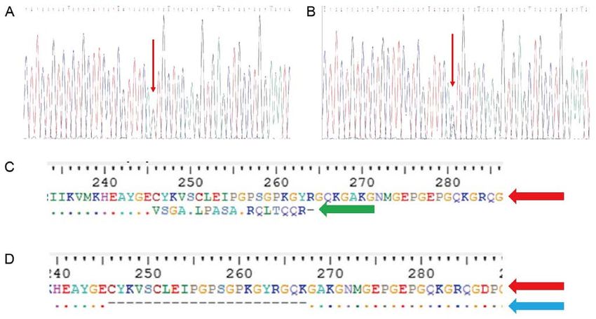
4 ZHONG et al: ABNORMAL COL6A2 GENE IN BETHLEM MYOPATHY
Universal (Anti‑Mouse/Rabbit) Detection reagent (HRP)
(cat. no. D‑3004; Thermo Fisher Scientific, Inc.) conjugated
to peroxidase in Tris‑HCI buffer containing carrier protein
and anti‑microbial agent. The experiment was performed
according to the manufacturer's protocol.
Results
Three patients in a family display typical features of BM. The
three patients in this study were from three generations of a
Chinese family (Fig. 1). The proband (II‑4) was 40 years old.
From 4 years of age, he experienced difficulty getting up from Figure 1. Pedigree of the family with Bethlem myopathy.
the floor and climbing stairs; at 8 years of age, he began toe
walking and needed to use a rail to climb stairs. He underwent
Achilles tendon release aged 8 years. He had mild weakness sons (Fig. 5A), while the spouse (II‑5), proband's siblings (II‑1,
in the shoulder girdle and upper limb muscles, and moderate 2 and 3), father (I‑1) and mother (I‑2) were normal (Fig. 5B).
weakness in the axial and hip girdle muscles. At the age of 40, The members who had the mutation in the COL6A2 gene had
he was able to walk unassisted but with a marked Trendelen a similar phenotype, while those without this mutation did not
burg gait. Physical examination revealed proximal muscle have muscle weakness or motor disability. This splice acceptor
weakness and amyotrophy. Joint contractures of the shoulders, site mutation observed in intron 4 was co‑segregated within
hips, knees and feet were observed (Fig. 2A). Skin examina‑ the family.
tion revealed that he had keratosis pilaris along the extensor
surfaces of his arms and legs. Splicing mutations can result in aberrant splicing
Two sons (III‑1 and III‑2) of the proband shared similar In silico analysis. In order to determine the effects of the
clinical symptoms. The father was the most severe case, the mutation on the splicing of COL6A2 mRNA, in silico and
younger brother the mildest. Examination in our hospital in vitro analyses, which involved RNA‑sequencing analysis
occurred when the older boy was 18 years old. He could not and RT‑PCR, was performed.
get up from the floor. The younger 14‑year‑old brother experi‑ Human Splicer Finder and Alternative Splice Site Predictor,
enced difficulty when getting up from the floor and climbing which are two freely available online bioinformatics tools,
stairs, and he walked on his toes (Table I). Both underwent were used to predict the effect of the c.736‑1G>C variation
Achilles tendon release 10 years of age. on COL6A2 mRNA splicing. Human Splicer Finder shown
To further demonstrate the patients' pattern of muscle that wild‑type sequencing existed a normal splice acceptor
involvement and to determine the potential relevance of these sites of 3' end; while the normal acceptor sites disappeared
unknown genetic variants, muscle MRI for the lower extremi‑ and replaced by a new one in the mutant‑type. The same result
ties was recommended. This revealed a distinct pattern of fatty was found by Alternative Splice Site Predictor. Wild‑type
infiltration in both lower extremities, and the anterior group sequencing existed a splice acceptor sites of 3' end; while the
muscles were more impaired than the rear groups in the thigh acceptor sites disappeared in the mutant‑type. These tools
(Fig. 3A‑C). This is consistent with a pattern of symptoms suggested that this mutation would probably result in the elim‑
termed ‘central shadow’ in the rectus femoris and a pattern ination of any wild‑type splice acceptor sites that would create
further described as ‘outside‑in’ in the vastus lateralis muscle. a new cryptic splice acceptor site. Thus, a 129 bp sequence at
Fatty infiltration of the thigh muscles varied greatly among the 3' end of intron 4 was retained (data not shown).
the three patients. The father (Fig. 3A) was the most severe
case, while the youngest son (Fig. 3C) exhibited the mildest Prediction of influence on the protein. To understand the influ‑
symptoms. ence of the two abnormal splicing variants, ExPASy was used
H&E staining of the gastrocnemius muscle of the proband to predict their impact on the protein that they would produce.
showed mild dystrophic features, such as muscle degeneration The new cryptic splice acceptor site obtained suggested that
and necrotic fibers. In addition, connective tissue hyperplasia the loss of the wild‑type splice site could result in a frame
was observed in the muscle fibers and these were unequal in size shift in amino acid 246, which would eventually lead to a
and had certain structural distortion (Fig. 4A). Abnormalities premature stop codon in amino acid 264 (p.Cys246fs264*) of
of glycogen deposition were not observed when tissue sections the protein. The skipping of exon 5 appeared to result in the
of muscle were stained with PAS staining (Fig. 4B). deletion of 21 amino acids, namely from 246 to 267, in the
protein (p.Cys246_Lys267del) (Fig. 5C and D). The truncated
Identification of a heterozygous splicing mutation in the protein lacked the triple‑helical domain (THD) region.
COL6A2 gene. To make a genetic diagnosis, 2.84 million
75‑bp single‑end sequence reads were generated from the RNA‑sequencing analysis. Integrative Genomics Viewer was
proband. Of these, 2.83 million reads (99.93%) aligned to used to display the genomic data. In addition to the normal
the human reference genome. Of the 169 selected genes base sequence, two abnormal splicing variants adjacent to the
(Table SI), a heterozygous splicing mutation (c.736‑1G>C) in mutation site were identified in the COL6A2 gene. One variant
the COL6A2 gene was identified in the proband and Sanger had a 141‑bp sequence retained at the 3' end of intron 4, causing
sequencing validated that the same mutation existed in the two a new cryptic splice acceptor site. Another variant was due to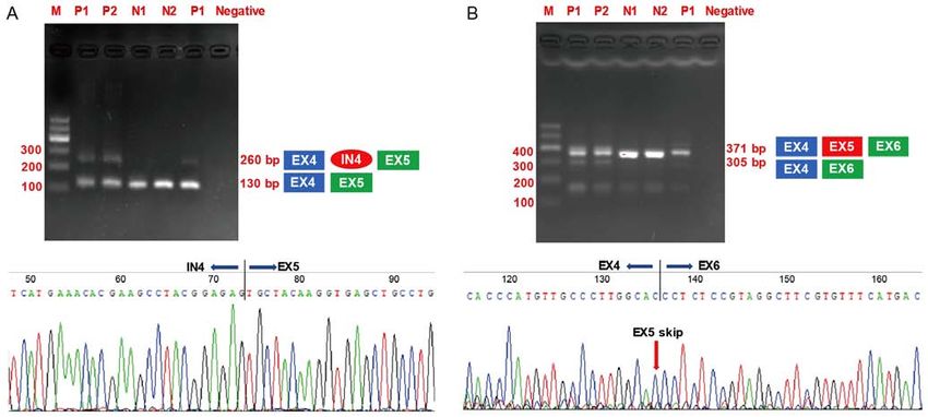
INTERNATIONAL JOURNAL OF MOlecular medicine 47: 28, 2021 5
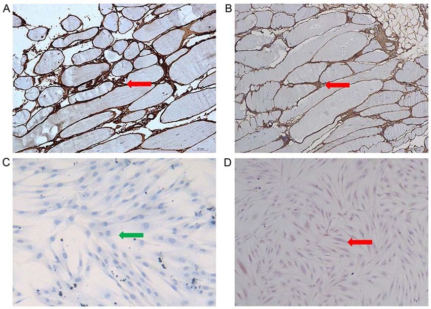
Figure 2. Typical clinical features of the family with BM. Typical finger flexor contractures in (A) II‑4 and (B) III‑1 with BM made it almost impossible to
put the fingers close together with the elbows extended. (C) The younger son could still put his fingers close together. Three patients shared similar clinical
symptoms. (D) Skin examination revealed scar and (E) and keratosis pilaris in proband. BM, Bethlem myopathy.
exon 5 skipping. The percentage of presence of the normal, 3' RT‑PCR products were subjected to gel extraction and Sanger
end of intron 4 retention and exon 5 skipping variants was 50, sequencing, which showed that a 129‑bp sequence from the 3'
36 and 14%, respectively (Table II). The coverage (reads) of end of intron 4 was retained between exon 4 and exon 5. This
the patient was lower than compared with that of the control. result was consistent with the in silico analysis. RT‑PCR ampli‑
Differential gene expression analysis revealed that the expres‑ fication of exon 3 to exon 6 also produced a 371‑bp product
sion of COL6A2 in the patient was ~5‑fold higher compared when using cDNA samples from either the control individuals
with that of the normal control. or the patients as templates. However, a smaller product of
305 bp was only detected in the patients' samples (Fig. 6B).
RT‑PCR. To validate the results demonstrated by Gel extraction and Sanger sequencing also revealed that exon 4
RNA‑sequencing analysis, RNA derived from cultured skin directly spliced the in‑frame to exon 6 and hence skiped exon 5.
fibroblasts from the proband and his elder son, and two healthy
controls were used to perform RT‑PCR experiments. The Collagen VI is absent in fibroblasts but is expressed normally
difference between the wild‑type transcripts and the mutant in muscle. To further investigate the consequences of the
transcripts in patients was that the wild‑type transcripts were c.736‑1G>C mutation on the protein product, immunohisto‑
longer (which was caused by a cryptic splice acceptor site chemical analysis was performed using a rabbit monoclonal
activation) or shorter (which was caused by exon skipping) antibody against collagen VI (cat. no. EPR17072). Collagen VI
compared with the mutant transcripts when identical primers immunohistochemistry indicated that this protein was absent
were used to amplify the PCR products that flanked the region in the skin fibroblasts derived from patients but showed a
of mutation. Therefore, this method can serve as a practical normal pattern in the fibroblasts derived from the controls.
tool to distinguish the two transcripts by when the samples In contrast, a normal pattern was found in the patient muscle
are subjected to gel electrophoresis. The amplification of fibers, attesting to the integrity of the basement membrane
exon 3 to exon 5 in COL6A2 generated a 130‑bp product for (Fig. 7C and D). Fig. 7C and D do not have the same back‑
all the cDNA samples from the controls and patients. Notably, ground with stain. The difference between the two figures
an additional larger product of 260 bp was only detected in may be due to non‑specific factors, for example the degree of
the cDNA samples obtained from the patients (Fig. 6A). The pathological staining.6 ZHONG et al: ABNORMAL COL6A2 GENE IN BETHLEM MYOPATHY
Table I. Clinical data and biochemical results of three patients in the family.
Patient
‑‑‑‑‑‑‑‑‑‑‑‑‑‑‑‑‑‑‑‑‑‑‑‑‑‑‑‑‑‑‑‑‑‑‑‑‑‑‑‑‑‑‑‑‑‑‑‑‑‑‑‑‑‑‑‑‑‑‑‑‑‑‑‑‑‑‑‑‑‑‑‑‑‑‑‑‑‑‑‑‑‑‑‑‑‑‑‑‑‑‑‑‑‑‑‑‑‑‑‑‑‑‑‑‑‑‑‑‑‑‑‑‑‑‑‑‑‑‑‑‑‑‑‑‑‑‑‑‑‑‑
Variable II:4 III:1 III:2
Age, years 40 18 14
Approximate age at which muscle weakness 4 4 5
was diagnosed, years
Capable of climbing stairs No No No
Capable of walking 100 m Yes Yes Yes
Capable of jumping up and down for 1 min No No No
Capable of standing for 10 min No No No
Skin appearance Some scarring Keratosis pilaris Normal
Rigidity of spine No No No
Plasma creatine kinase levels, U/l 576 747 759
Ultrasonic cardiogram Normal Normal Normal
Figure 3. Muscle MRI with axial cuts performed through the mid‑thigh region of (A) II:4, (B) III:1 and (C) III:2 and (D) the grandfather, I:1, with normal
muscle with a low‑signal intensity and high‑signal intensity fat intensity. Arrow 1 indicates vastus lateralis muscle; it reveals abnormal signaling along the
periphery of the vastus lateralis muscle (‘outside‑in’ pattern). Arrow 2 indicates rectus femoris; it reveals abnormal signaling in the central region of the rectus
femoris muscle (‘central shadow’ pattern). Arrow 3 indicates sartorius. Arrow 4 indicates gracilis muscles. L, left; R, right.
Discussion patients in the family shared similar classical BM presenta‑
tion and disease progression, presenting with an early onset,
BM is a well‑defined collagen VI disorder. BM has been asso‑ slow progression and a relatively mild disease. The severity
ciated with heterozygous, dominant and recessive mutations in of the clinical symptoms had a tendency to worsen with
one of the COL6A1, COL6A2 or COL6A3 genes, and is either advancing age.
inherited from an affected parent or occurs de novo (5). The The majority of patients with COL6‑RD exhibit a patho‑
present study was able to identify a heterozygous mutation genomic pattern in muscle MRI scans, and this is particularly
(c.736‑1G>C) in a splice‑site of the COL6A2 gene within a noticed in patients with certain degree of muscle weak‑
Chinese family. The pathogenicity of this mutation has not ness (17). Patients with BM can exhibit patterns that were
been reported previously, to the best of our knowledge. Three termed ‘central shadow’ in the rectus femoris and ‘outside‑in’INTERNATIONAL JOURNAL OF MOlecular medicine 47: 28, 2021 7
Figure 4. H&E and PAS staining of skew muscle in patient. (A) Patient muscle biopsy H&E staining reveals variations in the size of fibers (green arrow),
evidence of degeneration and connective tissue hyperplasia (red arrow). (B) PAS staining showed no abnormalities of glycogen deposition. Glycogen was
expressed normally in the cytoplasm. Magnification, x10. PAS, periodic acid‑Schiff; H&E, hematoxylin and eosin.
Figure 5. Sequence analysis of the Bethlem myopathy family. (A) Sequence analysis of heterozygous mutation c.736‑1G>C was found in proband (II:4), two
sons (III:1 and III:2). (B) Sequence analysis of the wife (II:5), proband's siblings (II‑1, 2 and 3) and his father (I‑1) and mother (I‑2). (C and D) Prediction of the
change of protein assessed by ExPASy. (C) A new cryptic splice acceptor site activation resulted in a frameshift in 246 amino acid and led to a premature stop
codon after 264 amino acids (green arrow). (D) Exon 5 skipping resulted in the deletion of amino acids between positions 246 to 267 (blue arrow).
in the vastus lateralis muscle in previous reports (2). In the pathogenic In silico analysis predicted that c.736‑1G>C, which
present study, the presentation of muscle MRI scans were is a splice site mutation, likely damages the splice acceptor
similar to the previous study; fatty infiltration of the thigh site. This evidence supports the likely pathogenicity of the
muscles varied greatly among the three patients studied. The novel splice‑site mutation.
degree of fatty infiltration observed in the MRI scans was To identify the aberrant splice variant caused by the
associated with the severity of the clinical symptoms. Thigh splice‑site mutation, RNA‑sequencing was used to analyze
muscle MRI scans can also be used to assess the progression the muscle obtained from the proband. The results revealed
of the disease in patients with BM (18). a 129‑bp retention of the 3' end of intron 4 and skipping of
To confirm the clinical findings, a myopathy panel exon 5. Finally, RT‑PCR was performed to validate the results
was performed on the proband. A heterozygous mutation of RNA‑sequencing. The splice site mutation (c.736‑1G>C)
(c.736‑1G>C) was identified in a splice‑site of the COL6A2 destroyed the normal splice acceptor site, which led to two
gene. This mutation has not been previously reported in main consequences. Firstly, the activation of a new cryptic
previous literature, to the best of our knowledge. The splice splice acceptor site in intron 4 caused a frameshift and
acceptor site mutations at positions 1 and 2 are considered ultimately resulted in premature termination of the protein.8 ZHONG et al: ABNORMAL COL6A2 GENE IN BETHLEM MYOPATHY
Table II. Coverage of splicing variants in proband and normal control.
Patient Normal control
‑‑‑‑‑‑‑‑‑‑‑‑‑‑‑‑‑‑‑‑‑‑‑‑‑‑‑‑‑‑‑‑‑‑‑‑‑‑‑‑‑‑‑‑‑‑‑‑‑‑‑‑‑‑‑‑‑‑‑‑‑‑‑‑‑ ‑‑‑‑‑‑‑‑‑‑‑‑‑‑‑‑‑‑‑‑‑‑‑‑‑‑‑‑‑‑‑‑‑‑‑‑‑‑‑‑‑‑‑‑‑‑‑‑‑‑‑‑‑‑‑‑‑‑‑‑‑‑‑‑‑
Splicing variant Reads Percentage, % Reads Percentage, %
Wild‑type transcripts sequence 18 50 4,846 99.5
Intron 4 retention 13 36 18 0.4
Exon 5 skipping 5 14 7 0.1
Figure 6. Gel electrophoresis of RT‑PCR product of RNA isolated from fibroblasts of patients and normal control. (A) Products amplified with primers intron
retention, with a larger PCR product (260 bp) with part of intron 4 found in patients. (B) Products were amplified with primers to analyze exon skipping. It
demonstrates a smaller PCR product (305 bp) lacking exon 5 found in patients. M, marker; P1, patient (II:4) fibroblasts cDNA; P2, patient (III:1) fibroblasts
cDNA; N1, normal control 1 fibroblasts cDNA; N2, normal control 1 fibroblasts cDNA; negative, negative control; blue indicates exon 4; green square indicates
exon 5; red oval indicates intron 4.
Secondly, there was a skipped sequence of 66 bp on exon 5, mRNA in the patient's fibroblasts was degraded, most likely
which introduced a deletion comprising of 22 amino acids. through nonsense‑mediated mRNA decay. This possibility
However, this resulted in no alteration of the open reading was further investigated. The patient's fibroblasts were
frame of COL6A2. As reported in a previous study, the mutant treated with cycloheximide to inhibit RNA degradation. Next,
mRNA, exon skipping or the retention of the intron in the RT‑PCR analysis of total RNA prepared from the patient's
mRNA was detected, which was similar to the mutant mRNA fibroblasts indicated that aberrant splicing had occurred.
in our study. It formed was degraded by the nonsense medi‑ Zhang et al (21) reported the existence of three patients from
ated decay (NMD) pathway (19). Even if the present protein two families with UCMD resulting from recessive muta‑
escaped the NMD pathway, the truncated protein lacked the tions of the COL6A2 gene. A splice acceptor site mutation
triple‑helical domain (THD) region. Hence, these variants in intron 17 was found in two of the patients, causing NMD
may result in aberrant splicing and affect the structure of in patients and in their unaffected parents. Fibroblasts from
collagen VI. the patients expressed low levels of COL6A2 mRNA. In the
Differential gene expression analysis revealed that the parents, the COL6A2 mRNA levels were reduced to 57‑73%
expression of COL6A2 in the patient was ~5‑fold higher of those of the control. In the present study, the splicing muta‑
compared with that of the normal control. The low level of tion led to a truncated protein. To avoid the accumulation of
COL6A2 mRNA most likely resulted from NMD in one the truncated protein, NMD may decrease the level of aber‑
allele. In a previous report, nonsense‑mediated decay was rant mRNA. This might be the reason why the patients in the
found in patients with splicing mutations. Lucarini et al (20) present study had a mild phenotype.
reported a patient with UCMD and a homozygous A to G In order to identify the consequence of the mutation at the
transition at position ‑10 of intron 12. Northern blot hybrid‑ protein level in the current study, immunohistochemical anal‑
ization reported that the COL6A2 mRNA level in the patient yses of the patients' muscles and fibroblasts were performed.
was substantially reduced compared with that in the normal Collagen VI was expressed differently between these two
control. The data suggested that the majority of the COL6A2 samples. This finding is consistent with those of previousINTERNATIONAL JOURNAL OF MOlecular medicine 47: 28, 2021 9
Figure 7. COL6 immunohistochemistry of patient and normal control. (A) Immunohistochemistry revealed COL6 was normally expressed in patient's muscle
and (B) in normal control. Magnification, x100. (C) COL6 was absent in patient's skin fibroblast, but (D) normally expressed in normal control. Magnification,
x40. Red arrow indicates COL6 protein expressed in the intercellular substance. Green arrow indicates no COL6 protein expressed. COL6, collagen VI.
studies. In patients with BM, muscle biopsies immunostained at the age of 8 years old. Hip dislocation, torticollis, extended
with various anti‑collagen VI antibodies tended to be positive talipes and skin changes were also present in this patient.
and therefore could not be used for diagnostic purposes (22). In contrast, the patients in the current study presented only
This phenomenon also occurs in other muscular dystrophies. with mild symptoms. The proband patient was 40 years old and
For example, in patients with limb‑girdle muscular dystro‑ still walking independently. It was hypothesized that the fact
phy1B, lamin A/C labeling of muscle tissue is a common that certain transcripts ‘escape’ exon 5 skipping (by retaining
event (23). The cause of this phenomenon is unclear. It can be intron 4 instead) contributes to reducing the load of the
hypothesized that the protein expression is normal but struc‑ dominant‑negative protein isoform, whereas the c.736‑1G>A
ture and function are impaired. Further research is needed mutation primarily produces the exon 5‑skipped variant,
to confirm this hypothesis. However, further tests, such as which could explain the reportedly severe phenotype. Finally,
western blotting, on the samples obtained from the patients, it should be noted that similar types of mutations or mutations
were not possible in our study, due to the limited quantity of within the same domains of the protein do not necessarily
sample available. result in comparable phenotypic severity of the disease, since
COL6A2 encodes the α2 chain of type VI collagen and these have been identified in patients with differing clinical
comprises a signaling peptide domain and three different von severity by the present group and other researchers (26).
Willebrand factor type A domains. These are typically protein The interpretation of numerous splicing variants of
binding domains that have been shown to bind ECM proteins unknown biological and clinical significance found in
as well as several collagen units (1). In the patients with BM general genetic screenings represents a major challenge
in the present study, the splice‑site mutation (c.736‑1G>C) was when attempting to assess the molecular diagnosis of genetic
located within the α2 THD region. The majority of mutations diseases (12). A fraction of splicing mutations may be delete‑
in the THD region reported previously were mostly associ‑ rious because they may eventually affect mRNA splicing (27).
ated with UCMD (24), and their phenotypes were different The majority of online splice prediction programs tend to offer
from those of the current patients. It is conceivable that tools that predict putative 5'‑splice donor and 3'‑splice acceptor
non‑penetrance and incomplete expression could be the causes sites (including several cryptic splice sites). Additionally,
for this difference. Therefore, the correlation between pheno‑ these will give probability scores or potential alterations of
type and genotype is not clear in patients with COL6‑RD. the splicing, including Splice‑Port, ESEfinder, BDGP Spice
Zhang et al (25) reported a mutation (c.736‑1G>A) at the same Site Prediction, Human Splicing Finder and Alternative Splice
position in a patient with UCMD, who acquired ambulation Site Predictor (28). However, these programs do not offer direct
after a delay of 16 months. Subsequently, he lost ambulation predictions of functional consequences of splice‑site mutations10 ZHONG et al: ABNORMAL COL6A2 GENE IN BETHLEM MYOPATHY
with regards to exon skipping, exon deletion or intron reten‑ intron 4 of the COL6A2 gene. Using a number of bioinfor‑
tion (10). Researchers often have to make their own predictions matics and experimental methods, the hypothesis that the
based on the calculated probability scores. Some splice‑site c.736‑1G>C mutation caused the BM phenotype in this family
mutations can result in both exon skipping and partial exon was supported. These findings extend our present knowledge
deletion, which is occasionally due to activation of certain of the possible mutations of the COL6A2 gene associated with
cryptic splice sites within the exon (29). Therefore, it appears BM. The splicing mutation c.736‑1G>C in the COL6A2 gene
to be unwise to predict the eventual functional consequences can produce two different mRNA splice variants: One retaining
of these alterations in the genomic sequence. Therefore, assays intron 4 and one skipping exon 5 (in‑frame), which possibly
for detecting mutant mRNA splicing isoforms are needed and causes aberrant splicing and leads to premature termination
can determine the mechanism of mutation at a splice site. and truncation of translation. Ultimately, this would affect the
According to the literature data, RT‑PCR analysis (29) of quaternary structure of collagen VI. RNA‑sequencing can be
patient mRNA and transfection of mini‑gene constructs (30) a powerful tool to determine the underlying mechanism of a
expressing the mutated exons in cell lines are two of the most disease‑causing mutation at a splice site.
widely used methods for validating mutant mRNA splicing
isoforms. RT‑PCR and mini‑gene assays can probe the specific Acknowledgements
variant in question but do not reveal the relative abundance of
each of the different isoforms. In addition, mini‑gene construct The authors would like to thank Dr Dev Sooranna, Imperial
expression only reveals the molecular consequence of a College London, for editing the manuscript.
splice‑site mutation in vitro, while alterations in vivo cannot
be detected by mini‑gene assays (31). RT‑PCR can identify any Funding
mutant mRNAs directly, but is sophisticated for certain genes,
such as the dystrophin gene, which contain several alternative This study was supported by The First Affiliated Hospital
promoters, markedly large introns and cryptic exons (32). of Guangxi Medical University Starting Fund for returnees
In the current study, RNA‑sequencing was successfully who had studied abroad (grant no. 2010001) and The Chinese
used to analyze the influence of splicing mutations. This Natural Science Foundation (grant no. 81760215).
approach can directly identify the sequences of aberrant
mRNAs and the relative abundance of each isoform of the Availability of data and materials
protein (12). This appears to be much simpler and faster
than RT‑PCR and mini‑gene assays. However, a limitation The datasets generated and/or analyzed during the current
of high‑throughput sequencing is the existence of sequencing study are available in the Genome Sequence Archive
errors (33). In the present study, discrepancies between repository (https://bigd.big.ac.cn/gsa‑human/). The results of
the RNA‑sequencing and RT‑PCR results were observed. Human Splicer Finder and Alternative Splice Site Predictor.
Specific RT‑PCR was needed to validate the results of Human Splicer Finder additional data are available upon
RNA‑sequencing. RNA‑sequencing holds expanded promise request.
for the splicing mutation pathogenesis in various diseases,
including myopathy, developmental disorders and neurodegen‑ Authors' contributions
erative disorders. RNA‑sequencing can be beneficial due to its
diagnostic, prognostic and therapeutic applicability (34). Our DL and JZ summarized the clinical information, analyzed
future studies will focus on processing additional cases of BM the genetic test results and drafted the manuscript. YX helped
in order to evaluate this method, so that the specific impact of to collect the clinical information and analyze the genetic
sequence variants on splicing can be assessed. test results. YD assessed the pathological biopsy results
Muscle biopsies are invasive operations (35). In our study, and performed the pathological biopsies. JZ collected the
limited muscle sample was obtained from the proband. The additional clinical information. YS performed the MRI. All
total RNA extracted from the muscle was consumed by the authors read and approved the final manuscript.
RNA‑sequencing and the rest of the sample was used hematox‑
ylin and eosin staining and immunochemistry. Unfortunately, Ethics approval and consent to participate
there was not enough RNA for RT‑PCR to validate the results
of RNA‑sequencing. BM is a disease involving both muscle All procedures performed in this study involving human
and skin. COL6 is abundantly expressed by skin fibroblasts, participants were conducted in accordance with the ethical
and human skin biopsies and in vitro primary skin fibroblast standards of the institutional and national research commit‑
cultures have been widely used to characterize COL6 defects tees and was approved by The Medical Ethics Committee of
in BM (5,15). Notably, fibroblast are relatively easy to culture the First Affiliated Hospital of Guangxi Medical University
from the skin biopsies (36), therefore the present study (Nanning, China; approval no. 2017‑KY‑E‑154). Written
performed reverse transcription PCR using RNA from fibro‑ informed consent was obtained from each participant included
blasts derived from two patients and two healthy individuals in the study or a parent.
as controls. Our future studies will extract more total RNA for
the RT‑PCR validation, aiming to compare the expression of Patient consent for publication
the mRNA in muscle and skin fibroblast.
In conclusion, the present study described a BM‑affected Consent for publication was also obtained from all participants
family resulting from a novel splice‑site mutation within included in this study or a parent.INTERNATIONAL JOURNAL OF MOlecular medicine 47: 28, 2021 11
Competing interests 20. Lucarini L, Giusti B, Zhang RZ, Pan TC, Jimenez‑Mallebrera C,
Mercuri E, Muntoni F, Pepe G and Chu ML: A homozygous
COL6A2 intron mutation causes in‑frame triple‑helical deletion
The authors declare that they have no competing interests. and nonsense‑mediated mRNA decay in a patient with Ullrich
congenital muscular dystrophy. Hum Genet 117: 460‑466, 2005.
21. Zhang RZ, Sabatelli P, Pan TC, Squarzoni S, Mattioli E,
References Bertini E, Pepe G and Chu ML: Effects on collagen VI mRNA
stability and microfibrillar assembly of three COL6A2 mutations
1. Cescon M, Gattazzo F, Chen PW and Bonaldo P: Collagen VI at in two families with Ullrich congenital muscular dystrophy.
a glance. J Cell Sci 128: 3525‑3531, 2015. J Biol Chem 277: 43557‑43564, 2002.
2. Bushby KMD, Collins J and Hicks D: Collagen type VI myopa‑ 22. Jimenez‑Mallebrera C, Maioli MA, Kim J, Brown SC,
thies. Adv Exp Med Biol 802: 185‑199, 2014. Feng L, Lampe AK, Bushby K, Hicks D, Flanigan KM,
3. Kim SY, Kim WJ, Kim H, Choi SA, Lee JS, Cho A, Jang SS, Bonnemann C, et al: A comparative analysis of collagen VI
Lim BC, Kim KJ, Kim JI, et al: Collagen VI‑related myopathy: production in muscle, skin and fibroblasts from 14 Ullrich
Expanding the clinical and genetic spectrum. Muscle Nerve 58: congenital muscular dystrophy patients with dominant and reces‑
381‑388, 2018. sive COL6A mutations. Neuromuscul Disord 16: 571‑582, 2006.
4. Foley AR, Hu Y, Zou Y, Columbus A, Shoffner J, Dunn DM, 23. Norwood F, de Visser M, Eymard B, Lochmüller H and
Weiss RB and Bönnemann CG: Autosomal recessive inheritance Bushby K; EFNS Guideline Task Force: EFNS guideline on
of classic Bethlem myopathy. Neuromuscular Disord 19: 813‑817, diagnosis and management of limb girdle muscular dystrophies.
2009. Eur J Neurol 14: 1305‑1312, 2007.
5. Gualandi F, Urciuolo A, Martoni E, Sabatelli P, Squarzoni S, 24. Lampe AK and Bushby KMD: Collagen VI related muscle disor‑
Bovolenta M, Messina S, Mercuri E, Franchella A, Ferlini A, et al: ders. J Med Genet 42: 673‑685, 2005.
Autosomal recessive Bethlem myopathy. Neurology 73: 25. Zhang YZ, Zhao DH, Yang HP, Liu AJ, Chang XZ, Hong DJ,
1883‑1891, 2009. Bonnemann C, Yuan Y, Wu XR and Xiong H: Novel collagen VI
6. Norwood FLM, Harling C, Chinnery PF, Eagle M, Bushby K mutations identified in Chinese patients with Ullrich congenital
and Straub V: Prevalence of genetic muscle disease in Northern muscular dystrophy. World J Pediatr 10: 126‑132, 2014.
England: In‑depth analysis of a muscle clinic population. 26. Yang HP, Zhang YZ, Ding J, Jiao H, Lü JL and Xiong H: Clinical
Brain 132: 3175‑3186, 2009. and mutation analyses of a Chinese family with Bethlem myop‑
7. Lee JH, Shin HY, Park HJ, Kim SH, Kim SM and Choi YC: athy. Zhonghua Yi Xue Za Zhi 92: 2820‑2824, 2012 (In Chinese).
Clinical, pathologic, and genetic features of collagen VI‑related 27. Vaz‑Drago R, Custódio N and Carmo‑Fonseca M: Deep intronic
myopathy in Korea. J Clin Neurol 13: 331‑339, 2017. mutations and human disease. Hum Genet 136: 1093‑1111, 2017.
8. Briñas L, Richard P, Quijano‑Roy S, Gartioux C, Ledeuil C, 28. Tang R, Prosser DO and Love DR: Evaluation of bioinformatic
Lacène E, Makri S, Ferreiro A, Maugenre S, Topaloglu H, et al: programmes for the analysis of variants within splice site
Early onset collagen VI myopathies: Genetic and clinical consensus regions. Adv Bioinformatics 2016: 5614058, 2016.
correlations. Ann Neurol 68: 511‑520, 2010. 29. BinEssa HA, Zou M, Al‑Enezi AF, Alomrani B, Al‑Faham MSA,
9. Burset M, Seledtsov IA and Solovyev VV: Analysis of canonical Al‑Rijjal RA, Meyer BF and Shi Y: Functional analysis of 22
and non‑canonical splice sites in mammalian genomes. Nucleic splice‑site mutations in the PHEX, the causative gene in X‑linked
Acids Res 28: 4364‑4375, 2000. dominant hypophosphatemic rickets. Bone 125: 186‑193, 2019.
10. Ohno K, Takeda J and Masuda A: Rules and tools to predict 30. Steffensen AY, Dandanell M, Jønson L, Ejlertsen B, Gerdes AM,
the splicing effects of exonic and intronic mutations. Wiley Nielsen FC and Hansen Tv: Functional characterization of
Interdiscip Rev RNA 9, 2018. BRCA1 gene variants by mini‑gene splicing assay. Eur J Hum
11. Demir K, Kattan WE, Zou M, Durmaz E, BinEssa H, Genet 22: 1362‑1368, 2014.
Nalbantoğlu Ö, Al‑Rijjal RA, Meyer B, Özkan B and Shi Y: Novel 31. Wang Y, Sun Y, Liu M, Zhang X and Jiang T: Functional charac‑
CYP27B1 gene mutations in patients with vitamin D‑dependent terization of argininosuccinate lyase gene variants by mini‑gene
rickets type 1A. PLoS One 10: e0131376, 2015. splicing assay. Front Genet 10: 436, 2019.
12. Ozsolak F and Milos PM: RNA sequencing: Advances, challenges 32. Zaum AK, Stüve B, Gehrig A, Kölbel H, Schara U, Kress W
and opportunities. Nat Rev Genet 12: 87‑98, 2011. and Rost S: Deep intronic variants introduce DMD pseudoexon
13. Chen R, Im H and Snyder M: Whole‑exome enrichment with the in patient with muscular dystrophy. Neuromuscul Disord 27:
agilent sureselect human all exon platform. Cold Spring Harb 631‑634, 2017.
Protoc 2015: 626‑633, 2015. 33. Lou DI, Hussmann JA, Mcbee RM, Acevedo A, Andino R,
14. Li H and Durbin R: Fast and accurate short read alignment with Press WH and Sawyer SL: High‑throughput DNA sequencing
Burrows‑Wheeler transform. Bioinformatics 25: 1754‑1760, errors are reduced by orders of magnitude using circle sequencing.
2009. Proc Natl Acad Sci USA 110: 19872‑19877, 2013.
15. Martoni E, Urciuolo A, Sabatelli P, Fabris M, Bovolenta M, 34. Lee H, Huang AY, Wang LK, Yoon AJ, Renteria G, Eskin A,
Neri M, Grumati P, D'Amico A, Pane M, Mercuri E, et al: Signer RH, Dorrani N, Nieves‑Rodriguez S, Wan J, et al:
Identification and characterization of novel collagen VI Diagnostic utility of transcriptome sequencing for rare Mendelian
non‑canonical splicing mutations causing ullrich congenital diseases. Genet Med 22: 490‑499, 2020.
muscular dystrophy. Hum Mutat 30: E662‑E672, 2009. 35. Alieva M, van Rheenen J and Broekman MLD: Potential impact
16. Wang C, Yue F and Kuang S: Muscle histology characterization of invasive surgical procedures on primary tumor growth and
using H&E staining and muscle fiber type classification using metastasis. Clin Exp Metastas 35: 319‑331, 2018.
immunofluorescence staining. Bio Protoc 7: e2279, 2017. 36. Barker SE, Grosse SM, Siapati EK, Kritz A, Kinnon C,
17. Fu J, Zheng YM, Jin SQ, Yi JF, Liu XJ, Lyn H, Wang ZX, Thrasher AJ and Hart SL: Immunotherapy for neuroblastoma
Zhang W, Xiao JX and Yuan Y: ‘Target’ and ‘Sandwich’ signs in using syngeneic fibroblasts transfected with IL‑2 and IL‑12. Brit
thigh muscles have high diagnostic values for collagen VI‑related J Cancer 97: 210‑217, 2007.
myopathies. Chin Med J (Engl) 129: 1811‑1816, 2016.
18. Mercuri E, Lampe A, Allsop J, Knight R, Pane M, Kinali M,
Bonnemann C, Flanigan K, Lapini I, Bushby K, et al: Muscle This work is licensed under a Creative Commons
MRI in Ullrich congenital muscular dystrophy and Bethlem Attribution-NonCommercial-NoDerivatives 4.0
myopathy. Neuromuscul Disord 15: 303‑310, 2005. International (CC BY-NC-ND 4.0) License.
19. Vanegas OC, Zhang RZ, Sabatelli P, Lattanzi G, Bencivenga P,
Giusti B, Columbaro M, Chu ML, Merlini L and Pepe G: Novel
COL6A1 splicing mutation in a family affected by mild Bethlem
myopathy. Muscle Nerve 25: 513‑519, 2002.You can also read