Mineral phosphorus drives glacier algal blooms on the Greenland Ice Sheet - GFZpublic
←
→
Page content transcription
If your browser does not render page correctly, please read the page content below
ARTICLE
                  https://doi.org/10.1038/s41467-020-20627-w                    OPEN

                  Mineral phosphorus drives glacier algal blooms
                  on the Greenland Ice Sheet
                  Jenine McCutcheon 1,2 ✉, Stefanie Lutz3, Christopher Williamson4,5, Joseph M. Cook6, Andrew J. Tedstone4,
                  Aubry Vanderstraeten7, Siobhan A. Wilson 8, Anthony Stockdale 1, Steeve Bonneville 7,
                  Alexandre M. Anesio 9, Marian L. Yallop5, James B. McQuaid 1, Martyn Tranter 4,9 &
                  Liane G. Benning 1,3,10
1234567890():,;

                  Melting of the Greenland Ice Sheet is a leading cause of land-ice mass loss and cryosphere-
                  attributed sea level rise. Blooms of pigmented glacier ice algae lower ice albedo and accel-
                  erate surface melting in the ice sheet’s southwest sector. Although glacier ice algae cause up
                  to 13% of the surface melting in this region, the controls on bloom development remain
                  poorly understood. Here we show a direct link between mineral phosphorus in surface ice and
                  glacier ice algae biomass through the quantification of solid and fluid phase phosphorus
                  reservoirs in surface habitats across the southwest ablation zone of the ice sheet. We
                  demonstrate that nutrients from mineral dust likely drive glacier ice algal growth, and thereby
                  identify mineral dust as a secondary control on ice sheet melting.

                  1 School of Earth & Environment, University of Leeds, Woodhouse Lane, Leeds LS2 9JT, UK. 2 Department of Earth and Environmental Sciences, University of

                  Waterloo, Waterloo N2L 3G1 ON, Canada. 3 GFZ German Research Centre for Geosciences, Telegrafenberg 14473 Potsdam, Germany. 4 Bristol Glaciology
                  Centre, University of Bristol, Bristol BS8 1QU, UK. 5 School of Biosciences, University of Bristol, Bristol BS8 1TQ, UK. 6 Institute of Biological, Environmental and
                  Rural Sciences, Aberystwyth University, Aberystwyth SY23 3DA, UK. 7 Department of Geosciences, Environment and Society, Université Libre de Bruxelles,
                  1050 Bruxelles, Belgium. 8 Department of Earth and Atmospheric Sciences, University of Alberta, Edmonton, AB T6G 2E3, Canada. 9 Department of
                  Environmental Science, Aarhus University, Frederiksborgvej 399, 4000 Roskilde, Denmark. 10 Department of Earth Sciences, Free University of Berlin, 12249
                  Berlin, Germany. ✉email: jenine.mccutcheon@uwaterloo.ca

                  NATURE COMMUNICATIONS | (2021)12:570 | https://doi.org/10.1038/s41467-020-20627-w | www.nature.com/naturecommunications                                            1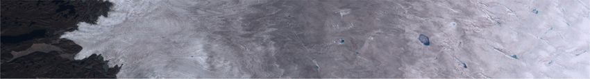
ARTICLE                                                                NATURE COMMUNICATIONS | https://doi.org/10.1038/s41467-020-20627-w

T
       he Greenland Ice Sheet (GrIS) comprises only 11.2% of                      a
       land ice on Earth1, yet surface melting and ice-calving from
       the GrIS accounted for 37% of cryosphere attributed sea
level rise between 2012 and 20162. Mass loss is predominantly
determined by the incoming shortwave radiation flux3,4 modu-                                     6                         3   2
                                                                                                                  4a,b
lated by surface albedo5. There has been a ~40% increase in                                                                                               1
surface melting and runoff from the GrIS over the last quarter of                                             5
a century6 as a north−south oriented band of low-albedo ice,
known as the Dark Zone, has developed along the western margin
of the ice sheet7. Albedo depends on the physical structure of                                                                                            25 km
surface ice8 and the presence of light absorbing particulates
(LAP), which include pigmented glacier snow and ice algae, black
                                                                                  b
carbon (BC), and mineral dust7. Glacier ice algae (hereafter gla-                           Clean Ice (CI)                        High algal biomass (Hbio) ice
cier algae) produce photoprotective phenolic pigments9–14 that
lower ice sheet albedo on the landscape-scale, thereby con-
tributing to melting15,16. Glacier algae were calculated to be
directly responsible for 9−13% of the surface melting in the Dark
Zone in 201616, and there are comparable indirect effects due to
water retention and changes to ice crystal fabric as a result of algal
growth8. While glacier algal blooms can cover up to 78% of the                               50 cm                                            50 cm
ice surface16, they exhibit a high degree of interannual variability
in intensity and spatial extent7 that is yet to be understood. Thus,
there is a pressing need to better quantify the parameters that                       Dispersed cryoconite (DCC)                    Cryoconite holes (CCH)
control glacier algal growth and constrain the impact of these
blooms on ice sheet albedo, melting, and contributions to sea-                               50 cm
level rise. Here we demonstrate, through nutrient addition
experiments and spatially resolved mineralogical and geochemical
data, that phosphorus is a limiting nutrient for glacier algae in the
Dark Zone. We identify phosphorus-bearing minerals (hydro-
xylapatite) as the likely phosphorous nutrient source fueling                                                                                              25 cm
glacier algal blooms. We also disentangle the biogeochemical
controls on ice sheet darkening by characterizing the source,                               Surface biofilm                       Supraglacial stream water
composition, and nutrient delivery capacity of mineral dust.
These results, in combination with nutrient addition experiments,
demonstrate that phosphorus from mineral dust is a limiting
nutrient for algal blooms in the Dark Zone.

Results and discussion
Phosphorus is a limiting nutrient for glacier algae. Glacier algal                                                 5 cm                                       1m
blooms were studied at five sites along a transect across the
ablation zone in southwest GrIS (Fig. 1a). Surface snow and ice               Fig. 1 Sample collection locations and habitats on the Greenland Ice
samples were collected at locations ~33−130 km from the ice                   Sheet. a Sample collection sites 1–5 across the ablation zone in southwest
margin in 2016 and 2017 (Sites 1−5), with reference rocks col-                Greenland Ice Sheet and rock sample collection site 6 near the terminus of
lected near the Russell Glacier terminus in 2018 (Site 6; Fig. 1a,            Russell Glacier viewed using Sentinel-2 imagery (cloud-free composite of
Supplementary Table 1). Targeted surface habitats included clean              all acquisitions between 01/07/2016 and 31/08/2016); b photographs of
ice (CI; free of macroscopically visible LAP), high algal biomass             surface ice habitats: clean ice (CI), high algal biomass ice (Hbio), dispersed
(Hbio) ice, Hbio snow, dispersed cryoconite (DCC) ice, cryoconite             cryoconite ice (DCC), cryoconite holes (CCH), surface biofilm, and
holes (CCH), a floating biofilm, and supraglacial stream water                  supraglacial stream water.
(Fig. 1b). To identify potential nutrient limitations on glacier algal
growth, we carried out a series of soluble nutrient addition
incubation experiments at Site 4b using Hbio ice (8.0 ± 2.1 × 10³             rETRmax measurements made for the phosphate and +ALL
cells mL−1) that was melted in the dark over 24 h, and re-                    treatments, and both of these treatments were significantly higher
incubated for 120 h across five treatments (phosphate, nitrate,                than each of the control, ammonium, and nitrate treatments, with
ammonium, phosphate+nitrate+ammonium (+ALL), control).                        no significant differences between the latter three treatments
Concomitantly, the health and productivity of glacier algae                   (Supplementary Tables 2 and 3). These results indicate that
assemblages were monitored using rapid light response curves17                phosphorus was the limiting nutrient for glacier algal growth at
performed with pulse amplitude modulation (PAM) fluorometery                   Site 4b. No significant differences in photophysiological para-
at 24, 72, 120 h. A significant response to phosphorus addition                meters were apparent after 24 or 72 h across treatments (Sup-
was apparent after 120 h of incubation (Fig. 2a), achieving the               plementary Tables 2 and 3, Supplementary Fig. 1, data for 72 h
maximum quantum efficiency (Fv/Fm: inverse proxy of micro-                     not shown). The delayed response to phosphorus addition until
algae stress) and maximum rates of electron transport (rETRmax:               120 h suggests a mechanism of phosphate storage sufficient to
proxy for photosynthesis rate). Both parameters displayed posi-               sustain glacier algal productivity for ~5 days, which is similar in
tive responses to increased P availability (PO43- and +ALL                    duration to the doubling time of 5.5 ± 1.7 days reported for gla-
treatments) compared to other treatments (Fig. 2b, c). Specifi-                cier algal populations from this region15. Due to the slow dou-
cally, after 120 h there were no significant differences between the           bling time of glacier algae, it is not surprising that a significant

2                NATURE COMMUNICATIONS | (2021)12:570 | https://doi.org/10.1038/s41467-020-20627-w | www.nature.com/naturecommunications
NATURE COMMUNICATIONS | https://doi.org/10.1038/s41467-020-20627-w ARTICLE
a increase in cell counts was not measured over the 120 h experi-
ment (Supplementary Table 4). Luxury uptake of phosphorus is
400
400 After 120 h incubation common among microorganisms, with phosphorus stored
intracellularly as polyphosphates that can act as an extraneous
source under limiting conditions18. Such a storage mechanism
300
300 would be beneficial for glacier algae inhabiting the oligotrophic
surface ice environments of the GrIS Dark Zone. The lack of a
TR
rETR
photophysiological response to the addition of nitrate or
200
200
rE
ammonium suggests that glacier algae are not limited by N.
Control
Although our measurements of dissolved inorganic N in surface
100
100 +ALL ice and snow samples were low or below detection limit (Sup-
+NH4+ plementary Table 8), inorganic and organic N have been docu-
+NO3-
+PO43-
mented in Hbio ice habitats in concentrations of 1.0 and 14 µM,
00 respectively19, indicating that N is available in the system.
0 1000
1000 2000
2000 3000
3000 4000
4000
2 1 Phosphorus limitation decreases with increasing mineral
PAR (µmol
PAR (µmolphotonsm
photonsm2s-1s
) )
phosphorus. To assess potential phosphorus sources, sequential P
b extractions were conducted (Steps I, III, IV, and V from Rut-
0.7
Control +ALL +NH4+ +NO3- +PO43- tenberg, et al.20) and revealed that organic phosphorus (Porg)
B b
B accounted for up to 86% of the solid-phase P in Hbio ice (Sup-
b
0.6 plementary Table 5), with exchangeable (Pexch;ARTICLE NATURE COMMUNICATIONS | https://doi.org/10.1038/s41467-020-20627-w
a b c
0.008 0.0008
r=0.80 r=0.83 r=0.94
12
Porg (E-06 mol·g-1)
TOC (mol·g-1)
0.006
TN (mol·g-1)
8
0.0004
0.004
4
0.002 0.0000 0
0 1 2 0 1 2 0 1 2
Pmin (E-06 mol·g-1) Pmin (E-06 mol·g-1) Pmin (E-06 mol·g-1)
d e Hbio ice DCC CCH f
8 100 250
Redfield(C/N/P)/Sample(C/N/P) (E-06)
r=0.94
200
Concentration (µg·L-1 )
6 75
Abundance (wt%)
150
4 50
100
2 25
50
0 0 0
0 1 2 5 4a 4b 3 2 4a 4b 4b 5 4a 4b 3 2
Pmin (E-06 mol·g-1) Site Site
a-d: e: f:
Site 2 Hbio ice Site 4a DCC ice Site 5 Hbio ice Quartz Pyroxenes Na+ Mg2+ Al3+ K+ Ca2+ Fe2+
Site 3 Hbio ice Site 4b CCH All sites, samples, years Plag. feldspars Phyllosilicates
K feldspars Hydroxylapatite
Site 3 Hbio snow Site 4b Hbio ice linear regression (all
sites, years, habitats) Amphiboles Organic matter
Site 4a Hbio ice Site 4b DCC ice
95% confidence interval
2
3
4a,b
5
10 km
Fig. 3 Dark Zone particulate matter nutrient concentrations, mineralogy, and major cation concentrations in associated meltwater. Pmin concentration
in particulates plotted against a total organic carbon (TOC), b total nitrogen (TN), c organic phosphorus (Porg), and d molar C:N:Porg ratio normalized to the
Redfield ratio (C:N:P 106:16:1). Linear regression and r-values (Pearson’s product-moment correlation) correspond to all data points from all sites (gray
dots). e Relative mineral and organic matter abundances across the ablation zone, including hydroxylapatite (bright yellow); f major cation concentrations
in meltwater from Hbio ice across the ablation zone. Hbio ice: high algal biomass ice; DCC ice: dispersed cryoconite ice; CCH: cryoconite hole. In a–d: colored
points: mean values for different sites, habitats, and years, ±SE (2016: solid fill; 2017: white-fill); solid line: linear regression; thin dashed lines: 95%
confidence interval. Site 2 Hbio ice n = 1; Site 3 Hbio ice n = 2; Site 3 Hbio snow n = 1; Site 4a Hbio ice n = 5; Site 4a DCC ice n = 2; Site 4b Hbio ice n = 2; Site
4b DCC ice n = 1; Site 4b CCH n = 1; Site 5 Hbio ice n = 1. In e: Hbio ice: site 5: n = 1; site 4a n = 5; site 4b: n = 4; site 3: n = 2; site 2 n = 1; DCC ice: site 4a
n = 4; site 4b n = 1; CCH site 4b n = 1. In f: site 5 n = 6; site 4a n = 5; site 4b n = 3; site 3 n = 2; site 2 n = 1.
including amphiboles (4−14 wt%) and pyroxenes (NATURE COMMUNICATIONS | https://doi.org/10.1038/s41467-020-20627-w ARTICLE Fig. 4 Composition of bacterial, fungal, and algal communities in surface habitats across the Dark Zone. NMDS plots showing the sample similarities for bacteria (a), fungi (c), and algae (e) and their respective community compositions based on relative abundances (b, d, f). Sites are represented by colors and habitats by point shapes. Samples cluster according to sites and dashed lines represent the 95% confidence interval. All samples with a sufficiently high number of sequences were used for the NMDS plots, whereas representative samples across sites and habitats were selected for the bar charts (details in Supplementary Tables 9–13). 2016 from Sites 2, 3, and 4a (Fig. 4). Since Ancylonema P to the organic reservoir. This accounts for the lower Pmin nordenskioeldii and Mesotaenium sp. comprised between 66 concentration at sites hosting more prolific algal blooms (4 and 5), and 99% of the algal community in all samples (Supplementary where a higher proportion of Pmin has been transformed into Porg Table 13), the assemblage of glacier algae used in the nutrient through bioweathering and algal biomass accumulation. Glacier addition experiments at site 4 was reasonably representative of algal productivity outstrips that of associated heterotrophic the region. Collectively, the microbial community (Fig. 5a–c) was assemblages23, and likely drives recycling of solubilized P. Site 4a intermixed with mineral dust and occurred as disseminated Hbio and DCC ice contained three to four times more dissolved P particulates in Hbio ice (Fig. 5c) and aggregated cryoconite than clean ice or supraglacial stream water (Supplementary Table 8), granules in DCC ice and cryoconite hole material (Fig. 5d). substantiating claims that P is retained in surface ice habitats19. Microbial exopolymer enables cells to both adhere to mineral Microbes may similarly be utilizing mineral iron; ferromagnesian dust (Fig. 5e) and trap and bind mineral grains (Fig. 5f). minerals are less abundant in Hbio ice than other habitats, and Heterotrophic bacteria26 and fungi27 can accelerate apatite decrease in abundance among Hbio samples by up to 60 % at sites weathering beyond abiotic rates, thereby transferring solid-phase hosting more prolific algal blooms (Fig. 3e). Dissolved iron NATURE COMMUNICATIONS | (2021)12:570 | https://doi.org/10.1038/s41467-020-20627-w | www.nature.com/naturecommunications 5
ARTICLE                                                                    NATURE COMMUNICATIONS | https://doi.org/10.1038/s41467-020-20627-w

               a                                                                b

                                                                     2 m                                                             5 m

               c                                                                d

                                                                    MD
                                            A

                                       MD

                                            A

                                                                    10 m                                                          200 m

               e                                                                f

                                                                    1 m                                                            1 m

Fig. 5 Scanning electron micrographs of cell-mineral associations in Dark Zone surface habitats. Scanning electron microscopy (SEM) micrographs
showing a bacteria, b fungi, and c glacier algae that comprise the Dark Zone microbial community. Microbes in c) Hbio ice are more disseminated than
those in d cryoconite granules. In all surface ice habitats, exopolymer enables microbial cells to e adhere to mineral surfaces, and f trap and bind mineral
grains (arrows). In c A: algal cells, MD: mineral dust. Images in a–f are representative of the n = 12 samples observed using SEM.

concentrations in Hbio ice show a concomitant inverse trend (Fig. 3f,           African33 dust) as significant contributors (Fig. 6a, b). A local
Supplementary Table 8). Specifically, at Site 4a, the concentration of           mineral source means that the hydroxylapatite in our particulates
iron was two and four-time higher in Hbio ice (69 ± 20 μg L−1) than             was derived from local apatite-bearing lithologies34. Local deliv-
DCC ice (36 ± 5 μg L−1) and clean ice (16 ± 13 μg L−1), respectively            ery of mineral dust to the GrIS is consistent with ice core records
(Supplementary Table 8). If iron is extracted as a micronutrient, this          that indicate delivery of Greenlandic dust to the ice sheet during
has downstream implications for export of bioavailable iron from                interglacial periods35. Furthermore, analysis of the grain-size
the ice sheet to the marine system28.                                           distribution of our particulate dust fractions revealed that 99% of
                                                                                all grains were
NATURE COMMUNICATIONS | https://doi.org/10.1038/s41467-020-20627-w ARTICLE
a b 2.0
Site 3 & 4
3
Site 6 Forefield W Greenland
1.5
Sample/UCC
sediment kimberlites
2
Eu/Eu*
Dark Zone
cryoconite
1.0
1 Africa
Asia
0 0.5
La Ce Pr Nd Sm Eu Gd Tb Dy Ho Er Tm Yb Lu 0 1 2
La/Sm
Greenland Ice Sheet West Greenland forefield Global dust sources
Dark Zone LAP (Site 3 & 4, this study) Forefield lithologies (Site 6, this study) Asia32
Dark Zone cryoconite29 Forefield sediment30 + Africa33
West Greenland kimberlites31
c d
1000
0.4
site 2 Hbio site 3 Hbio site 4a DCC
site 4a Hbio site 5 Hbio
LAP loading ( g·mL-1)
0.3
100
Frequency
0.2
10
0.1
0.0
1
Clean ice Hbio ice DCC ice
0 5 10 15 20 25
Diameter ( m) Habitat
Fig. 6 Rare Earth element signature and grain size distribution for mineral dust in Dark Zone particulates. a, b Rare Earth element (REE) normalized to
the upper continental crust (UCC) compared to potential sources; c mineral dust size distribution; and d particulate mass loading by habitat. In d plot
shows mean ± SE, clean ice: n = 4 samples, high algal biomass ice (Hbio) ice: n = 3 samples, dispersed cryoconite (DCC) ice: n = 1 sample.
(Fig. 3e, Supplementary Table 5). In spite of its dominance by pigmented algal cells in the same manner that mineral grains
mass, mineral dust is not the primary cause of ice surface dar- enhance light absorption by black carbon nanoparticles through
kening in the GrIS Dark Zone. In situ spectral reflectance mea- lensing41. This effect depends on the structure and mixing ratio of
surements at Site 4 combined with refractive index and mineral heterogeneous LAP aggregates in snow and ice. Its potential to
dust grain-size distribution measurements in a radiative transfer contribute to ice sheet albedo reduction remains unexplored.
model indicate that mineral dust has a negligible effect on albedo
reduction compared to pigmented glacier algae16. Rather, our
findings indicate that the presence of Pmin may have a second- Microbes, minerals, and melting: a positive feedback system.
order effect on albedo. If this is the case, it follows that the spatial Previous studies have made links between snow algae and mineral
extent and melt rate of P-bearing ice may in part constrain the derived nutrients42–44, demonstrated that snow algae respond to
spatial distribution of the algal blooms producing the darkening the addition of fertilizer45, and inferred that glacier algal abun-
observed on the landscape-scale. dance correlate with mineral dust loading24. Here we demonstrate
The limited first order effect of minerals on albedo is likely due how glacier algae respond in situ to the addition of specific
to dark-colored ferromagnesian phases (7−21 wt%) being inter- nutrients and link this response to collocated nutrient-bearing
mixed with felsic phases (79−93 wt%), namely feldspars and mineral dust. Our findings demonstrate that mineral nutrient
quartz, which are adept at scattering light. The measured availability is a second-order control on albedo by modulating
refractive index of the dust16 indicates that light scattering by glacier algal bloom development. Comparable datasets spanning
felsic mineral grains supersedes light absorption by their the GrIS are therefore required to incorporate mineral dust as a
ferromagnesian counterparts. Note, these findings may not apply factor in the positive feedback between algal growth and surface
to all locations; mineral dust can lower snow and ice albedo in melting. Algae induced melting liberates ice-bound dust, from
glacier37 and ice sheet environments38,39. This is dependent on which heterotrophs can extract micronutrients. Recycled nutri-
the bulk complex refractive index of the dust, which is a product ents augment glacier algal blooms, thereby further reducing
of dust composition and grainsize. This is compounded by factors albedo and promoting additional melting. Furthermore, trapping
such as the distribution of dust within the ice matrix, mass mixing and binding of mineral dust by microbial exopolymers (Fig. 5f),
ratio, ice grain size and shape, and ambient illumination helps retain valuable nutrients in the ice habitat. Notably, the
conditions40. The high proportion of felsic mineral grains may glacier algal biofilm contained the highest abundance of hydro-
also indirectly contribute to albedo reduction via lensing of xylapatite of all samples (1.1 wt%; Supplementary Table 6),
NATURE COMMUNICATIONS | (2021)12:570 | https://doi.org/10.1038/s41467-020-20627-w | www.nature.com/naturecommunications 7ARTICLE NATURE COMMUNICATIONS | https://doi.org/10.1038/s41467-020-20627-w
suggesting preferential entrapment of hydroxylapatite in these depth surface ice areas containing a conspicuous loading of glacier algae were
complex microbial colonies. Microbial retention of mineral dust sampled on June 22nd 2017 into sterile Whirl-Pak bags, and melted in the dark for
24 h under ambient on-ice conditions (5–10 °C). These samples were used as the
reinforces the biological-albedo reducing feedback by prolonging inoculum for the incubation experiments and had algal counts of 8.0 ± 2.1 × 10³
colocation of algae and essential nutrients on the ice surface. cells mL−1 (mean ± SD, n = 5). Algal cells were counted using a modified Fuchs-
Nevertheless, factors controlling the timing, spatial extent, and Rosenthal haemocytometer (Lancing, UK) on a Leica DM 2000 epifluorescence
intensity of algal blooms remain knowledge gaps limiting our microscope with attached MC120 HD microscope camera (Leica, Germany). The
ability to project biological albedo reduction and melt. Bloom inoculum was incubated in 30 mL microalgal culturing flasks (Corning, UK) in
quadruplicates across five nutrient treatments: control (no nutrient addition),
initiation depends on bare ice exposure following snowpack +NH4+ (10 µM final concentration), +NO3− (10 µM final concentration), +PO43−
retreat, as indicated by glacier algal cell counts for the same (10 µM final concentration), and +ALL nutrients (10 µM of each NH4 and NO3; 2
locations9, and the fact that growth of cryophilic algae can be µM of PO4 to maintain a 10:1 N:P ratio across treatments). Nutrient concentrations
stimulated by the presence of liquid water45. It is essential to were designed to provide ~10-fold ambient dissolved inorganic nitrogen (DIN) and
dissolved inorganic phosphorus (DIP) concentrations previously reported for GrIS
constrain the timing of bloom development following snowpack supraglacial ice52,53.
retreat because satellite imagery indicates that surface darkening After 24 h, 72 h, and 120 h, measurements of variable chlorophyll fluorescence
occurs within days of snow clearance7. Since 2000, the melt were performed on 3 mL incubation sub-samples with a WaterPAM fluorometer
season in the GrIS Dark Zone has progressively started earlier, and attached red-light emitter/detector cuvette system (Walz GmBH, Germany).
During the experiment, the ambient air temperature ranged between −4 and +4 °C.
lasted longer, and exhibited greater albedo reduction7,46. Years Rapid light response curves (RLCs) were performed to constrain glacier algae
experiencing earlier winter snowpack retreat yield more expan- photophysiology17, providing information on energy use from limiting through to
sive algal blooms47. The higher Pmin measured at inland sites (2 saturating levels of irradiance54. All samples were dark-adapted for 20 minutes prior
and 3) indicate that these locations are geochemically primed to to RLC assessment, which was undertaken with a saturating pulse of ca. 8,600 μmol
photons m−2 s−1 for 600 ms duration and nine 20 s incremental light steps ranging
host future glacier algal blooms. These trends may be exacerbated from 0 to 4000 μmol photons m−2 s−1. Maximum quantum efficiency (Fv/Fm) was
by increased atmospheric delivery of mineral dust to the ice sheet calculated from minimum (Fo) and maximum (Fm) fluorescence yields in the dark-
through increased windblown dust from exposed forefield adapted state as Fv/Fm = (Fm − Fo)/Fm. Electron transport through photosystem II
lithologies48, and by projected increased snowfall over the GrIS49. (PSII) was calculated from PSII quantum efficiency (YPSII) in relative units
The complexity of this rapidly changing Arctic system makes it (rETR = YPSII × PAR × 0.5) assuming an equal division of photosynthetically active
radiation (PAR) between photosystem I and PSII. Analysis of rapid light curves
difficult to anticipate future changes to ice sheet albedo, melt (rETR ~ PAR) followed17 with iterative curve fitting in R (v.3.6.0) and calculation of
rates, and contributions to sea level. Our data provide a the relative maximum electron transport rate (rETRmax), theoretical maximum light
quantitative link between mineral-derived nutrients and glacier utilization coefficient (α), and light saturation coefficient (Ek) following Eilers and
algae blooms, and demonstrate that mineral dust is an essential Peeters55. Statistical differences in photophysiological parameters were assessed
using two-way ANOVA with the fixed variables of treatment (5 levels) and date
nutrient source for glacier algae. This biogeochemical process (2 levels: 24 and 120 h) and the interaction term, following tests of homogeneity of
must therefore be incorporated into predictive models thereby variance and normality of distribution. Tukey HSD tests were used to assess
improving our understanding of how glacier algal blooms will statistically significant differences in quantum efficiency and relative maximum
contribute to ice sheet melting in the future. electron transport rate between nutrient treatments. Simultaneous to
photophysiological measurements, a further 5 mL of homogenized sample was fixed
using 25% glutaraldehyde at 2% final concentration and transported back to the
Methods University of Bristol, UK, to assess glacier algal cell abundance (cells ml−1)9,
Sample collection and processing. Surface snow and ice samples were collected completed within 1 month of sample return. Detailed statistical outputs and final
along a transect across the ablation zone of the southwestern margin of the cell count data can be found in Supplementary Tables 2–4).
Greenland Ice Sheet during the 2016 (July 27–August 17) and 2017 (June 1–28)
melt seasons (Fig. 1). Site 4 was the basecamp location in both seasons, differ- Phosphorus extractions. The phosphorus content of the Hbio ice, DCC ice, Hbio
entiated as 4a (2016) and 4b (2017). Sites 1, 2, 3, and 5 were sampled only in snow, and CCH samples was determined using a modified version of the SEDEX
2016. The data presented are for samples representing a range of surface snow sequential extraction protocol20. Steps I, III, IV, and V were completed as a means
and ice habitats. The clean snow sample (GR16_1) collected at site 1 provides a of quantifying loosely bound/exchangeable P (Pexch), mineral P (Pmin), and organic
reference for snow chemistry from the accumulation zone. The sample did not P (Porg). The extracted P was measured in the fluid phase as described below for the
contain sufficient particulate mass for solid-phase chemical and mineralogical melted ice samples. Detailed results can be found in Supplementary Table 5.
analyses. The collected samples were classed into the following categories based
on macroscopically visible characteristics: clean snow (n = 1), clean surface ice
(CI, n = 4), high algal biomass ice (Hbio; n = 19), high algal biomass snow (n = Meltwater fluid chemistry. Melted surface samples and supraglacial stream water
2), and dispersed cryoconite ice (DCC; n = 5). In addition, supraglacial stream samples were filtered using 0.22 μm single use syringe filters into acid-washed
water (n = 2), a sample of cryoconite hole (CCH) material (n = 1), a cryoconite Nalgene bottles. Inductively-coupled plasma mass spectroscopy (ICP-MS; Thermo
hole layer from an ice core (n = 1), and a floating algal biofilm (n = 1) were Fisher iCAPQc) was used to measure fluid phase cations in the filtered water
collected. Details of sample types and collection locations are in Supplementary samples that were acidified using Aristar HNO3. ICP-MS was conducted by Stephen
Table 1. Clean was defined as surface snow and ice containing no macro- Reid at the University of Leeds, UK. Phosphorus was either measured using seg-
scopically visible particulates, Hbio ice and snow consisted of surface ice and mented flow-injection analysis (AutoAnalyser3, Seal Analytical), or for samples
snow containing visible glacier algal and particulate material, and DCC consisted containing lower concentration of phosphorus using a 100 cm WPI Liquid Wave-
of ice surfaces covered in disseminated particulate material from melted out guide Capillary Cell in conjunction with an Ocean Optics USB2000 + spectro-
cryoconite holes. Cryoconite hole material was sampled to provide a biogeo- photometer with a precision of 1.6% and a LOD of 2 nmol L−1. Aliquots of the non-
chemical reference for the DCC samples. The biofilm sample consisted of a semi- acidified 0.22 μm filtered samples were also analyzed in replicates by ion chroma-
coherent slick of aggregated microbial and particulate material floating on the tography by Andrea Viet-Hillebrand at the German Research Centre for Geos-
surface of ponded meltwater. Ice and snow samples were collected from the top ciences, Potsdam, Germany. Analyses were carried out with a conductivity detector
3–5 cm of surface into sterile plastic bags, melted at ambient temperatures on a Dionex ICS 3000 system, equipped with an AS 11 HC Dionex analytical
(5–10 °C) (details in50,51). Aliquots of filtered melted samples were processed as column run at 35 °C for chromatographic separation of the anions. Standards
described below for fluid chemistry analyses. While on the ice, melted samples containing all investigated inorganic ions (F−, Cl−, SO42−, NO3−, PO43−) were
were filtered through glass fiber filters (GFF, pore size: 0.7 μm), from which the analyzed and all replicate samples had a standard deviationNATURE COMMUNICATIONS | https://doi.org/10.1038/s41467-020-20627-w ARTICLE
the German Research Centre for Geosciences, Potsdam, Germany. Detailed below this threshold were discarded (16 S: 5500, ITS2: 15000, 18 S: 15000). Further,
results found in Supplementary Table 5. Pearson’s product-moment correlation only ASVs with a minimum frequency count of 10 were retained in the feature
r-values in Fig. 3 and supplementary Fig. 2 were calculated using Excel (v16.xx). tables. Detailed results can be found in Supplementary Tables 9–13.
The filtered feature tables were imported into R (v.3.6.0) to create bar charts
representing the respective community compositions based on their relative
Mineralogy. The mineralogy of the dust was determined using a Bruker D8 abundances. Non-metric multidimensional scaling (NMDS) analyses were
Advance Eco X-ray diffractometer (Bruker, Billerica, USA) with a Cu source, performed using the “metaMDS” function (Bray-Curtis distances) of the R package
operated at 40 kV and 40 mA at the University of Leeds, UK. Samples were “vegan” and plots were created using the package “ggplot2”. Analysis of similarities
hand-milled in an agate mortar and pestle prior to loading in 5 or 10 mm low- (ANOSIM) was carried out using the “anosim” function of the “vegan” package
background silicon mounts. The small quantity of material per sample neces- and “sites” and “habitats” as treatment groups.
sitated the use of shallow sample mounts, thereby making the sample not infi-
nitely thick with respect to X-rays, and thus rendering this analysis semi-
quantitative. Furthermore, because it was necessary to rely on hand grinding, Scanning electron microscopy (SEM). Hbio ice, cryoconite, DCC ice, and bio-
XRD patterns exhibit the effects of non-ideal particle size statistics and preferred film samples were fixed using 2.5 % v/v glutaraldehyde and stored at 4 °C.
orientation on some phases, which can result in higher Rwp values. The 2016 Samples were dehydrated via an ethanol dehydration series (25%, 50%, 75%,
COD and 1996 ICDD databases were used to complete phase identification for 100%, 100%, 100%) for 15 min at each step, followed by 10 min in each of: 50:50
each sample, in conjunction with DIFFRACplus Eva v.2 software56. Topas V 4.256 ethanol: hexamethyldisilazane (HMDS), and 2 × 100% HMDS. The HMDS was
and the fundamental parameters approach57 were used to complete Rietveld removed and the samples were air-dried prior to being mounted on stainless
refinements25,58,59. No preferred orientation corrections were used because steel stubs using adhesive carbon tabs. Samples were coated with 5 nm of iridium
refinements are typically more accurate for samples containing many phases that using an Agar High Resolution sputter coater. SEM characterization of the
are known to exhibit severe preferred orientation (e.g., phyllosilicates) when samples was conducted using a Hitatchi 8230 SEM at the Leeds Electron
such corrections are excluded60. In some cases, the use of multiple K-feldspar, Microscopy and Spectroscopy Centre (LEMAS), University of Leeds, UK.
plagioclase feldspar, and orthopyroxene structures were used in a single
refinement because this approach provided substantially improved fit statistics Rare Earth Element analysis and data compilation. Rare Earth Element (REE)
and visual fits to observed XRD patterns. This may reflect the incorporation of analysis was conducted on Hbio ice, DCC ice, and cryoconite hole particulate
dust from multiple source rocks of differing mineralogical composition. Mineral solid materials filtered onto 0.7 µm GFF and hand-milled in an agate mortar and
phases identified using XRD were grouped into the following classes: quartz, pestle. To remove organic matter, samples were ashed in a slightly open ceramic
plagioclase feldspars (albite/andesine/anorthite), amphiboles (refined using the crucible in a muffle furnace at 450 °C for 4 h. Rock samples collected from site 6
structure of actinolite), potassium feldspars (orthoclase/microcline), pyroxene by Gilda Varliero and Gary Barker (University of Bristol, UK) (representing
(enstatite/augite/diopside), and micas (refined using the structure of muscovite). lithologies of the catchment area) were cut into centimeter-sized cubes prior to
Detailed results are found in Supplementary Tables 6 and 7. milling in a tungsten ring mill. Acid dissolution of the mineral fraction was
achieved in Savillex® beakers using pro-analysis acids previously purified by
Microbial community composition. A total of 26 samples comprising 15 high distillation and sub-boiling. Dissolution was first performed with 2 mL 14 M
algal biomass ice (Hbio ice), two high algal biomass snow (Hbio_snow), one HNO3 and 1 mL 23 M HF on a hot plate at 120 °C for 48 h and later, after
biofilm, four dispersed cryoconite (DCC) (macroscopically visible particles), and evaporation to dryness, with 2 mL 6 M HCl on a hot plate at 120 °C for 24 h. REE
four clean ice (CI) samples (without macroscopically visible particles) were concentrations were determined using HR-ICP-MS (ThermoFisher Element 2)
collected into sterile 50 mL centrifuge tubes (Hbio ice, Hbio snow, DCC, Biofilm) at the Vrije Universiteit Brussel, Belgium. Trace element concentrations were
or sterile sampling bags (CI). After gentle thawing at field-lab temperatures calibrated using elemental standard solutions and USGS reference material
(~5–10 °C), and concentrating by gravimetric settling of particles (for Hbio ice, (AGV-2). Precision for all elements is better than 2% RSD. Detailed results can
Hbio snow, DCC, Biofilm) or filtering (CI) through sterile Nalgene single-use be found in Supplementary Table 14.
filtration units (pore size 0.22 µm), up to 5 replicate from the concentrates or 1
filter per sampling event were transferred to 5 ml cryo-tubes and immediately Mineral dust particle size distribution analysis. Approximately 100 mg of each
frozen in liquid nitrogen. Samples were returned to the German Research Centre sample was transferred to a 50 mL centrifuge tube, to which 35 mL of 30% H2O2
for Geosciences in Potsdam, Germany in a cryo-shipper at liquid nitrogen (w/w) (Honeywell Fluka™) was added in order to remove the organic content. The
temperatures and stored at −80 °C until processing. tubes were sonicated (VWR ultrasonic cleaner) for 10 min to disaggregate the
DNA was extracted from all samples using the PowerSoil (Hbio ice, Hbio snow, solids. The samples were agitated in an orbital shaking incubator operating at 100
DCC, Biofilm) or PowerWater (CI) DNA Isolation kits (MoBio Laboratories). The rpm at 35 °C. After 72 h, the samples were centrifuged at 4000 rpm for 10 min
16 S rRNA, 18 S rRNA and ITS amplicons were prepared according to the Illumina (Eppendorf centrifuge 5810). The supernatant was removed and replaced with new
“16 S Metagenomic Sequencing Library Preparation” guide. 16 S rRNA genes were H2O2. This was repeated six times until no more organic oxidation was observed.
amplified using the bacterial primers 341 F (5′-CCTACGGGNGGCWGCAG) and The mineral fraction was washed three times in water (Sartorius arium pro
785 R (5′-GACTACHVGGGTATCTAATCC) spanning the V3-V4 hypervariable ultrapure water) for 24 h, with centrifugation succeeding each wash. The organic-
regions. 18 S rRNA genes were amplified using the eukaryotic primers 528 F free mineral fractions were dried at 35 °C prior to particle size analysis measured by
(5′ GCGGTAATTCCAGCTCCAA) and 706 R (5’ AATCCRAGAATTTCACCT Kerstin Jurkschat using a DC24000 CPS disc centrifuge65 at Oxford Materials
CT; Cheung et al., 2010) spanning the V4-V5 hypervariable regions. ITS amplicons Characterisation Services, Oxford, UK.
were amplified using the primers 5.8SbF (5′ CGATGAAGAACGCAGCG) and
ITS4R (5′ TCCTCCGCTTATTGATATGC) spanning the ITS2 region. All primers
LAP mass loading quantification. Aliquots of melted snow and ice samples of
were tagged with the Illumina adapter sequences. Polymerase chain reactions
known volumes were filtered in the field successively through pre-weighed 5 μm
(PCR) were performed using KAPA HiFi HotStart ReadyMix. Initial denaturation
and 0.2 μm polycarbonate filters. The filters were returned to the University of
at 95 °C for 3 min was followed by 25 cycles of denaturation at 95 °C for 30 s,
Leeds, UK where they were dried and weighed to determine the mass of LAP per
annealing at 55 °C for 30 s, and elongation at 72 °C for 30 s. The final elongation
volume of melted sample. The sum of the total organic carbon and nitrogen was
was at 72 °C for 5 min. All PCRs were carried out in reaction volumes of 25 µL. All
used as a proxy to indicate the biomass fraction of each sample, with the remaining
pre-amplification steps were done in a laminar flow hood with DNA-free certified
sample mass allocated to mineral dust. These values were used to calculate the
plastic ware and filter tips. Amplicons were barcoded using the Nextera XT Index
mineral dust mass loading per unit of melted ice.
kit. The pooled library was sequenced on the Illumina MiSeq using paired 300-bp
reads at the University of Bristol Genomics Facility, Bristol, UK.
The sequenced 16 S, 18 S, and ITS2 libraries were individually imported into Reporting summary. Further information on research design is available in the Nature
Qiime2 (v.2019.1)61. Itsxpress was used to extract the precise ITS2 region, and thus Research Reporting Summary linked to this article.
removing the conserved regions, from the ITS2 libraries before further processing
(--p-region ITS2, _--p-taxa ALL). The imported libraries were quality-filtered using
the dada2 pipeline (16 S: --p-trunc-len-f = 280, --p-trunc-len-r = 200, --p-trim- Data availability
feft-f = 10, --p-trim-left-r = 10; 18 S: --p-trunc-len-f = 250, --p-trunc-len-r = 200, Detailed microbial community, and fluid and solid phase chemistry results are available
--p-trim-feft-f = 10, --p-trim-left-r = 10; ITS2: --p-trunc-len-f = 0, --p-trunc-len-r in the supplementary information file. The microbial community data is available
= 0, --p-trim-feft-f = 0, --p-trim-left-r = 0). The amplicon sequence variants through the sequence read archive under accession number PRJNA564214. The COD
(ASV) in the filtered libraries were classified using classify-sklearn and the database is available here: http://www.crystallography.net/cod/. Figures that have
respective databases Greengenes (16 S, “gg-13-8-99-nb-classifier”)62, Silva (18 S, associated raw data: 2,3,4,6.
“silva-132-99-nb-classifier”)63, and Unite (ITS2, “unite_ver8_99_02.02.2019”)64.
ASVs skewing the results were removed from each data set (16 S: --p-exclude Code availability
Chloroplast, mitochondria; 18 S: --p-exclude Archaea, Bacteria). Feature tables Code for processing the rapid light curves (RLC) was produced by C Williamson and is
containing solely algal (18 S: --p-include Chloroplastida, Ochrophyta) or fungal
available here: https://github.com/chrisjw18/rlcs
(ITS2: --p-include Fungi) sequences were created. Subsequently, all feature tables
were rarefied to the lowest yet sufficient sample size and low-coverage samples
NATURE COMMUNICATIONS | (2021)12:570 | https://doi.org/10.1038/s41467-020-20627-w | www.nature.com/naturecommunications 9ARTICLE NATURE COMMUNICATIONS | https://doi.org/10.1038/s41467-020-20627-w
Received: 26 February 2020; Accepted: 2 December 2020; 28. Hawkings, J. R. et al. Ice sheets as a significant source of highly reactive
nanoparticulate iron to the oceans. Nat. Commun. 5, 3929 (2014).
29. Wientjes, I. G. M., Van de Wal, R. S. W., Reichart, G. J., Sluijs, A. &
Oerlemans, J. Dust from the dark region in the western ablation zone of the
Greenland ice sheet. Cryosphere 5, 589–601 (2011).
30. Tepe, N. & Bau, M. Distribution of rare earth elements and other high field
References strength elements in glacial meltwaters and sediments from the western
1. Vaughan D. G. et al. Climate Change 2013: The Physical Science Basis. Greenland Ice Sheet: Evidence for different sources of particles and
Contribution of Working Group I to the Fifth Assessment Report of the nanoparticles. Chem. Geol. 412, 59–68 (2015).
Intergovernmental Panel on Climate Change. Observations: cryosphere. 31. Nielsen, T. F. D., Jensen, S. M., Secher, K. & Sand, K. K. Distribution of
317–382 (Cambridge, UK, 2013). kimberlite and aillikite in the Diamond Province of southern West Greenland:
2. Bamber, J. L., Westaway, R. M., Marzeion, B. & Wouters, B. The land ice a regional perspective based on groundmass mineral chemistry and bulk
contribution to sea level during the satellite era. Environ. Res. Lett. 13, 063008 compositions. Lithos 112, 358–371 (2009).
(2018). 32. Ferrat, M. et al. Improved provenance tracing of Asian dust sources using rare
3. Fettweis, X. et al. Important role of the mid-tropospheric atmospheric earth elements and selected trace elements for palaeomonsoon studies on the
circulation in the recent surface melt increase over the Greenland ice sheet. eastern Tibetan Plateau. Geochim. Cosmochim. Acta 75, 6374–6399 (2011).
Cryosphere 7, 241–248 (2013). 33. van der Does, M., Pourmand, A., Sharifi, A. & Stuut, J.-B. W. North African
4. Hofer, S., Tedstone, A. J., Fettweis, X. & Bamber, J. L. Decreasing cloud cover mineral dust across the tropical Atlantic Ocean: insights from dust particle
drives the recent mass loss on the Greenland Ice Sheet. Sci. Adv. 3, e1700584 size, radiogenic Sr-Nd-Hf isotopes and rare earth elements (REE). Aeolian Res.
(2017). 33, 106–116 (2018).
5. Box, J. E. et al. Greenland ice sheet albedo feedback: thermodynamics and 34. Japsen, P., Bonow, J. M., Green, P. F., Chalmers, J. A. & Lidmar-Bergström, K.
atmospheric drivers. Cryosphere 6, 821–839 (2012). Elevated, passive continental margins: Long-term highs or Neogene uplifts?
6. van den Broeke, M. et al. Greenland ice sheet surface mass loss: recent New evidence from West Greenland. Earth Planet. Sci. Lett. 248, 330–339
developments in observation and modeling. Curr. Clim. Change Rep. 3, (2006).
345–356 (2017). 35. Simonsen, M. F. et al. East Greenland ice core dust record reveals timing of
7. Tedstone, A. J. et al. Dark ice dynamics of the south-west Greenland Ice Sheet. Greenland ice sheet advance and retreat. Nat. Commun. 10, 4494 (2019).
Cryosphere 11, 2491–2506 (2017). 36. Kok, J. F., Parteli, E. J. R., Michaels, T. I. & Karam, D. B. The physics of wind-
8. Tedstone, A. J. et al. Algal growth and weathering crust structure drive blown sand and dust. Rep. Prog. Phys. 75, 106901 (2012).
variability in Greenland Ice Sheet ice albedo. Cryosphere 14, 521–538 37. Oerlemans, J., Giesen, R. & Van Den Broeke, M. Retreating alpine glaciers:
(2020). Increased melt rates due to accumulation of dust (Vadret da Morteratsch,
9. Williamson, C. J. et al. Ice algal bloom development on the surface of the Switzerland). J. Glaciol. 55, 729–736 (2009).
Greenland Ice Sheet. FEMS Microbiol. Ecol. 94, fiy025 (2018). 38. Dumont, M. et al. Contribution of light-absorbing impurities in snow to
10. Lutz, S., McCutcheon, J., McQuaid, J. B. & Benning, L. G. The diversity of ice Greenland’s darkening since 2009. Nat. Geosci. 7, 509 (2014).
algal communities on the Greenland Ice Sheet as revealed by oligotyping. 39. Bøggild, C. E., Brandt, R. E., Brown, K. J. & Warren, S. G. The ablation zone in
Microbial Genomics 4, 1–10 (2018). northeast Greenland: ice types, albedos and impurities. J. Glaciol. 56, 101–113
11. Yallop, M. L. et al. Photophysiology and albedo-changing potential of the ice (2010).
algal community on the surface of the Greenland ice sheet. ISME J. 6, 40. Wiscombe, W. J. & Warren, S. G. A model for the spectral albedo of snow. I:
2302–2313 (2012). pure snow. J. Atmos. Sci. 37, 2712–2733 (1980).
12. Uetake, J., Naganuma, T., Hebsgaard, M. B., Kanda, H. & Kohshima, S. 41. Liu, D. et al. Black-carbon absorption enhancement in the atmosphere
Communities of algae and cyanobacteria on glaciers in west Greenland. Polar determined by particle mixing state. Nat. Geosci. 10, 184–188 (2017).
Sci. 4, 71–80 (2010). 42. Hamilton, T. L. & Havig, J. Primary productivity of snow algae communities
13. Remias, D., HolzingerS, A., Aigner, I. & Lütz, C. Ecophysiology and on stratovolcanoes of the Pacific Northwest. Geobiology 15, 280–295 (2017).
ultrastructure of Ancylonema nordenskiöldii (Zygnematales, Streptophyta), 43. Havig, J. R. & Hamilton, T. L. Snow algae drive productivity and weathering at
causing brown ice on glaciers in Svalbard (high arctic). Polar Biol. 35, 899–908 volcanic rock-hosted glaciers. Geochim. Cosmochim. Acta 247, 220–242
(2012). (2019).
14. Williamson, C. et al. Algal photophysiology drives darkening and melt of the 44. Phillips-Lander, C. M. et al. Snow algae preferentially grow on Fe-containing
Greenland Ice Sheet. Proc. Natl Acad. Sci. USA 117, 5694–5705 (2020). minerals and contribute to the formation of Fe phases. Geomicrobiol. J. 37,
15. Stibal, M. et al. Algae drive enhanced darkening of bare ice on the Greenland 572–581 (2020).
Ice Sheet. Geophys. Res. Lett. 44, 11,463–411,471 (2017). 45. Ganey, G. Q., Loso, M. G., Burgess, A. B. & Dial, R. J. The role of microbes in
16. Cook, J. M. et al. Glacier algae accelerate melt rates on the south-western snowmelt and radiative forcing on an Alaskan icefield. Nat. Geosci. 10,
Greenland Ice Sheet. Cryosphere 14, 309–330 (2020). 754–759 (2017).
17. Perkins, R. G., Mouget, J. L., Lefebvre, S. & Lavaud, J. Light response curve 46. Shimada, R., Takeuchi, N. & Aoki, T. Inter-annual and geographical variations
methodology and possible implications in the application of chlorophyll in the extent of bare ice and dark ice on the Greenland Ice Sheet derived from
fluorescence to benthic diatoms. Mar. Biol. 149, 703–712 (2006). MODIS satellite images. Front. Earth Sci. 4, 43 (2016).
18. Keenan, J. D. & Auer, M. T. The influence of phosphorus luxury uptake on 47. Cook, J. M. et al. Quantifying bioalbedo: a new physically based model and
algal bioassays. J. Water Pollut. Control Fed. 46, 532–542 (1974). discussion of empirical methods for characterising biological influence on ice
19. Holland, A. T. et al. Nutrient cycling in supraglacial environments of the Dark and snow albedo. Cryosphere 11, 2611–2632 (2017).
Zone of the Greenland Ice Sheet. Biogeosciences 6, 3283–3296 (2019). 48. Derksen, C. & Brown, R. Spring snow cover extent reductions in the
20. Ruttenberg, K. C. et al. Improved, high-throughput approach for phosphorus 2008–2012 period exceeding climate model projections. Geophys. Res. Lett. 39,
speciation in natural sediments via the SEDEX sequential extraction method. L19504 (2012).
Limnol. Oceanogr. Methods 7, 319–333 (2009). 49. Tedesco, M. & Fettweis, X. 21st century projections of surface mass balance
21. Redfield, A. C. The biological control of chemical factors in the environment. changes for major drainage systems of the Greenland ice sheet. Environ. Res.
Am. Sci. 46, 230A–221 (1958). Lett. 7, 045405 (2012).
22. Montagnes, D. J. S., Berges, J. A., Harrison, P. J. & Taylor, F. J. R. Estimating 50. Lutz, S., Anesio, A. M., Edwards, A. & Benning, L. G. Microbial diversity on
carbon, nitrogen, protein, and chlorophyll a from volume in marine Icelandic glaciers and ice caps. Front. Microbiol. 6, 307–307 (2015).
phytoplankton. Limnol. Oceanogr. 39, 1044–1060 (1994). 51. Lutz, S., Anesio, A. M., Jorge Villar, S. E. & Benning, L. G. Variations of algal
23. Nicholes, M. J. et al. Bacterial dynamics in supraglacial habitats of the communities cause darkening of a Greenland glacier. FEMS Microbiol. Ecol.
Greenland Ice Sheet. Front. Microbiol. 10, 1366 (2019). 89, 402–414 (2014).
24. Stibal, M. et al. Microbial abundance in surface ice on the Greenland Ice Sheet. 52. Hawkings, J. et al. The Greenland Ice Sheet as a hot spot of phosphorus
Front. Microbiol. 6, 225 (2015). weathering and export in the Arctic. Glob. Biogeochem. Cycles 30, 191–210
25. Rietveld, H. M. A profile refinement method for nuclear and magnetic (2016).
structures. J. Appl. Crystallogr. 2, 65–71 (1969). 53. Wadham, J. L. et al. Sources, cycling and export of nitrogen on the Greenland
26. Welch, S. A., Taunton, A. E. & Banfield, J. F. Effect of microorganisms and Ice Sheet. Biogeosciences 13, 6339–6352 (2016).
microbial metabolites on apatite dissolution. Geomicrobiol. J. 19, 343–367 54. Ralph, P. J. & Gademann, R. Rapid light curves: A powerful tool to assess
(2002). photosynthetic activity. Aquat. Bot. 82, 222–237 (2005).
27. Smits, M. M., Bonneville, S., Benning, L. G., Banwart, S. A. & Leake, J. R. 55. Eilers, P. H. C. & Peeters, J. C. H. A model for the relationship between light
Plant-driven weathering of apatite – the role of an ectomycorrhizal fungus. intensity and the rate of photosynthesis in phytoplankton. Ecol. Model. 42,
Geobiology 10, 445–456 (2012). 199–215 (1988).
10 NATURE COMMUNICATIONS | (2021)12:570 | https://doi.org/10.1038/s41467-020-20627-w | www.nature.com/naturecommunicationsNATURE COMMUNICATIONS | https://doi.org/10.1038/s41467-020-20627-w ARTICLE
56. Topas V. 3.0: General Profile and Structure Analysis Software for Powder collected samples and conducted the incubation experiments. J.M.C. and A.J.T. aided
Diffraction Data (Bruker AXS, Germany, 2004). sample collection and provided valuable discussions regarding albedo and ice sheet
57. Cheary, R. W. & Coelho, A. A fundamental parameters approach to X-ray processes. S.A.W. aided XRD data interpretation and writing Rietveld refinement code.
line-profile fitting. J. Appl. Crystallogr. 25, 109–121 (1992). A. S. conducted P extractions and measurements. A.V. and S.B. conducted REE analysis
58. Bish, D. L. & Howard, S. A. Quantitative phase analysis using the Rietveld and data interpretation. A.M.A., M.L.Y., J.B.M., M.T., and L.G.B. acquired the funding,
method. J. Appl. Crystallogr. 21, 86–91 (1988). collected samples, and guided data interpretation. All authors contributed to the
59. Hill, R. & Howard, C. Quantitative phase analysis from neutron powder manuscript.
diffraction data using the Rietveld method. J. Appl. Crystallogr. 20, 467–474
(1987).
60. Wilson, S. A., Raudsepp, M. & Dipple, G. M. Quantifying carbon fixation in Competing interests
trace minerals from processed kimberlite: a comparative study of quantitative The authors declare no competing interests.
methods using X-ray powder diffraction data with applications to the Diavik
Diamond Mine, Northwest Territories, Canada. Appl. Geochem. 24,
2312–2331 (2009). Additional information
61. Bolyen, E. et al. Reproducible, interactive, scalable and extensible microbiome Supplementary information is available for this paper at https://doi.org/10.1038/s41467-
020-20627-w.
data science using QIIME 2. Nat. Biotechnol. 37, 852–857 (2019).
62. McDonald, D. et al. An improved Greengenes taxonomy with explicit ranks
for ecological and evolutionary analyses of bacteria and archaea. ISME J. 6, Correspondence and requests for materials should be addressed to J.M.
610–618 (2012).
Peer review information Nature Communications thanks Jon Telling, Christine
63. Quast, C. et al. The SILVA ribosomal RNA gene database project: improved data
Foreman, and other, anonymous, reviewers for their contributions to the peer review of
processing and web-based tools. Nucleic Acids Res. 41, D590–D596 (2013).
this work.
64. Nilsson, R. H. et al. The UNITE database for molecular identification of fungi:
handling dark taxa and parallel taxonomic classifications. Nucleic Acids Res.
47, D259–D264 (2018). Reprints and permission information is available at http://www.nature.com/reprints
65. Neumann, A. et al. New method for density determination of nanoparticles
Publisher’s note Springer Nature remains neutral with regard to jurisdictional claims in
using a CPS disc centrifuge™. Colloids Surf. B Biointerfaces 104, 27–31 (2013).
published maps and institutional affiliations.
Acknowledgements
We thank G Varliero and G Barker for sample collection and S Reid, B Plessen, S Open Access This article is licensed under a Creative Commons
Pinkerneil, and A Viet-Hillebrand for their technical support. We thank Caroline Attribution 4.0 International License, which permits use, sharing,
Peacock, Dominique Tobler, and Eric Oelkers for their feedback on the manuscript. We adaptation, distribution and reproduction in any medium or format, as long as you give
acknowledge funding from UK Natural Environment Research Council Consortium appropriate credit to the original author(s) and the source, provide a link to the Creative
Grant, Black and Bloom (NE/M020770/1 and NE/M021025/1). LGB and SL acknowledge Commons license, and indicate if changes were made. The images or other third party
funding from the German Helmholtz Recruiting Initiative (award number: I-044-16- material in this article are included in the article’s Creative Commons license, unless
01). LGB, AMA, and MT were also supported through an ERC Synergy Grant (ʻDeep indicated otherwise in a credit line to the material. If material is not included in the
Purpleʼ grant # 856416) from the European Research Council (ERC). article’s Creative Commons license and your intended use is not permitted by statutory
regulation or exceeds the permitted use, you will need to obtain permission directly from
the copyright holder. To view a copy of this license, visit http://creativecommons.org/
Author contributions licenses/by/4.0/.
J.M. collected and processed samples, conducted the XRD, P extractions, and SEM,
processed XRD and geochemistry data, drafted the manuscript. S.L. collected and
processed samples, and produced the microbial community data. C.W. and A.M.A. © The Author(s) 2021
NATURE COMMUNICATIONS | (2021)12:570 | https://doi.org/10.1038/s41467-020-20627-w | www.nature.com/naturecommunications 11You can also read