A human amygdala site that inhibits respiration and elicits apnea in pediatric epilepsy - JCI Insight
←
→
Page content transcription
If your browser does not render page correctly, please read the page content below
CLINICAL MEDICINE

                                         A human amygdala site that inhibits
                                         respiration and elicits apnea in pediatric
                                         epilepsy
                                         Ariane E. Rhone,1,2 Christopher K. Kovach,1,2 Gail I.S. Harmata,1,2,3,4,5,6 Alyssa W. Sullivan,7
                                         Daniel Tranel,2,7,8 Michael A. Ciliberto,9 Matthew A. Howard,1,2,3 George B. Richerson,2,3,4,8,10
                                         Mitchell Steinschneider,11 John A. Wemmie,1,2,3,4,6,8,10,12 and Brian J. Dlouhy1,2,3
                                         Department of Neurosurgery, 2Iowa Neuroscience Institute, 3Pappajohn Biomedical Institute, 4Interdisciplinary Graduate
                                         1

                                         Program in Neuroscience, 5Pharmacological Sciences Training Program, 6Department of Psychiatry, 7Department of
                                         Psychological and Brain Sciences, 8Department of Neurology, 9Department of Pediatrics, and 10Department of Molecular
                                         Physiology and Biophysics, Carver College of Medicine, University of Iowa, Iowa City, Iowa, USA. 11Department of
                                         Neurology, Albert Einstein College of Medicine, New York, New York, USA. 12Department of Veterans Affairs Medical
                                         Center, Iowa City, Iowa, USA.

                                                 BACKGROUND. Seizure-induced inhibition of respiration plays a critical role in sudden unexpected
                                                 death in epilepsy (SUDEP). However, the mechanisms underlying seizure-induced central apnea in
                                                 pediatric epilepsy are unknown.

                                                 METHODS. We studied 8 pediatric patients with intractable epilepsy undergoing intracranial
                                                 electroencephalography. We recorded respiration during seizures and during electrical stimulation
                                                 mapping of 174 forebrain sites. A machine-learning algorithm was used to delineate brain regions
                                                 that inhibit respiration.

                                                 RESULTS. In 2 patients, apnea coincided with seizure spread to the amygdala. Supporting a role for
                                                 the amygdala in breathing inhibition in children, electrically stimulating the amygdala produced
                                                 apnea in all 8 subjects (3–17 years old). These effects did not depend on epilepsy type and were
                                                 relatively specific to the amygdala, as no other site affected breathing. Remarkably, patients
                                                 were unaware that they had stopped breathing, and none reported dyspnea or arousal, findings
                                                 critical for SUDEP. Finally, a machine-learning algorithm based on 45 stimulation sites and 210
                                                 stimulation trials identified a focal subregion in the human amygdala that consistently produced
                                                 apnea. This site, which we refer to as the amygdala inhibition of respiration (AIR) site includes the
                                                 medial subregion of the basal nuclei, cortical and medial nuclei, amygdala transition areas, and
                                                 intercalated neurons.

                                                 CONCLUSIONS. A focal site in the amygdala inhibits respiration and induces apnea (AIR site) when
                                                 electrically stimulated and during seizures in children with epilepsy. This site may prove valuable for
                                                 determining those at greatest risk for SUDEP and as a therapeutic target.

                                                 FUNDING. National Institute of Neurological Disorders and Stroke — Congress of Neurological
                                                 Surgeons, National Institute of General Medical Sciences, Roy J. Carver Charitable Trust.

Conflict of interest: The authors have
declared that no conflict of interest
exists.

Copyright: © 2020, American Society      Introduction
for Clinical Investigation.              Sudden unexpected death in epilepsy (SUDEP) is a major health problem, recognized as the most com-
Submitted: November 13, 2019             mon cause of death in patients with medically intractable epilepsy (1). Because of its increased incidence
Accepted: February 26, 2020              in early life compared with other neurological conditions, SUDEP ranks second only to stroke among all
Published: March 12, 2020.               neurological disease in years of potential life lost (2). Recent evidence indicates SUDEP is more frequent
                                         in pediatric cases than previously thought (3). Moreover, SUDEP accounts for more than one-third of all
Reference information: JCI Insight.
2020;5(6):e134852.                       causes of mortality in pediatric patients with epilepsy (4). Major risk factors include generalized tonic-clon-
https://doi.org/10.1172/jci.             ic seizures (GTCS), medication intractability, and seizure frequency (5), although SUDEP can also occur
insight.134852.                          in children with more typical epilepsy syndromes, such as juvenile myoclonic epilepsy, as well as children

insight.jci.org   https://doi.org/10.1172/jci.insight.134852                                                                                                      1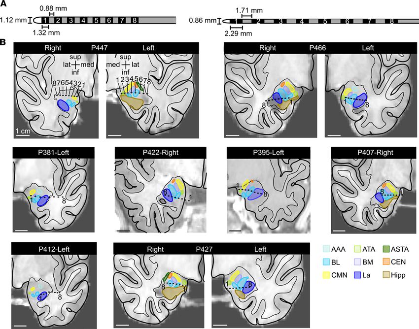
CLINICAL MEDICINE

                                  who have never had a GTCS (6–8). To prevent such devastating outcomes, it will be critical to identify the
                                  mechanisms underlying SUDEP in children as well as adults and in various types of epilepsy.
                                       Although the mechanisms underlying SUDEP remain unclear, multiple lines of evidence suggest
                                  inhibition of breathing plays a critical role (9–11). Supporting this possibility, central apnea is frequent-
                                  ly observed during seizures (12–14). Moreover, in a study of SUDEP cases that occurred in epilep-
                                  sy-monitoring units, seizures resulted in terminal apnea that preceded cardiac asystole, with the result-
                                  ing oxygen desaturation likely worsened by a lack of arousal and being prone, face down in bed, and
                                  without timely intervention (9). And, although sites within the brainstem are essential for breathing, a
                                  number of forebrain sites commonly affected by epilepsy have also been implicated in breathing con-
                                  trol, including the amygdala, hippocampus, and cingulate cortex (15–17). In humans, forebrain control
                                  of breathing is thought to be important for speaking, singing, voluntary breath holding, arousal, and
                                  emotional control and expression such as anxiety, fear, panic, laughing, and crying (18–22). Forebrain
                                  sites likely influence brainstem respiratory networks through neural projections, which might provide
                                  pathways for seizure-induced apnea. Consistent with these possibilities, recent studies indicate that loss
                                  of breathing in adult patients with epilepsy coincides with seizure spread to the amygdala (16, 23). In
                                  adults, electrical stimulation of the amygdala reproduced this apnea (16, 17). Thus far, no studies have
                                  examined forebrain control of breathing in children or seizure-induced apnea in pediatric epilepsy.
                                       Because the amygdala undergoes functional and structural connectivity changes from birth through
                                  adolescence (24–26), questions arise about the importance of the amygdala in seizure-induced apnea and
                                  SUDEP in children. First, does the amygdala play a role in seizure-induced loss of breathing in children
                                  as in adults? Second, does the amygdala inhibit breathing? Third, are these effects specific to epilepsy type?
                                  Fourth, might other forebrain sites have a similar function? And finally, do specific regions of the amygdala
                                  mediate these effects? The latter being an important possibility, as the amygdala is composed of multiple
                                  nuclei with distinct functional roles (27, 28). Previous studies did not delineate nucleus-specific effects on
                                  breathing, in part because they used electrodes with widely spaced contacts not suited for precise mapping
                                  (16, 17, 29). Thus, it is unknown if stimulation of every site in the amygdala can inhibit breathing or if it is
                                  restricted to a specific site or nuclei. To answer these questions, we recorded from and functionally mapped
                                  forebrain sites in 8 pediatric patients with intractable epilepsy undergoing intracranial electroencephalogra-
                                  phy (iEEG; see Table 1 and Figure 1 for patient characteristics and electrode locations, respectively).

                                  Results
                                  Apnea occurs in patients with pediatric epilepsy when seizures spread to the amygdala. Six of the eight patients with
                                  pediatric epilepsy participated in 2–3 weeks of continuous respiratory monitoring while undergoing sei-
                                  zure mapping with intracranial electrodes. During this monitoring period, all 6 patients had interpretable
                                  respiratory signals during seizures (Table 2). Of these 6 patients, P407 and P466 had apnea during 12 of
                                  20 captured seizures. During these 12 apneic seizures, apnea coincided with seizure spread to the amygda-
                                  la. In P407, cessation of breathing occurred on average within 0.4 seconds of seizure propagation to the
                                  amygdala and prior to spread of epileptic activity to the hippocampus or other cortical sites (Table 2 and
                                  Figure 2). Similar findings were observed in P466, as cessation of breathing occurred on average within
                                  0.7 seconds of seizure propagation to the amygdala (Table 2). In P407 and P466, in all seizures in which
                                  apnea occurred, seizure activity was observed in the amygdala. Conversely, in all patients and all seizures
                                  in which apnea did not occur, seizure activity was not observed in the amygdala.
                                       Amygdala stimulation induced apnea in all pediatric subjects. The iEEG seizure data suggested that seizure
                                  activity within the amygdala of patients with pediatric epilepsy was associated with apnea. Although these
                                  findings indicated a relationship between the amygdala and apnea, they did not prove causation. To test
                                  whether the amygdala may be responsible for seizure-induced apnea, we electrically stimulated amygdala
                                  electrode contact sites in all 8 pediatric subjects using a bipolar biphasic stimulus (Figure 3A). Because 3 sub-
                                  jects had bilateral amygdala electrodes and 5 subjects had 1 amygdala electrode, in total, sites in 11 amygda-
                                  lae were stimulated. In each subject, stimulation of the amygdala elicited apnea, albeit to different degrees.
                                  For example, in P381, electrical stimulation between the most medial contacts in the amygdala (contacts
                                  1–2) (Figure 3, B and C) induced apnea that persisted throughout the duration of stimulation, beginning
                                  shortly after stimulation onset and terminating at the end of the 31.7-second stimulation, at which point
                                  normal breathing resumed (Figure 3D). Stimulation of the amygdala via an electrode contact pair positioned
                                  slightly more lateral (contacts 2–3) induced apnea that was transient (17.1 s), with the resumption of normal

insight.jci.org   https://doi.org/10.1172/jci.insight.134852                                                                                         2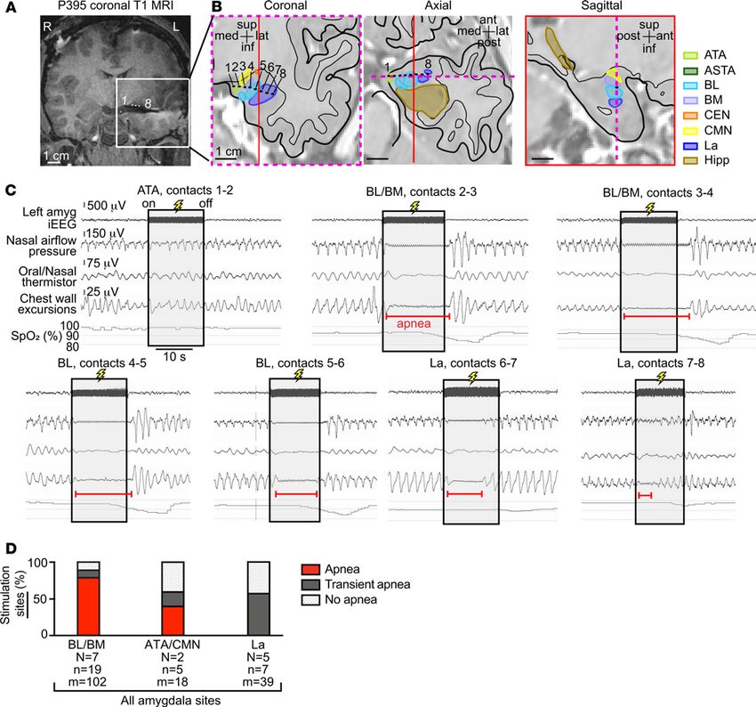
CLINICAL MEDICINE

Table 1. Patient characteristics and epilepsy history

Patient no.    Age (yr),     Handedness            Comorbidities         Imaging findings (MRI, Epilepsy onset (yr) Epilepsy duration Seizure frequency
                 Sex                                                              PET)                                     (yr)
P447             3, F      Not established      Tuberous sclerosis      Multiple bilateral tubers         1.1                 1.9              1–4/day
P466             5, M             L                History of             Ventriculomegaly                3.4                 2.1             1/2 weeks
                                                myelomeningocele
P381             5, M              L                  None            Normal brain MRI                     1.2                4.6              1–5/day
P422             9, F              R                  None             R frontal cortical                   5                  4               1–11/day
                                                                           dysplasia
P395            13, M              R              ADHD, anxiety      L frontal cavernoma                   9                   4               1–8/day
P407            14, M              R          Anxiety and depression  R frontotemporal                     6                   8               1/week
                                                                      hypometabolism;
                                                                      normal brain MRI
P412             17, M             R           Generalized anxiety   Bilateral gray matter                 5                   12            5–10/month
                                                                          heterotopia
P427             17, M          Mixed          Panhypopituitarism,         R frontal                       9                   8              4–20/day
                                                     previous         encephalomalacia
                                               craniopharyngioma
                                                    resection
Demographic information and epilepsy history for the patients reported in this study. Age refers to the child’s age at time of electrode implantation. All
patients are White and non-Hispanic. F, female; M, male; L, left; R, right; ADHD, attention-deficit/hyperactivity disorder.

                                    breathing prior to the end of the 31.8-second stimulation (Figure 3E). In each participant, stimulation of the
                                    amygdala elicited apnea or transient apnea in multiple trials and across multiple different electrode contact
                                    pairs (sites) (Figure 3, F and G). Interestingly, stimulation of either the right or left amygdala induced apnea,
                                    indicating that inhibition of breathing is not a function lateralized to one hemisphere. Overall, these findings
                                    indicate that either the right or left amygdala can induce apnea in patients with pediatric epilepsy across a
                                    diverse cohort with varied age, epilepsy etiology, seizure type, and seizure focus (Tables 1 and 2).
                                         Amygdala stimulation induces apnea without dyspnea. Because the amygdala plays a critical role in emo-
                                    tional processing and undergoes developmental changes through adolescence, we asked our subjects wheth-
                                    er they experienced any emotional changes during stimulation. Remarkably, the patients did not report any
                                    emotional change or display any signs of emotional effects. Because even seconds without breathing can
                                    be distressing (30), we asked our patients about their lack of breathing. Strikingly, the pediatric patients
                                    were completely unaware that they had stopped breathing and did not report any shortness of breath, air
                                    hunger, desire to breathe, or display any visible signs of respiratory distress during or after the periods of
                                    stimulation-induced apnea. The lack of distress or alarm during apnea suggests that amygdala stimulation
                                    may not only inhibit breathing, but also the sensation of the need to breathe and threat of suffocation when
                                    breathing is impaired. Such striking effects were also observed in adult patients described previously (16).
                                         The amygdala was the only stimulated forebrain structure that consistently induced apnea. In total, 342
                                    stimulation trials were conducted at 174 stimulation sites across the 8 pediatric epilepsy research par-
                                    ticipants (Figure 4). These included 159 trials at 31 amygdala sites and 181 trials at 143 nonamygdala
                                    candidate sites (Figure 4A). Of amygdala stimulation trials, 53% (84 of 159) produced apnea (Figure
                                    4F). In stark contrast, stimulating sites outside the amygdala failed to induce apnea or transient apnea
                                    in all but 2 trials. Stimulation lateral to the amygdala and within the hippocampus, cingulate gyrus,
                                    gyrus rectus, frontal cortex, temporal cortex, and parietal cortex all failed to induce apnea or transient
                                    apnea (Figure 4, B–F). In all these structures, normal breathing persisted throughout the period of stim-
                                    ulation. Thus, the amygdala was the only forebrain structure that consistently induced apnea (Figure
                                    4, A and F). The sole exception was in subject P447, where stimulation between a specific electrode
                                    contact pair 1 mm anterior to the amygdala caused transient apnea in 2 trials and no apnea in 3 trials.
                                    In all trials that resulted in apnea or transient apnea, breathing inhibition began within 1.9 seconds of
                                    electrical stimulation onset and normal breathing resumed within 5.5 seconds after stimulation ended
                                    (Figure 4A), suggesting that even short 5-second stimulations were sufficient to induce apnea at sen-
                                    sitive sites. Together, these findings suggest that the ability of forebrain stimulation to induce central
                                    apnea is relatively specific to the amygdala.

insight.jci.org   https://doi.org/10.1172/jci.insight.134852                                                                                                 3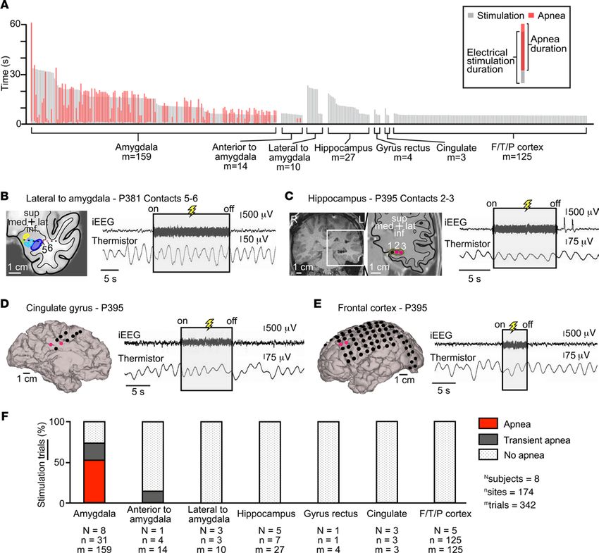
CLINICAL MEDICINE

Figure 1. High-density amygdala-depth electrodes were placed in 8 pediatric patients. (A) On the left, a high-density electrode with 0.88 mm distance
between contacts measuring 1.32 mm long was placed in all pediatric patients except P381 (left). In P381, a high-density 8 contact electrode with 1.71 mm
spacing between 2.29-mm-long electrode contacts was placed (right). (B) Anatomical localization of the amygdala-depth electrode contacts in the coronal
plane: unilaterally in 5 patients and bilaterally in 3 patients. Each amygdala was parcellated into their individual nuclei. Numbers 1–8 specify the electrode
contacts from medial to lateral. La, lateral nucleus (royal blue); BL, basolateral nucleus (light blue); BM, basomedial nucleus (lavender); CEN, central nucle-
us, orange; CMN, cortical and medial nuclei (yellow); ATA, amygdala transition areas (light green); ASTA, amygdalostriatal transition area (forest green);
AAA, anterior amygdala area (aqua); Hipp, hippocampus (brown).

                                         Stimulation-induced effects on breathing varied by contact location within the amygdala. Because the amygdala
                                    is composed of multiple nuclei that have distinct connectivity and functions, we hypothesized that specific
                                    locations within the amygdala may be responsible for stimulation-induced apnea. To test this hypothesis,
                                    we analyzed effects of stimulating different locations via different electrode pairs. Focal stimulation of a
                                    single nucleus or adjacent nuclei was technically feasible because high-density electrodes were used that
                                    contained closely spaced contacts (Figure 1). Due to the small size of the amygdala nuclei, adjacent elec-
                                    trode contacts spanned 2 nuclei in some cases. Except for the lateral nucleus (La), this necessitated group-
                                    ing adjacent nuclei together when analyzing the site-specific effects of electrical stimulation.
                                         The highly localized nature of stimulation-induced effects on breathing was exemplified by findings
                                    in P395 (Figure 5, A–C). In this subject, apnea was induced only when electrical stimuli were delivered
                                    through contacts located in the basal nuclei (basolateral nucleus/basomedial nucleus [BL/BM]). Stimula-
                                    tion of the La induced transient apnea, and stimulation of the amygdala transitional area (ATA) failed to
                                    disrupt breathing (Figure 5C).
                                         Across patients, stimulation elicited apnea at 55% (17 of 31) of all amygdala sites. Apnea was most
                                    consistently observed when stimulation spanned the basal nuclei (Figure 5D), where 79% of sites pro-
                                    duced apnea that persisted throughout the duration of stimulation and 11% produced transient apnea.

insight.jci.org   https://doi.org/10.1172/jci.insight.134852                                                                                                 4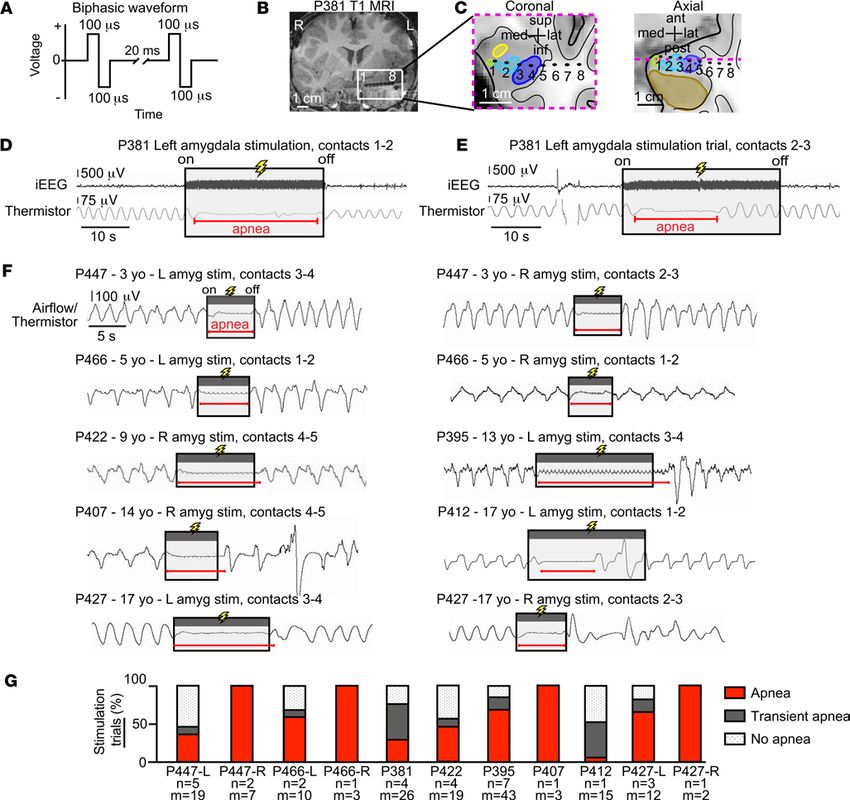
CLINICAL MEDICINE

Table 2. Results of iEEG and respiratory monitoring

Patient no.            Seizure type       Seizure focus     Clinical seizures      Seizures with      Seizures with     Average delay       Average delay
                                             (iEEG)                             respiratory signals       apnea           from onset        from onset to
                                                                                                                         to spread to         apnea (s)
                                                                                                                        amygdala (s)
P447                 FOA (myoclonic L anterior parietal            24                   24                 0                   –                   –
                       jerks); FOIA       lobe (FOA
                                      myoclonic jerks);
                                       R frontal lobe
                                            (FOIA)
P466                  FOIA (clonic)    R hemispheric-              12                   10                 9                 24.7                25.4
                                      multifocal, some
                                      with amygdala/
                                        hippocampal
                                        involvement
P381                       FOIA       R frontoparietal              5                    0                 –                   –                  –
P422                  FOA (motor)      R frontal lobe               2                    2                 0                   –                  –
P395                  FOA (motor)       L frontal lobe            >100                   0                 –                   –                  –
P40                   FOIA (clonic)    R hemispheric-              10                   10                 3                  27.7               28.1
                                      multifocal, some
                                      with early spread
                                        to amygdala
P412                  FOA (clonic)      L frontal lobe             8                     1                 0                   –                   –
P427                 FOA (myoclonic R frontal lobe                 53                   52                 0                   –                   –
                   jerks); FOIA; GTCS
Details of seizures observed during clinical monitoring. P381 and P395 did not comply with respiratory belts during their monitoring period. In the other
patients, respiratory signals during seizures could be compromised by motor artifact resulting from seizure, nonstationary status (e.g., walking, speaking),
or from clinical intervention. FOA, focal onset aware; FOIA, focal onset impaired awareness; GTCS, generalized tonic-clonic seizure; L, left; R, right; iEEG,
intracranial electroencephalography.

                                   A significantly lower number of sites spanning the ATA and cortical and medial nuclei (CMN) and
                                   within the La elicited apnea when electrically stimulated, at 40% and 0% respectively (Fisher’s exact
                                   test, P = 0.0011). These findings suggested a region near the BL/BM may be most sensitive to stimula-
                                   tion in eliciting apnea.
                                        Across subjects, amygdala stimulation-induced apnea localized to the medial aspect of the basal nuclei, ATA,
                                   and CMN. To examine whether a site near or within the basal nuclei may be responsible for stimula-
                                   tion-induced apnea, we plotted all electrode pairs within the amygdala and immediately adjacent to the
                                   amygdala, including the hippocampus (45 stimulation sites), for all subjects in a common anatomical
                                   space using Montreal Neurological Institute (MNI) coordinates (Figure 6). Electrode pairs that induced
                                   apnea were focally located in the medial portions of the amygdala and overlapped the BL/BM and
                                   ATA/CMN, consistent with the analysis using individual subject anatomy. More specifically, the elec-
                                   trode pairs that induced apnea appeared to localize to a more medial BL/BM region and within the ATA
                                   and CMN. Sites that induced transient apnea were lateral to the apnea-inducing sites. Sites that failed to
                                   induce apnea were located at even more lateral sites in the amygdala or in the hippocampus.
                                        Machine-learning algorithm predicts an amygdala inhibition of respiration site. To model the amygdala
                                   region responsible for inhibition of respiration, we trained a support vector machine to predict the
                                   respiratory outcome (apnea, transient apnea, or no effect) using the MNI coordinates of 45 tested sites
                                   in the mesial temporal lobe as predictors. A machine-learning algorithm predicted areas responsible
                                   for each respiratory outcome, including an apneic area located in the medial portion of the amyg-
                                   dala overlapping the medial portion of BL/BM as well as ATA and CMN (Figure 7A and Supple-
                                   mental Table 2; supplemental material available online with this article; https://doi.org/10.1172/
                                   jci.insight.134852DS1). Because this area in the amygdala inhibited respiration, we refer to it as the
                                   amygdala inhibition of respiration (AIR) site (Figure 7B and Supplemental Video 1).

insight.jci.org   https://doi.org/10.1172/jci.insight.134852                                                                                                5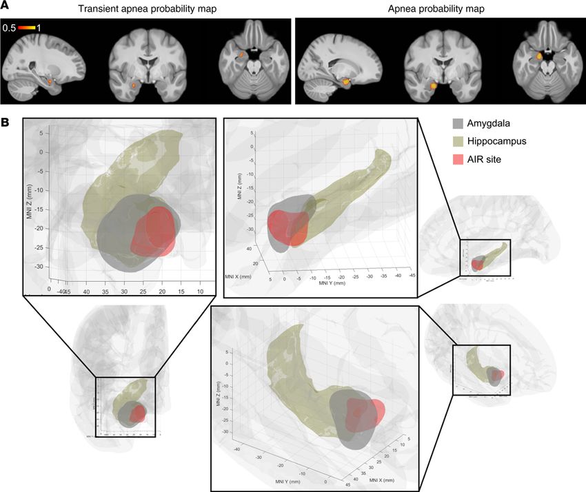
CLINICAL MEDICINE

Figure 2. Apnea coincided with seizure spread to the amygdala. (A) 3D surface-rendered MR image of P407’s right convexity with locations of
cortical electrode contacts (black circles) in the frontal, temporal, and parietal lobes. (B) T1-weighted structural MR images showing the hippocampal
and amygdala-depth electrodes and artifact produced by each electrode contact. (C) Schematic illustrating seizure spread from the lateral tempo-
ral cortex (purple circle, seizure onset) to the amygdala (red circle). (D) iEEG of seizure originating in lateral temporal cortex (purple circle in A) and
spreading quickly to the amygdala (red circle in B). Breathing is normal until seizure activity spreads to the amygdala, which coincides with apnea
and loss of normal breathing (inspiration is plotted up). The seizure spreads 10 seconds later to multiple other recording contacts in the hippocam-
pus, temporal cortex, and parietal cortex, as colored in A and B, and then generalizes, at which point motion artifact obscures the respiratory signal.
Normal breathing is observed after seizure end.

                                   Discussion
                                   In this report we make use of the rare opportunity to study pediatric patients implanted with intra-
                                   cranial electrodes undergoing continuous respiratory and seizure monitoring to directly examine how
                                   epileptic seizures and electrical stimulation of the amygdala alters breathing in children. Thus, we
                                   were able to test a number of important questions about the amygdala in seizure-induced apnea and
                                   SUDEP in children.
                                       First, we explored in children across a range of ages whether the amygdala plays a role in seizure-in-
                                   duced apnea, as in adults (16, 23), and whether amygdala stimulation might be sufficient to inhibit respi-
                                   ration. The amygdala undergoes functional and structural connectivity changes throughout adolescence
                                   (24–26), raising the possibility that effects in adults might not occur in children. Nevertheless, here we
                                   observed seizure-induced apnea in 1 patient who was 5 years old and in another who was 14 years old;

insight.jci.org   https://doi.org/10.1172/jci.insight.134852                                                                                              6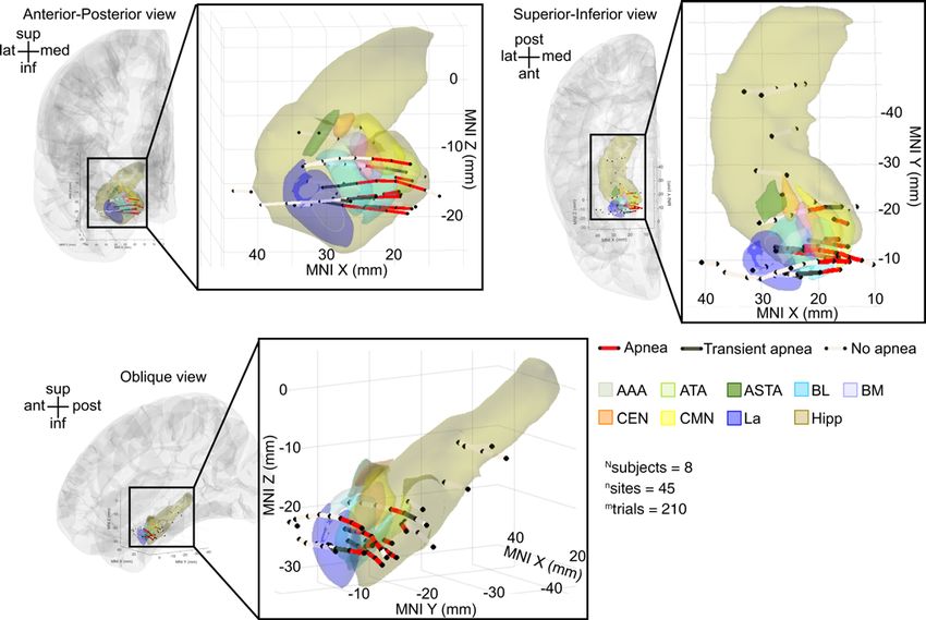
CLINICAL MEDICINE

Figure 3. Electrical stimulation of the amygdala induced varying degrees of apnea in all 8 patients with pediatric epilepsy. (A) Focal electrical stimulation was
delivered using a bipolar biphasic square waveform, 200-μs pulse width, 50-Hz frequency, and constant voltage (see Methods). (B) Coronal T1-weighted struc-
tural MR image showing the left amygdala-depth electrode in subject P381. Artifacts produced by each contact from most medial to lateral are labeled 1 to 8,
respectively. (C) Location of electrode contacts superimposed upon an anatomic diagram of P381’s left temporal lobe with amygdala nuclei, hippocampus, and
surrounding areas in coronal and axial planes. The dotted magenta line within the axial view represents coronal plane outlined with same dotted line. Amygdala
nuclei defined as in Figure 1B. (D) Amygdala stimulation (contacts 1 and 2) in P381 resulted in apnea that was almost immediate in onset and lasted the duration
of stimulation. iEEG signal shown on top and respiratory signal from oral/nasal thermistor shown below (inspiration plotted up; duration of stimulation depict-
ed by shaded gray box; duration of apnea shown by horizontal red line; conventions remain the same for E and F). (E) Stimulation of more lateral sites in the
amygdala (contacts 2 and 3) in P381 resulted in transient apnea: apnea was almost immediate in onset (
CLINICAL MEDICINE

Figure 4. Stimulation-induced apnea was relatively specific to the amygdala and not seen with stimulation of any other forebrain site. (A) Plot illustrating
durations of stimulation and durations of apnea for each trial, shown by gray and red vertical lines, respectively. All trials of stimulation were at least 5 seconds
in duration. Stimulation always induced apnea within 1.9 seconds after onset. Apnea was only observed with stimulation of the amygdala. (B) Location of elec-
trode contacts superimposed upon an anatomic diagram of P381’s left temporal lobe with amygdala nuclei and surrounding areas in coronal plane. Amygdala
nuclei defined as in Figure 1B. Stimulation outside and immediately lateral to the amygdala (contacts 5 and 6, pink) failed to elicit apnea. (C) Coronal T1-weight-
ed structural MR image showing the left hippocampus depth electrode in subject P395. Electrode contacts from most medial to lateral are labeled 1–3. Location
of electrode contacts superimposed upon an anatomic diagram of the left temporal lobe with hippocampus and surrounding areas in coronal plane. Stimulation
of the hippocampus (contacts 2 and 3, pink) failed to elicit apnea. (D) 3D surface-rendered MR image of P395’s left medial hemisphere with superimposed
electrode contacts in P395. Stimulation of the cingulate gyrus (pink circles) failed to elicit apnea. (E) 3D surface-rendered MR image of P395’s left convexity with
superimposed electrode contacts. Stimulation of the left frontal cortex (pink circles) failed to elicit apnea. (F) Bar graph illustrating the percentage of trials elic-
iting each of the 3 types of breathing effects (apnea, red; transient apnea, dark gray; no apnea, dot shading) for all patients. Beneath each bar are the number of
subjects (N), number of sites stimulated (n), and number of trials (m). Except for 1 electrode pair immediately anterior to the amygdala where stimulation led to
transient apnea, stimulation at all other sites and all other trials failed to induce any measurable changes in breathing.

                                      a connection between the amygdala and brainstem respiratory network is established in humans prior to
                                      age 3, either in early postnatal development or in utero. This functional connection likely underlies at least
                                      some cases of seizure-induced central apnea in children as well as adults. Moreover, although it remains
                                      to be proven, we speculate that this connection may have important implications for SUDEP and possibly

insight.jci.org   https://doi.org/10.1172/jci.insight.134852                                                                                                           8
CLINICAL MEDICINE
Figure 5. Stimulation-induced effects on breathing varied by contact location in the amygdala. (A) Coronal T1-weighted structural MR image showing the
left amygdala-depth electrode in subject P395. Electrode contacts from most medial to lateral are labeled 1 to 8, respectively. (B) The location of electrode
contacts superimposed upon anatomic diagrams of P395’s left temporal lobe with amygdala nuclei, hippocampus, and surrounding areas in coronal, axial
and sagittal planes. The dotted magenta line within the axial and sagittal view represents coronal plane outlined with the same dotted line. The red line in
the axial and coronal view represents the sagittal plane outlined with the same red line. Amygdala nuclei defined as in Figure 1B. (C) Stimulation of bipolar
contacts 1–8 from medial to lateral within the amygdala nuclei elicited different effects on breathing. Apnea was observed with stimulation of the basal
nuclei (BL/BM) and only transient apnea was observed with stimulation of the lateral amygdala. No apnea was observed with ATA stimulation. Oxygen
desaturation corresponded with the length of apnea and transient apnea at each site. (D) Bar graph depicting across-subject respiratory effects in the 3
grouped amygdala nuclei for which stimulation elicited effects on breathing. Within the BL/BM, 79% of stimulated electrode contact pairs were charac-
terized by apnea, and 11% were characterized by transient apnea. Fewer sites in the ATA/CMN showed breathing disruption (40% apnea, 20% transient
apnea). Transient apnea (57% of sites) or no apnea occurred with electrical stimulation of the La. Omnibus Fisher’s exact test showed significant association
between the 3 nuclei and the 3 respiratory outcomes (P = 0.00137). Post hoc planned comparisons showed significant association between sites in BL/BM
and apnea (BL/BM versus other areas, and apnea versus other respiratory outcome, Fisher’s exact test, P = 0.0011). Oxygen desaturation nadir occurred after
amygdala stimulation because oxygen saturation was measured with a pulse oximeter on the extremities resulting in a 15- to 20-second delay.
sudden infant death syndrome (SIDS) and sudden unexplained death in childhood (SUDC), conditions
that parallel SUDEP (31–33).
Despite disrupted breathing, it was striking that no child ever reported shortness of breath or dys-
pnea and no child displayed any visible signs of respiratory distress, emotional distress, or arousal.
insight.jci.org https://doi.org/10.1172/jci.insight.134852 9CLINICAL MEDICINE
Figure 6. Across-subject analysis localized amygdala stimulation-induced apnea to the medial aspect of BL/BM and the ATA/CMN. Anterior-posterior,
superior-inferior, and oblique views of all stimulated electrode pairs in the amygdala and hippocampus across subjects plotted in a common coordinate
system (MNI). Electrode contact pairs that produced apnea were located in the medial BL/BM and ATA/CMN. Electrode contact pairs that produced tran-
sient apnea were typically located just lateral or adjacent to this medial BL/BM region. Electrode contact pairs that failed to induce apnea were located in
La, outside the amygdala, or in the hippocampus. Electrode pairs that induced apnea are denoted by dark red lines; those that produced transient apnea
are depicted in dark gray lines; sites that did not induce apnea are depicted in light gray. For clarity, electrode sites on the lateral convexity, in the cingulate
gyrus, and gyrus rectus are not shown; no sites omitted from this figure demonstrated effects of electrical stimulation on breathing. See Supplemental
Table 1 for a list of MNI coordinates and the respiratory effect for each contact pair. Nuclei are color-coded with the same convention as in Figure 1B. Elec-
trode contacts may appear outside of the template brain due to anatomical variation across subjects relative to the MNI coordinate system. All electrode
contacts were plotted in the right hemisphere for simplicity, because no differences were observed between right and left amygdala stimulation.
These findings were consistent with previous observations in adults (16) and were remarkably similar
to the lack of apparent struggle or arousal observed in nonhuman primates upon amygdala stimulation
(34) and in patients who died from SUDEP (9). Thus, seizure spread to the amygdala may suppress
emotional distress and air hunger at the same time that it inhibits breathing. Supporting a coordinated
control of arousal and breathing by the amygdala, CO2-evoked arousal and respiration were increased
in patients with bilateral amygdala lesions (35). An absence of dyspnea and/or emotional alarm could
be critical during seizures, as it could interfere with a patient’s ability to maintain and protect their air-
way, especially if face down in bed, which could be catastrophic if they are alone and no one is nearby
to intervene (9).
Because epileptic seizures can induce plasticity and alter brain connectivity (36), it is conceivable that
different epilepsy types may induce different patterns of brain connectivity and enhance or reduce the
amygdala’s ability to suppress breathing. Therefore, we asked whether the amygdala’s effects on breath-
ing would be observed across epilepsy types. Interestingly, amygdala stimulation consistently induced
apnea across a spectrum of epilepsies, varying in seizure foci and pattern of spread. Thus, apnea did not
depend on epilepsy type. Moreover, in a previous study, amygdala stimulation induced apnea even in a
patient without epilepsy (16). These observations indicate that the connection between the amygdala
and brainstem respiratory networks is intrinsic, robust, and independent of epilepsy. However, they do
insight.jci.org https://doi.org/10.1172/jci.insight.134852 10CLINICAL MEDICINE
Figure 7. Machine-learning algorithm localizes an amygdala inhibition of respiration site. (A) Probability map of transient apnea (left) and apnea
(right) resulting from support vector machine classification of respiratory effects predicted from MNI coordinates of 45 tested mesial temporal elec-
trode contact pairs. (B) The amygdala inhibition of respiration (AIR) site (red) predicts apnea based on the results of A, overlaid on amygdala (gray,
FSL) and hippocampus (brown, FreeSurfer). Probability map is plotted in right hemisphere only for simplicity, because no differences were observed
between right and left amygdala stimulation.
not rule out the possibility that over time repeated seizures might strengthen this circuit and increase the
likelihood or severity of seizure-induced apnea.
Breathing is essential for life, and thus the ability of the amygdala to inhibit breathing might be sur-
prising. However, it has long been recognized that forebrain structures in humans can control breathing,
and in many cases this control involves emotion, such as in speech, laughing, and crying (18–22). Emotion
is thought to depend at least in part on the amygdala. The amygdala is critical for mediating defensive
responses to a variety of threats, including freezing and fight-and-flight responses (37, 38), where altering
respiration can be crucial for survival. Thus, a pathway by which the amygdala influences breathing might
be advantageous from an evolutionary perspective.
In addition to the amygdala, other forebrain sites have been implicated in breathing control, stimu-
lation-evoked apnea, and apnea during seizures (15–17, 19, 23, 29, 39). Thus, it is conceivable that stim-
ulation of other forebrain sites might also induce apnea. Therefore, we investigated whether apnea was
relatively specific to amygdala stimulation,or if it would be seen with other sites. Interestingly, however,
stimulating numerous sites outside the amygdala, including the hippocampus and cingulate gyrus, failed
to induce any effect on breathing in this cohort. The only exception was a site immediately anterior to the
insight.jci.org https://doi.org/10.1172/jci.insight.134852 11CLINICAL MEDICINE
amygdala in P447, which elicited transient apnea in 2 trials likely due to current spread from stimulation
within immediate proximity to the amygdala.
The amygdala is composed of multiple nuclei with different functions. Therefore, we asked if specific
regions of the amygdala mediate apnea. Stimulating a circumscribed zone in the medial amygdala consis-
tently produced apnea. This site, which we call the AIR site, overlaps the very medial aspect of the basal
nuclei (BL/BM), CMN, and ATA and also likely includes intercalated (ITC) neurons, a cluster of inhibitory
neurons lying between the BL/BM (40–42). From animal studies, several projections from neurons within the
AIR site might mediate apnea. First, BM and ITC neurons project directly to the parabrachial nucleus, a part
of the brainstem central pattern generator (43–50) controlling timing and phase transitions of the respiratory
cycle via the pre-Bötzinger complex, a key driver of inspiration and respiratory rhythm (51–53). A second
pathway by which neurons in the AIR site might affect breathing is via the central nucleus (CEN); the BL,
BM, medial nucleus, and ITC project to the CEN (27, 49, 54, 55), which in turn also projects to sites in the
breathing central pattern generator (43–50), including the pre-Bötzinger complex (51–53). A third pathway
by which breathing may be affected is via projections from the medial nucleus to both the hypothalamus and
bed nucleus of the stria terminalis (55, 56), which both in turn project to the pre-Bötzinger complex (48). As
mentioned above, the absence of dyspnea and emotional alarm during the apneic episodes was surprising
and suggests that dyspnea and arousal might also be suppressed by electrical stimulation. An inhibitory con-
nection to midbrain neurons sensitive to CO2/pH might suppress dyspnea and arousal (57), although con-
nections between these neurons and the AIR site are not clear. Alternatively, the parabrachial nucleus, which
receives direct projections from AIR site neurons, promotes arousal when CO2 concentrations rise, and might
be ideally positioned to affect dyspnea and arousal, as well as breathing (58). Finally, it is conceivable that AIR
site projections to cortical, limbic, and paralimbic sites might also suppress dyspnea (59).
There are limitations to the methods required by these studies that deserve consideration. With electri-
cal stimulation, we cannot precisely target specific neurons and circuits. Using finite element modeling of
bipolar stimulation, with the parameters we used in this study, current spread is localized to a small discrete
region around the bipolar electrode contacts (60). Regardless, we cannot exclude that apnea was induced
from current spread to immediately nearby sites or white matter tracts; nor is it known that the electrical
stimulation that elicits apnea is activating, inhibiting, or a combination of both. Further, we were unable to
test whether stimulation of sites within the CEN or AAA could elicit apnea because electrode contacts were
not located here. Focal stimulation of these small nuclei as well as other forebrain sites will require further
study. Although high-resolution MRI and CT allow high-quality imaging of individual brain structures and
precise location of electrode contacts, the human amygdala atlas (28) used to define the nuclei was derived
from a study of normal human subjects and thus may differ from our epilepsy cohort. Supporting the use of
this atlas, no structural abnormalities were detected by T1- and T2-weighted imaging in any amygdala of the
patients reported here, suggesting that amygdala anatomy was likely similar to the normal population. Nev-
ertheless, it remains possible that chronic epilepsy might induce functional or structural connectivity changes
in the amygdala over time. And finally, because unilateral amygdala seizures without contralateral amygdala
involvement can elicit apnea (P466), seizures in or stimulation of only 1 amygdala appears sufficient to inhibit
breathing. Nevertheless, we cannot rule out that both amygdalae are required for breathing inhibition.
Importantly, localizing brain sites responsible for seizure-induced loss of breathing in patients with
epilepsy could be key to preventing SUDEP. Novel imaging strategies might reveal abnormalities within
the AIR site or differences in connectivity between the AIR site and brainstem respiratory sites. Such efforts
could help predict who might be at the greatest risk for seizure-induced apnea and SUDEP, for example,
if it were found that connections between the amygdala and respiratory sites were abnormally strong in
specific individuals. Moreover, localizing the AIR site provides a potential therapeutic target. Developing
methods to inhibit the AIR site and its connection to brainstem respiratory networks via drug based or
surgical strategies may limit apnea during seizures and thus help prevent SUDEP.
Methods
Patients. Eight pediatric patients with medically intractable epilepsy were studied while undergoing
iEEG during a 2- to 3-week monitoring period for seizure focus localization (age range, 3–17 years old;
2 females; see Table 1 for patient characteristics and epilepsy history). Intracranial electrodes (Ad-Tech
Medical Instrument Corporation) were implanted at sites determined by the multidisciplinary epilepsy
team at the University of Iowa. During the monitoring period, antiepileptic drugs were tapered to allow
insight.jci.org https://doi.org/10.1172/jci.insight.134852 12CLINICAL MEDICINE
seizures to occur to identify seizure foci; antiepileptic drugs were restarted prior to electrical stimulation
used in the current study.
Data acquisition, imaging, and electrode localization. iEEG data were acquired using a Nihon Kohden
JE-120 256 channel amplifier and analyzed using NeuroWorkbench software. Patients were monitored at
the University of Iowa Stead Family Children’s Hospital. Experimental recordings did not interfere with
the collection of clinically relevant data. Seizure foci and timing of seizure onset and spread were deter-
mined by analysis of iEEG by a pediatric epileptologist with independent assessment by a pediatric epilep-
sy neurosurgeon. Electrode localization was performed using MR and CT imaging using techniques previ-
ously reported (16, 61). For each patient, whole-brain high-resolution T1-weighted structural 3T MRI scans
(resolution and slice thickness, ≤1.0 mm) were obtained before and after electrode implantation. CT and
MR structural scans after implantation were linearly coregistered to MR scans before implantation using
the FLIRT module of FSL (62). Images were corrected for displacement and deformation from surgery
using nonlinear thin-plate spline warping using manually selected control points (63). Finally, FreeSurfer
was used to identify the cortical surface and white-gray boundaries within T1 images before implantation,
which were corrected manually based on visual inspection where necessary (64). High-density depth elec-
trodes containing closely spaced contacts (Figure 1A) were placed in all 8 pediatric patients. In 5 patients,
only 1 amygdala was implanted, and in 3 patients, both amygdalae were implanted. In total, 11 amygdalae
contained depth electrodes across 8 subjects (Figure 1B).
Amygdala nuclei parcellation. Amygdala nuclei were delineated and parcellated using the CIT168 human
brain template (28). This was conducted using a high-precision nonlinear volumetric coregistration of pre-
operative structural T1 and T2 imaging onto the template brain. Eight amygdala nuclei were delineated,
and the structural relationships between electrode contacts and nuclei were determined from the project-
ed contact locations within the preoperative brain images. For more clarity and simplicity, as described
in Tyszka and Pauli (28), the BL as labeled in this study includes the dorsal, intermediate, and ventral/
paralaminar subnuclei of the BL. The CIT168 atlas is derived from in vivo imaging in 168 subjects and
thus provides a much finer parcellation of the amygdala with more accurate external boundary definition
than current ex vivo histology-based atlases or other nuclei parcellations from other sources. Moreover,
amygdala volume (both right and left) does not significantly vary across age, as observed in a study of 166
participants from 5 to 30 years old (26), thus supporting the use of this anatomic atlas in children.
Electrical stimulation. Electrical stimulation was conducted at the bedside while the subjects were awake
and in a relaxed state (16). Electrical stimulation for functional brain mapping is a well-established and
safe method used routinely for clinical and research purposes in neurosurgery patients (16, 65). Bipolar
biphasic stimulation was conducted with parameters set at 200-μs pulse width, a frequency of 50 Hz, and
constant voltage using a Grass SD9 stimulator. Pairs of adjacent electrode contacts (sites) were stimulated.
A dose-response curve for each subject was obtained by increasing stimulation voltage beginning at 2.5
V until breathing was affected up to a maximum of 15 V (the typical threshold for motor movement with
stimulation of the motor cortex in these subjects). No afterdischarges occurred during experimental stim-
ulation trials. With increasing voltage during the establishment of the dose-response curve, apnea was an
all or none effect. Apnea occurred at 10 V in all patients except P381 and P412, where apnea was observed
with 15 V, consistent with previous findings (16). Stimulation duration was varied between patients based
on the proximity of the stimulation site to the patient’s seizure focus (range, 5–60 seconds). Sites in close
proximity to the seizure focus were stimulated for shorter durations. To allow for recovery between trials,
we maintained an interval of 1.5–2.5 minutes from the end of one trial to the beginning of the next. No
subjects showed evidence of, or complained of, any pain or discomfort during stimulation. Patients and
their parents/guardians were blinded to the delivery of the electrical stimulation.
Respiratory measurements. To record breathing, patients wore a chest/abdominal plethysmography
belt (ProTech zRIP) during their 2- to 3-week monitoring period for seizure focus localization. Oral and
nasal airflow were additionally measured with a BiNAPS nasal pressure transducer and a ThermiSense
oral/nasal thermistor (Salter Labs) and were worn as tolerated. Video recordings of the patient and
respiratory plethysmography were obtained in synchrony with iEEG using the Nihon Kohden system.
During the stimulation session, subjects wore the chest/abdominal plethysmography belt, nasal pres-
sure transducer, and oral/nasal thermistor. Capillary oxyhemoglobin saturation (SpO2) was recorded
using a BluPro SpO2 sensor. A breath was defined as a complete inspiratory and expiratory cycle. Apnea
durations were tabulated in Microsoft Excel based on measurements derived from NeuroWorkbench
insight.jci.org https://doi.org/10.1172/jci.insight.134852 13CLINICAL MEDICINE
(Nihon Kohden). Central apnea was defined as at least 1 missed breath with a flattened airflow trace
(oral/nasal thermistor or nasal pressure transducer) and verified by absence of chest wall movement
measured with plethysmography belts or video. Trials were categorized as “apnea” when breathing
was interrupted for the entire stimulation duration and as “transient apnea” when normal breathing
resumed prior to the end of stimulation.
Experimental design and statistical analysis. Multiple stimulation trials were conducted at different fore-
brain electrode contact pairs (sites). The outcome of each stimulation trial was categorized as apnea, tran-
sient apnea, or no apnea and each stimulated site was categorized as apnea, transient apnea, or no apnea
based on respiratory outcome elicited by the majority of trials. If stimulation of a site resulted in an equal
distribution of respiratory outcomes over all trials at that site, it was categorized as the greater inhibitory
effect of the 2 (e.g., if stimulation resulted in apnea and transient apnea in an equal number of trials, the
site was categorized as apnea). If all 3 outcomes were observed with equal proportion, that site was catego-
rized as the middle outcome (i.e., transient apnea); this occurred at only 1 site. After stimulation trials, all
subjects except P447 (age 3) were asked to report any noticeable change in breathing, difficulty breathing,
or changes in emotional state.
Statistics. Statistical and data analyses were conducted using Matlab (Mathworks), Prism 7.0 (Graph-
pad Software Inc.), and Microsoft Excel. For classification analyses, we used a multiclass support vector
machine (66) with a 4-mm Gaussian kernel and 5-fold cross validation implemented in the Classification
Learner App in Matlab R2018b (fitcecoc.m). The kernel standard width was selected to be 4 mm based on
cross-validation performance.
Study approval. Experimental protocols were approved by the University of Iowa Institutional Review
Board. Informed consent was obtained from the parents or legal guardians of all patients. Verbal assent
was obtained from children 5 to 9 years of age, and written assent was obtained from all older children.
No assent was obtained from the 3 year old, but she was tested in the company of her parents who had
the option of terminating the experimental protocol at any time. Consent could be rescinded at any time
without interfering with the child’s clinical evaluation. Similarly, children could rescind assent at any time.
Author contributions
BJD, GISH, JAW, and AER conceived and designed the study. AER, CKK, GISH, AWS, DT, MAC,
MAH, GBR, MS, JAW, and BJD acquired and/or analyzed data. BJD, AER, JAW, and MS drafted a sig-
nificant portion of the manuscript or figures.
Acknowledgments
Support for this work was provided by the Roy J. Carver Charitable Trust (to BJD), the National Institute of
Neurological Disorders and Stroke — Congress of Neurological Surgeons K12 Getch Scholar Award (to BJD),
the National Institute of General Medical Sciences training grant T32 GM067795 (to GISH), and the National
Institute of General Medical Sciences training grant T32 GM108540 (to AWS). The graphical abstract was
created with BioRender.
Address correspondence to: Brian J. Dlouhy, Department of Neurosurgery, University of Iowa Carver
College of Medicine, University of Iowa Stead Family Children’s Hospital, 200 Hawkins Drive, Iowa City,
Iowa 52242, USA. Phone: 319.356.2542; Email: brian-dlouhy@uiowa.edu.
1. Shorvon S, Tomson T. Sudden unexpected death in epilepsy. Lancet. 2011;378(9808):2028–2038.
2. Thurman DJ, Hesdorffer DC, French JA. Sudden unexpected death in epilepsy: assessing the public health burden. Epilepsia.
2014;55(10):1479–1485.
3. Sveinsson O, Andersson T, Carlsson S, Tomson T. The incidence of SUDEP: A nationwide population-based cohort study.
Neurology. 2017;89(2):170–177.
4. Sillanpää M, Shinnar S. Long-term mortality in childhood-onset epilepsy. N Engl J Med. 2010;363(26):2522–2529.
5. Dlouhy BJ, Gehlbach BK, Richerson GB. Sudden unexpected death in epilepsy: basic mechanisms and clinical implications for
prevention. J Neurol Neurosurg Psychiatry. 2016;87(4):402–413.
6. Doumlele K, Friedman D, Buchhalter J, Donner EJ, Louik J, Devinsky O. Sudden unexpected death in epilepsy among patients
with benign childhood epilepsy with centrotemporal spikes. JAMA Neurol. 2017;74(6):645–649.
7. Harden C, et al. Practice guideline summary: Sudden unexpected death in epilepsy incidence rates and risk factors: Report of
the Guideline Development, Dissemination, and Implementation Subcommittee of the American Academy of Neurology and
the American Epilepsy Society. Neurology. 2017;88(17):1674–1680.
insight.jci.org https://doi.org/10.1172/jci.insight.134852 14CLINICAL MEDICINE
8. Verducci C, et al. SUDEP in the North American SUDEP Registry: The full spectrum of epilepsies. Neurology. 2019;93(3):e227–e236.
9. Ryvlin P, et al. Incidence and mechanisms of cardiorespiratory arrests in epilepsy monitoring units (MORTEMUS): a retrospec-
tive study. Lancet Neurol. 2013;12(10):966–977.
10. Kim Y, et al. Severe peri-ictal respiratory dysfunction is common in Dravet syndrome. J Clin Invest. 2018;128(3):1141–1153.
11. Bateman LM, Spitz M, Seyal M. Ictal hypoventilation contributes to cardiac arrhythmia and SUDEP: report on two deaths in
video-EEG-monitored patients. Epilepsia. 2010;51(5):916–920.
12. Bateman LM, Li CS, Seyal M. Ictal hypoxemia in localization-related epilepsy: analysis of incidence, severity and risk factors.
Brain. 2008;131(Pt 12):3239–3245.
13. Watanabe K, et al. Seizures with apnea in children. Pediatrics. 1982;70(1):87–90.
14. Nashef L, Walker F, Allen P, Sander JW, Shorvon SD, Fish DR. Apnoea and bradycardia during epileptic seizures: relation to
sudden death in epilepsy. J Neurol Neurosurg Psychiatry. 1996;60(3):297–300.
15. Kaada BR, Jasper H. Respiratory responses to stimulation of temporal pole, insula, and hippocampal and limbic gyri in man.
AMA Arch Neurol Psychiatry. 1952;68(5):609–619.
16. Dlouhy BJ, et al. Breathing inhibited when seizures spread to the amygdala and upon amygdala stimulation. J Neurosci.
2015;35(28):10281–10289.
17. Lacuey N, Hampson JP, Harper RM, Miller JP, Lhatoo S. Limbic and paralimbic structures driving ictal central apnea. Neurology.
2019;92(7):e655–e669.
18. McKay LC, Evans KC, Frackowiak RS, Corfield DR. Neural correlates of voluntary breathing in humans. J Appl Physiol.
2003;95(3):1170–1178.
19. Murphy K, et al. Cerebral areas associated with motor control of speech in humans. J Appl Physiol. 1997;83(5):1438–1447.
20. Masaoka Y, Homma I. Amygdala and emotional breathing in humans. Adv Exp Med Biol. 2004;551:9–14.
21. Rochet-Capellan A, Fuchs S. Changes in breathing while listening to read speech: the effect of reader and speech mode. Front
Psychol. 2013;4:906.
22. Shea SA. Behavioural and arousal-related influences on breathing in humans. Exp Physiol. 1996;81(1):1–26.
23. Nobis WP, et al. The effect of seizure spread to the amygdala on respiration and onset of ictal central apnea. J Neurosurg. 2019:1–11.
24. Scherf KS, Smyth JM, Delgado MR. The amygdala: an agent of change in adolescent neural networks. Horm Behav.
2013;64(2):298–313.
25. Jiang Y, Tian Y, Wang Z. Age-related structural alterations in human amygdala networks: reflections on correlations between
white matter structure and effective connectivity. Front Hum Neurosci. 2019;13:214.
26. Saygin ZM, et al. Structural connectivity of the developing human amygdala. PLoS One. 2015;10(4):e0125170.
27. Janak PH, Tye KM. From circuits to behaviour in the amygdala. Nature. 2015;517(7534):284–292.
28. Tyszka JM, Pauli WM. In vivo delineation of subdivisions of the human amygdaloid complex in a high-resolution group tem-
plate. Hum Brain Mapp. 2016;37(11):3979–3998.
29. Nobis WP, et al. Amygdala-stimulation-induced apnea is attention and nasal-breathing dependent. Ann Neurol. 2018;83(3):460–471.
30. Gigliotti F. Mechanisms of dyspnea in healthy subjects. Multidiscip Respir Med. 2010;5(3):195–201.
31. Buchanan GF. Impaired CO2-induced arousal in SIDS and SUDEP. Trends Neurosci. 2019;42(4):242–250.
32. Richerson GB, Buchanan GF. The serotonin axis: shared mechanisms in seizures, depression, and SUDEP. Epilepsia.
2011;52 Suppl 1:28–38.
33. Hefti MM, et al. Hippocampal malformation associated with sudden death in early childhood: a neuropathologic study: Part 2
of the investigations of The San Diego SUDC Research Project. Forensic Sci Med Pathol. 2016;12(1):14–25.
34. Reis DJ, McHugh PR. Hypoxia as a cause of bradycardia during amygdala stimulation in monkey. Am J Physiol.
1968;214(3):601–610.
35. Feinstein JS, et al. Fear and panic in humans with bilateral amygdala damage. Nat Neurosci. 2013;16(3):270–272.
36. Maccotta L, et al. Impaired and facilitated functional networks in temporal lobe epilepsy. Neuroimage Clin. 2013;2:862–872.
37. Taugher RJ, et al. The amygdala differentially regulates defensive behaviors evoked by CO2. Behav Brain Res. 2020;377:112236.
38. Hagenaars MA, Oitzl M, Roelofs K. Updating freeze: aligning animal and human research. Neurosci Biobehav Rev. 2014;47:165–176.
39. Lacuey N, Zonjy B, Londono L, Lhatoo SD. Amygdala and hippocampus are symptomatogenic zones for central apneic sei-
zures. Neurology. 2017;88(7):701–705.
40. Millhouse OE. The intercalated cells of the amygdala. J Comp Neurol. 1986;247(2):246–271.
41. Zikopoulos B, John YJ, García-Cabezas MÁ, Bunce JG, Barbas H. The intercalated nuclear complex of the primate amygdala.
Neuroscience. 2016;330:267–290.
42. Nitecka L, Ben-Ari Y. Distribution of GABA-like immunoreactivity in the rat amygdaloid complex. J Comp Neurol.
1987;266(1):45–55.
43. Geerling JC, Loewy AD. Aldosterone-sensitive neurons in the nucleus of the solitary tract: bidirectional connections with the
central nucleus of the amygdala. J Comp Neurol. 2006;497(4):646–657.
44. Hopkins DA, Holstege G. Amygdaloid projections to the mesencephalon, pons and medulla oblongata in the cat. Exp Brain Res.
1978;32(4):529–547.
45. Moga MM, Gray TS. Peptidergic efferents from the intercalated nuclei of the amygdala to the parabrachial nucleus in the rat.
Neurosci Lett. 1985;61(1-2):13–18.
46. Verner TA, Pilowsky PM, Goodchild AK. Retrograde projections to a discrete apneic site in the midline medulla oblongata of
the rat. Brain Res. 2008;1208:128–136.
47. Jongen-Rêlo AL, Amaral DG. Evidence for a GABAergic projection from the central nucleus of the amygdala to the brainstem
of the macaque monkey: a combined retrograde tracing and in situ hybridization study. Eur J Neurosci. 1998;10(9):2924–2933.
48. Yang CFK, Kim EJ, Callaway EM, Feldman JL. Monosynaptic projections to excitatory and inhibitory preBötzinger complex
neurons. bioRxiv. https://doi.org/10.1101/694711. Published July 7, 2019. Accessed March 9, 2020.
49. Petrovich GD, Risold PY, Swanson LW. Organization of projections from the basomedial nucleus of the amygdala: a PHAL
study in the rat. J Comp Neurol. 1996;374(3):387–420.
50. Subramanian HH, Balnave RJ, Holstege G. The midbrain periaqueductal gray control of respiration. J Neurosci.
insight.jci.org https://doi.org/10.1172/jci.insight.134852 15You can also read