Linking Amygdala Persistence to Real-World Emotional Experience and Psychological Well-Being
←
→
Page content transcription
If your browser does not render page correctly, please read the page content below
The Journal of Neuroscience, 0, 2021 • 00(00):000 • 1

Behavioral/Cognitive

Linking Amygdala Persistence to Real-World Emotional
Experience and Psychological Well-Being
    Nikki A. Puccetti,1 Stacey M. Schaefer,2 Carien M. van Reekum,3 Anthony D. Ong,4                                                                               David M. Almeida,5
    Carol D. Ryff,6 Richard J. Davidson,2,6 and Aaron S. Heller1
1
  Department of Psychology, University of Miami, Coral Gables, Florida 33124, 2Center for Healthy Minds, University of Wisconsin-Madison,
Madison, Wisconsin 53703, 3School of Psychology and Clinical Language Science, University of Reading, Reading RG6 6AL, United Kingdom,
4
  Department of Human Development, Cornell University, Ithaca, New York 14853, 5Department of Human Development and Family Studies and
Center for Healthy Aging, The Pennsylvania State University, University Park, Pennsylvania 16802, and 6Department of Psychology, University of
Wisconsin-Madison, Madison, Wisconsin 53706

Neural dynamics in response to affective stimuli are linked to momentary emotional experiences. The amygdala, in particular,
is involved in subjective emotional experience and assigning value to neutral stimuli. Because amygdala activity persistence
following aversive events varies across individuals, some may evaluate subsequent neutral stimuli more negatively than
others. This may lead to more frequent and long-lasting momentary emotional experiences, which may also be linked to self-
evaluative measures of psychological well-being (PWB). Despite extant links between daily affect and PWB, few studies have
directly explored the links between amygdala persistence, daily affective experience, and PWB. To that end, we examined
data from 52 human adults (67% female) in the Midlife in the United States study who completed measures of PWB, daily
affect, and functional MRI (fMRI). During fMRI, participants viewed affective images followed by a neutral facial expression,
permitting quantification of individual differences in the similarity of amygdala representations of affective stimuli and neu-
tral facial expressions that follow. Using representational similarity analysis, neural persistence following aversive stimuli was
operationalized as similarity between the amygdala activation patterns while encoding negative images and the neutral facial
expressions shown afterward. Individuals demonstrating less persistent activation patterns in the left amygdala to aversive
stimuli reported more positive and less negative affect in daily life. Further, daily positive affect served as an indirect link
between left amygdala persistence and PWB. These results clarify important connections between individual differences in
brain function, daily experiences of affect, and well-being.
Key words: amygdala; daily affect; emotion; fMRI; neural dynamics; representational similarity analysis

      Significance Statement
      At the intersection of affective neuroscience and psychology, researchers have aimed to understand how individual differences
      in the neural processing of affective events map onto to real-world emotional experiences and evaluations of well-being.
      Using a longitudinal dataset from 52 adults in the Midlife in the United States (MIDUS) study, we provide an integrative
      model of affective functioning: less amygdala persistence following negative images predicts greater positive affect (PA) in
      daily life, which in turn predicts greater psychological well-being (PWB) seven years later. Thus, day-to-day experiences of PA
      comprise a promising intermediate step that links individual differences in neural dynamics to complex judgements of PWB.

                                                                                                                     Introduction
Received June 29, 2020; revised Feb. 3, 2021; accepted Feb. 24, 2021.                                                Psychological well-being (PWB) captures one’s perceived self-ac-
   Author contributions: S.M.S., C.M.v.R., A.D.O., D.M.A., C.D.R., and R.J.D. designed research; S.M.S., C.M.v.R.,   ceptance, positive relations with others, autonomy, environmen-
A.D.O., D.M.A., C.D.R., and R.J.D. performed research; N.A.P. and A.S.H. analyzed data; N.A.P. and A.S.H. wrote      tal mastery, purpose in life, and personal growth (Ryff, 1989;
the paper.
   Publicly available data from the MIDUS study were used for this research. Since 1995, the MIDUS study has
                                                                                                                     Ryff and Keyes, 1995). Relatively more enduring, trait-like judge-
been funded by the following: John D. and Catherine T. MacArthur Foundation Research Network and                     ments of PWB are linked to relatively more transient positive
National Institute on Aging Grants P01-AG020166 and U19-AG051426.                                                    and negative emotional states in the real-world (Burns and Ma,
   R.J.D. serves on the board of directors for the non-profit organization Healthy Minds Innovations. All other      2015; Rush et al., 2019). Specifically, higher PWB is associated
authors declare no competing financial interests.
   Correspondence should be addressed to Aaron S. Heller at aheller@miami.edu.
                                                                                                                     with greater positive affect (PA; Burns and Ma, 2015) and lower
   https://doi.org/10.1523/JNEUROSCI.1637-20.2021                                                                    negative affect (NA; Rush et al., 2019) in daily life. A key question
Copyright © 2021 the authors                                                                                         is what neurobiological processes give rise to these subjective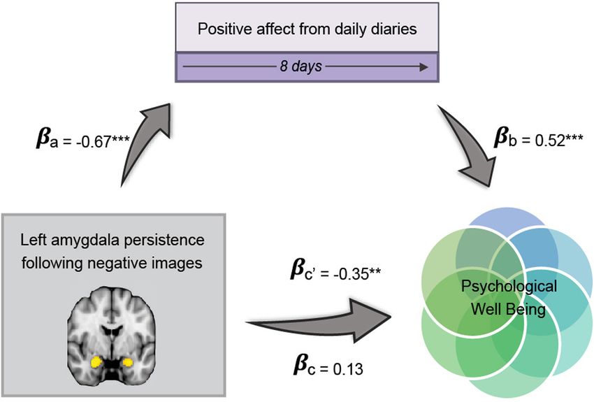
2 • J. Neurosci., 0, 2021 • 00(00):000                                                        Puccetti et al. · Amygdala Persistence, Daily Affect, and Well-Being

affective experiences and judgements. One promising source of          we tested whether daily affect provided a pathway by which
these individual differences may be the time course of amygdala        amygdala persistence was linked with PWB.
activity. (Sander et al., 2003).
     The amygdala is necessary for threat detection and threat         Materials and Methods
conditioning–the assigning of value to neutral stimuli (LeDoux,        Participant characteristics
1996). The amygdala is also implicated in a range of affective         Data were collected from 2004 to 2009 as a part of the MIDUS-II
processes, including general detection of salience (Davis and          Longitudinal Study of Health and Well-being (http://www.midus.wisc.
Whalen, 2001; Sander et al., 2003; Kober et al., 2008), facial proc-   edu/), which recruited a national sample (ages 35–85) through random
essing (Todorov et al., 2008), and experiencing fear (LeDoux,          digit dialing. Participants first completed self-report questionnaires
2000). In particular, the amygdala supports negative appraisals of     (n = 4963), a random subsample then completed a series of eight daily
putatively neutral stimuli when they are preceded by an unre-          consecutive telephone interviews to assess daily affect (n = 2022), and a
lated, aversive stimulus (Lapate et al., 2016). Individual differen-   further subgroup able and willing to travel to the Midwest included neu-
ces in amygdala response to fearful faces predict how negatively       roscience assessments, including an fMRI scan (n = 72). Two participants
                                                                       did not complete all five functional runs of the MRI task; eight were
a subsequent, unrelated neutral stimulus is appraised (Lapate et
                                                                       removed for excessive motion (sudden spikes, gradual drift, or both, that
al., 2016, 2017). This biasing effect may be because of persistence
                                                                       exceeded 2 mm). Nine of the remaining participants did not complete
of amygdala activity following emotionally evocative stimuli that      the daily diary study. Finally, one participant was excluded from the
“spills over” into the encoding of the subsequent neutral stimulus     analysis for reporting levels of daily NA greater than 3 SDs from the
(Tambini et al., 2017; Grupe et al., 2018).                            group mean. This resulted in 52 participants that completed MRI, daily
     Functional MRI (fMRI) research suggests that individual differ-   diary, and self-reported PWB for the analyses [39–76 years old
ences in the degree of amygdala activity persistence after aversive    (M = 57.74, SD = 10.5), 67% female, 69% white, 29% African American,
events is associated with individual differences in affective style.   2% Native American or Alaskan Native]. There were five sets of twins in
Schuyler et al. (2014) found that more persistent amygdala activity    the sample. Lastly, 31 of these subjects repeated the self-report question-
following negative images predicted higher levels of neuroticism.      naires pertaining to PWB again seven years later (SD = 1.6, range = 5–
Additionally, greater amygdala persistence following negative          9 years).
                                                                           On average, the first measurement of PWB was completed 729 d, or
images was linked to lower likeability ratings of subsequent neutral
                                                                       24.3 months, before the daily diary procedure (SD = 430 d, range = 136–
faces. Similarly, greater amygdala persistence following negative
                                                                       1533 d) and 1230 d, or 41 months, before the fMRI scan (SD = 372 d,
words has been found in patients with major depressive disorder        range = 348–1669 d). Thirty-one participants completed the daily diaries
(Siegle et al., 2002). These studies demonstrate that amygdala per-    before the fMRI session by an average of 999 d (SD = 347 d, range = 408–
sistence is linked to individual differences in affective style.       1491 d). The other 21 participants completed the fMRI scan before the
     However, the existing literature has relied on univariate         daily diary procedure by an average of 231 d (SD = 59, range = 161–
amygdala analyses that cannot account for the multivariate rep-        386 d). Because significant time passed between each measurement and
resentation of affective stimuli. Assessing persistence by averag-     time lag varied by participants, each relevant time lag is included as a
ing activity over the amygdala removes fine-grained, spatial           covariate in the analysis. Therefore, significant relationships from the
activity patterns that carry detailed information about stimulus       analyses are likely to reflect stable, meaningful connections that with-
properties. In contrast, multivariate approaches, such as repre-       stand long and variable measurement gaps. Data and documentation for
sentational similarity analysis (RSA), preserve the voxel-wise pat-    MIDUS 1, MIDUS 2, MIDUS 3, and all MIDUS projects are available to
                                                                       the public for downloading at the Inter-university Consortium for
tern of activation and compare the neural representations of
                                                                       Political and Social Research (ICPSR) website (https://www.icpsr.umich.
different types of stimuli. Although RSA was initially used to         edu/web/ICPSR/series/203).
examine similarity in visual perception (Kriegeskorte and
Bandettini, 2007; Kriegeskorte et al., 2008), it has been applied to   Experimental design and statistical analyses
identify similarity in affective processing (Ölander et al., 2017;     PWB
Brooks and Freeman, 2018). Given that we rarely encounter a            Participants completed a 42-item measure of PWB (Ryff, 1989; Ryff and
single stimulus in isolation, the dynamics of amygdala represen-       Keyes, 1995). This measure contains subscales for six domains of PWB,
tation across temporally-related stimuli are paramount to under-       including self-acceptance, positive relations with others, autonomy, envi-
standing individual differences in well-being. Therefore, by           ronmental mastery, purpose in life, and personal growth. Each subscale
capturing the commonalities in the voxel-wise pattern of amyg-         is comprised of seven items that are rated on a 1–7 scale (“strongly
dala activity among affective stimuli, compared with the pattern       agree” to “strongly disagree”). Items that indicate greater PWB are
                                                                       reverse coded such that higher overall scores reflect higher PWB. The
of activity among subsequent neutral facial expressions, RSA
                                                                       total score for the measure is calculated by averaging the score from each
could yield a more accurate and ecologically valid metric of           of the subscales. This measure of PWB has been shown to be reliable
amygdala persistence.                                                  over the span of six weeks (test-retest reliability for the six scales .0.8;
     To that end, the current study used RSA to test whether indi-     Ryff, 1989) and stable over a 9- to 10-year period (Christensen and
vidual differences in amygdala persistence following affective         Mendoza, 1986; Heller et al., 2013; Radler et al., 2018). The subset of 31
images was linked with daily affective experience and PWB. We          subjects that completed this measure again seven years later show an
analyzed a Midlife in the United States (MIDUS) subsample that         intraclass correlation of 0.797 between their two PWB scores. Thus,
completed a PWB questionnaire, daily telephone interviews of           PWB scores likely reflect stable individual differences in well-being that
affective experience, and an fMRI scan. We hypothesized that           can be linked with daily diary and fMRI data collected months or years
those with less amygdala persistence following negative visual         later.
stimuli, reflected in less similarity between voxel-wise neural rep-
                                                                       Daily diary procedure
resentations of negative stimuli and neutral facial expressions        The daily diary protocol included telephone interviews each evening for
that follow negative stimuli, would report higher daily PA and         eight consecutive evenings. Participants reported on stressful events and
less daily NA. In line with previous work, we further hypothe-         a range of positive and negative emotions since the time they woke up.
sized that higher reports of daily PA and less NA would corre-         The frequency with which participants experienced each emotion that
spond to greater PWB. Lastly, using a mediational framework,           day was rated on a five-point scale (0 = none of the time, 4 = all of the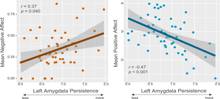
Puccetti et al. · Amygdala Persistence, Daily Affect, and Well-Being                                                                                             J. Neurosci., 0, 2021 • 00(00):000 • 3

Figure 1. Study design schematic. A, fMRI task design. Participants viewed 60 negative, neutral, and positive IAPS images for 4 s. When viewing the image, participants indicated whether
the image was negative, neutral, or positive via a button press. Images were followed by either a neutral facial expression displayed for 0.5 s or a black screen. On neutral facial expression tri-
als, the face was displayed 3 s after the offset of the IAPS image. We note that there were 20 trials with faces displayed 1 s after the image, which are not pictured here, that were not ana-
lyzed in relation to the affective outcomes of interest. Images here are examples of images seen not actual images used. B, Quantifying amygdala persistence. An RSA was conducted to
calculate the voxel-wise pattern similarity in the amygdala between the negative images and the neutral faces that followed them. C, Daily affective experience. Participants reported on the
degree to which they experienced 13 positive and 14 negative emotions throughout their day. This information was collected over the telephone to an interviewer for eight consecutive days. Individual
differences in daily affective experience were used as a mediator linking the metric of amygdala persistence with PWB. PWB, psychological well-being; RSA, representational similarity analysis.

time). Thirteen PA items were rated: “in good spirits,” “cheerful,”                                  60°; 262 whole-brain volumes per run]. Additionally, a high-resolution
“extremely happy,” “calm and peaceful,” “satisfied,” “full of life,” “close to                       T1-weighted anatomic image was obtained (T1-weighted inversion re-
others,” “like you belong,” “enthusiastic,” “attentive,” “proud,” “active,” and                      covery fast gradient echo; 256  256 in-plane resolution; 240-mm FOV;
“confident”; and fourteen NA items were rated: “restless or fidgety,”                                124  1.1-mm axial slices).
“nervous,” “worthless,” “so sad nothing cheered you up,” “everything
required effort,” “hopeless,” “lonely,” “afraid,” “jittery,” “irritable,”                            fMRI analysis
“ashamed,” “upset,” “frustrated,” and “angry.” Cronbach’s a for the                                  fMRI data were resampled to 2  2  2 mm, then preprocessed and ana-
PA scale = 0.96 and for the NA scale = 0.91, based on reliability calcu-                             lyzed using AFNI (Cox, 1996), ANTs (Avants et al., 2011), and FSL
lations recommended by Raudenbush and colleagues (Raudenbush et                                      (Jenkinson et al., 2012). T1 images were skull-stripped using FSL’s BET
al., 1991; Charles et al., 2016, 2019). These items were averaged to cre-                            function. For functional data, the first four volumes of each run were dis-
ate a daily PA and NA score. The average across all of a participant’s                               carded before analysis and then functional images were despiked using
daily interviews was taken to reflect their mean PA and NA.                                          the 3dDespike program from the AFNI toolbox. Motion correction was
                                                                                                     performed with the ANTs toolbox; functional images were first aligned
fMRI procedure and acquisition                                                                       within each run to the mean image, then all runs were aligned to the
Participants completed an image-viewing task in which they saw 60 posi-                              mean image from the first functional run. Next, using ANTs, nonlinear
tive, 60 negative, and 60 neutral images from the International Affective                            spatial normalization was applied to the functional data to match the
Picture System (IAPS; Fig. 1; Lang et al., 2008). These images were matched                          MNI152 template. The normalized functional images were then brain-
for luminosity, picture complexity (using the jpeg file size as an index of                          masked and scaled using FSL.
complexity) and social content (van Reekum et al., 2018), and the positive                               The preprocessed data were input to AFNI’s 3dDeconvolve to test a
and negative picture sets were equally arousing on average. Following a 1 s                          general linear model (GLM) with task and nuisance regressors. The 5 func-
fixation screen, each image was presented for 4 s, and participants indicated                        tional runs were concatenated, and the model was estimated using these
with a manual response whether the image was positive, negative or neutral                           concatenated data. There were nine task regressors: three regressors for the
in valence. Following IAPS slides was either a neutral facial expression (1 or                       IAPs (one for each of the positive, negative, neutral conditions; convolved
3 s following the IAPS image offset), or a black screen with a fixation cross                        for 4 s), three regressors for neutral facial expressions 1 s after an image
but no face. Faces were one of 30 male faces from the XM2VTSDB multi-                                (one for each of the positive, negative, neutral images it follows; convolved
modal face database (Messer et al., 1999) presented for 0.5 s (Fig. 1).                              for 0.5 s), and three for neutral facial expressions 3 s later (again one for
Participants were asked to simply view the faces and made no response to                             each of the positive, negative, neutral images it follows; convolved for 0.5 s).
these stimuli. The total intertrial interval (including the face presentations                       The six standard motion regressors were included, as well as their deriva-
for trials where faces followed an IAPS picture) was selected from an expo-                          tives, and the square of each of those 12 parameters, resulting in 24 motion
nential distribution and varied from 5.5–17.6 s with an average                                      parameters in the model to remove sources of variance because of motion
duration = 8.89 s, and consisted of a black screen with a white fixation cross.                      (Satterthwaite et al., 2013). At the individual level, this GLM yielded whole-
    A standard clinical whole-head quadrature head coil was used. Five                               brain maps of b coefficients for each voxel for each task condition.
functional runs were collected. Functional images were obtained using a
T2p-weighted, echoplanar images [EPIs; 30 sagittal slices, 4-mm thick-                               RSA
ness with 1-mm gap; 3.75  3.75 mm in-plane (64  64 voxels);                                        We used RSA to measure individual differences in the persistence of
FOV = 240; repetition time (TR)/echo time (TE)/flip, 2000 ms/30 ms/                                  amygdala activity patterns in response to different classes of stimuli:
4 • J. Neurosci., 0, 2021 • 00(00):000 Puccetti et al. · Amygdala Persistence, Daily Affect, and Well-Being
Table 1. Means, SDs, and Pearson bivariate correlation coefficients between affective variables and amygdala persistence
Variable M (SD) 1 2 3 4 5 6 7 8
1. PWB 39.87 (5.70)
2. Mean daily PA 2.89 (0.62) 0.51ppp
3. Mean daily NA 0.34 (0.20) 0.18 0.66ppp
4. LAmyg. persistence: negative images 0.88 (0.38) 0.16 –0.47pp 0.37p
5. LAmyg. persistence: neutral images 0.81 (0.36) 0.03 0.11 0.12 0.04
6. LAmyg. persistence: positive images 0.86 (0.37) 0.14 0.06 0.16 0.16 0.13
7. RAmyg. persistence: negative images 0.74 (0.38) 0.05 0.12 0.21 0.36p 0.30 0.23
8. RAmyg. persistence: neutral images 0.80 (0.35) 0.18 0.06 0.35p 0.07 0.36p 0.40p 0.01
9. RAmyg. persistence: positive images 0.91 (0.38) 0.03 0.13 0.06 0.10 0.10 0.45pp 0.05 0.23
N = 52. Mean daily NA is square root transformed here. Correlations are FDR-corrected. PWB, psychological well-being; NA, negative affect; PA, positive affect; Amyg., amygdala; L, left; R, right; pp , 0.05, ppp , 0.01,
pppp , 0.001.
negative images and neutral facial expression that follow negative persistence following neutral and positive images as covariates in all
images. In essence, this analysis quantifies the how closely the represen- models to confirm that any effects were specific to amygdala persistence
tation of neutral face expressions that are presented after negative stimuli following negative images. We conducted an identical analysis, but
resemble the representation of actual negative images in the amygdala. with patterns of activity in the left and right occipital pole, the
To accomplish this, the subject-level, whole-brain b maps from the right and left nucleus accumbens (NAcc), and the medial prefron-
GLM were inputted to the RSA MATLAB toolbox (Nili et al., 2014). b tal cortex (mPFC) to test whether effects were specific to the amyg-
maps were masked to isolate voxels of the left and right amygdala thresh- dala. Similar to the amygdala, these regions were taken from the
olded at 50% using the Harvard Oxford Subcortical Brain Atlas (Desikan Harvard Oxford Brain Atlas and thresholded at 50% (Desikan et
et al., 2006). Given evidence from fMRI studies for amygdala laterality in al., 2006). We selected the occipital pole because we did not expect
some processing of affective stimuli (Baas et al., 2004; Dyck et al., 2011; persistence in these visual regions to be associated with daily
Murphy et al., 2020), we examined the left and right amygdala sepa- affect. However, we selected the NAcc and the mPFC to explore
rately. The 2 2 2-mm left amygdala mask contained 240 voxels and whether other regions that are often implicated in affective proc-
the right amygdala mask contained 280 voxels. These masks correspond essing show similar effects as the amygdala. False discovery rate
to ;27 and 32 voxels, respectively, in the native sampling space. The (FDR; Benjamini and Hochberg, 1995) correction was applied to
persistence of left and right amygdala activity from affective images to these correlation matrices by using the raw p value vector as input
subsequent neutral facial expressions was quantified in the following to the p.adjust command in the stats package (i.e., p.adjust(uncor-
way: we extracted the pattern of left and right amygdala activity in rected_ps, method = “fdr”). p.adjust also requires an argument for
response to the IAPS image as well as in response to the neutral faces. the number of unique pairwise comparisons of interest. In the 9 by
Both patterns were reshaped into a 1 n vector and correlated with one 9 correlation matrix shown in Table 1, for example, the number of
another. These correlation values were Fisher Z transformed then sub- tests is 36 or the length of the raw p value vector. Further, p values
tracted from 1 to calculate the representational distance, or dissimilarity, obtained from multiple regression models for relationships involv-
between the two patterns. This typical dissimilarity metric yields values ing variables of interest (daily NA, PA, PWB, and amygdala
which range from 0 to 2, with greater numbers reflecting greater dissimi- persistence) were concatenated into a single vector and FDR-cor-
larity. Because this analysis was focused on persistence (a measure of rected using p.adjust.
similarity rather than dissimilarity), we inverted the dissimilarity metrics To test whether the RSA-derived persistence was superior to a uni-
from the RSA analysis. Also, we elected to use the neutral facial expres- variate measure of persistence in predicting affect, we computed the dif-
sions presented at 3 s post-IAPS offset, rather than 1s, for our metric of ference between univariate amygdala responses to the IAPs images and
amygdala persistence given the sluggish nature of the BOLD signal the neutral facial expressions. Although it has been argued that within-
(Lindquist et al., 2009). person difference scores in neural activity may be unreliable as individ-
For the primary analyses, this persistence metric was calculated by ual difference measures (Infantolino et al., 2018), this metric was most
including all stimuli from all runs into a single GLM. However, because analogous to the RSA-derived persistence metric. We calculated mean b
within-run temporal autocorrelation could bias similarity metrics, we value across the voxels in the left and right amygdala for negative, neu-
performed additional analyses in which each fMRI run was analyzed tral, and positive IAPs images and also for those faces that followed those
separately. This alternative method reduces potential biases introduced images. We then took the absolute value of the difference between the
by temporal collinearity among events within the same run. Once each IAPs images and the face stimuli for each valence condition to obtain
run was modeled with a separate GLM, this similarity metric was cal- scores analogous to similarity, or representational distance. These uni-
culated by individually estimating the dissimilarity among all variate differences scores were then examined as predictors of daily affect
between-run, image-face comparisons (e.g., similarity of amygdala and PWB in correlations and multiple regression models similar to the
IAPS activity of run 1 with amygdala face activity in run 2, run 3, multivariate persistence metrics.
etc.). Once dissimilarity metrics for all 20 IAPS-face permutations Finally, path analyses were conducted using version 0.6–4 of the ‘lav-
were calculated, they were averaged together. Importantly, this aver- aan’ package in R (Rosseel, 2012) to test whether amygdala persistence
age value excludes the within-run comparisons (e.g., there is no com- predicted PWB, via daily affect. First, with the full sample, we assessed
parison among run 1 IAPS slides with run 1 faces). Replicating our whether daily affect mediated the relationship between amygdala persist-
analyses using this potentially less biased metric demonstrates that ence and the first measurement of PWB. While this model takes advant-
the primary effects are unlikely to be a product of biased estimates age of all available data, the outcome precedes the predictors in time. To
driven by within-run temporal correlation. overcome this challenge, we also tested a similar model with the subset
of participants who had completed a measurement of PWB after their
Associations between amygdala persistence and daily affect fMRI and diaries and used this second measurement of PWB as the out-
After assessing zero-order correlations, we tested whether amygdala per- come. Further, while traditional mediation frameworks required that a
sistence following negative images was associated with mean daily PA significant total effect be present for amygdala persistence and PWB
and NA in multiple linear regressions. These regressions included cova- (Baron and Kenny, 1986), current methodological models of mediation
riates of age, gender, race, twin status, time between visits, and number suggest that this direct pathway is not necessary to detect mediation
of telephone diary interviews completed. Specifically, dummy-coded var- (Hayes, 2009). The significance of the path coefficients in these models
iables were included for race categories and each twin pair. We included was determined by using the Wald z-statistic, which divides thePuccetti et al. · Amygdala Persistence, Daily Affect, and Well-Being J. Neurosci., 0, 2021 • 00(00):000 • 5
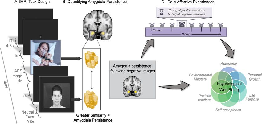
Figure 2. Left amygdala persistence following negative images is associated with daily negative and positive affect. Zero-order Pearson correlation plots between left amygdala persistence
following negative images and mean negative and positive affect from the daily diaries with a gray standard error ribbon. The square root transformed mean NA is shown here. N = 52.
parameter estimate by its standard error value. The path model also Links between amygdala persistence and daily affect
included the identical covariates as above. Pearson bivariate correlations (Table 1) revealed that left amyg-
dala persistence following negative images was associated with
daily mean NA (r = 0.37, p = 0.040; Fig. 2A) and daily mean PA
Results (r = 0.47, p , 0.001; Fig. 2B). Left amygdala persistence follow-
Descriptive statistics ing neutral images was not associated with daily mean NA
The mean of the composite PWB score was 39.87 (SD = 5.70, (r = 0.12, p = 0.626) or PA (r = 0.11, p = 0.645). Similarly, per-
range = 22.17–48.50). For the 31 participants that reported on sistence following positive images was unrelated to mean NA (r
PWB approximately seven years later, the mean was 40.33 = 0.16, p = 0.540) or PA (r = 0.06, p = 0.810). In contrast with
(SD = 4.63, range = 28.75–48.50). Participants completed an aver- the left amygdala, right amygdala persistence following negative
age of 7.5 daily telephone interviews (SD = 0.87, range = 4–8). images was not significantly related to daily mean NA (r = 0.21,
The mean daily NA was 0.15 (SD = 0.17, range = 0–0.76), mean p = 0.370) or daily mean PA (r = 0.12, p = 0.626). The relation-
daily PA was 2.89 (SD = 0.62, range = 1.35–3.99). Daily affect var- ship between amygdala persistence and daily NA was not signifi-
iables were assessed for acceptable skew and kurtosis based on cantly different between the left and right amygdala (t(51) = 1.04,
recommendations by Kline (i.e., skewness ,|2| and kurtosis ,| p = 0.305) in a test of the difference between two correlated cor-
7|; Kline, 2015) with the ‘psych’ R package, version 1.8.12 relations in the psych package in R (version 1.8.12; Steiger, 1980;
(Revelle, 2020). While mean PA demonstrated acceptable skew Revelle, 2020). However, the relationship between amygdala per-
( 0.23) and kurtosis ( 0.22), mean NA showed a positive skew sistence and daily PA was significantly stronger in the left amyg-
that approached unacceptable (1.89) with acceptable kurtosis dala compared with the right (t(51) = 2.45, p = 0.018).
(3.34). As a result, mean NA was square root transformed (trans- Given the associations between left amygdala persistence
formed skewness = 0.59 and kurtosis = 0.25). Results from all following negative images and daily affect, we tested the
analyses remained unchanged when tested with the raw NA val- robustness of these effects in a multiple regression model
ues. Descriptive statistics for all variables are shown in Table 1. that included relevant covariates. The effect of left amygdala
Left amygdala persistence following a negative image ranged persistence following the negative images on daily affect held
from 0.03 to 1.97, with a mean of 0.88 (SD = 0.43). Left amygdala when including covariates in multiple regressions. Greater
persistence activity following a neutral image ranged from 0.06 to amygdala persistence predicted more daily NA (b = 0.24,
1.78, with a mean of 0.80 (SD = 0.41). Left amygdala persistence fol- t(40) = 3.46, p = 0.003, B = 0.50) and less daily PA (b = 0.97,
lowing a positive image ranged from 0.01 to 1.74, with a mean of t(40) = 4.84, p , 0.001, B = 0.67) when controlling for age,
0.84 (SD = 0.42). gender, race, twin status, the time between visits, and the
Right amygdala persistence following a negative image ranged number of diaries completed. The relationship between left
from 0.00 to 1.79 (M = 0.71, SD = 0.44). Right amygdala persistence amygdala persistence following negative images and daily
following a neutral image ranged from 0.10 to 2.14 (M = 0.79, NA was also specific, as persistence following neutral images
SD = 0.40) and right amygdala persistence following a positive (b = 0.07, t(38) = 1.00, p = 0.448, B = 0.014), or following posi-
image ranged from 0.01 to 1.97 (M = 0.89, SD = 0.44). A repeated tive images (b = 0.03, t(38) = 0.41, p = 0.730, B = 0.06),
measures ANOVA assessed differences in persistence by hemi- did not attenuate the relationship between left amygdala per-
sphere (left and right) across the three image valence conditions. sistence following negative images and daily NA (b = 0.23,
There was no main effect of hemisphere (F(1,51) = 0.80, p = 0.372) or t(38) = 3.19, p = 0.006, B = 0.48) when included in the model
valence F(2,102) = 0.97, p = 0.382, nor was there an interaction (adjusted R2 = 0.17; Table 2). Similarly, left amygdala persist-
between hemisphere and valence F(2,102) = 1.80, p = 0.167). ence following neutral images (b = 0.08, t(38) = 0.40,
Similarity in left and right amygdala persistence was also p = 0.730, B = 0.06), or following positive images (b =
observed in Pearson bivariate correlations. There were positive 0.11, t(38) = 0.56, p = 0.700, B = 0.08), did not attenuate
correlations between left and right amygdala persistence follow- the effect of persistence following negative images on daily
ing a negative image (r = 0.36, p = 0.030), a neutral image PA (b = 0.97, t(38) = 4.69, p , 0.001, B = 0.67; full model
(r = 0.36, p = 0.030), and a positive image (r = 0.45, p = 0.003). adjusted R2 = 0.25; Table 2).6 • J. Neurosci., 0, 2021 • 00(00):000 Puccetti et al. · Amygdala Persistence, Daily Affect, and Well-Being
Table 2. Multiple regression models of daily negative and PA predicted by left amygdala persistence following negative images
Predictor b (SE) b t p value
NA model:
Left amygdala persistence: negative images 0.23 (0.07) pp 0.48 3.20 0.006
Left amygdala persistence: neutral images 0.07 (0.07) 0.14 1.00 0.45
Left amygdala persistence: positive images 0.03 (0.07) 0.06 0.41 0.73
Time between fMRI and diaries 0.00 (0.00) 0.29 1.22 0.23
Diary days completed 0.07 (0.03) 0.28 1.89 0.07
Race 0.04 (0.09) 0.08 0.39 0.70
Gender 0.03 (0.06) 0.08 0.54 0.60
Age 0.00 (0.00) 0.07 0.47 0.64
PA model:
Left amygdala persistence: negative images 0.97 (0.21)ppp 0.67 4.69 , 0.001
Left amygdala persistence: neutral images 0.08 (0.21) 0.06 0.40 0.73
Left amygdala persistence: positive images 0.11 (0.20) 0.08 0.56 0.70
Time between fMRI and diaries 0.00 (0.00) 0.11 0.31 0.63
Diary days completed 0.02 (0.10) 0.02 0.15 0.88
Race 0.23 (0.27) 0.18 0.86 0.40
Gender 0.13 (0.18) 0.10 0.69 0.50
Age 0.01 (0.00) 0.18 1.28 0.21
df = 37, pp , 0.05, ppp , 0.01, pppp , .001. Mean daily NA is square root transformed in this model. Five dummy-coded variables to represent the five sets of twins in the sample were included in the model and not
shown here. Race was coded as 1 = White; 0 = African American. Gender was coded as 0 = male; 1 = female. NA, negative affect; PA, positive affect.
Links between amygdala persistence and PWB Table 3. Path model of left amygdala persistence following negative images
Pearson bivariate correlations (Table 1) revealed that left amyg- on the first measure of PWB through daily PA
dala persistence following negative images was not directly asso- Path b (SE) b Z 95% lower CI 95% upper CI p value
ciated with PWB (r = 0.16, p = 0.540). Similarly, right amygdala a 0.97 (0.18) ppp 0.67 5.49 1.32 0.62 ,0.001
persistence following negative images was not associated with b 4.83 (1.28) ppp 0.52 3.79 2.33 7.32 ,0.001
PWB (r = 0.05, p = 0.810). Left amygdala persistence following c 1.75 (2.09) 0.13 0.84 2.34 5.84 0.517
neutral images was also not associated with PWB (r = 0.03, c’ 4.68 (1.50) pp 0.35 3.12 7.63 1.74 0.005
p = 0.874), nor was left amygdala persistence following positive Total 2.93 (1.89) 0.22 1.55 6.64 0.77 0.120
images related to PWB (r = 0.14, p = 0.600). Further, right N = 52. Path a is the effect of left amygdala persistence on mean daily PA, path b is the effect of mean
daily PA on PWB, and path c is the effect of left amygdala persistence on PWB. Path c’ is the indirect effect
amygdala persistence following neutral images was not signifi- of left amygdala persistence on PWB through mean daily PA. Covariates included in the model were age,
cantly associated with PWB (r = 0.18, p = 0.480), nor was right gender, race, twin status, number of diaries completed, and time between assessments; PWB, psychological
amygdala persistence following positive images related to PWB well-being; PA, positive affect pp , 0.05, ppp , 0.01, pppp , 0.001.
(r = 0.03, p = 0.868).
diaries. A similar pattern of effects was found when repeating the
Links between daily affect and PWB model. There were significant effects of both left amygdala per-
Pearson bivariate correlations (Table 1) showed that PWB was sistence on mean PA (b = 0.88, z = 3.29, p = 0.003), and
significantly associated with daily mean PA (r = 0.51, p , 0.001), mean PA on PWB (b = 5.64, z = 4.34, p , 0.001) yielding a signif-
but not mean NA (r = 0.18, p = 0.480). icant indirect effect of left amygdala persistence on PWB through
mean PA (b = 4.94, z = 2.62, p = 0.016). Unlike the previous
Daily PA links left amygdala persistence following negative model, there was a direct effect of left amygdala persistence fol-
images to PWB lowing negative images on the second PWB measurement
Because there was not a significant direct association between (b = 4.72, z = 2.19, p = 0.047). This model accounted for 46% of
PWB and daily mean NA, we only included daily mean PA in the variance in mean PA and 57% of the variance in the first
the full path models. In the first model using the full sample, and measurement of PWB, suggesting that amygdala persistence is
in line with the bivariate correlations reported above, there was indirectly related to future PWB years later through daily PA.
no direct effect of left amygdala persistence following negative
images on the first PWB measurement (b = 1.75, z = 0.84, Univariate amygdala difference scores between images and
p = 0.517; Table 3). However, there were significant effects of neutral facial expressions
both left amygdala persistence on mean PA (b = 0.97, z = Overall, we did not find that univariate differences scores
5.49, p , 0.001), and mean PA on PWB (b = 4.83, z = 3.79, between IAPS images and subsequent faces were related to daily
p , 0.001). Furthermore, there was a significant indirect effect of affect or PWB. Specifically, the left amygdala difference scores
left amygdala persistence on PWB through mean PA (b = 4.68, between negative images and the neutral facial expressions that
z = 3.12, p = 0.005; Fig. 3). This model accounted for 43% of followed were not significantly related to daily NA (r = 0.03,
the variance in mean PA and 44% of the variance in the first p = 0.94), daily PA (r = 0.03, p = 0.94), or PWB (r = 0.27,
measurement of PWB. p = 0.20). Left amygdala difference scores between the neutral
We also tested whether the indirect effect was also present IAPs images and the faces that follow were also not associated
using the second PWB measurement approximately seven years with daily NA (r = 0.09, p = 0.79), daily PA (r = 0.08, p = 0.84), or
later as the outcome in a subset of 31 participants. This model PWB (r = 0.18, p = 0.46). Further, left amygdala difference
overcomes the temporal limitations of using the first measure- scores between the positive IAPs images and the subsequent
ment of PWB, which was collected before the fMRI and daily faces were not significantly related to daily NA (r = 0.01,Puccetti et al. · Amygdala Persistence, Daily Affect, and Well-Being J. Neurosci., 0, 2021 • 00(00):000 • 7
Similar to the RSA persistence metric, we
tested whether the left amygdala univariate dif-
ference scores would predict individual differ-
ences in daily affect using multiple regression
with relevant covariates. The univariate differ-
ence between negative images and neutral fa-
cial expressions that followed did not
significantly predict daily mean NA (b = 0.04,
t(38) = 0.21, p = 0.839, B = 0.04) in a model that
also included the univariate difference between
neutral images and neutral facial expressions that
followed (b = 0.02, t(38) = 0.11, p = 0.912, B =
0.02), the univariate difference between positive
images and neutral facial expressions that fol-
lowed (b = 0.007, t(38) = 0.03, p = 0.973, B =
0.005), and the same covariates included in the
other analyses (age, gender, race, twin status, the
time between visits, and the number of diaries
completed). A similar lack of effect resulted from
an identical model with daily mean PA as the out-
Figure 3. Path model of left amygdala persistence following negative images predicting PWB via daily PA. come. The univariate difference between nega-
Standardized path coefficients displayed. Covariates, including age, gender, number of diaries completed, race, twin tive images and neutral facial expressions
status, time between assessments and persistence following neutral and positive images are not shown here for sim- that followed did not significantly predict
plicity. The indirect path is denoted with c’. N = 52; PWB, psychological well-being; PA, positive affect pp , 0.05, daily mean PA (b = 0.40, t(38) = 0.63,
ppp , 0.01, pppp , 0.001. p = 0.534, B = 0.12), nor did the difference
between neutral images and neutral facial
Table 4. Pearson bivariate correlations between PWB, daily affect, and persist- expressions that followed (b = 0.11, t(38) =
ence within other brain regions 0.18, p = 0.861, B = 0.03), or the difference between posi-
Variable 1 2 3 tive images and neutral facial expressions that followed (b =
0.60, t(38) = 0.91, p = 0.370, B = 0.16). Together, these results suggest
1. PWB that our RSA persistence metric captured unique individual var-
2. Mean daily PA 0.51pp
iance related to daily affective experience.
3. Mean daily NA 0.18 0.66pp
4. L. Occ. persistence: negative images 0.05 0.18 0.09
5. L. Occ. persistence: neutral images 0.20 0.07 0.23 Exploratory analyses addressing spatial specificity
6. L. Occ. persistence: positive images 0.29 0.11 0.07 To assess whether the association between daily affect and amyg-
7. R. Occ. persistence: negative images 0.12 0.11 0.07 dala persistence following negative images was unique to the
8. R. Occ. persistence: neutral images 0.04 0.05 0.14 amygdala, we tested whether these effects were present in other
9. R. Occ. persistence: positive images 0.20 0.24 0.17 regions of the brain. To do this, we extracted the patterns of the
10. L. NAcc. persistence: negative images 0.57pp 0.51pp 0.27 left occipital pole (LOcc) and right occipital pole (ROcc), the left
11. L. NAcc. persistence: neutral images 0.09 0.25 0.21 NAcc (LNAcc) and right NAcc (RNAcc), and the mPFC, then
12. L. NAcc. persistence: positive images 0.02 0.09 0.04 conducted an identical RSA analysis using these regions. The
13. R. NAcc. persistence: negative images 0.10 0.10 0.16 Pearson bivariate correlations are displayed in Table 4.
14. R. NAcc. persistence: neutral images 0.20 0.05 0.17
Results revealed that persistence in the LOcc and ROcc following
15. R. NAcc. persistence: positive images 0.23 0.02 0.12
16. mPFC persistence: negative images 0.07 0.11 0.09 negative images were not associated with daily mean NA (ROcc
17. mPFC persistence: neutral images 0.04 0.03 0.04 r = 0.07, p = 0.837; LOcc r = 0.09, p = 0.810), or mean PA (ROcc r =
18. mPFC persistence: positive images 0.25 0.14 0.01 0.11, p = 0.774; LOcc r = 0.18, p = 0.667), or PWB (ROcc
N = 52. Mean daily NA is square root transformed here. R, right; L, left; Occ, occipital; NAcc, nucleus accum- r = 0.12, p = 0.774; LOcc r = 0.05, p = 0.872). Occipital persistence
bens; mPFC, medial prefrontal cortex; NA, negative affect; PA, positive affect pp , 0.05, ppp , 0.01, following neutral images was also not associated with daily mean
pppp , 0.001. NA (ROcc r = 0.14, p = 0.765; LOcc r = 0.23, p = 0.495), mean
PA (ROcc r = 0.05, p = 0.872; LOcc r = 0.07, p = 0.837), or PWB
(ROcc r = 0.04, p = 0.873; LOcc r = 0.20, p = 0.650). Also, occipital
p = 0.96), daily PA (r = 0.20, p = 0.42), or PWB (r = 0.12, p = 0.66). persistence following positive images was unrelated to daily mean
Similarly, univariate difference scores in the right amygdala NA (ROcc r = 0.17, p = 0.671; LOcc r = 0.07, p = 0.837), mean
between negative images and the neutral facial expressions were PA (ROcc r = 0.24, p = 0.495; LOcc r = 0.11, p = 0.774), or PWB
not associated with daily NA (r = 0.05 p = 0.94), daily PA (ROcc r = 0.20, p = 0.644; LOcc r = 0.29, p = 0.322).
(r = 0.02, p = 0.96), or PWB (r = 0.16, p = 0.52). Additionally, Similarly, mPFC persistence following negative images was
right amygdala difference scores between neutral images and the unrelated to daily NA (r = 0.09, p = 0.810), daily PA (r = 0.11,
faces that followed were not related to daily NA (r = 0.01, p = 0.774), or PWB (r = 0.07, p = 0.837). Also, mPFC persist-
p = 0.96), daily PA (r = 0.04, p = 0.94), or PWB (r = 0.22, ence following neutral images was not significantly associated
p = 0.36). Finally, right amygdala difference scores between the with daily NA (r = 0.04, p = 0.873), daily PA (r = 0.03, p = 0.896),
positive IAPs images and the subsequent faces were not signifi- or PWB (r = 0.04, p = 0.873) nor was mPFC persistence following
cantly related to daily NA (r = 0.05, p = 0.94), daily PA positive images related to daily NA (r = 0.01, p = 0.970), daily
(r = 0.13, p = 0.66), or PWB (r = 0.05, p = 0.94). PA (r = 0.14, p = 0.765), or PWB (r = 0.25, p = 0.400).8 • J. Neurosci., 0, 2021 • 00(00):000 Puccetti et al. · Amygdala Persistence, Daily Affect, and Well-Being
Results also showed that RNAcc persistence following nega- critical. Previous research has shown that more persistent amygdala
tive images was not associated with daily NA (r = 0.16, activity in response to aversive stimuli predicts higher levels of traits
p = 0.700), daily PA (r = 0.10, p = 0.810), or PWB (r = 0.10, including neuroticism (Schuyler et al., 2014). We extended this liter-
p = 0.782). Persistence following neutral images in the RNAcc ature by examining how experimentally-obtained individual differ-
was similarly not related to daily NA (r = 0.17, p = 0.668), daily ences in amygdala persistence, quantified using RSA, map onto
PA (r = 0.05, p = 0.873), or PWB (r = 0.20, p = 0.644). naturalistic daily affect and PWB assessed over long periods of time.
Further, RNAcc persistence following positive images was unre- Individuals with greater left amygdala persistence following
lated to daily NA (r = 0.12, p = 0.774), daily PA (r = 0.02, negative images reported more NA and less PA in daily life. This
p = 0.912), or PWB (r = 0.23, p = 0.495). We found that LNAcc aligns with previous research demonstrating that the amygdala
persistence following negative images was significantly correlated facilitates rapid, negative appraisals of neutral stimuli preceded
with daily PA (r = 0.51, p , 0.001) as well as PWB (r = 0.57, by an unrelated, aversive stimulus (Lapate et al., 2016; Tambini
p , 0.001), but not with daily NA (r = 0.27, p = 0.364). However, et al., 2017; Grupe et al., 2018). Additionally, previous work doc-
LNAcc persistence following neutral images was not related to umenting individual differences in amygdala persistence follow-
any outcomes, including daily NA (r = 0.21, p = 0.612), daily PA ing emotional stimuli (Siegle et al., 2002; Schuyler et al., 2014)
(r = 0.25, p = 0.400), and PWB (r = 0.09, p = 0.810). LNAcc suggests that some individuals may be more susceptible to the
persistence following positive image was also not significantly biasing of neutral stimuli by unrelated, negative stimuli. In the
associated with daily NA (r = 0.04, p = 0.873), daily PA (r = 0.09, current study, we cannot confirm the neutral facial expressions
p = 0.810), and PWB (r = 0.02, p = 0.941). These correlations sug- were perceived as neutral, however, individual differences in
gest that greater persistence (i.e., similarity between representa- neutral-face perception were accounted for by including neutral-
tions of negative images and faces that follow after) in the LNAcc image and positive-image persistence as covariates. These results
is also related to less PA in daily life and even less PWB. demonstrate that amygdala similarity between negative images
These outcomes indicate that individual differences in persist- and neutral facial expressions that follow such images is
ence following negative images in the amygdala, but not cortical linked to subjective emotional experience outside of the lab-
regions, is linked to daily affect. Additionally, the LNAcc, a sub- oratory. It may be that for individuals with greater amygdala
cortical structure that can mediate both appetitive and defensive persistence, negative moments may become amplified or
behaviors (Al-Hasani et al., 2015; Berridge, 2019), shows some prolonged by imbuing unrelated moments that follow with a
overlap with the left amygdala in that it is also correlated with negative appraisal. Ultimately, this persistence could result
daily PA but not NA. in more NA and less PA, on average, in daily life. This brain-
behavior link between left amygdala persistence and daily
Exploratory analyses to reduce impact of temporal affect can inform our understanding of more enduring, long-
autocorrelation of fMRI term evaluations of well-being.
Because within-run temporal autocorrelation of fMRI could PWB was indirectly linked to left amygdala persistence
impact metrics of similarity, we recomputed left amygdala per- through daily affect. Those who exhibited less amygdala persist-
sistence by calculating IAPS, face similarity permutations sepa- ence, reported higher daily PA, which in turn, was associated
rately for each scan run and then averaged them together. This with more positive PWB evaluations. A significant indirect effect
metric reflects the mean of all possible between-run image-face between two variables in the absence of a direct effect can occur
comparisons computed separately for negative, neutral, and posi- for a myriad of reasons (MacKinnon et al., 2000; Shrout and
tive image comparisons with the neutral facial expressions pre- Bolger, 2002; Hayes, 2009; Pardo and Román, 2013). One reason
sented 3 s later. is the length of time between variable measurements (James et
Links between left amygdala persistence and daily affect were al., 2006; Pardo and Román, 2013). It is possible that the
similar, though slightly weaker for daily NA. Specifically, the rela- extended time interval separating amygdala persistence and
tionship between this alternative metric of persistence and daily NA PWB measurements (M = 41 months) limited our ability to
(b = 0.56, t(36) = 2.00, p = 0.053) was not quite significant. However, detect a significant direct effect. Another possibility is that amyg-
this alternative metric of persistence following negative images was dala persistence contributes to momentary stimulus appraisals,
which are proximal to day-to-day affect. In contrast, PWB is
associated with daily PA (b = 2.82, t(36) = 3.22, p = 0.003) in iden-
determined by an adjoining set of complex integrative processes
tical models from the primary analysis, which included age, gender,
that govern how one recollects, integrates, and judges the mean-
race, twin status, the time between visits, and the number of diaries
ing of these daily experiences. These results build on previous
completed as covariates. Also, similar to the primary analyses, this al-
links between daily PA and greater PWB (Burns and Ma, 2015)
ternative metric of persistence following negative image was not
by tying variability in complex, cognitive evaluations about well-
directly associated with PWB (b = 10.45, t(37) = 1.52, p = 0.128), but
being to the emotional landscape of daily life and individual dif-
the indirect effect of left amygdala persistence on PWB through daily
ferences in brain function. Critically, this indirect effect held
PA was replicated using this alternative metric of persistence (b =
regardless of whether we examined the first measurement of
12.95, z = 2.76, p = 0.006).
PWB in the full sample or the second measurement of PWB
seven years after the fMRI collected in a subset of participants. In
Discussion our study, lower daily NA was not significantly associated with
What gives rise to individual differences in both transient emotional PWB. It is difficult to know how negative moments are inte-
experiences and enduring PWB across months and years? The field grated into broader evaluations of one’s well-being, but one pos-
of translational neuroscience asserts that real-world, affective out- sibility is that less variability in mean NA relative to mean PA in
comes can be predicted by neural activity observed in controlled, ex- this sample limited our ability to explain individual differences
perimental settings (Berkman and Falk, 2013; Shackman et al., in NA. Another possibility is that the null result between daily
2016). In particular, individual differences in neural activity in NA and PWB may be due a lack of power, as previous research
regions encoding value and salience, like the amygdala, may be with over 2000 subjects (Rush et al., 2019) found a link betweenPuccetti et al. · Amygdala Persistence, Daily Affect, and Well-Being J. Neurosci., 0, 2021 • 00(00):000 • 9
daily NA and PWB. Therefore, additional research should probe to better understand possible influences on enduring emotional
the directionality and role of daily NA in linking individual dif- representations over time.
ferences in brain function with PWB. Beyond the fMRI task design, additional research should further
Our persistence results were found in the left amygdala, indi- probe the mechanisms by which amygdala persistence translates to
cating potential differential affective processing between the left daily affect by exploring the cortical-subcortical relationships that
and right amygdala. However, hemispheric differences should be explain individual differences in amygdala persistence. Moreover,
interpreted with caution as only the correlation between amyg- the amygdala persistence effects here should be replicated with
dala persistence and PA (not NA) was significantly stronger in higher spatial resolution scans. In this study, the voxel size was rela-
left amygdala. Still, the left amygdala has been reportedly more tively large and sampling amygdala activity with higher spatial reso-
involved with cognitive processing of emotional information lution would increase the number of voxels included yielding a
compared with the right amygdala, which facilitates automatic richer analysis. Beyond the amygdala, other brain regions should be
stimulus detection and processing (Gläscher and Adolphs, 2003; further investigated. Although persistence in cortical regions did not
Dyck et al., 2011). Thus, left amygdala persistence may reflect show significant relationships with daily affect and PWB, LNAcc
more elaborative, cognitive appraisal of the negative stimuli persistence was negatively related to daily PA, similar to the left
(Phelps et al., 2001). Another possibility is that the left-lateralized amygdala. This suggests that persistence-affect relationships may not
amygdala findings may be an artifact of data preprocessing pro- be specific to the amygdala. Also, while this subset of the MIDUS
sample captured a range of adults (ages 39–76), it would be useful to
cedures, such as smoothing and motion correction (Murphy et
examine these effects during periods of critical development, such as
al., 2020). Given the inconsistencies in the current study and the
adolescence (Steinberg, 2005). Finally, although daily diary protocols
broader literature, additional research is needed to clarify the
often map on well to momentary experience (Kahneman et al.,
potential lateralization in amygdala persistence.
2004), ecological momentary assessments across a longer timescale
These findings also extend the previous literature by using
might provide a more fine-grained measurement of daily affective
RSA to quantify amygdala persistence in relationship to psycho- experiences.
logical functioning. We applied RSA, a multivariate technique In summary, individual differences in amygdala persistence
(Kriegeskorte et al., 2008; Brooks and Freeman, 2018), to capture were linked to meaningful, ecological affective outcomes.
individual differences in neural persistence, or the discriminabil- Specifically, the results indicated that those with less persistence
ity of neural representations of temporally related stimulus in the left amygdala following negative images may be less sus-
classes. In other words, we quantified, for a given person, how ceptible to the biasing of neutral facial expression by unrelated,
much the voxel-wise representation of negative image stimuli negative stimuli. Protection against this vulnerability to negative
resembled the representation of neutral facial expressions that biasing may bring about more PA as well as less NA in daily life.
followed negative stimuli in the amygdala. Previous research on Critically, it is through greater daily PA that left amygdala per-
amygdala persistence that averages across amygdala voxels sistence is related to PWB. As PWB is a complex, cognitive self-
ignores differential activation patterns across the spatially heter- evaluation requiring one to integrate across a lifetime of experi-
ogenous amygdala structure. Indeed, we found that univariate ences, such an evaluative process is likely supported by distrib-
left amygdala responses to neutral facial expressions following uted brain networks rather than any one individual region.
negative images were not associated with daily NA or PA.
However, our persistence metric, derived from an RSA compar-
ing voxel-wise patterns of activity in the amygdala between affec- References
Al-Hasani R, McCall JG, Shin G, Gomez AM, Schmitz GP, Bernardi JM, Pyo
tive stimuli and neutral facial expressions that followed, did
CO, Park SI, Marcinkiewcz CM, Crowley NA, Krashes MJ, Lowell BB,
predict daily NA and PA. These dynamic comparisons better Kash TL, Rogers JA, Bruchas MR (2015) Distinct subpopulations of nu-
match real-world experiences than isolating response to a single cleus accumbens dynorphin neurons drive aversion and reward. Neuron
stimulus, making them prime targets for quantifying amygdala 87:1063–1077.
persistence. Avants BB, Tustison NJ, Song G, Cook PA, Klein A, Gee JC (2011) A repro-
These results suggest numerous avenues for future research. ducible evaluation of ANTs similarity metric performance in brain image
Specifically, additional task features could provide a more com- registration. Neuroimage 54:2033–2044.
Baas D, Aleman A, Kahn RS (2004) Lateralization of amygdala activation: a
prehensive account of neural persistence. The post-IAPS image
systematic review of functional neuroimaging studies. Brain Res Brain
stimuli used here were limited to neutral facial expressions, how- Res Rev 45:96–103.
ever we cannot be certain that subjects perceived these stimuli as Baron RM, Kenny DA (1986) The moderator–mediator variable distinction
neutral. Because the amygdala is involved in facial processing in social psychological research: conceptual, strategic, and statistical con-
(Todorov et al., 2008), the social nature of these stimuli may siderations. J Pers Soc Psychol 51:1173–1182.
have influenced our metric of persistence. It is important to Benjamini Y, Hochberg Y (1995) Controlling the false discovery rate: a prac-
examine whether other types of putatively neutral stimuli influ- tical and powerful approach to multiple testing. J R Stat Soc Series B Stat
Methodol 57:289–300.
ence amygdala persistence and to collect concurrent subjective Berkman ET, Falk EB (2013) Beyond brain mapping: using neural measures
ratings of these stimuli. Additionally, including greater variation to predict real-world outcomes. Curr Dir Psychol Sci 22:45–50.
in the time between affective and neutral stimuli (i.e., 3, 5, and 7 Berridge KC (2019) Affective valence in the brain: modules or modes? Nat
s) could pinpoint the optimal lag for detecting individual varia- Rev Neurosci 20:225–234.
tion in persistence. Although we used individuals’ neutral and Brooks JA, Freeman JB (2018) Conceptual knowledge predicts the representa-
positive persistence values as covariates, greater exploration of tional structure of facial emotion perception. Nat Hum Behav 2:581–591.
individual factors that influence amygdala persistence, such as Burns RA, Ma J (2015) Examining the association between psychological
wellbeing with daily and intra-individual variation in subjective well-
attentional state and hemodynamic response function shape, being. Pers Individ Dif 82:34–39.
could deepen our understanding of the mechanisms of neural Charles ST, Mogle J, Urban EJ, Almeida DM (2016) Daily events are impor-
persistence. Additional work should examine persistence both in tant for age differences in mean and duration for negative affect but not
the presence and the absence of subsequent, intervening stimuli positive affect. Psychol Aging 31:661–671.You can also read