BLAST INJURIES - Guidelines for Burn Care Under Austere Conditions - American Burn Association
←
→
Page content transcription
If your browser does not render page correctly, please read the page content below
4/6/2020

          Guidelines for Burn Care
          Under Austere Conditions
          Special Etiologies: Blast, Radiation, and Chemical
          Injuries

1

    BLAST INJURIES

2

                                                                     1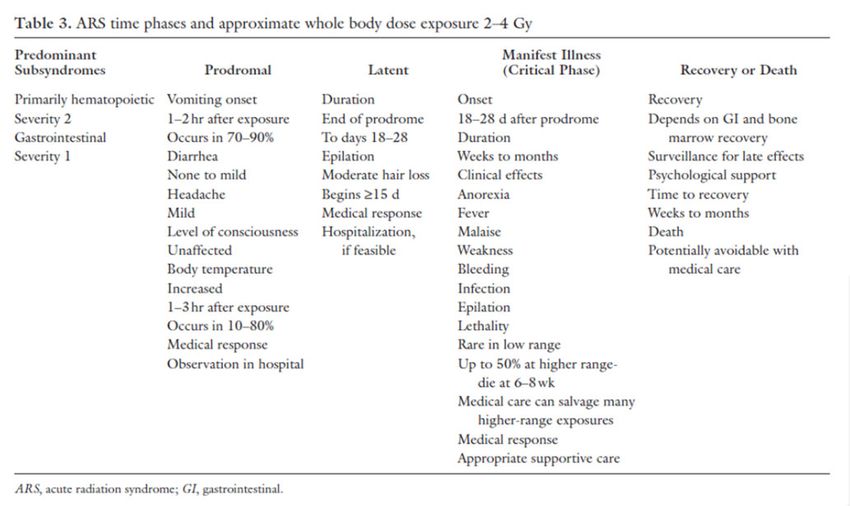
4/6/2020

    Introduction

    • Recent events, such as terrorist attacks in Boston, Madrid, and London, highlight the
      growing threat of explosions as a cause of mass casualty disasters.
    • Several major burn disasters around the world have been caused by accidental
      explosions.
    • During the recent conflicts in Iraq and Afghanistan, explosions were the primary
      mechanism of injury (74% in one review).
    • Furthermore, explosions were the leading cause of injury in burned combat casualties
      admitted to the U.S. Army Burn Center during these wars, who frequently manifested
      other consequences of blast injury.
    • Thus, providers responding to burn care needs in austere environments should be
      familiar with the array of blast injuries which may accompany burns following an
      explosion.

3

    Classification of Blast Injuries

    • Blast injuries are classified as follows:
        • Primary: Direct effects of blast wave on the body (e.g., tympanic membrane rupture, blast lung injury,
          intestinal injury)
        • Secondary: Penetrating trauma from fragments
        • Tertiary: Blunt trauma from translation of the casualty against an object
        • Quaternary: Burns and inhalation injury
        • Quinary: Bacterial, chemical, radiological contamination (e.g., “dirty bomb”)
    • In any given explosion, these types of injuries overlap.
    • Primary blast injury is more common in explosion survivors inside structures or
      vehicles because of blast-wave physics.
    • By far, secondary blast injury is more common.

4

                                                                                                                         2
4/6/2020

    Classification of Blast Injuries (cont.)

    • A study of 4623 explosion episodes in a Navy database identified the following
      injuries among U.S. service members:
       • mild traumatic brain injury (mTBI, 10.8%)
       • open wounds in the lower extremity (8.8%)
       • open wounds of the face (8.2%) to include tympanic membrane rupture
    • In these casualties, in whom torso body armor use was common, the extremities
      (41.3%) and head and neck (37.4%) were much more frequently injured than the
      torso (8.8%).
    • In the U.S. military’s Joint Theater Trauma Registry for 2003 to 2006, Ritenour and
      colleagues estimated that 12% of the 4765 service members injured in explosions
      had primary blast injury.
    • Nine percent of the 4765 had tympanic membrane rupture, 3.6% had blast lung injury,
      and 0.1% had blast injury of the intestines.
    • The latter two injuries were identified more frequently on autopsy: 2.3% of those killed
      in action had blast lung injury, and 2.0% had intestinal injury.

5

    Physics of Blast Injury

    • The Lovelace Foundation in Albuquerque, New Mexico, and others conducted
      extensive experiments on blast effects during the 1950s to 1960s, and defined the
      effects of overpressure and blast duration on survival.
    • These studies concluded that mortality is
       • 1) a function of maximum incident overpressure, and
       • 2) an inverse function of the duration of overpressure (e.g., a pressure wave of shorter duration is
         more lethal).
    • In the body, both stress and shear waves induce tissue damage.
       • Stress refers to microscopic effects at interfaces between tissues of different densities, of which
         spalling is displacement and fragmentation of more dense into less dense tissue, and implosion is
         the opposite.
       • Shear is macroscopic tissue damage which occurs as energy travels at different velocities through
         adjacent tissues of different densities.

6

                                                                                                                      3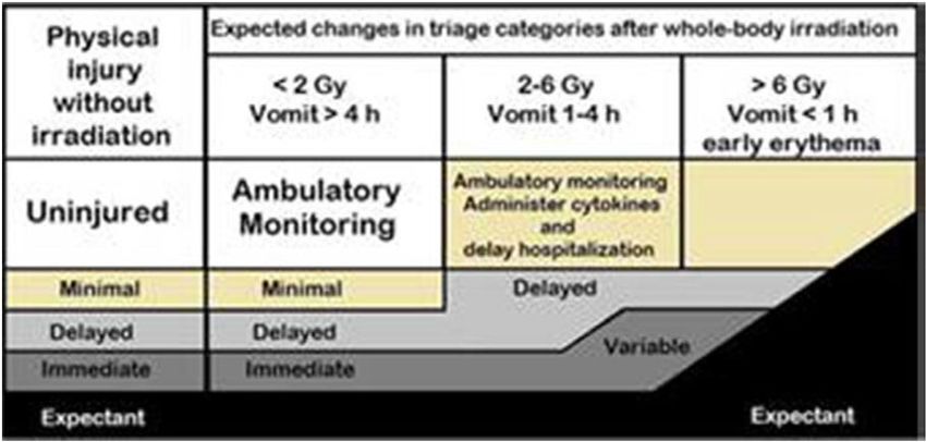
4/6/2020

    Physics of Blast Injury (cont.)

    • The most striking effect of a blast wave on the body occurs in organs with air–water
      interfaces, which is why the tympanic membranes, lungs, and the gastrointestinal (GI)
      tract are vulnerable.
    • However, other interfaces in the body also affect the course taken by waves in the
      body, and may attenuate or focus them.
    • For example, internal membranes within the cranium, such as the falx cerebri and the
      tentorium cerebelli, reflect and focus shear waves in the brain.
    • The above concepts are dramatically impacted by the circumstances surrounding the
      explosion.
    • The use of helmets and torso body armor influences the injury pattern by altering both
      overpressure and wave shape/duration.
    • For example, in one study, hard body-armor plates reduced the behindarmor
      overpressure by a factor of more than 50.

7

    Physics of Blast Injury (cont.)

    • The resultant decreased risk of death from lung injury increases the likelihood of
      death from TBI.
    • Consistent with this point of view, a review of thoracic injuries among U.S. service
      members in the Joint Theater Trauma Registry suggested that blast lung constituted
      only 1.4% of such injuries; by contrast, pneumothorax and pulmonary contusion
      predominated in that database.
    • Explosions within buildings or vehicles are more lethal than explosions in the open air
      because structures focus and reflect blast waves, generate more secondary
      fragments, ignite secondary fires, and create the risk of entrapment or of structural
      collapse.
    • As the distance increases from the explosion, the likelihood of primary blast injury
      drops, and secondary blast injuries predominate.

8

                                                                                                      4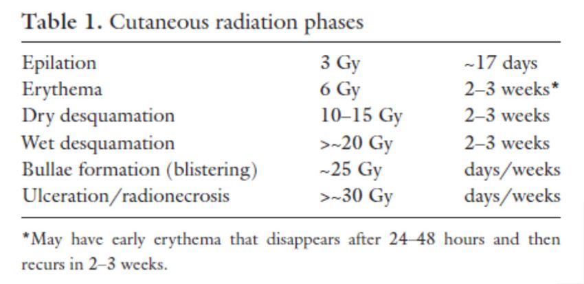
4/6/2020

     Clinical Care: Emergency Care

     • The immediate physiologic response to a blast wave includes a triad of bradycardia, apnea,
       and hypotension, which are mediated in part by the vagus nerve and specifically by pulmonary
       C fibers.
     • Additionally, hypotension is multifactorial, reflecting nitric oxide release by the injured lung14
       and a decrease in systemic vascular resistance lasting several hours.
     • The initiation of positivepressure mechanical ventilation in patients with primary blast lung
       injury should be done with caution to avoid barotrauma, which can worsen pulmonary injury or
       induce arterial gas embolism (AGE).
     • Fluid resuscitation of the patient with primary blast injury must strike a balance between
       overresuscitation, which will worsen pulmonary edema, and under-resuscitation, which is
       poorly tolerated in animal models of blast injury accompanied by hemorrhage.
     • These cautionary statements about patient resuscitation in primary blast injury should not
       distract from the large body of evidence from the recent conflicts in Iraq and Afghanistan,
       which point to the importance of damage control resuscitation and damage control surgery.

9

     Clinical Care: Emergency Care (cont.)

     • The work up of victims of explosions is greatly facilitated by the use of a computed
       tomography (CT) scanner.
     • Faced with multiple victims with injuries from head to toe, the ability to rapidly
       evaluate by CT scan is invaluable.
     • This resource, however, is a potential bottleneck in a mass casualty scenario and
       may be unavailable in an austere environment.

10

                                                                                                                  5
4/6/2020
Clinical Care: Traumatic Brain Injury
• Explosions are associated with a spectrum of traumatic brain injuries, from mild to severe.
• The mechanisms by which a blast wave injures the brain are complex and poorly understood.
• As of 2007, the incidence of mTBI in service members returning from the recent wars in Iraq
and Afghanistan was estimated at 19.5%.
• About 5% of veterans had a combination of mTBI, posttraumatic stress disorder, and
depression.
• Accordingly, all persons exposed to an explosion should undergo screening for TBI using a
brief survey tool.
• The U.S. military uses a Military Acute Concussion Evaluation tool to evaluate any service
member who is dazed, confused, ‘saw stars,’ or had transient loss of consciousness, following
an explosion, fall, motor vehicle accident, or other blow to the head.
• The latest version was released in 2012.
• Eighteen of 63 combat casualties with a clinical diagnosis of mTBI and with normal CT scans
had abnormal diffusion tensor imaging (an advanced form of magnetic resonance imaging)
indicating an axonal injury; thus, the diagnosis of mTBI remains primarily clinical.
11
Clinical Care: Otologic Injuries
• Patients who are injured in explosions require otologic examination.
• The tympanic membrane is the most sensitive structure in the body to blast.
• The overpressure which causes tympanic membrane rupture in 50% of persons
exposed is only 15 psi.
• Thus, patients with tympanic membrane rupture should also be evaluated for
delayed-onset lung or intestinal injury (see below).
• Nevertheless, rupture is not seen in all explosion casualties; rupture is affected by
factors such as head position relative to the blast and by ear canal contents.
• Thus, the presence of an intact tympanic membrane does not exclude other blast
injuries.
• Eighty percent of tympanic membrane ruptures heal spontaneously; most within 3
months of injury.
12
64/6/2020
Clinical Care: Otologic Injuries (cont.)
• The surface area of rupture predicts spontaneous healing success.
• Healing is unlikely without surgery if the area is greater than 80%.
• Other findings in patients with blast injury to the ear may include hearing loss
(temporary or permanent), tinnitus, otalgia, otorrhea, and bleeding from the ear.
• Less commonly, vertigo or gait disturbance may reflect inner ear injury, but may also
point to temporal bone fracture or brain injury.
• In addition to otoscopy, patients should be examined for bilateral hearing acuity, for
sensorineural hearing loss with a tuning fork, and for facial nerve injury.
• Long-term follow-up, to include audiometry, should be performed for all persons
exposed to explosions.
13
Clinical Care: Blast Lung
• This is the second most common primary blast injury.
• “Blast lung syndrome” features dyspnea, cough, and hypoxia.
• The process involves alveolar-capillary disruption, intraparenchymal hemorrhage,
hemo- and pneumothorax, pneumomediastinum, subcutaneous emphysema, and/or
bronchopleural fistula.
• Blast lung injury may become manifest 6–8 hours after injury; thus, asymptomatic but
at-risk patients should be observed for delayed onset of symptoms for that period of
time.
• The chest radiograph typically shows bilateral hilar infiltrates in a “butterfly” pattern.
• Blast lung injury may also cause AGE, manifested, for example, by focal neurologic
deficits.
• Physical findings of AGE may include retinal arterial air on ophthalmoscopy, tongue
blanching, or livedo reticularis (lacy skin mottling).
• Likewise, all of these findings may be absent.
14
74/6/2020
Clinical Care: Intestinal Injuries
• These are the third most common primary blast injuries.
• A meta-analysis showed that the prevalence of intestinal injuries in 1040 survivors of
air-blast explosions was 3%.
• The terminal ileum and cecum were the most commonly injured sites, but any of the
other hollow organs, as well as solid viscera, can be injured.
• Intestinal injury appears as subserosal hemorrhage, which on histopathology is
shown to be submucosal.
• With increased loading, immediate perforation may occur.
• Lesions which do not perforate at the time of injury may (in about 5% of cases)
undergo necrosis and delayed perforation, most often more than the ensuing 3–5
days.
• In the absence of perforation, diagnosis by CT scan may be erroneous, and frequent
reexamination of the patient is recommended.
15
Clinical Care: Eye Injuries
• All explosion-injured patients should undergo an examination of the eyes.
• One study of 46 veterans with TBI demonstrated that 20 also had closed-eye
(nonpenetrating) injuries, many of which were previously undiagnosed; 3 of these
required medical or surgical intervention.
• U.S. Army clinical practice guidelines on emergency eye care emphasize:
• 1) documentation of eye examination to include visual acuity
• 2) Wood’s lamp examination of the corneas with fluorescein
• 3) careful examination for globe penetration
• 4) use of a metal Fox shield and avoidance of pressure or dressings to protect the patient with an
open globe injury
• 5) prompt specialist referral for all suspected globe injuries
• In the acute setting, ultrasound (performed with the lids closed and with care to avoid
pressure on the globe) has been used for detecting injuries such as blood in the
globe, vitreous foreign body, retinal detachment, or globe collapse.
16
84/6/2020
Clinical Care: Extremity and Pelvic/Perineal Injuries
• The extremities remain the leading site of battlefield injury, even during the recent
conflicts in which explosions predominated.
• Wartime injuries to the extremities are particularly destructive.
• In comparison to civilian casualties, combat casualties have worse limb salvage rates
for Gustilo-Anderson open tibia fractures grades IIIB and IIIC.
• The blast mechanism notoriously destroys more tissue than initially may be apparent,
and injects foreign material proximally along tissue planes; when in doubt, aggressive
debridement and early amputation is often the best course of action.
• Costly and prolonged attempts at limb salvage may be ill-advised in an austere
scenario.
17
Clinical Care: Extremity and Pelvic/Perineal
Injuries (cont.)
• A particularly high-risk scenario is the blast victim with open pelvic fractures and/or
bilateral high above-the-knee amputations.
• Immediate activation of a massive transfusion protocol, rapid transportation to the
operating room, a multiple-team approach to surgery, damage control laparotomy with
proximal vascular control, and pelvic external fixation are among the maneuvers that
may be required to salvage such patients.
• In an austere environment, this type of care may or may not be possible.
• Injuries by improvised explosive devices which use ball bearings or other small
metallic projectiles may precipitate extremity compartment syndrome, with or without
fractures.
18
94/6/2020
Clinical Care: Triage of Multiple Casualties
• Multiple blast injuries are common in terrorist attacks, combat, and industrial
accidents.
• Care involves an initial evaluation often supplemented with CT imaging, multiple
surgical procedures, critical care, and blood-bank resources.
• Patients suffering blast are at risk for delayed manifestations of their injuries and
therefore require monitoring over time by skilled providers.
• This resource set is simply not available in some austere circumstances, which
mandates assignment of a skilled triage officer empowered to make difficult decisions
about patient prioritization.
• Initial surgical care is determined by physical exam and patient stability.
19
Clinical Care: Triage of Multiple Casualties (cont.)
• When imaging is not practically available, difficult decisions about diagnostic
exploration of wounds and body cavities must be made based on physical findings,
again supporting the need for an experienced triage officer.
• Blastinjured patients that are transported from the scene of injury early after the
incident are at risk for delayed presentation of life-threatening injuries, particularly to
the lung or bowel and should be so monitored during transport.
• Finally, subspecialty care is rarely available in austere settings, recommending the
use of Clinical Practice Guidelines such as those promulgated by the U.S. Army
Institute of Surgical Research.
• A particularly common example involves the eye.
• Subtle globe injuries are common in blast-injured patients, but specialty ophthalmologic consultation
is rarely promptly available.
20
104/6/2020
Clinical Care: Conclusion
• The rising use of explosives as weapons of terror throughout the world means that
burn providers working in austere environments or mass casualty disasters are likely
to encounter patients with the spectrum of injuries described here.
• The multisystem response to blast injury, the complex rehabilitation needs of patients
with TBI, and the large wound burden associated with blast-induced injuries of the
extremities mean that the multidisciplinary burn team is well suited to take care of
these types of patients.
21
Clinical Care: Recommendations
• Evaluate burn patients injured in explosions for other manifestations of blast injuries.
• Inspect the tympanic membranes; understand that intact tympanic membranes do not
rule out other primary blast injuries.
• Be vigilant for delayed presentation of lung and GI tract injuries.
• Screen for TBI using a tool such as the military acute concussion evaluation.
• Evaluate the eyes in all patients injured in an explosion.
22
114/6/2020
RADIATION INJURY
23
RADIATION INJURY: Introduction
• Situations in which ionizing radiation may play a role in patient care in the austere
environment include nuclear reactor accidents, military grade thermonuclear
detonations, and terrorist deployment of an improvised nuclear device or radiologic
dispersal device.
• An explosive radiologic dispersal device is commonly known as a “dirty bomb.”
• One needs to only look back to the Fukushima Daiichi nuclear reactor disaster of
2011 to be reminded of the potential for such an incident.
• This guideline discusses special issues which must be addressed in the care of the
burn patient who has also sustained radiation injury, including acute radiation
syndrome (ARS); internal contamination with radioactive materials through inhalation,
ingestion, absorption through open wounds/burns, or absorption across normal skin;
and external contamination.
24
124/6/2020
RADIATION INJURY: Rationale
• Austere medical care occurs when hospital resources, medical supplies, and
personnel are limited or unavailable.
• Providing care under austere conditions may result in a deviation from the expected
standard of care.
• When treating radiation-injured patients, limited resources should be used only for
patients who have suffered survivable doses of exposure, and offer palliative care to
those who are not likely to survive, if possible.
• Modified triage algorithms that consider the amount of exposure, in some cases as
little as 2 to 6 Gy, should be used to determine survivability when resources have
become scarce (Figure 1).
25
RADIATION INJURY: Rationale (cont.)
• When radiation-injured patients overwhelm local resources, the Radiation Injury
Treatment Network (RITN; www.ritn.net) should be alerted by calling (612)884–8276.
• RITN can assist with coordinating medical response to a radiation incident, and
providing comprehensive evaluation and treatment for patients at participating centers
around the United States.
• RITN centers specialize in treatment of radiation injuries and hematopoietic stem cell
transplantation.
• The Radiation Emergency Assistance Center/Training Site (REAC/TS) located in Oak
Ridge, Tennessee, is available 24/7 for consultation on patient management at
(865)576–1005, or http://orise.orau.gov/reacts.
• When assistance is required for radiation-specific injuries, contacting REAC/TS as
early as possible is imperative to ensure that time-sensitive issues have been
appropriately addressed.
• Both RITN and REAC/TS are excellent sources for additional information on radiation
injury and management.
26
134/6/2020
• Figure 1. Expected changes in triage categories after whole-body
irradiation based on injury severity. (Modified from Armed Forces
Radiobiology Research Institute.)
27
RADIATION INJURY: Types of Ionizing Radiation
• There are several primary types of ionizing radiation that are important considerations
in disaster planning including α, β, and γ radiation.
• Alpha and beta radiation occur in particulate form and can become contaminants,
whereas γ radiation is electromagnetic in nature and does not cause contamination.
• However, γ-emitting radioactive materials can become contaminants.
• For example, a thermonuclear explosion may release massive amounts of γ radiation
in the initial blast zone and also disperse radioactiveα and β particles into the
atmosphere, which could then be distributed in a plume across a wide geographic
area.
• Neutrons are also emitted when there is a criticality or a nuclear detonation, but their
contribution to radiation dose is minor.
28
144/6/2020
RADIATION INJURY: Types of Ionizing Radiation (cont.)
• Alpha and beta particles are very small and in the event of an explosion or dispersion
will be distributed as a powder or dust.
• Alpha radiation penetrates only a few microns, is easily shielded, and therefore does
not pose a threat on normal skin.
• Beta particles are moderately penetrating but can be shielded by a sheet of foil.
• Beta radiation has the potential to cause significant burns even on normal skin.
• The primary threat of α and β particles is through inhalation or ingestion, where they
irradiate the sensitive tissues of the eye, respiratory tract, and GI mucosa, or
contaminate wounds (to include burns), which could delay or prevent wound healing.
• This delay or prevention of wound healing, specifically in the austere environment, will
significantly complicate patient management.
29
RADIATION INJURY: Types of Ionizing Radiation (cont.)
• The presence of contamination should be determined as soon as feasible, preferably
before transport or admission to a health care facility.
• A common question from medical providers dealing with a contaminated patient is “is
it safe for us to treat this patient?”
• The answer to this question is almost always “yes.”
• Contaminated patients generally pose no threat to health care workers as long as
standard personal protective equipment is worn and the principles of ALARA (As Low
As Reasonably Achievable) are followed.
• The principles of ALARA are: limit the time spent in the presence of radioactive
materials; maximize the distance from radioactive materials; and maximize shielding
from radioactive materials.
• Life-saving medical or surgical treatment of a casualty should not be delayed pending
decontamination.
30
154/6/2020
RADIATION INJURY: Types of Ionizing Radiation (cont.)
• Care should be taken to minimize contaminants in ambulances and health care
facilities.
• The presence of radiation is determined by a radiation meter (if available), such as a
Geiger-Mueller meter with a pancake probe.
• Readings of greater than two times background in counts per minute (cpm) are
considered positive for contamination.
• Every emergency department or receiving facility should have detection equipment
available which should be checked for operational status on a regular basis.
• If no detection equipment is readily available, patients should be considered
contaminated and appropriate responder protection and patient decontamination
initiated.
31
RADIATION INJURY: Types of Ionizing Radiation (cont.)
• Patients having facial contamination with radioactive materials should also be evaluated for
the potential of internal contamination, most likely in the lung.
• Internal contamination may warrant lavage of the contaminated organ system
(bronchoalveolar, gastric), and decorporation therapy, if the amount inhaled exceeds the
annual limit on intake.
• Decorporation therapy is the removal of radioactive isotopes from the body using a drug
specific for the radioactive contaminant.
• Although lavage and decorporation are highly unlikely to be performed in the austere
environment, it is possible to determine inhaled dose via analysis of a nasal swab with a
Geiger-Mueller pancake probe.
• Currently, there are no specific alternatives to decorporation therapy.
• When a patient has a burn or wound that is contaminated, potentially all secretions should be
considered contaminated, and isolated and disposed of according to local policies and
procedures.
• Access to these patients should be restricted to employees who are not pregnant and
appropriately badged with a thermoluminescent dosimeter.
32
164/6/2020
RADIATION INJURY: Radiation Burns
• After a mass casualty radiation incident, both thermal and radiation burns may be seen.
• The incidence of radiation burns will greatly increase in those found closer to “ground zero” in
the setting of a thermonuclear detonation.
• The differentiation of the cause of burn (thermal vs radiation) is not of great importance as the
treatment of the wound is largely the same.
• Cutaneous radiation syndrome or local radiation injury occurs primarily when the acute local
dose is at least 3 Gy, and depending on dose, development of visible skin changes is delayed
by days to weeks.
• Sudden onset of radiation burns is seen only in very high, un-survivable doses of radiation.
• A radiation injury > 2 Gy in the presence of a burn > 20% TBSA worsens the triage category by
one level (Figure 1).
• This is contingent on conventional vs crisis standards of care and normal vs poor resource
availability.
• Phases of manifest illness in cutaneous radiation syndrome with associated acute doses and
timing of onset depicted in Table 1 and range from epilation to ulceration and radionecrosis.
33
34
174/6/2020
RADIATION INJURY:
Decontamination Recommendations
• Radiation burn surface area and depth are estimated using the same methods traditionally
used for thermal burns. American Burn Association burn center referral criteria should be
adhered to if possible.
• The radioactively contaminated patient should be decontaminated, with special attention given
to open wounds, including burns.
• In the austere environment, there is a strong possibility that a radiation detector may not be
available, in which case wounds should be assumed contaminated.
• Decontamination should be gentle and sharp debridement should be avoided if at all possible
because radiation-injured skin and subcutaneous tissues are exquisitely sensitive to physical
trauma.
• In the event of embedded radioactive shrapnel, special care should be taken to limit the
spread of radioactive contaminants during irrigation and debridement.
• This can be done with waterproof dressings and drapes.
• It should also be assumed that these fragments will cause uptake (internal contamination).
35
RADIATION INJURY:
Decontamination Recommendations
• Decontamination of the wound should include
• Determine presence of contamination if possible
• Irrigate with water or normal saline
• Scrub gently with a cloth and tepid soapy water
• Perform minor debridement if there is visible debris in the burn/wound
• Contain runoff and supplies contacting the wound (gauze, cloths) in a plastic garbage bag or similar,
marked as contaminated, and disposed of accordingly
• Principles of radiation burn care in the field are consistent with care of thermal burns
and include:
• simple, clean dressings
• topical antimicrobials (silver sulfadiazine, bacitracin)
• elevation of burned extremities
• traditional surgical burn intervention if resources permit
36
184/6/2020
RADIATION INJURY: Acute Radiation Syndrome
• In caring for the radiation burn patient, the presence of ARS is highly likely and
deserves mention.
• ARS manifests following irradiation to the total body or a significant proportion
thereof.
• Organ systems affected include hematopoietic, GI, and neurovascular.
• Typically management of ARS is done in tertiary care medical centers with rapid and
ample availability of blood products, intravenous (IV) fluids, antibiotics, nutritional
support, and laboratory testing.
• Managing ARS in the austere environment will pose a considerable challenge and
may involve unconventional and unproven methods because of limitations in supplies
of blood products, antibiotics, and other supplies.
37
RADIATION INJURY: Acute Radiation Syndrome (cont.)
• The whole body LD50 dose, even with supportive care, is 6 to 7 Gy.
• Patients may survive up to a 10 Gy whole body dose if they can be evacuated to a
hematopoietic stem cell transplant center.
• Doses greater than 10 Gy are largely nonsurvivable.
• In the austere environment, with limited resources, nonsurvivable doses may be in
the range of 2 to 6 Gy.
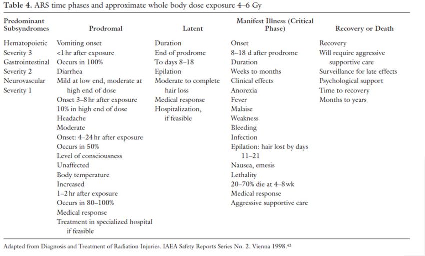
• Tables 2–4 depict general guidelines for the management of ARS.
• The syndromes include the following.
38
194/6/2020
Hematopoietic Syndrome
• Seen in absorbed doses of 1 Gy or greater and requires 7 days or more to manifest,
depending upon dose.
• The bone marrow is exquisitely sensitive to ionizing radiation, and the development of
hematopoietic syndrome is the result of bone marrow hypoplasia/aplasia.
• Decrements in the absolute lymphocyte count will be used to corroborate the estimated whole
body dose originally based on time to vomiting and predict how low the white blood cell will
drop in 1 to 2 weeks during the “critical period” of neutropenia (absolute neutrophil count of
less than 500/mm3).
• Neutropenia in the critical period can result in serious infections which may need an infectious
disease consultant if available.
• Neutropenic patients should receive prophylactic antiviral, antimicrobial, and antifungal
medications.
• Depending on the length of time spent in the austere environment with poor resource
availability and crisis standards of care, it may not be possible to follow any of these
recommendations.
• Blood products (platelets, packed red blood cells) will not be needed within the first few days
following a radiation exposure but may be needed for physical trauma and/or thermal burns.
39
GI Syndrome
• Seen in doses of 2 to 4 Gy or greater.
• GI syndrome results from massive cell death throughout the epithelium of the GI tract.
• Nausea and vomiting are the earliest indicators of GI syndrome and can occur within
1 hour at very high doses.
• Patients will then develop anorexia, abdominal pain, diarrhea, hematemesis,
hematochezia, fluid and electrolyte shifts, hypovolemia, and eventual renal failure and
cardiovascular collapse.
• This symptomatology will present extreme fluid and resuscitation challenges even for
burn patients with a small TBSA injury.
• Rectal administration of medications and fluids is not recommended in patients with
neutropenia as this can damage friable rectal mucosa and cause bacteremia.
40
204/6/2020
41
Neurovascular Syndrome
• Seen in high-dose exposures (20–30 Gy).
• Neurovascular syndrome can present as cognitive and neurologic deficits, ataxia,
seizures, and hypotension all from cerebral edema.
• Symptoms present hours to days from exposure and are generally fatal within days.
• Initial management in the field will probably be without knowledge of dose received,
and management will be based on symptoms.
• A useful field dose estimator is time to vomiting (Tables 2–4), but the estimated
absorbed dose using this parameter needs to be corroborated with serial complete
blood counts with white blood cell differentials looking for, in particular, decrements in
the absolute lymphocyte counts.
• Beware of psychogenic vomiting, which is usually not persistent as is the vomiting
that results from radiation exposure.
42
214/6/2020
43
Conclusions: Growth Factors
• Preventing profound neutropenia and subsequent infection will be critical in the
healing process for the radiation-injured burn patient.
• Patients who are known to have received an acute whole body dose of 2 to 3 Gy
should be treated with cytokines (colony stimulating factors, or CSFs).
• They should be given filgrastim (granulocyte-colony stimulating factor, or G-CSF;
Neupogen®, Amgen, Thousand Oaks, CA) at a dose of 5 μg/kg subcutaneous
injection daily, or pegfilgrastim (Neulasta®, Amgen, Thousand Oaks, CA) 6 mg
subcutaneous injection weekly as long as neutropenia persists.
• In infants, children, and adolescents < 45 kg, calculate the dose at 100 μg/kg. G-CSF
should be available through the Strategic National Stockpile in the event of a radiation
disaster.
44
224/6/2020
Conclusions: Blood Products
• Patients may develop lifethreatening anemia or thrombocytopenia.
• If this occurs and transfusion is possible, blood products should be irradiated in order
to prevent transfusion associated graft vs host disease. In a medical emergency,
including austere conditions, nonirradiated blood products may be given.
• Blood product administration should be given per AABB guidelines.
• Transfusion of whole blood and red blood cell products should be judicious and based
on clinical findings.
• Typically, platelets are transfused prophylactically in the nonhemorrhagic patient for
platelet counts of less than 10,000.
• In the setting of limited resources or inability to perform platelet counts, the field
provider may elect to transfuse platelets only for active bleeding.
• Conversely, in the presence of open wounds, or bleeding, a higher parameter may
need to be considered to prevent life-threatening hemorrhage.
45
Conclusions: Infection
• Patients who are known to be neutropenic should receive prophylactic broad spectrum antibiotics.
• A commonly accepted standard of care is a flouroquinolone, acyclovir, and an antifungal such as
fluconazole.
• In the febrile patient, antibacterial coverage must be expanded to cover the wide array of gram-
positive and gram-negative bacteria that infect neutropenic patients.
• Typically, this is done with a carbapenem (imipenem or meropenem) or fourth-generation
cephalosporin such as cefepime.
• In the austere environment, the clinician may have significant limits on choice of antibiotics.
• If this is the case, attempt to provide coverage for staphylococcus, streptococcus, and enteric
gram-negative organisms from available antibiotics.
• More detailed topical antimicrobial coverage can be found in Guidelines for Burn Care under
Austere Conditions: Wound Care.
• Patients with ARS are immunodeficient and efforts should be made to isolate them from infectious
sources, including large crowds and dusty environments.
• Patients should also wear protective face masks anytime they are unable to be isolated from public
areas or previously mentioned environments.
46
234/6/2020
Conclusions: Fluid, Electrolyte, and Nutrition
• In GI syndrome, large volume fluid losses from vomiting and diarrhea, complicated by
anorexia, will be encountered.
• Patients should be given maintenance fluid requirements plus compensation for fluid
losses with normal saline or Lactated Ringer’s IV.
• Oral replacement may not be possible due to the injured GI mucosa.
• Severe electrolyte imbalance may also be seen and should be monitored for if
possible.
• Antiemetics should be given if indicated; most effective would be odansetron or other
5HT3 antagonist.
• Effective alternatives include lorazepam, promethazine, prochlorperazine,
diphenhydramine, scopolamine, and dronabinol.
• Any of these agents can be used in addition to odansetron for breakthrough nausea.
• Nutritional support is not critical early on; however, over time (days to weeks) may be
required in order for proper healing of burns and other trauma to occur.
47
Conclusions: Fluid, Electrolyte, and Nutrition (cont.)
• Patients with GI syndrome are not likely to do well with enteral feedings until healing
of the gut tissue takes place, which will be evident by resolution of nausea, vomiting,
and diarrhea.
• Therefore, if nutrition is required, a parenteral formula should be used if available.
• In the austere environment when standard of care is replaced by crisis standard of
care, it may not be appropriate to give critical supplies (growth factors, blood
products, antibiotics, IV fluids) to patients who have absorbed doses of radiation that
will require intensive resources in order to survive.
• Patients may need to be triaged as expectant in order to direct medical supplies and
material to patients with a greater chance of survival.
48
244/6/2020
Conclusions: Fluid, Electrolyte, and Nutrition (cont.)
• It must also be remembered that austere conditions will most likely be temporary, and
the expected duration of austere conditions will have to be considered in use of
medical supplies.
• Clinical reassessment and repeat triage are critical, as resource scarcity worsens or
improves.
• This guideline is in no way a comprehensive reference for management of the patient
who has been irradiated in the austere environment.
• This document is intended to be the catalyst for future research.
• Comprehensive guidance on diagnosis and treatment for health care providers of
irradiated patients can be found at the U.S. Department of Health and Human
Services, Radiation Emergency Medical Management website,
http://www.remm.nlm.gov/.
49
50
254/6/2020
Conclusions: Recommendations
• Irradiated patients will likely need decontamination verified by a handheld radiation
detector (Geiger-Muller meter) if possible.
• Traumatic injuries are the number one priority in an irradiated patient.
• In the austere setting, the triage category may be significantly worsened in an
irradiated patient.
• Radiation burns will present as delayed onset, and should be treated as a thermal
burn.
• Burn surgical intervention should be accomplished sooner, rather than later, because
of the impaired wound healing and neutropenia of ARS.
• Development of ARS is dose dependent. Irradiated patients may develop ARS and
will require intensive management of hematologic, infectious disease, and
fluid/electrolyte issues.
51
TOXIC INDUSTRIAL
CHEMICALS
52
264/6/2020
TOXIC INDUSTRIAL CHEMICALS: Introduction
• Burn providers in an austere environment may encounter a variety of chemical
injuries.
• Some of these are caused by accidental exposure to toxic industrial chemicals (TICs),
some by the employment of chemical warfare agents, and some by the use of TICs
as improvised chemical weapons.
• In this section, the diagnosis and treatment of inhalation injury and burns caused by
the most common chemicals will be discussed.
• Other chemical agents which could be encountered, such as nerve agents, are
outside the scope of this review; more information is available from the Centers for
Disease Control at http://www.bt.cdc.gov/agent/agentlistchem.asp, from the Agency
for Toxic Substances & Disease Registry (ATSDR) at
http://www.atsdr.cdc.gov/MMG/index.asp, and in the Textbook of Military Medicine at
http://www.bordeninstitute.army.mil/cwbw/default.htm.
53
TOXIC INDUSTRIAL CHEMICALS: Introduction (cont.)
• Inhalation injury is a common route of exposure for TICs related to burn injury.
• In general, the treatment of acute lung injury secondary to TICs is similar to that for smoke
inhalation injury.
• Treatment is supportive and includes:
• 1) airway management
• 2) lung-protective ventilation
• 3) pulmonary toilet
• 4) avoidance of volume overload or excessively rapid fluid infusion that might worsen pulmonary edema
• Close monitoring for the development of acute lung injury and ventilator-associated
pneumonia is required for all patients requiring mechanical ventilation related to inhaled TICs.
• A specific drug treatment or antidote for TICs is likely to work best immediately after injury.
• Finally, these patients should be transferred to a center with expertise in inhalation injury, if
feasible.
54
274/6/2020
Pathophysiology and Clinical Care: Chlorine (Cl2)
• This gas is used abundantly in industry and is a common cause of industrial and
transportation accidents.
• Chlorine was one of the first chemical weapons deployed during World War I (WWI).
• In Iraq in 2006–2007, chlorine was used as a component of improvised explosive
devices by insurgents to attack both civilian and military/political targets.
• According to some models, release of a large quantity of chlorine in an urban area
could cause numerous deaths.
• Long-term health effects include reactive airway disease, dermal burns, and
posttraumatic stress disorder.
• Chlorine dissolves in water to form hydrochloric (HCl) and hypochlorous (HOCl)
acids; all three species participate in pathogenicity.
• Inhalation of chlorine causes both small airway and alveolar injuries.
55
Pathophysiology and Clinical Care: Chlorine (Cl2)
(cont.)
• Proposed treatments include inhaled (nebulized) or IV corticosteroids,
nebulized sodium bicarbonate in water (e.g., 3.75–4.2%), and nebulized β
agonists.
• Nebulized sodium bicarbonate has not shown clear benefit in existing reports.
• Chlorine rapidly depletes levels of endogenous antioxidants (ascorbate,
glutathione, and urate) in the airways.
• Recent research has focused on antioxidant strategies, to include IV and
aerosolized delivery of ascorbic acid and deferoxamine.
56
284/6/2020
Pathophysiology and Clinical Care: Phosgene (COCl2)
• Phosgene has a characteristic new-mown hay smell. It is used commercially in the production
of plastics, drugs, pesticides, isocyanates, and polyurethane.
• It is also released during structural fires and welding near or combustion of chlorinated
hydrocarbons.
• It was the most lethal chemical agent used during WWI.
• The classic presentation is that of delayed-onset pulmonary edema; the casualty may be seen
and discharged, only to come back in 6 to 12 hours with lethal edema triggered by exertion.
• Patients may be hypovolemic due to rapid loss of plasma volume into the lungs.
• Consider IV corticosteroids if the patient presents soon after exposure; administer
bronchodilators.
• The pathophysiology of phosgene inhalation injury includes oxidative stress and influx of
neutrophils into the lung.
• Proposed new treatments focus on these mechanisms and include N-acetylcysteine,
ibuprofen, aminophylline, isoproterenol, and colchicine, but none have been proved effective in
humans.
57
Pathophysiology and Clinical Care: Hydrogen
Sulfide (H2S)
• This gas has a “rotten eggs” smell.
• It is commonly experienced in the petroleum, natural gas, animal husbandry and
waste management industries and has been called “dung lung.”
• It enters the bloodstream via the lungs, binds to cytochrome c oxidase, and (like
cyanide) prevents oxygen use by the cells.
• An additional proposed mechanism of action (at high doses) involves the formation of
reactive oxygen species.
• H2S is metabolized to thiosulfate; it also binds to hemoglobin to form sulfhemoglobin.
• “Knockdown” is a sudden loss of consciousness and cessation of breathing due to the
effect of H2S on brainstem mitochondria.
• Other effects are seizures and myocardial ischemia.
• Its direct toxic effect on the lungs causes pulmonary edema.
• A direct effect on the cornea causes keratoconjunctivitis (“gas eye”).
58
294/6/2020
Pathophysiology and Clinical Care: Hydrogen
Sulfide (H2S) (cont.)
• Treat the patient with supportive care, to include IV fluids, oxygen, and, if obtunded,
mechanical ventilation.
• After initial resuscitation, patients who were unconscious for a prolonged period may
manifest brain anoxia, acute lung injury, and/or multiorgan failure.
• Antidotes have been used but without clear evidence of benefit. Consider IV sodium
nitrite (as for cyanide poisoning).
• IV nitrites can produce hypotension and low levels of methemoglobinemia.
• The cyanide antidote hydroxocobalamin has also been used.
• Some authors have proposed hyperbaric oxygen.
59
Pathophysiology and Clinical Care: Anhydrous
Ammonia (NH3)
• Ammonia is commonly used in the fertilizer, refrigeration, food processing, petroleum, and
explosives industries.
• It has a strong odor, which is an effective warning sign for exposure.
• White and colleagues described the treatment of five casualties who were injured during a
battle in which a container of ammonia was struck by a projectile and exploded.
• Ammonia is transported in liquid form at subzero temperatures.
• It reacts quickly on release to form NH4, ammonium hydroxide, a strong base, which is water
soluble.
• Thus, it causes alkali skin and eye burns, as well as frostbite.
• It may also cause rapid, severe tracheobronchial or pulmonary inflammation and obstruction if
inhaled, followed by pulmonary edema.
• Provide supportive care with intubation and ventilation and decontaminate the patients.
• Inhaled corticosteroids were not effective in animal models, suggesting the directly destructive
mechanism of this chemical.
• Copiously irrigate patients with skin and ocular involvement, as for any alkali injury.
60
304/6/2020
Pathophysiology and Clinical Care: Mustard
Agent (HD)
• Although mustard agents are not considered traditional TICs, their effects cause
extensive dermal burns and their exposure occurs in combat or austere environments
in countries that maintain these chemicals.
• Two types of mustard agent have been used as weapons.
• One, nitrogen mustard, was also used as a chemotherapeutic agent.
• The other, sulfur mustard (HD), has only been used as a weapon.
• Although HD was developed during the latter portion of WWI, it caused more
casualties than all the other agents combined.
• Traces of HD and other chemical weapons are still occasionally encountered in
places where munitions
• were dumped following WWI.
• HD was used by Iraq against Iran during the Iran–Iraq War of 1980–1988.
61
Pathophysiology and Clinical Care: Mustard
Agent (HD) (cont.)
• HD is troublesome for the following reasons:
• 1) It is relatively easy to make and is stockpiled in various third world countries, to
include some which are currently experiencing civil war.
• 2) Symptoms are delayed 2 to 24 hours after exposure.
• 3) It is persistent in the environment, placing medical personnel and others at risk of
cross-contamination.
• 4) It generates incapacitated casualties without causing a high death rate, thus posing
logistical challenges.
• 5) There is no specific antidote.
62
314/6/2020
Pathophysiology and Clinical Care: Mustard
Agent (HD) (cont.)
• HD predominantly affects moist areas of the body (eyes, airways, axilla, groin).
• HD quickly cyclizes in tissue and alkylates cell components (DNA and proteins).
• This DNA damage causes cell death, as well as mutations which affect the health of survivors
for years to come.
• Other mechanisms include release of reactive oxygen species, depletion of glutathione,
generation of reactive nitrogen species by iNOS, and production of proinflammatory cytokines
like tumor necrosis factor α.
• HD is radiomimetic, meaning that it predominantly affects rapidly dividing cells in the GI tract
and marrow.
• Basal keratinocytes are particularly vulnerable to HD, which is why skin injuries feature
dermal–epidermal separation (similar histologically to toxic epidermal necrolysis syndrome).
• Wound healing, compared to thermal injuries of similar depth, is greatly prolonged.
• Because HD is a persistent chemical warfare agent, the first principal in management is to
protect caregivers and patients through sound protection measures and effective casualty
decontamination.
63
Pathophysiology and Clinical Care: Mustard
Agent (HD) (cont.)
• A treatment plan for mustard agent casualties was developed by the U.S. Army Burn
Center in preparation for Operation Iraqi Freedom, which assumed that all U.S. forces
injured with HD would be decontaminated in the field, then evacuated to the Burn
Center in San Antonio, Texas.
• Triage of mustard casualties uses the following indicators of high-dose exposure:
• 1) rapid onset of pulmonary symptoms, that is, within 2 to 6 hours
• 2) ≥25% TBSA cutaneous injury (not just erythema)
• 3) heavy vomiting within 24 hours of exposure
• 4) a lymphocyte drop of ≥ 50% within 24 hours of exposure
• Treatment includes airway support, close monitoring of those with lung injuries for
pneumonia, ophthalmology evaluation, atropine and antiemetics for vomiting,
cutaneous management based on depth of injury, and granulocyte-colony-stimulating
factor (GCSF) for those with decreased lymphocyte counts.
• Lymphopenia is an early marker for impending pancytopenia; the main effect of
GCSF is to prevent the neutropenia.
64
324/6/2020
Pathophysiology and Clinical Care: Hydrogen
Fluoride
• Hydrogen fluoride and the aqueous form, hydrofluoric acid, are common chemicals in
the gasoline, glassware, and semiconductor industries.
• Chemical suppressants with fluorinated hydrocarbons may produce HF used and
could produce toxicity in a confined-space, long-exposure duration.
• HF dissolves in the epithelial lining to create hydrofluoric acid.
• Low doses cause pulmonary irritation, and large doses can cause bronchial and
pulmonary parenchymal destruction.
• Systemic toxicity may develop and result in hypocalcemia, hyperkalemia, and sudden
cardiac death.
• Pulmonary complications are treated supportively.
• Systemic toxicity is treated with IV calcium, and local burns due to hydrofluoric acid
are treated with topical calcium.
• Nebulized calcium has been used for treatment of hydrogen fluoride inhalation injury.
65
Pathophysiology and Clinical Care:
Other chemicals
• Hydrogen chloride gas is produced by the pyrolysis of polyvinyl chloride, a plastic
used for pipes.
• It is an occupational hazard for firefighters.
• Pulmonary toxicity can develop and is treated supportively.
• Isocyanates, such as methylisocyanate, can be produced during pyrolysis of
chemicals and polymers.98 Toluene diisocyanate (TDI) and diphenyl methane
diisocyanate (MDI) are also produced from polymer plants during a fire.
• Isocyanates produce pulmonary toxicity (Figure 2).
66
334/6/2020
• Figure 2. Triage category for combined
injury and trauma based on injury
severity. (Modified from Radiation
Emergency Medical Management—
Triage Tools.)
67
Conclusion
• Any of the chemical agents discussed in the manuscript may be encountered by burn
care providers working in austere, mass casualty, or battlefield environments.
• Incorporation of agents like chlorine into improvised explosive devices indicates the
willingness of terrorists to use TICs as agents of opportunity.
• Thoroughly decontaminate casualties and avoid secondary contamination of
providers and medical facilities.
• Employ the same principles of care as for burns and smoke inhalation injury caused
by conventional means.
• Finally, know the specific antidotes or treatments available for many of these TICs.
• The burn-care team is uniquely qualified to care for casualties with TIC-related
injuries.
68
344/6/2020
Recommendations
• Be prepared to perform decontamination of chemical casualties in the
deployed environment
• To avoid spread of toxic chemicals, decontaminate casualties outside of the
hospital
• Consider specific therapies based on identification of the chemical, for
example:
• Chlorine: antioxidants
• Phosgene: early IV corticosteroids; observation for delayed onset pulmonary edema
• Hydrogen sulfide: hydroxocobalamin or nitrites
• Ammonia: copious prolonged decontamination
• Mustard agent: monitor lymphocyte count; GCSF for lymphopenia
• Hydrogen fluoride: calcium
69
ACKNOWLEDGMENTS
• Special thanks to Doran M. Christensen, Senior Medical Advisor, at the Radiation
Emergency Assistance Center/Training Site (REAC/TS) for his assistance with this
article.
• Work provided by Dr. Christensen and REAC/TS were provided under contract with
the U.S. Department of Energy, Contract # DE-AC05-06OR23100.
70
35You can also read