EVMS CRITICAL CARE COVID-19 MANAGEMENT PROTOCOL - FMDA
←
→
Page content transcription
If your browser does not render page correctly, please read the page content below
EVMS CRITICAL CARE
  COVID-19 MANAGEMENT PROTOCOL
                           Developed and updated by Paul Marik, MD
                          Chief of Pulmonary and Critical Care Medicine
                          Eastern Virginia Medical School, Norfolk, VA
                                        August 1st, 2020

This is our recommended approach to COVID-19 based on the best (and most recent) literature. This is a
very dynamic situation; therefore, we will be updating the guideline as new information emerges. Please
check on the EVMS website for updated versions of this protocol.
EVMS COVID website: https://www.evms.edu/covid-19/medical_information_resources/
Short url: evms.edu/covidcare

Disclaimer: The information provided in this protocol is primarily to educate physicians on a
protocol that we found to be highly effective in damping down the hyper-inflammatory
cytokine “storm” that is the cause of mortality and morbidity in COVID-19. Our guidance
should only be used by medical professionals in formulating their approach to COVID-
19. Patients should always consult with their physician before starting any medical treatment.

FLCC website: https://covid19criticalcare.com/

 Page 1 of 25 | EVMS Critical Care COVID-19 Management Protocol 08-01-2020 | evms.edu/covidcare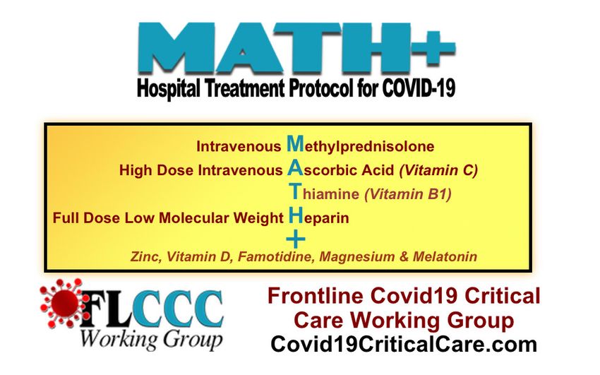
Figure 1. The course of COVID-19 and General Approach to treatment

         THIS IS A STEROID RESPONSIVE DISEASE:
                  HOWEVER, TIMING IS CRITICAL

 Page 2 of 25 | EVMS Critical Care COVID-19 Management Protocol 08-01-2020 | evms.edu/covidcare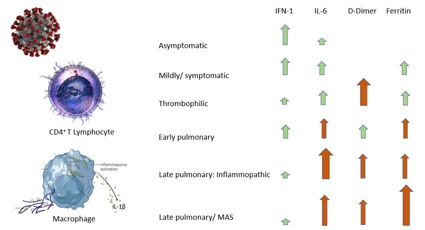
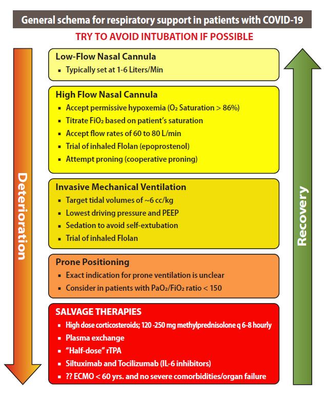
Figure 2. Timing of the initiation of anti-inflammatory therapy

 Page 3 of 25 | EVMS Critical Care COVID-19 Management Protocol 08-01-2020 | evms.edu/covidcare
Prophylaxis
While there is extremely limited data, the following “cocktail” may have a role in the
prevention/mitigation of COVID-19 disease. It should however be noted that a recent publication
suggests that melatonin my reduce the risk of COVID-19 infection.[1] This cocktail is cheap, safe, and
widely available.
    •   Vitamin C 500 mg BID (twice daily) and Quercetin 250-500 mg BID [2-8]
    •   Zinc 50-75 mg/day (elemental zinc). Zinc lozenges are preferred. After 1 month, reduce the
        dose to 30-50 mg/day. [2,9-13]
    •   Melatonin (slow release): Begin with 0.3mg and increase as tolerated to 2 mg at night [1,14-17]
    •   Vitamin D3 2000-4000 u/day [18-25]
    •   Optional: Famotidine 20-40 mg/day [26]

Symptomatic patients (at home):
    •   Vitamin C 500 mg BID and Quercetin 250-500 mg BID
    •   Zinc 75-100 mg/day (elemental zinc)
    •   Melatonin 6-12 mg at night (the optimal dose is unknown)
    •   Vitamin D3 2000-4000 u/day
    •   ASA 81 -325 mg/day (unless contraindicated)
    •   Optional: Famotidine 20-40 mg/day
    •   Optional: Ivermectin 150-200 ug/kg orally (dose can be repeated on day 2) [27-31]
    •   In symptomatic patients, monitoring with home pulse oximetry is recommended. Baseline or
        ambulatory desaturation < 94% should prompt hospital admission. [32]
    •   Not recommended: Hydroxychloroquine (HCQ). The use of HCQ is extremely controversial.[33]
        The best scientific evidence to date suggest that HCQ has no proven benefit for post exposure
        prophylaxis, for the early symptomatic phase and in hospitalized patients. [34-39] It should be
        noted that these studies did not include Zinc, and it is possible that the efficacy of HCQ requires
        the co-administration of Zinc. [40,41] However, considering the unique pharmacokinetics of
        HCQ, it is unlikely that HCQ is of benefit (takes about 10 days to achieve adequate plasma and
        lung concentrations).[42-44] The benefit derived from the co-administration of Zinc may be due
        to the effects of zinc alone. This is however, a very “volatile” situation, so stay tuned.

Mildly Symptomatic patients (on floor):
    •   Vitamin C 500 mg q 6 hourly and Quercetin 250-500 mg BID (if available)
    •   Zinc 75-100 mg/day
    •   Melatonin 6-12 mg at night (the optimal dose is unknown)
    •   Vitamin D3 4000 u/day
    •   Enoxaparin 60 mg daily [31,45-54] Consider increasing the dose to 1mg/kg q 12 hourly in those
        with a high D-Dimer or an increasing D-Dimer (see Xa monitoring below).
    •   Methylprednisolone 40 mg q 12 hourly ; increase to 80 mg q 12 hourly in patients with
        progressive symptoms and increasing CRP. [55-61] The role of inhaled corticosteroids
        (budesonide) is unclear and appears to be rather limited.
    •   Famotidine 40 mg daily (20 mg in renal impairment)
    •   Optional: Ivermectin 150-200 ug/kg (dose can be repeated on day 2)

 Page 4 of 25 | EVMS Critical Care COVID-19 Management Protocol 08-01-2020 | evms.edu/covidcare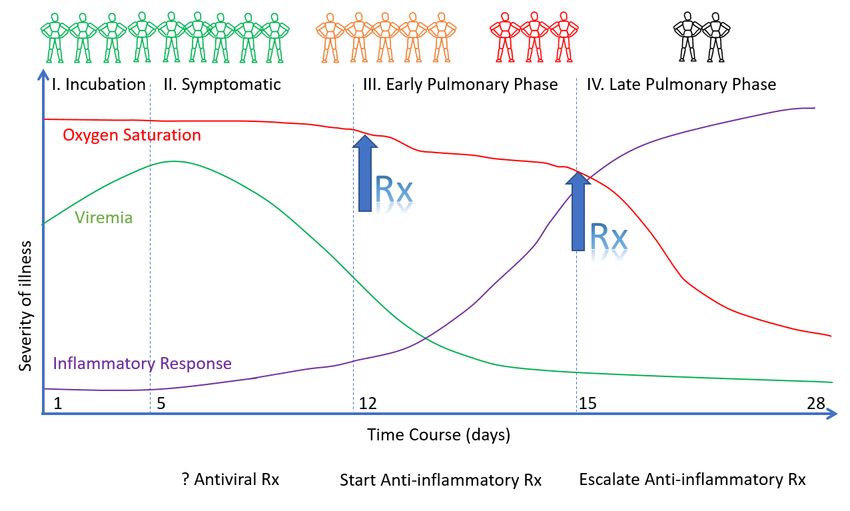
•   Optional: Vascepa (Ethyl eicosapentaenoic acid) 4g daily or Lovaza (EPA/DHA) 4g daily;
         alternative DHA/EPA 4g daily. Vascepa and Lovaza tablets must be swallowed and cannot be
         crushed, dissolved or chewed.
     •   Optional: Remdesivir, 200 mg IV loading dose D1, followed by 100mg day IV for 9 days. [62,63]
         This agent has been reported to reduce time to recovery (based on an ordinal scale). [63] The
         benefit of this agent on patient centered outcomes is unclear.
     •   N/C 2L /min if required (max 4 L/min; consider early t/f to ICU for escalation of care).
     •   Avoid Nebulization and Respiratory treatments. Use “Spinhaler” or MDI and spacer if required.
     •   T/f EARLY to the ICU for increasing respiratory signs/symptoms, increasing oxygen requirements
         and arterial desaturation.

Progressive Respiratory symptoms (hypoxia- requiring N/C ≥ 4 L min: admit to ICU):
Essential Treatment (dampening the STORM); MATH +
1. Methylprednisolone 80 mg loading dose then 40 mg q 12 hourly for at least 7 days and until
   transferred out of ICU. In patients with an increasing CRP or worsening clinical status increase the
   dose to 80 mg q 12 hourly (then 125mg q 12 hourly), then titrate down as appropriate. [55-61]
2. Ascorbic acid (Vitamin C) 3g IV q 6 hourly for at least 7 days and/or until transferred out of ICU.
   Note caution with POC glucose testing (see below). [64-72]. Oral absorption is limited by saturable
   transport and it is difficult to achieve adequate levels with PO administration. However,
   unfortunately, IV Vitamin C is not available in many hospitals; in this situation attempts should be
   made to administer PO vitamin C at a dose of 1g every 4-6 hours.
3. Full anticoagulation: Unless contraindicated we suggest FULL anticoagulation (on admission to the
   ICU) with enoxaparin, i.e 1 mg kg s/c q 12 hourly (dose adjust with Cr Cl < 30mls/min). [45-54]
   Heparin is suggested with CrCl < 15 ml/min. Due to augmented renal clearance patients may have
   reduced anti-Xa activity despite standard dosages of LMWH.[73] We therefore recommend
   monitoring anti-Xa activity in underweight and obese patients, those with chronic renal failure and
   in those patients with an increasing D-dimer, aiming for an anti-Xa activity of 0.6-1.1 IU.ml.

     Note: A falling SaO2 despite respiratory symptoms should be a trigger to start anti-inflammatory
     treatment (see Figure 2).
     Note: Early termination of ascorbic acid and corticosteroids will likely result in a rebound effect with
     clinical deterioration (see Figure 3).

Additional Treatment Components (the Full Monty)

4. Melatonin 6-12 mg at night (the optimal dose is unknown).
5. Famotidine 40 mg daily (20 mg in renal impairment)
6. Vitamin D 4000 u PO daily
7. Thiamine 200 mg IV q 12 hourly [74-78]
8. Magnesium: 2 g stat IV. Keep Mg between 2.0 and 2.4 mmol/l. Prevent hypomagnesemia (which
   increases the cytokine storm and prolongs Qtc). [79-81]
9. Atorvastatin 80 mg/day. Statins have pleotropic anti-inflammatory, immunomodulatory,
   antibacterial, and antiviral effects. In addition, statins decrease expression of PAI-1. Simvastatin has

 Page 5 of 25 | EVMS Critical Care COVID-19 Management Protocol 08-01-2020 | evms.edu/covidcare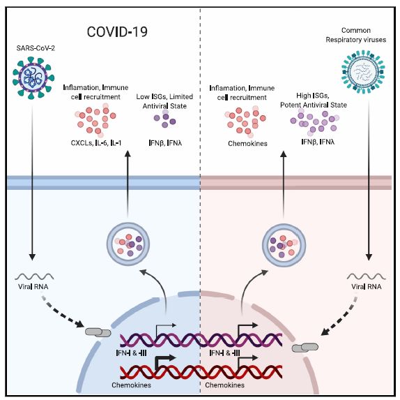
been demonstrated to reduce mortality in the hyper-inflammatory ARDS phenotype. [82]
      Preliminary data suggests atorvastatin may improve outcome in patients with COVID-19.[83,84]
      Due to numerous drug-drug interactions simvastatin should be avoided.
10.   Optional: Vascepa, Lovaza or DHA/EPA 4g day (see above).
11.   Optional: Azithromycin 500 mg day 1 then 250 mg for 4 days (has immunomodulating properties
      including downregulating IL-6; has anti-viral properties and in addition, Rx of concomitant bacterial
      pneumonia). [31,85] The benefit of azithromycin in COVID-19 is however unproven.
12.   Optional: Remdesivir. The role of this agent in patients with more advanced pulmonary
      involvement appears to be limited.
13.   Broad-spectrum antibiotics if superadded bacterial pneumonia is suspected based on procalcitonin
      levels and resp. culture (no bronchoscopy). Due to the paradox of hyper-inflammation and immune
      suppression (a major decrease of HLA-DR on CD14 monocytes) secondary bacterial infection is not
      uncommon.
14.   Maintain EUVOLEMIA (this is not non-cardiogenic pulmonary edema). Due to the prolonged
      “symptomatic phase” with flu-like symptoms (6-8 days) patients may be volume depleted. Cautious
      rehydration with 500 ml boluses of Lactate Ringers may be warranted, ideally guided by non-
      invasive hemodynamic monitoring. Diuretics should be avoided unless the patient has obvious
      intravascular volume overload. Avoid hypovolemia.
15.   Early norepinephrine for hypotension. It should however be appreciated that despite the cytokine
      storm vasodilatory shock is distinctly uncommon in uncomplicated COVID-19 (not complicated by
      bacterial sepsis). This appears to be due to the fact TNF-α which is only moderately elevated in
      COVID-19, is “necessary” for vasodilatory shock.

16. Escalation of respiratory support (steps); Try to avoid intubation if at all possible, (see Figure 4)
           • Accept “permissive hypoxemia” (keep O2 Saturation > 84%); follow venous lactate and
               Central Venous O2 saturations (ScvO2) in patents with low arterial O2 saturations
           • N/C 1-6 L/min
           • High Flow Nasal canula (HFNC) up to 60-80 L/min
           • Trial of inhaled Flolan (epoprostenol)
           • Attempt proning (cooperative repositioning-proning) [86,87]
           • Intubation … by Expert intubator; Rapid sequence. No Bagging; Full PPE.
               Crash/emergency intubations should be avoided.
           • Volume protective ventilation; Lowest driving pressure and lowest PEEP as possible.
               Keep driving pressures < 15 cmH2O.
           • Moderate sedation to prevent self-extubation
           • Trial of inhaled Flolan (epoprostenol)
           • Prone positioning.
   There is widespread concern that using HFNC could increase the risk of viral transmission. There is
   however, no evidence to support this fear. HFNC is a better option for the patient and the health
   care system than intubation and mechanical ventilation. CPAP/BiPAP may be used in select
   patients, notably those with COPD exacerbation or heart failure.

   A sub-group of patients with COVID-19 deteriorates very rapidly. Intubation and mechanical
   ventilation may be required in these patients.

Page 6 of 25 | EVMS Critical Care COVID-19 Management Protocol 08-01-2020 | evms.edu/covidcare
17. Salvage Treatments
        • High dose corticosteroids; 120 -250 mg methylprednisolone q 6-8 hourly
        • Plasma exchange [88-90]. Should be considered in patients with progressive
            oxygenation failure despite corticosteroid therapy as well as in patients with severe
            MAS. Patients may require up to 5 exchanges. FFP is required for the exchange; giving
            back “good humors” appears to be more important than taking out “bad humors”.
        • In patients with a large dead-space ventilation i.e. high PaCO2 despite adequate minute
            ventilation consider “Half-dose rTPA” to improve pulmonary microvascular blood flow;
            25mg of tPA over 2 hours followed by a 25mg tPA infusion administered over the
            subsequent 22 hours, with a dose not to exceed 0.9 mg/kg followed by full
            anticoagulation.[91,92]
        • Janus Kinase inhibitors downregulate cytokine expression and may have a role in this
            disease. [93-95]
        • Convalescent serum; the role and timing of convalescent serum are uncertain. [96-99]
            COVID-19 pulmonary disease is immune mediated, and it would therefore appear
            paradoxical to enhance the antibody response with convalescent serum. [100]

     Salvage treatments of unproven benefit.
         • Siltuximab and Tocilizumab (IL-6 inhibitors).[101,102] Roche™ recently announced the
            results of the COVACTA study, which demonstrated that Tocilizumab did not improve
            patient outcome.
         • Convalescent serum: the role and timing of convalescent serum are uncertain. [96-99]
            COVID-19 pulmonary disease is immune mediated, and it would therefore appear
            paradoxical to enhance the antibody response with convalescent serum. [100]
         • CVVH with cytokine absorbing/filtering filters [103] This treatment strategy appears to
            have a very limited role.
         • ECMO [104]. Unlike “typical ARDS” patients do not progress into a resolution phase.
            Rather, patients with COVID-19 progress to a severe fibro-proliferative phase and
            ventilator dependency. ECMO in these patients would likely serve little purpose.

 18. Treatment of Macrophage Activation Syndrome (MAS)

     •   A sub-group of patients will develop MAS. This appears to be driven by SARS-CoV-2 induced
         inflammasome activation and increased IL-1 β production (see Figure 5). [105,106]
     •   A ferritin > 4400 ng/ml is considered diagnostic of MAS. Other diagnostic features include
         increasing AST/ALT and increasing CRP. [107]
     •   “High dose corticosteroids.” Methylprednisolone 120 mg q 6-8 hourly for at least 3 days,
         then wean according to Ferritin, CRP, AST/ALT (see Figure 6). Ferritin should decrease by at
         least 15% before weaning corticosteroids.
     •   Consider plasma exchange.
     •   Anakinra (competitively inhibits IL-1 binding to the interleukin-1 type I receptor) can be
         considered in treatment failures. [108,109]

Page 7 of 25 | EVMS Critical Care COVID-19 Management Protocol 08-01-2020 | evms.edu/covidcare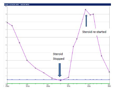
19. Monitoring
   • On admission: PCT, CRP, IL-6, BNP, Troponins, Ferritin, Neutrophil-Lymphocyte ratio, D-dimer
      and Mg. D-dimer is the most important prognostic marker.
   • Daily: CRP, Ferritin, D-Dimer and PCT. CRP and Ferritin track disease severity closely (although
      ferritin tends to lag behind CRP). Early high CRP levels are closely associated with the degree of
      pulmonary involvement and the CT score. [110]
   • Thromboelastogram (TEG) in patients with high D-dimer and repeated as indicated.
   • In patients receiving IV vitamin C, the Accu-Chek™ POC glucose monitor will result in spuriously
      high blood glucose values. Therefore, a laboratory glucose is recommended to confirm the
      blood glucose levels. [111,112]
   • Monitor QTc interval if using azithromycin and monitor Mg++ (torsades is uncommon in
      monitored ICU patients)
   • No routine CT scans, follow CXR and chest ultrasound.
   • ECHO as clinically indicated; Pts may develop a severe cardiomyopathy.

20. Post ICU management
           a. Enoxaparin 40-60 mg s/c daily
           b. Methylprednisolone 40 mg day, then wean slowly (follow CRP)
           c. Vitamin C 500 mg PO BID
           d. Melatonin 3-6 mg at night

Figure 3. Premature discontinuation of corticosteroids and IV vitamin C (after 4
day) and the effect of reinitiation of this combination on the CRP profile.

 Page 8 of 25 | EVMS Critical Care COVID-19 Management Protocol 08-01-2020 | evms.edu/covidcare
Figure 4.
Page 9 of 25 | EVMS Critical Care COVID-19 Management Protocol 08-01-2020 | evms.edu/covidcareFigure 5. SARS-CoV-2 induced Macrophage Activation Syndrome (MAS) treated with Vitamin C 3g IV q 6 and increased methylprednisolone (125 mg q 8 hourly) Page 10 of 25 | EVMS Critical Care COVID-19 Management Protocol 08-01-2020 | evms.edu/covidcare
Key Concepts of the EVMS Treatment Protocol
This is a very complex disease; many of the mysteries are still unravelling. However, a number of
concepts are key to the management of this “treatable disease; they include.

    1. Patients transition through a number of different phases (clinical stages). The treatment of each
       phase is distinct ... this is critically important (see Figures 1 & 2).
    2. As patients, progress down the pulmonary cascade the disease becomes more difficult to
       reverse. The implications of this are twofold.
             a. Early treatment is ESSENTIAL to a good outcome (this is critical)
             b. Treatment in the late pulmonary phase my require escalation of the dose of
                corticosteroids as well as the use of salvage methods (i.e. plasma exchange).
    2. It is important to recognize that COVID-19 patients present with a variety of phenotypes, likely
       dependent on genetic heterogeneity, blood type, sex and androgen status, age, viral load,
       immunological and nutritional status, and co-morbidities (see Figure 6).[58,113-118] The
       phenotype at presentation likely determines the optimal approach to treatment.
    3. COVID-19 is a treatable disease; it is inappropriate to limit therapy to “supportive care” alone.
       Furthermore, it is likely that there will not be a single “magic bullet” to treat COVID-19. Rather,
       we should be using multiple drugs/interventions that have synergistic and overlapping biological
       effects that are safe, cheap and “readily” available. The impact of COVID-19 on middle- and low-
       income countries will be enormous; these countries will not be able to afford expensive
       designer molecules.
    4. The pulmonary phase is characterized by immune dysregulation, [93,95,102,105,106,116,119-
       127] a pulmonary microvascular injury (endothelialitis),[127-130] with activation of clotting and
       a pro-coagulant state together with the characteristics of an organizing pneumonia.
    5. It should be noted that SARS-CoV-2 as compared to all other respiratory viruses, upregulates
       cytokines and chemokines while at the same time down regulating the expression of Interferon
       alpha (the hosts primary antiviral defence mechanism). [105,120] This factor is critical to
       understanding the treatment of COVID-19 organizing pneumonia. (see Figure 7).[120]
    6. THIS is NOT ARDS (at least initially). The initial
       pulmonary phase neither looks like, smells like nor is
       ARDS.[131-133] The ground glass infiltrates are
       peripheral and patchy, and do not resemble the
       dependent air space consolidation (sponge/baby lung)
       seen with “typical ARDS”.[134] Extravascular lung
       water index (EVLWI) is normal or only slightly
       increased; this by definition excludes non-cardiogenic
       pulmonary edema (ARDS). Lung compliance is normal
       (this excludes ARDS). Patients are PEEP unresponsive.
       Treating patients as if they ARDS is a very dangerous
       approach. The hypoxia is due to severe
       ventilation/perfusion mismatch likely due to the
       microvascular narrowing, thrombosis and vasoplegia.
    7. The core principles of the pulmonary phase (MATH+) is the use of anti-inflammatory agents to
       dampen the “cytokine storms” together with full anticoagulation to limit the microvascular and
       macrovascular clotting and supplemental oxygen to help overcome the hypoxia.

 Page 11 of 25 | EVMS Critical Care COVID-19 Management Protocol 08-01-2020 | evms.edu/covidcare
8. Patients in whom the cytokine storm is not “dampened” will progress into the “H phenotype”
characterized by poor lung compliance, severe oxygenation failure and PEEP recruitability (see
Figure 8). Progression to this phase is exacerbated by ventilator induced lung injury (VILI). The
histologic pattern of the “H Phenotype” is characterized by an acute fibrinous and organizing
pneumonia (AFOP), with extensive intra-alveolar fibrin deposition called fibrin “balls” with
absent hyaline membranes.[118,135-138] Corticosteroids seem to be of little benefit in
established AFOP. High dose methylprednisolone should be attempted in the “early phase” of
AFOP, however many patients will progress to irreversible pulmonary fibrosis with prolonged
ventilator dependency and ultimately death.
9. The combination of steroids and ascorbic acid (vitamin C) is essential. Both have powerful
synergistic anti-inflammatory actions. [65] Vitamin C protects the endothelium from oxidative
injury.[66,139-141] Furthermore, vitamin C Increases the expression of interferon-alpha (this is
critical) ([5] while corticosteroids (alone) decease expression of interferon-alpha. [142-145] It
should however be noted that when corticosteroids are used in the pulmonary phase (and not
in the viral replicative phase) they do not appear to increase viral shedding or decrease the
production of type specific antibodies.
10. Notwithstanding the very important and impressive results of the Recovery-Dexamethasone
study, methylprednisolone is the corticosteroid of choice for the pulmonary phase of COVID-19.
This is based on pharmacokinetic data (better lung penetration),[146] genomic data specific for
SARS-CoV-2,[147] and a long track record of successful use in inflammatory lung diseases.
11. For prophylaxis and treatment of the early symptomatic phase we suggest the combination of
Quercetin (a plant polyphenol), Vitamin C and Zinc. This is based on intriguing basic science data
which indicates that:
a. Zinc is essential for innate and adaptive immunity.[10] In addition, Zinc inhibits RNA
dependent RNA polymerase in vitro against SARS-CoV-2 virus.[9]
b. Quercetin has direct viricidal properties against a range of viruses, including SARS-CoV-
2.[3,7] In addition, quercetin acts as a zinc ionophore. [148]
c. Vitamin C improves the potency of Quercetin and has antiviral activity.[3]
12. It should also be noted that Vitamin D may be a very powerful prophylactic and treatment
strategy against COVID-19. [18-25] Vitamin D deficiency explains, in part, the enormous
geographic variation in mortality of this disease.
Page 12 of 25 | EVMS Critical Care COVID-19 Management Protocol 08-01-2020 | evms.edu/covidcareFigure 6. COVID-19 Subtypes of Infections (Phenotypes) Page 13 of 25 | EVMS Critical Care COVID-19 Management Protocol 08-01-2020 | evms.edu/covidcare
Figure 7. Imbalanced host response to SARS-CoV-2 drives development of COVID-
19. [120] Open Access Publication.

Page 14 of 25 | EVMS Critical Care COVID-19 Management Protocol 08-01-2020 | evms.edu/covidcare
Scientific Rationale for MATH+ Treatment Protocol (pulmonary phase)
Three core pathologic processes lead to multi-organ failure and death in COVID-19:

    1) Hyper-inflammation (“Cytokine storm”) – a dysregulated immune system whose cells infiltrate
       and damage multiple organs, namely the lungs, kidneys, and heart. It is now widely accepted
       that SARS-CoV-2 causes aberrant T lymphocyte and macrophage activation resulting in a
       “cytokine storm.” [93,95,102,105,106,116,119,121-126]
    2) Hyper-coagulability (increased clotting) – the dysregulated immune system damages the
       endothelium and activates blood clotting, causing the formation of micro and macro blood clots.
       Clotting activation may occur directly due to increased expression of Factor Xa as well as
       endothelial injury with the release of large aggregates of van Willebrand factor. These blood
       clots impair blood flow. [45-54,129,130,149,150]
    3) Severe Hypoxemia (low blood oxygen levels) –lung inflammation caused by the cytokine storm,
       together with microthrombosis in the pulmonary circulation severely impairs oxygen absorption
       resulting in oxygenation failure.

The above pathologies are not novel, although the combined severity in COVID-19 disease is
considerable. Our long-standing and more recent experiences show consistently successful treatment if
traditional therapeutic principles of early and aggressive intervention is achieved, before the onset of
advanced organ failure. It is our collective opinion that the historically high levels of morbidity and
mortality from COVID-19 is due to a single factor: the widespread and inappropriate reluctance amongst
hospitalists and intensivists to employ anti-inflammatory and anticoagulant treatments, including
corticosteroid therapy early in the course of a patient’s hospitalization. It is essential to recognize that
it is not the virus that is killing the patient, rather it is the patient’s overactive immune system. [95,100]
The flames of the “cytokine fire” are out of control and need to be extinguished. Providing supportive
care (with ventilators that themselves stoke the fire) and waiting for the cytokine fire to burn itself out
simply does not work… this approach has FAILED and has led to the death of tens of thousands of
patients.

  “If what you are doing ain’t working, change what you are doing”- PEM
The systematic failure of critical care systems to adopt corticosteroid therapy resulted from the
published recommendations against corticosteroids use by the World Health Organization
(WHO) [151,152]. This recommendation was then perpetuated by the Centers for Disease Control and
Prevention (CDC), the American Thoracic Society (ATS), Infectious Diseases Association of America (IDSA)
amongst others. A very recent publication by the Society of Critical Care Medicine and authored one of
the members of the Front Line COVID-19 Critical Care (FLCCC) group (UM), identified the errors made by
these organizations in their analyses of corticosteroid studies based on the findings of the SARS and
H1N1 pandemics.[55,153] Their erroneous recommendation to avoid corticosteroids in the treatment of
COVID-19 has led to the development of myriad organ failures which have overwhelmed critical care
systems across the world and led to excess deaths. The recently announced results of the RECOVERY-
DEXAMETHASONE study provides definitive and unambiguous evidence of the lifesaving benefits of
corticosteroids and strong validation of the MATH + protocol. The RECOVERY-DEXAMETHASONE study,
randomized 2104 patients to receive dexamethasone 6 mg (equivalent to 32 mg methylprednisolone)
once per day (either by mouth or by intravenous injection) for ten days and were compared with 4321
patients randomized to usual care alone. Dexamethasone reduced deaths by one-third in ventilated

 Page 15 of 25 | EVMS Critical Care COVID-19 Management Protocol 08-01-2020 | evms.edu/covidcare
patients (rate ratio 0.65 [95% confidence interval 0.48 to 0.88]; p=0.0003) and by one fifth in other patients receiving oxygen only (0.80 [0.67 to 0.96]; p=0.0021). There was no benefit among those patients who did not require respiratory support (1.22 [0.86 to 1.75; p=0.14). The results of this study STRONGLY support the EVMS protocol which recommends the use of corticosteroids for the “pulmonary phase” of COVID-19. It should be noted that we would consider the non-titratable ‘fixed” dose of dexamethasone used in the RECOVERY-DEXAMETHASONE study to be very low. Furthermore, as indicated above we consider methylprednisolone to be the corticosteroid of choice for the treatment of COVID-19 pulmonary disease. Our treatment protocol targeting the key pathologic processes has achieved near uniform success, if begun within 6 hours of a COVID19 patients presenting with shortness of breath and/or arterial desaturation and requiring supplemental oxygen. If such early initiation of treatment could be systematically achieved, the need for mechanical ventilators and ICU beds will decrease dramatically. The systematic use of the MATH+ protocol in 2 hospitals in the USA has reduced the hospital mortality from COVID-19 to approximately 6% (the average hospital mortality for COVID-19 across the world is reported to be 21%). Page 16 of 25 | EVMS Critical Care COVID-19 Management Protocol 08-01-2020 | evms.edu/covidcare
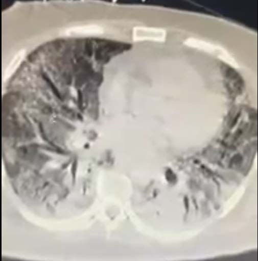
Figure 8. The consequences of “steroid” avoidance”. CT scan after 23 days of
“supportive care” demonstrating the late fibroproliferative (irreversible) phase of
COVID-19 lung disease (Image kindly provide by Dr. Pierre Kory, from NYC).

 Page 17 of 25 | EVMS Critical Care COVID-19 Management Protocol 08-01-2020 | evms.edu/covidcare
References

  1. Jehi L, Ji X, Milinovich A et al. Individualizing risk prediction for positive COVID-19 testing. Results
     from 11,672 patients. Chest 2020.
  2. Maggini S, Beveridge S, suter M. A combination of high-dose vitamin C plus zinc for the common
     cold. Journal of International Medical Research 2012; 40:28-42.
  3. Colunga Biancatelli RM, Berrill M, Catravas JD et al. Quercetin and Vitamin C: experimental
     therapy for the prevention and treatment of SARS-CoV-2 via synergistic action. Front Immunol
     2020.
  4. Kyung Kim T, Lim HR, Byun JS. Vitamin C supplementaion reduces the odds of developing a
     common cold in Republic of Korea Army recruits: a randomised controlled trial. BMJ Mil Health
     2020.
  5. Colunga Biancatelli RM, Berrill M, Marik PE. The antiviral properties of vitamin C. Expert Rev Anti
     Infect Ther 2020; 18:99-101.
  6. Khaerunnisa S. Potential inhibitor of COVID-19 main protease (Mpro) from several medicinal
     plant compuns by molecular docking study. medRxiv 2020.
  7. Chen L, Li J, Luo C et al. Binding interaction of quercetin-3-B-galactoside and its synthetic
     derivatives with SARS-CoV 3CL: structure-activity relationship reveal salient pharmacophore
     features. Bioorganic & Medicinal Chemistry Letters 2006; 14:8295-306.
  8. Yi L, Li Z, Yuan K et al. Small molecules blocking the entry of severe respiratory syndrome
     coronavirus into host cells. J Virol 2020; 78:11334-39.
  9. te Velthuis AJ, van den Worm SH, Sims AC et al. Zn2+ inhibits Coronavirus and Arterivirus RNA
     polymerase activity In Vitro and Zinc ionophores block the replication of these viruses in cell
     culture. PLos Pathog 2010; 6:e1001176.
 10. Gammoh NZ, Rink L. Zinc in Infection and Inflammation. Nutrients 2017; 9.
 11. Hemila H. Zinc lozenges and the common cold: a meta-analysis comparing zinc acetate and zinc
     gluconate, and the role of zinc dosage. J Royal Soc Med Open 2017; 8:1-7.
 12. Singh M, Das RR. Zinc for the common cold. Cochrane Database of Syst Rev 2013; 6:CD001364.
 13. Hoeger J, Simon TP, Beeker T et al. Persistent low serum zinc is associated with recurrent sepsis
     in critically ill patients - A pilot study. PloS ONE 2017; 12:e0176069.
 14. Colunga Biancatelli RM, Berrill M, Mohammed YH et al. Melatonin for the treatment of sepsis:
     the scientific rationale. J Thorac Dis 2020; 12 (Suppl 1):S54-S65.
 15. Reiter RJ, Abreu-Gonzalez P, Marik PE et al. Therapeutic algorithm for use of melatonin in
     patients with COVID-19. Front Med 2020; 7:226.
 16. Reiter RJ, Sharma R, Ma Q et al. Melatonin inhibits COVID-19-induced cytokine storm by
     reversing aerobic glycolysis in immune cells: A mechanistic analysis. Medicine in Drug Discovery
     2020; 6:100044.
 17. Zhang R, Wang X, Ni L et al. COVID-19: Melatonin as a potential adjuvant treatment. Life Sci
     2020; 250:117583.
 18. Grant WB, Lahore H, McDonnell SL et al. Evidence that Vitamin D supplementation could reduce
     risk of influenza and COVID-19 infections and deaths. Nutrients 2020; 12:988.
 19. Lau FH, Majumder R, Torabi R et al. Vitamin D insufficiency is prevalent in severe COVID-19.
     medRxiv 2020.
 20. Marik PE, Kory P, Varon J. Does vitamin D status impact mortlality from SARS-CoV-2 infection?
     Medicine in Drug Discovery 2020.
 21. Rhodes JM, Subramanian S, Laird E et al. Editorial: Low population mortality from COVID-19 in
     countries south of 35 degrees North - supports vitamin D as a factor determining severity.
     Alimentary Pharmacology & Therapeutics 2020; (in press).

Page 18 of 25 | EVMS Critical Care COVID-19 Management Protocol 08-01-2020 | evms.edu/covidcare
22. Dancer RC, Parekh D, Lax S et al. Vitamin D deficiency contributes directly to the acute
respiratory distress syndrome (ARDS). Thorax 2015; 70:617-24.
23. LLie PC, Stefanescu S, Smith L. The role of vitamin D in the prevention of coronavirus disease
2019 infection and mortality. Aging Clin Exp Res 2020.
24. Daneshkhah A, Eshein A, Subramanian H. The role of vitamin D in suppressing cytokine storm of
COVID-19 patients and associated mortality. medRxiv 2020.
25. Bergman P, Lindh AU, Bjorkhem-Bergman L et al. Vitamin D and respiartory tract infections: A
systematic review and meta-analysis of randomized controlled trials. PloS ONE 2013; 8:e65835.
26. Freedberg DE, Conigliaro J, Sobieszczyk ME et al. Famotidine use is associated with impoved
clinical outcomes in hospitalized COVID-19 patients: A propensity score matched retrospective
cohort study. medRxiv 2020.
27. Caly L, Druce JD, Catton MG et al. The FDA-approved drug Ivermectin inhibits the replication of
SARS-CoV-2 in vitro. Antiviral Res 2020.
28. Patel AN, Desai SS, Grainger DW et al. Usefulness of ivermectin in COVID-19 illness. medRxiv
2020.
29. Rajter JC, Sherman MS, Fatteh N et al. ICON (Ivermectin in COvid Ninteen) study: Use of
ivermectin is associated with lower mortality in hospitalized patients with COVID-19. medRxiv
2020.
30. Scheim DE. Ivermectin for COVID-19 treatment: clinical response at quasi-threshold doses via
hypothesized alleviation of CD147-mediated vascular occlusion. medRxiv 2020.
31. Dayer MR. Coronavirus (2019-nCoV) deactivation via spike glycoprotein shielding by old drugs,
bioinformatic study. Preprints 2020.
32. Jouffroy R, Jost D, Prunet B. Prehospital pulse oximetry: a red flag for early detection of silent
hypoxemia in COVID-19 patients. Crit Care 2020; 24:313.
33. Risch HA. Early outpatient treatment of symptomatic, high-risk Covid-19 patients that should be
ramped-up immediately as key to the pandemic crisis. medRxiv 2020.
34. Borba MG, Val FF, Sampaio S. Effect of High vs Low Doses of chloroquine diphosphate as
adjunctive therapy for patietns hospitalized with severe acute respiratory syndrome coronavirus
2 (SARS-CoV-2) infection. A randomized clinical trial. JAMA Network Open 2020.
35. Boulware DR, Pullen MF, Bangdiwala AS et al. A randomized trial of hydroxychloroquine as
postexposure prophylaxis for Covid-19. N Engl J Med 2020.
36. Mitja O, Corbacho-Monne M, Ubals M et al. Hydroxychloroquine for early treatment of adults
with mild Covid-19: A randomized-controlled trial. Clin Infect Dis 2020.
37. Mitja O, Ubals M, Corbach-Monne M et al. A cluster-randomized trial of hydroxychloroquine as
prevention of Covid-19 transmission and disease. medRxiv 2020.
38. Cavalcanti AB, Zampieri FG, Rosa RG et al. Hydroxychloroquine with or without azithromycin in
mild-to-moderate Covid-19. N Engl J Med 2020.
39. Skipper CP, Pastick KA, Engen NW. Hydroxychlooquine in nonhospitalized adults with early
COVID-19. Ann Intern Med 2020.
40. Shittu MO, Afolami OI. Improving the efficacy of chloroquine and hydroxychloroquine against
SARS-CoV-2 may require zinc additives - A better synergy for future COVID-19 clinical trials. Le
Infezioni in Medicine 2020; 2:192-97.
41. Carlucci PM, Ahuja T, Petrilli C et al. Hydroxychloroquine and azithromycin plus zinc vs
hydroxychloroquine and azithromycin alone: outcomes in hospitalized COVID-19 patients.
medRxiv 2020.
42. MacGowan A, Hamilton F, Bayliss M et al. Hydroxychloroquine serum concentrations in non-
critical care patients infected with SARS-CoV-2. medRxiv 2020.
Page 19 of 25 | EVMS Critical Care COVID-19 Management Protocol 08-01-2020 | evms.edu/covidcare43. Tett SE, Cutler DJ, Day RO et al. Bioavailability of hydroxychloroquine tablets in healthy
volunteers. Br J Clin Pharmac 1989; 27:771-79.
44. Nicol MR, Joshi A, Rizk ML et al. Pharmacokinetic and pharmacological properties of chloroquine
and hydroxychloroquine in the context of COVID-19 infection. medRxiv 2020.
45. Bikdeli B, Madhavan MV, Jimenez et al. COVID-19 and thrombotic or thromboembolic disease:
Implications for prevention, antithrombotic therapy, and follow-up. J Am Coll Cardiol 2020.
46. Connors JM, Levy JH. COVID-19 and its implications for thrombosis and anticoagulation. Blood
2020.
47. Klok FA, Kruip MJ, van der Meer NJ et al. Incidence of thrombotic complications in critically ill
ICU patients with COVID-19. Thrombosis Research 2020.
48. Zhai Z, Li C, Chen Y et al. Prevention and treatment of venous thromboembolism assocaited with
Coronavirus Disease 2019 Infection: A consensus statement before guidelines. Thromb Haemost
2020.
49. Paranjpe I, Fuster V, Lala A et al. Association of treatment dose anticoagulation with in-hospital
survival among hospitalized patietns with COVID-19. J Am Coll Cardiol 2020.
50. Iba T, Levy JH, Levi M et al. Coagulopathy of coronavirus disease 2019. Crit Care Med 2020.
51. Joly BS, Siguret V, Veyradier A. Understanding pathophysiology of hemostasis disorders in
critically ill patients with COVID-19. Intensive Care Med 2020; 46:1603-6.
52. Helms J, Tacquard C, Severac F et al. High risk of thrombosis in patients with severe SARS-CoV-2
infection: a multicenter prospective cohort study. Intensive Care Med 2020; 46:1089-98.
53. Varatharajah N, Rajah S. Microthrombotic complications of COVID-19 are likely due to embolism
of circulating endothelial derived ultralarge Von Willebrand Factor (eULVWF) decorated-platelet
strings. Federal Practitioner 2020.
54. Du L, Kao RY, Zhou Y et al. Cleavage of spike protein of SARS coronavirus by protease factor Xa is
associated with viral infectivity. Biochemical & Biophysical Research Communications 2007;
359:174-79.
55. Villar J, Confalonieri M, Pastores SM et al. Rationale for prolonged corticosteroid tratment in the
acute respiratory distress syndrome (ARDS) caused by COVID-19. Crit Care Expl 2020; 2:e0111.
56. Fadel R, Morrison AR, Vahia A et al. Early course corticosteroids in hospitalized patients with
COVID-19. medRxiv 2020.
57. Chroboczek T, Lacoste M, Wackenheim C et al. Beneficial effect of corticosteroids in severe
COVID-19 pneumonia: a propensity score matching analysis. medRxiv 2020.
58. Wu C, Chen X, Cai Y et al. Risk factors associated with acute respiratory distress syndrome and
death in patients with Coronavirus disease 2019 pneumonia in Wuhan,China. JAMA Intern Med
2020.
59. Cruz AF, Ruiz-Antoran B, Gomez AM et al. Impact of glucocorticoid treatment in SARS-CoV-2
infection mortality: A retrospective controlled cohort study. medRxiv 2020.
60. Liu J, Zheng X, Huang Y et al. Successful use of methylprednisolone for treating severe COVID-19.
J Allergy Clin Immunol 2020.
61. Meduri GU, Bridges L, Shih MC et al. Prolonged glucocorticoid treatment is associated with
improved ARDS outomces: analysis of individual patients' data from four randomized trials and
trial-level meta-analysis of the updated literature. Intensive Care Med 2016; 42:829-40.
62. Wang Y, Zhang D, Du G et al. Remdesivir in adults with severe COVID-19: a randomised, double-
blind, placebo-controlled, multicenter trial. Lancet 2020.
63. Beigel JH, Tomashek KM, Dodd LE et al. Remdesivir for the treatment of Covid-19-Preliminary
report. N Engl J Med 2020.
Page 20 of 25 | EVMS Critical Care COVID-19 Management Protocol 08-01-2020 | evms.edu/covidcare64. Marik PE, Khangoora V, Rivera R et al. Hydrocortisone, Vitamin C and Thiamine for the
treatment of severe sepsis and septic shock: A retrospective before-after study. Chest 2017;
151:1229-38.
65. Barabutis N, Khangoora V, Marik PE et al. Hydrocortisone and Ascorbic Acid synergistically
protect and repair lipopolysaccharide-induced pulmonary endothelial barrier dysfunction. Chest
2017; 152:954-62.
66. Marik PE. Hydrocortisone, Ascorbic Acid and Thiamine (HAT therapy) for the treatment of sepsis.
Focus on ascorbic acid. Nutrients 2018; 10:1762.
67. Marik PE. Vitamin C for the treatment of sepsis: The scientific rationale. Pharmacol Therapeut
2018; 189:63-70.
68. Cheng RZ. Can early and high-dose vitamin C prevent and treat coronavirus disease 2019
(COVID-19). Medicine in Drug Discovery 2020.
69. Wang Y, Lin H, Lin BW et al. Effects of different ascorbic acid doses on the mortality of critically
ill patients: a meta-analysis. Ann Intensive Care 2019; 9:58.
70. Fowler AA, Truwit JD, Hite D et al. Vitamin C Infusion for TReatment In Sepsis-Induced Acute
Lung Injury- CITRIS-ALI: A Randomized, Placebo Controlled Clinical Trial. JAMA 2018; 322:1261-
70.
71. Boretti A, Banik BK. Intravenous vitamin C for reduction of cytokines storm in acute respiratory
distress syndrome. PharmaNutrition 2020; 12:100190.
72. Iglesias J, Vassallo AV, Patel V et al. Outcomes of metabolic resuscitation using ascorbic acid,
thiamine, and glucocorticoids in the early treatment of sepsis. Chest 2020; 158:164-73.
73. Tomasa-Irriguible TM, Martinez-Vega S, Mor-Marco E et al. Low molecular weight heparins in
COVID-19 patients: beware of augmented renal clearance! Crit Care 2020; 24:325.
74. Menezes RR, Godin AM, Rodrigues FF et al. Thiamine and riboflavin inhibit production of
cytokines and increase the anti-inflammatory activity of a corticosteroid in a chronic model of
inflammation induced by complete Freund's adjuvant. Pharmacological Reports 2020; 69:1036-
43.
75. Mallat J, Lemyze M, Thevenin D. Do not forget to give thiamine to your septic shock patient! J
Thorac Dis 2016; 8:1062-66.
76. Moskowitz A, Donnino MW. Thiamine (vitamin B1) in septic shock: a targeted therapy. J Thorac
Dis 2020; 12 (suppl 1):S78-S83.
77. Woolum JA, Abner EL, Kelly A et al. Effect of thiamine administration on lactate clearance and
mortality in patients with septic shock. Crit Care Med 2018; 46:1747-52.
78. Marik PE. Thiamine: An essential component of the metabolic resuscitation protocol. Crit Care
Med 2018; 46:1869-70.
79. Lee CY, Jan WC, Tsai PS et al. Magnesium sulfate mitigates acute lung injury in endotoxemia rats.
J Trauma 2011; 70:1177-85.
80. Salem M, Kasinski N, Munoz R et al. Progressive magnesium deficiency inceases mortality from
endotoxin challenge:Protective effects of acute magnesium replacement therapy [abstract]. Crit
Care Med 1995;A260.
81. Jiang P. Does hypomagnesemia impact on the outcome of patients admitted to the intensive
care unit? A systematic review and meta-analysis. Shock 2019; 47:288-95.
82. Calfee CS, Delucchi KL, Sinha P et al. Acute respiratory distress syndrome subphenotypes and
differential response to simvastatin: secondary analysis of a randomised controlled trial. Lancet
Resp Med 2018; 6:691-98.
83. Zhang XJ, Qin JJ, Cheng X et al. In-hospital use of statins is associated with a reduced risk of
mortality among individuals with COVID-19. Cell Metabolism 2020.
Page 21 of 25 | EVMS Critical Care COVID-19 Management Protocol 08-01-2020 | evms.edu/covidcare84. Rodriguez-Nava G, Trelles-Garcia DP, Yanez-Bello MA et al. Atorvastatin associated with
decreased hazard for death in COVID-19 patients admitted to an ICU: a retrospective cohort
study. Crit Care 2020; 24:429.
85. Hung IF, To KK, Chan JF et al. Efficacy of Clarithromycin-Naproxen-Oseltamivir combination in
the treatment of patients hospitalized for influenza A (H3N2) infection. An open-label
randomized, Controlled, Phase IIb/II trial. Chest 2017; 151:1069-80.
86. Xu Q, Wang T, Quin X et al. Early awake prone position combined with high-flow nasal oxygen
therapy in severe COVID-19; a case series. Crit Care 2020; 24:250.
87. Elharrar X, Trigui Y, Dois AM et al. Use of prone positioning in nonintubated patients with
COVID-19 and hypoxemic acute respiratory failure. JAMA 2020.
88. Keith P, Day M, Perkins L et al. A novel treatment approach to the novel coronavirus: an
argument for the use of therapeutic plasma exchange for fulminant COVID-19. Crit Care 2020.
89. Keith P, Wells AH, Hodges J et al. The therapeutic efficacy of adjunct therapeutic plasma
exchange for septic shock with multiple organ failure: A single center retrospective review. Crit
Care 2020.
90. Busund R, Koukline V, Utrobin U et al. Plasmapheresis in severe sepsis and septic shock: a
prospective, randomised, controlled trial. Intensive Care Med 2002; 28:1434-39.
91. Poor HD, Ventetuolo CE, Tolbert T et al. COVID-19 critical illness pathophysiology driven by
diffuse pulmonary thrombi and pulmonary endothelial dysfuncion responsive to thrombolysis.
medRxiv 2020.
92. Wang J, Najizadeh N, Moore EE et al. Tissue plasminogen activator (tPA) treatment for COVID-19
associated respiratory distress syndrome (ARDS): A case series. J Thromb Haemost 2020.
93. Wu D, Yang XO. TH17 responses in cytokine storm of COVID-19: An emerging target of JAK2
inhibitor Febratinib. J Microbiol Immunol Infect 2020.
94. Favalli EG, Biggioggero M, Maioli G et al. Baricitinib for COVID-19: a suitable treatment? Lancet
Infect Dis 2020.
95. Mehta P, McAuley DF, Brown M et al. COVID-19: consider cytokine storm syndromes and
immunosuppression. Lancet 2020; 395:1033-34.
96. Zeng QL, Yu ZJ, Gou JJ et al. Effect of convalescent plasma therapy on viral shedding and survival
in COVID-19 patients. Clin Infect Dis 2020.
97. Li L, Zhang W, Hu Y et al. Effect of convalescent plasma therapy on time to clinical improvement
in patients with severe and life-threatening COVID-19. A randomized clinical trial. JAMA 2020.
98. Fleming AB, Raabe V. Current studies of convalescent plasma therapy for COVID-19 may
underestimate risk of antibody-dependent enhancement [letter]. J Clin Virol 2020; 127:104388.
99. Duan K, Liu B, Li C et al. Effectiveness of convalescent plasma therapy in severe COVID-10
patients. PNAS 2020.
100. Jacobs JJ. Neutralizing antibodies mediate virus-immue pathology of COVID-19. Med Hypotheses
2020; 143:109884.
101. Xu X, Han M, Li T et al. Effective treatment of severe COVID-19 patietns with Tocilizumab.
ChinaXiv 2020.
102. Zhang C, Wu Z, Li JW et al. The cytokine release syndrome (CRS) of severe COVID-19 and
interleukin-6 receptor (IL-6R) antagonsit Tocilizumab may be the key to reduce the mortality. Int
J Antimicrob Agents 2020.
103. Brouwer WP, Duran S, Kuijper M et al. Hemoadsorption with CytoSorb shows a decreased
observed versus expected 28-day all-cause mortality in ICU patients with septic shock: a
propensity-score-weighted retrospective study. Crit Care 2019; 23:317.
Page 22 of 25 | EVMS Critical Care COVID-19 Management Protocol 08-01-2020 | evms.edu/covidcare104. Henry MB, Lippi G. Poor survival with extracorporeal membrane oxygenation in acute
respiratory distress syndrome (ARDS) due to coronavirus disease 2019 (COVID-19): Pooled
analysis of early reports. J Crit Care 2020; 58:27-28.
105. Giamarellos-Bouboulis EJ, Netea MG, Rovina N et al. Complex immune dysregulation in COVID-
19 patients with severe respiratory failure. Cell Host & Microbe 2020.
106. McGonagle D, Sharif K. The role of cytokines including interleukin-6 in COVID-19 induces
pneumonia and macrophage activation syndrome-like disease. Autoimmunity Reviews 2020.
107. Kyriazopoulou E, Leventogiannis K, Norrby-Teglund A et al. Macrophage activation-like
syndrome: an immunological entity associated with rapid progression to death in sepsis. BMC
Medicine 2017; 15:172.
108. van de Veerdonk FL, Netea MG. Blocking IL-1 to prevent respiratory failure in COVID-19. Crit
Care 2020; 24:445.
109. Cavalli G, De Luca G, Campochiaro C et al. Interleukin-1 blockade with high-dose anakinra in
patients with COVID-19, acute respiratory distress syndrome, and hyperinflammation: a
retrospective cohort study. Lancet Rheumatol 2020.
110. Tan C, Huang Y, Shi F et al. C-reactive protein correlates with computed tomographic findings
and predicts severe COVID-19 early. J Med Virol 2020.
111. Howell AP, Parrett JL, Malcom DR. Impact of high-dose intravenous vitamin C for treatment of
sepsis on point-of-care blood glucose readings. J Diabetes Sci Technol 2019.
112. Stephenson E, Hooper MH, Marik PE. Vitamin C and Point of Care glucose measurements: A
retrospective, Observational study [Abstract]. Chest 2018; 154 (suppl.):255a.
113. Zhao J, Yang Y, Huang H et al. Relationship between ABO blood group and the COVID-19
susceptibility. medRxiv 2020.
114. Banerjee A, Pasea L, Harris S et al. Estimating excess 1-year mortality associated with the COVID-
19 pandemic according to underlying conditions and age: a population-based cohort study.
Lancet 2020; 395:1715-25.
115. Goren A, Vamo-Galvan S, Wambier CG et al. A preliminary observation: Male pattern hair loss
among hospitalized COVID-19 patients in Spain- A potential clue to the role of androgens in
COVID-19 severity. J Cosmetic Dermatol 2020.
116. Huang C, Wang Y, Li X et al. Clinical features of patients infected with 2019 novel coronavirus in
Wuhan,China. Lancet 2020.
117. Guan W, Ni Z, Hu Y et al. Clinical characteristics of Coronavirus disease 2019 in China. N Engl J
Med 2020.
118. von der Thusen J, van der Eerden M. Histopathology and genetic susceptibility in COVID-19
pneumonia. Eur J Clin Invest 2020.
119. Zhou Y, Fu B, Zheng X et al. Pathogenic T cellls and inflammatory monocytes incite inflammatory
storm in severe COVID-19 patients. Immunology 2020.
120. Blanco-Melo D, Nilsson-Payant BE, Liu WC et al. Imbalanced host response to SARS-CoV-2 drives
development of COVID-19. Cell 2020.
121. Zhou F, Yu T, Du R et al. Clinical course and risk factor for mortality of adult inpatients with
COVID-19 in Wuhan, China: a retrospective cohort study. Lancet 2020.
122. Giamarellos-Bourboulis EJ, Netea MG, Rovina N et al. Complex immune dysregulation in COVID-
19 patients with severe respiratory failure. medRxiv 2020.
123. Qin C, Zhou L, Hu Z et al. Dysregulation of the immune response in patiens with COID-19 in
Wuhan, China. Lancet Infect Dis 2020.
124. Ye Q, Wang B, Mao J. The pathogenesis and treatment of the "Cytokine Storm" in COVID-19. J
Infection 2020.
125. Moore JB, June CH. Cytokine release syndrome in severe COVID-19. Science 2020.
Page 23 of 25 | EVMS Critical Care COVID-19 Management Protocol 08-01-2020 | evms.edu/covidcare126. Tay MZ, Poh CM, Renia L et al. The trinity of COVID-19: immunity, inflammation and
intervention. Nature Reviews 2020; 20:363-74.
127. Leisman DE, Deutschman CS, Legrand M. Facing COVID-19 in the ICU: vascular dysfunction,
thrombosis, and dysregulated inflammation. Intensive Care Med 2020; 46:1105-8.
128. Teuwen LA, Geldhof V, Pasut A et al. COVID-19: the vasculature unleashed. Nature Reviews
2020.
129. Varga Z, Flammer AJ, Steiger P et al. Endothelial cell infection and endotheliitis in COVID-19.
Lancet 2020.
130. Ackermann M, Verleden SE, Kuehnel M et al. Pulmonary vascular endothelialitis, Thrombosis,
and Angiogenesis in COVID-19. N Engl J Med 2020; 383:120-128.
131. Gattinoni L, Chiumello D, Caironi P et al. COVID-19 pneumonia: different respiratory treatment
for different phenotypes? Intensive Care Med 2020; 46:1099-102.
132. Chiumello D, Cressoni M, Gattinoni L. Covid-19 does not lead to a "typical" Acute Respiratory
Distress syndrome. Lancet 2020.
133. Gattinoni L, Chiumello D, Rossi S. COVID-19 pneumonia: ARDS or not? Crit Care 2020; 24:154.
134. Gattinoni L, Pesenti A. The concept of "baby lung". Intensive Care Med 2005; 31:776-84.
135. Carsana L, Sonzogni A, Nasr A et al. Pulmonary post-mortem findings in a large series of COVID-
19 cases from Northern Italy. medRxiv 2020.
136. Copin MC, Parmentier E, Duburcq T et al. Time to consider histologic pattern of lung injury to
treat critically ill patietns with COVID-19 infection [letter]. Intensive Care Med 2020.
137. Menter T, Haslbauer JD, Nienhold R et al. Post-mortem examination of COVID19 patients reveals
diffuse alveolar damage with severe capillary congestion and variegated findings of lungs and
other organs suggesting vascular dysfunction. medRxiv 2020.
138. Xu Z, Shi L, Wang Y et al. Pathological findings of COVID-19 associated with acute respiratory
distress syndrome. Lancet Resp Med 2020.
139. May JM, Qu ZC. Ascorbic acid prevents oxidant-induced increases in endothelial permeability.
Biofactors 2011; 37:46-50.
140. Utoguchi N, Ikeda K, Saeki K et al. Ascorbic acid stimulates barrier function of cultured
endothelial cell monolayer. Journal of Cellular Physiology 1995; 163:393-99.
141. Han M, Pendem S, Teh SL et al. Ascorbate protects endothelial barrier function during septic
insult: Role of protein phosphatase type 2A. Free Radic Biol Med 2010; 48:128-35.
142. Elenkov IJ. Glucocorticoids and the Th1/Th2 balance. Ann N Y Acad Sci 2004; 1024:138-46.
143. Shodell M, Siegal FP. Corticosteroids depress INF-alpha-producing plasmacytoid dentritic cells in
human blood. J Allergy Clin Immunol 2001; 108:446-48.
144. Thomas BJ, Porritt RA, Hertzog PJ et al. Glucocorticosteroids enhance replication of respiratory
viruses: effect of adjuvant interferon. Scientific Reports 2014; 4:7176.
145. Singanayagam A, Glanville N, Girkin JL et al. Corticosteroid suppression of antiviral immunity
increases bacterial loads and mucus production in COPD exacerbations. Nature Communications
2018; 9:2229.
146. Braude AC, Rebuck AS. Prednisone and methylprednisolone disposition in the lung. Lancet
1983;995-97.
147. Draghici S, Nguyen TM, Sonna LA et al. COVID-19: disease pathways and gene expression
chnages predict methylprednisolone can improve outcome in severe cases. Nature Reviews
2020.
148. Dabbagh-Bazarbachi H, Clergeaud G, Quesada IM et al. Zinc ionophore activity of Quercetin and
Epigallocatechin-gallate:From Hepa 1-6 cells to a liposome model. J Agric Food Chem 2014;
62:8085-93.
Page 24 of 25 | EVMS Critical Care COVID-19 Management Protocol 08-01-2020 | evms.edu/covidcare149. Tang N, Bai H, Chen X et al. Anticoagulant treatment is associated with decreased mortality in
severe coronavirus disease 2019 with coagulopathy. medRxiv 2020.
150. Sardu C, Gambardella J, Morelli MB et al. Is COVID-19 an endothelial disease? Clinical and basic
evidence. medRxiv 2020.
151. World Health Organization: Coronavirus Disease 2019 (COVID-19): Situation Report -54 (14th
March 2020). https://www.who.int/docs/default-source/coronaviruse/situation-
reports/20200314-sitrep-54-covid-19.pdf . 2020. Acessed 7-9-2020.
152. Clinical management of COVID-19. Interim guidance. 27th May 2020.
https://www.who.int/publications/i/item/clinical-management-of-covid-19 WHO/2019-
nCoV/clinical/2020.5 . 2020. World Health Organization. Acessed 7-10-2020.
153. Yam LY, Lau AC, Lai FY et al. Corticosteroid treatment of severe acute respiratory syndrome in
Hong Kong. J Infection 2007; 54:28-39.
Page 25 of 25 | EVMS Critical Care COVID-19 Management Protocol 08-01-2020 | evms.edu/covidcareYou can also read