ACE2: EVIDENCE OF ROLE AS ENTRY RECEPTOR FOR SARS-COV-2 AND IMPLICATIONS IN COMORBIDITIES - ELIFE
←
→
Page content transcription
If your browser does not render page correctly, please read the page content below
REVIEW ARTICLE

                                   ACE2: Evidence of role as entry receptor
                                   for SARS-CoV-2 and implications in
                                   comorbidities
                                   Natalia Zamorano Cuervo1, Nathalie Grandvaux1,2*
                                   1
                                    CRCHUM - Centre Hospitalier de l’Université de Montréal, Québec, Canada;
                                   2
                                    Department of Biochemistry and Molecular Medicine, Faculty of Medicine,
                                   Université de Montréal, Québec, Canada

                                   Abstract Pandemic severe acute respiratory syndrome coronavirus 2 (SARS-CoV-2) causes
                                   coronavirus 19 disease (COVID-19) which presents a large spectrum of manifestations with fatal
                                   outcomes in vulnerable people over 70-years-old and with hypertension, diabetes, obesity,
                                   cardiovascular disease, COPD, and smoking status. Knowledge of the entry receptor is key to
                                   understand SARS-CoV-2 tropism, transmission and pathogenesis. Early evidence pointed to
                                   angiotensin-converting enzyme 2 (ACE2) as SARS-CoV-2 entry receptor. Here, we provide a critical
                                   summary of the current knowledge highlighting the limitations and remaining gaps that need to be
                                   addressed to fully characterize ACE2 function in SARS-CoV-2 infection and associated
                                   pathogenesis. We also discuss ACE2 expression and potential role in the context of comorbidities
                                   associated with poor COVID-19 outcomes. Finally, we discuss the potential co-receptors/
                                   attachment factors such as neuropilins, heparan sulfate and sialic acids and the putative alternative
                                   receptors, such as CD147 and GRP78.

*For correspondence:
nathalie.grandvaux@umontreal.ca
                                   Introduction
                                   Severe acute respiratory syndrome (SARS)-Coronavirus 2 (SARS-CoV-2) has recently been identified
Competing interests: The           as the causative agent of a new severe respiratory disorder, known as coronavirus 19 disease
authors declare that no            (COVID-19), which was first detected in Wuhan, China, in December 2019 (Zhu et al., 2020). SARS-
competing interests exist.
                                   CoV-2 belongs to the Coronaviridae family, which includes evolutionary related enveloped (+) strand
Funding: See page 16               RNA viruses of vertebrates, such as seasonal common coronaviruses, SARS-CoV and Middle East
Received: 27 July 2020             Respiratory Syndrome (MERS)-CoV, which are responsible for severe clinical syndromes (Lu et al.,
Accepted: 29 October 2020          2020).
Published: 09 November 2020           SARS-CoV-2 is highly transmissible (Sanche et al., 2020; Zheng, 2020). By the end of September
                                   2020, SARS-CoV-2 had already infected more than 34 million people worldwide (Worldometers.
Reviewing editor: Frank L van
                                   info, 2020). Transmission occurs primarily through inhalation of droplets emitted from an infected
de Veerdonk, Radboud
                                   person or through contact with contaminated surfaces on which viruses can remain infectious for sev-
University Medical Center,
Netherlands
                                   eral days (van Doremalen et al., 2020). The difficulty in controlling the spread of SARS-CoV-2 is in
                                   part due to the unusual shedding of the virus by asymptomatic individuals (Gandhi et al., 2020).
   Copyright Zamorano Cuervo       While most infected people only experience mild to moderate respiratory symptoms after an incuba-
and Grandvaux. This article is
                                   tion period of up to 14 days, others face severe symptoms ultimately leading to acute respiratory
distributed under the terms of
                                   distress syndrome (ARDS) associated with a cytokine storm syndrome (CSS) (Wadman and Kaiser,
the Creative Commons
Attribution License, which         2020). The pulmonary pathobiology of COVID-19 differs from other severe respiratory infections
permits unrestricted use and       with typical ground glass opacities and vascular endotheliitis and angiogenesis (Ackermann et al.,
redistribution provided that the   2020). Although COVID-19 is a disease initiated in the lungs, it is clear that the symptoms affect
original author and source are     other organs and that the most severe cases present multisystem involvement (Wadman and Kaiser,
credited.                          2020; Varga et al., 2020). Understanding of these disorders outside the lungs is still limited, but

Zamorano Cuervo and Grandvaux. eLife 2020;9:e61390. DOI: https://doi.org/10.7554/eLife.61390                                      1 of 25
Review Article Cell Biology
probably essential to suggest appropriate treatment strategies. Over 1, 000,000 individuals have
died from COVID-19 at the end of September 2020 (Worldometers.info, 2020).
SARS-CoV-2 is closely related to SARS-CoV, which was responsible for a global outbreak in 2003.
The high similarities seen in the spike (S) glycoproteins exposed on the surface of virions quickly sug-
gested that SARS-CoV-2 could use the membrane protein angiotensin two converting enzyme
(ACE2) as an entry receptor similarly to SARS-CoV (Wan et al., 2020; Li, 2013; Li et al., 2006;
Hoffmann et al., 2020; Zhou et al., 2020).
Research on SARS-CoV-2 and COVID-19 is advancing at unprecedented speed. Much data has
already been accumulated, some still in the pre-printing stage, that supports ongoing research
aimed at understanding pathogenesis and provides information on potential therapeutic strategies.
Here, we offer a critical summary of the current knowledge highlighting the remaining gaps that
need to be filled to fully characterize the function of ACE2 in the infection by SARS-CoV-2 and the
associated pathogenesis. Current evidence supports the low expression of ACE2 in the human respi-
ratory system, which raises questions about the exact role of ACE2 in SARS-CoV-2 infection and has
given rise to the hypothesis that co-receptors/attachment factors, such as neuropilins, heparan sul-
fate, and sialic acids or putative alternative receptors, such as CD147 and GRP78, could be involved
in the entry of SARS-CoV-2 and contribute to tropism.
Evidence supporting ACE2 as an entry receptor for SARS-CoV-2
1.1 In-silico and in-vitro studies supporting interaction between ACE2 and
SARS-CoV-2 spike protein
The surface glycoprotein S of coronaviruses mediating the attachment and entry into target cells is
composed of 2 subunits, S1 and S2. The S1 subunit contains a N-terminal domain (NTD) and a
receptor-binding domain (RBD) encompassing the receptor-binding motif (RBM) (Figure 1). The S2
contains a fusion peptide (FP), heptad repeat 1 (HR1) and 2 (HR2) domains, and a transmembrane
(TM) and a cytoplasmic (CP) domain (Xia et al., 2020a; Li et al., 2005; Ou et al., 2020). After S1
binding to a membrane receptor, the FP is inserted into the cell membrane to promote fusion with
the viral membrane, a process that depends on proteolytic cleavages at the S1/S2 site to separate
S1 and S2 and at the S2 site to generate a mature FP (Hoffmann et al., 2018).
After SARS-CoV outbreak in 2003, ACE2 was identified as the receptor for entry into lung epithe-
lial cells (Li et al., 2003). Homologies between the RBD/RBM of SARS-CoV-2 and SARS-CoV led to
the hypothesis that ACE2 could also serve as a receptor for SARS-CoV-2. RBDs share 75% similarities
(Wan et al., 2020). Amino acid-based structure-function predictive framework analyses revealed sim-
ilarities specifically in the hot spot regions responsible for the stability of SARS-CoV binding to ACE2
that may play a role in the zoonotic and human-to-human transmissions (Li, 2013; Li et al., 2006).
However, 6 of the 14 amino acids of these hot spots differ in SARS-CoV-2 (L455, F486, Q493, S494,
N501, and Y505). A plausible hypothesis is that these residues are the result of natural selection and
may contribute to a greater affinity for human ACE2 (Wang et al., 2020c; Andersen et al., 2020).
Subsequent in-vitro studies, notably by resolution of cryo-EM structures, confirmed the structural
similarities of the interaction between the S proteins of SARS-CoV and SARS-CoV-2 and ACE2, but
also delineated further the divergences (Wan et al., 2020; Li, 2013; Li et al., 2006; Wrapp et al.,
2020). The in-depth analysis of the interaction between the peptidase (PD) domain of ACE2 (19–615
aa) and the RBD of SARS-CoV (306–527 aa) highlighted the role of the amino acids involved in polar
(A475, N487, E484, Y453), ionic (K417) and hydrophobic (Y489, F486) interactions and hydrogen
bonds (G446, Y449, G496, Q498, T500, G502). On the other hand, these interactions are not
engaged with the RBD of SARS-CoV-2 (319–541 aa) as predicted in part by the in-silico modeling
(Wan et al., 2020; Wang et al., 2020a; Ortega et al., 2020). Additionally, the X-ray crystallographic
structure of the PD domain (19–615 aa) in complex with the RBDs (319–541 aa) suggests the involve-
ment of K417, G446, A475, and Q493 in the interaction with SARS-CoV-2, but not with SARS-CoV
(Lan et al., 2020). A limitation of these studies is the use of ACE2 fragments lacking the C-terminal
collectrin (CLD) and TM-like domains, which probably affects the interaction with the S protein. The
cryo-EM analysis of the complete recombinant ACE2 suggests that the dimer coexists between an
open and a closed state, while in the presence of the RBD of SARS-CoV-2 (319–541 aa) only the
Zamorano Cuervo and Grandvaux. eLife 2020;9:e61390. DOI: https://doi.org/10.7554/eLife.61390 2 of 25Review Article Cell Biology
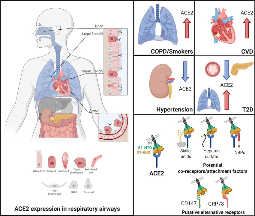
Figure 1. Angiotensin-converting enzyme 2 (ACE2), the proposed receptor of SARS-CoV-2, is expressed in the
respiratory airways at low levels (blue) compared to the intestine, kidney, heart, and pancreas. Low levels are also
observed in the liver. In nasal and bronchial tissues, ACE2 is mainly expressed by ciliated, club, and goblet cells. It
is also found in type-2 pneumocytes of alveoli and in endothelial cells of pulmonary capillaries. In comorbidities
associated with a severity and poor prognosis of COVID-19, ACE2 levels are increased in the lungs of COPD and
smokers and in the heart of patients with cardiovascular diseases (CVD). In contrast, patients with hypertension
exhibit decreased levels of ACE2 in the kidney. In T2D patients, ACE2 is decreased in the pancreas and the
vascular system but increased in the lungs. Current evidence supports a possible role of co-receptors or
attachment factors, such as neuropilins, heparan sulfate, and sialic acids. The low detection of ACE2 in respiratory
tissues also led to the speculation of a role of alternative receptors, such as CD147 and GRP78.
closed conformation is observed (Yan et al., 2020). This study also highlighted the involvement of
K417 and G446 in the ACE2/SARS-CoV-2 interaction. As expected, one RBD domain interacted with
the PD domain of one ACE2 monomer with an interface similar to the one found with SARS-CoV S
(Wang et al., 2020a). The structure of the full-length SARS-CoV-2 S determined by Walls et al. also
revealed open and closed conformational states, which were not observed using the 319–541 aa
fragment (Yan et al., 2020; Walls et al., 2020). This structure also demonstrated the role of F486
and N501, which were predicted in silico, and of K417 in the interaction with ACE2 (Wan et al.,
2020).
Several reports using surface plasmon resonance (SPR) to study the interaction between recombi-
nant ACE2 and S proteins have determined a higher Kd for SARS-CoV S than for SARS-CoV-2 S
(Table 1). Although these studies point to higher affinity of ACE2 for SARS-CoV-2 S than for SARS-
CoV S, they are in contrast with results obtained using a biolayer interferometry binding (BLI)
approach (Table 1). At this point, one cannot exclude that these discrepancies result from the analy-
sis of distinct domains. Additionally, currently available measures rely only on protein fragments
Zamorano Cuervo and Grandvaux. eLife 2020;9:e61390. DOI: https://doi.org/10.7554/eLife.61390 3 of 25Review Article Cell Biology
Table 1. Measure of the dissociation constant (Kd) of ACE2 bound to immobilized SARS-CoV or SARS-CoV-2 S proteins by surface
plasmon resonance (SPR) or biolayer interferometry binding (BLI) approaches.
Reference ACE2 protein PD domain SARS-CoV S SARS-CoV2 S Method Measured kd
Wrapp et al., 2020 1–615 aa 306–577 aa SPR 325.8 nM
1–1208 aa 14.7 nM
Wang et al., 2020a 19–615 aa 306–527 aa SPR 408.7 nM
319–541 aa 133.3 nM
Lan et al., 2020 19–615 aa 306–527 aa SPR 31.6 nM
319–541 aa 4.7 nM
Walls et al., 2020 1–614 aa 306–575 aa BLI 1.2 nM
328–533 aa 5 nM
Wrapp et al., 2020 1–615 aa 306–577 aa BLI 13.7 nM
319–591 aa 34.6 nM
lacking the TM domains that likely alter the dynamics of the three-dimensional protein structure,
prompting a careful interpretation of the available data.
Overall, in-silico predictions and structural analyses support an interaction between ACE2 and
SARS-CoV-2 S with additional contact points compared to the interaction with SARS-CoV S, which
are associated with a higher affinity that may explain the easy human-to-human spread
(Wrapp et al., 2020). Future studies should aim to further characterize the interaction between full-
length ACE2 and SARS-CoV-2 proteins to provide the most precise ground for the development of
therapies aimed at disrupting this interaction.
1.2 In cellulo evidence of ACE2 acting as SARS-CoV-2 entry receptor
The extraordinary speed of research following the identification of SARS-CoV-2, particularly the
development of pseudotyped viruses (Hoffmann et al., 2020; Ou et al., 2020; Walls et al., 2020;
Tai et al., 2020; Lei et al., 2020a), the isolation of the virus from patient samples and the establish-
ment of an in-vitro culture as early as January 2020 (Zhou et al., 2020), quickly made it possible to
tackle the question of the identity of the entry receptor in cellular models (Hoffmann et al., 2020).
Pseudotyped vesicular stomatitis virus expressing SARS-CoV-2 S (VSV-SARS-S2) efficiently infects
only a limited number of cell lines, with Calu-3 human lung adenocarcinoma epithelial cell line,
CaCo-2 human colorectal adenocarcinoma colon epithelial cell line and Vero African grey monkey
kidney epithelial cell line being the most permissive (Hoffmann et al., 2020; Ou et al., 2020). While
permissive cell lines all express ACE2, as previously demonstrated by indirect immunofluorescence
(IF) or by immunoblotting (IB, antibody AF933) (Ren et al., 2006; Tseng et al., 2005; Liao et al.,
2013; Lu et al., 2008), the role of ACE2 in virus entry was demonstrated by the observation that its
ectopic expression is sufficient to make the BHK-21 fibroblast and HEK293 cell lines permissive to
VSV-SARS-S2 (Hoffmann et al., 2020; Ou et al., 2020). Importantly, the preincubation of VSV-
SARS-S2 with the soluble form of ACE2 prevents the infection likely by blocking the site of S protein
interaction. The use of VSV pseudovirions expressing the SARS-CoV S protein (VSV-SARS-S) modi-
fied to contain the RBD from other coronaviruses, including MERS-CoV and SARS-CoV-2, allowed to
underscore the importance of the RBD in the interaction with ACE2 in cellular models (Letko et al.,
2020). Murine leukemia viruses (MLV)-based pseudoviruses expressing SARS-CoV and SARS-CoV-2
S were also found to infect Vero E6 cells (Walls et al., 2020), which express endogenous ACE2
(Sheahan et al., 2008), and ACE2-transfected BHK-21 cells (Walls et al., 2020).
Although pseudotyped viruses are a great tool to document the need for ACE2 for the entry
steps mediated by the S protein, they do not make it possible to assess the contribution of other
virions characteristics, such as envelope or membrane proteins, on the cell tropism (Joglekar and
Sandoval, 2017). Access to SARS-CoV-2 patient isolates allowed to further evaluate the need of
ACE2 for virus entry. Indeed, infection of HeLa, BHK-21 or A549 cells with isolates requires transfec-
tion of ACE2 (Hoffmann et al., 2020; Blanco-Melo et al., 2020; Zhou et al., 2020). While direct
protein interaction in cellular model is difficult to assess, cell surface colocalization of ectopically
Zamorano Cuervo and Grandvaux. eLife 2020;9:e61390. DOI: https://doi.org/10.7554/eLife.61390 4 of 25Review Article Cell Biology
expressed GFP-tagged ACE2 and SARS-CoV-2 S protein fragments containing the RBD was
observed (Wang et al., 2020a). GFP-ACE2 expressed at the cell surface was also shown to bind sol-
uble SARS-CoV-2 RBD; an interaction that was inhibited by incubation with a soluble form of ACE2
(Tai et al., 2020). Collectively, evidence obtained using cell lines further supports in-silico data that
ACE2 binds to SARS-CoV-2 S and points to a model in which ACE2 is necessary for virus entry. More
physiologically relevant studies are awaited to strengthen our understanding of the exact role of
ACE2 in vivo and help determine if co-receptors or attachment factors act in concert with ACE2 and
if alternative receptors may play a role in SARS-CoV-2 entry.
ACE2 expression in the respiratory apparatus
2.1 Basal expression of ACE2 in the human respiratory system
ACE2 was first identified in 2000 as a gene encoding a single isoform of a new angiotensin enzyme
(Donoghue et al., 2000). Recently, due to the great interest generated around ACE2, bioinformatics
and RNA-seq analyses have revisited the human ACE2 genomic region and identified the existence
of an alternative splice site (Rehman and Tabish, 2020; Blume et al., 2020) that results in the tran-
scription of a previously unknown isoform of ACE2 (deltaACE2 or dACE2). The dACE2 isoform lacks
the first N-terminal 356 aa region compared to full-length ACE2 (flACE2) (Blume et al., 2020;
Onabajo et al., 2020) and therefore is unable to bind to SARS-CoV-2 S (Onabajo et al., 2020). Con-
sidering the recent discovery of dACE2, the majority of ACE2 mRNA analyses reported below have
not taken into account its existence. However, when possible, we indicated when the design discrim-
inated specific ACE2 isoforms.
High-throughput gene promoter activity and mRNA expression data available in public reposito-
ries, that is functional annotation of the mammalian genome (FANTOM 5, flACE2), the Human Pro-
tein Atlas (HPA) and the genome-based tissue expression consortia (GTEx), provide evidence of
ACE2 mRNA expression in the respiratory system (Yu et al., 2015; Uhlén et al., 2015; Keen and
Moore, 2015). As pointed out in two recent reports, levels of ACE2 mRNA in the respiratory system
are low compared to other organs such as the small intestine, kidney or the myocardium
(Table 2; Hikmet et al., 2020; Aguiar et al., 2020). An additional transcriptomic dataset of various
human tissues confirmed ACE2 mRNA expression throughout the respiratory tract, including fetal
and adult lungs, olfactory bulbs and trachea, with the highest expression in the lung (Leung et al.,
2020). RNA-seq and microarray datasets from nasal and bronchial epithelial cells obtained through
brushings demonstrate ACE2 detection with low levels in the nose, trachea and small and large air-
ways (Leung et al., 2020; Su et al., 2004). Low expression of ACE2 (flACE2) in the nasal respiratory
epithelium and at even lower levels in the trachea, bronchioles and alveoli was confirmed by RNA in-
situ hybridization (RNA-ISH) of lung and nasal sections (n = 7) (Hou et al., 2020). RT-qPCR of brush
samples from the same donors confirmed a gradient of ACE2 mRNA with the strongest expression
in the nasal tissue and the lowest in the bronchioles and alveoli (Hou et al., 2020). It is noteworthy
that this gradient of ACE2 expression mirrors the profile of permissiveness to SARS-CoV-2. Indeed,
infection of primary cells led to higher viral titers in cells from the nasal epithelium and from the
large airways than in cells from lower airways or Alveolar type II cells (AT2). Analysis of the specific
expression of dACE2 and flACE2 in lung explants and nasal tissue reported similar expression profile
at basal levels (Blume et al., 2020; Onabajo et al., 2020). Further analyses are needed to evaluate
levels of expression of both isoforms in other respiratory tissues. While altogether these studies
Table 2. mRNA levels of ACE2 found in the lungs, small intestine, kidney, and heart muscle reported
in the Human Protein Atlas (HPA) consortium (Uhlén et al., 2015), the genome-based tissue
expression (GTEx) consortium (Keen and Moore, 2015).
Activity levels of the promoter of ACE2 assembled in the Fantom (FANTOM5) consortium (Yu et al.,
2015). Protein-transcripts per million (pTPM). Scaled Tags Per Million (sTPM).
Lung Small Intestine Kidney Heart Muscle
HPA (pTPM) 1.7 31.1 107.2 31.1
GTEx (pTPM) 1.1 5.4 6.8 5.4
FANTOM5 (sTPM) 2.8 21.7 31.5 21.7
Zamorano Cuervo and Grandvaux. eLife 2020;9:e61390. DOI: https://doi.org/10.7554/eLife.61390 5 of 25Review Article Cell Biology
provide clues for ACE2 mRNA expression in the respiratory apparatus that is the primary target by
SARS-CoV-2, they also shed light on the overall low levels detected at basal levels when performing
bulk analyzes of the whole tissues (Figure 1).
The low levels of ACE2 mRNA detected in tissues of the respiratory system likely reflects
restricted expression in specific cell subpopulations (Figure 1). This has indeed been documented in
several studies using scRNA-seq and snRNA-seq strategies, which make it possible to identify the
cell populations expressing ACE2 mRNA. A first report describing the integrated analysis of 107
scRNA-seq and snRNA-seq datasets, including 22 from lungs and airways and 85 from other organs
and encompassing 164 donors, revealed ACE2 expression in multi-ciliated epithelial and goblet cells
of the proximal airways, and in AT2 cells in the distal lung epithelium (Muus et al., 2020). Expression
of ACE2 in AT2 cells (defined as HTII280+ or PRO-SFTPC+ cells) was further confirmed by proximity
ligation in-situ hybridization (PLISH) performed on airway and alveoli tissue from healthy donors. A
second report describing the re-analysis of five scRNA-seq datasets from the lung, bronchus and
nasal tissues, also concluded that ACE2 is enriched in lung AT2 cells and in bronchial and nasal cili-
ated epithelial and goblet cells (Hikmet et al., 2020). While the two reports led to similar qualitative
conclusions, they differed in the quantitative report of the estimated % of ACE2+ AT2 cells, that is
6.2% and less than 1%, respectively. The use of the scRNA-ISH technique that combines scRNA-seq
and ISH data, showed 20% of nasal and bronchial cells expressing ACE2 (Okochi et al., 2020).
ACE2 mRNA was also detected in a fraction of nasal and bronchial ciliated (FOXJ1+) and secretory
(MUC5B+) cells and in AT2 cells (SFTPC+) in the alveoli (Hou et al., 2020).
Several studies attempted to detect ACE2 protein by immunohistochemistry (IHC) in lung histo-
logical sections. Results exhibit more discrepancies than mRNA analyses, making it difficult to draw
firm conclusions. Significant variations between studies are likely due to poor antibody validation
practices, different antigen retrieval methodologies or to low antibody sensitivity. In Table 3, we
attempted to summarize the available validation data for the antibodies used in the studies
described in this review in accordance to the pillars defined by the Antibodypedia validation initia-
tive (Uhlen et al., 2016). Importantly, all antibodies still await validation by siRNA or CRISPR tech-
nologies. Use of HPA000288 and MAB933 antibodies, which allow high detection of ACE2 in kidney
and intestine, only showed very low to no detection in lung tissues (Uhlén et al., 2015;
Hikmet et al., 2020). Positive staining of ACE2 was only observed in a very small percentage of indi-
viduals and corresponded to the surface of nasal ciliated cells, glandular cells of the trachea or AT2
cells consistent with mRNA expression. In a systematic study, six anti-ACE2 antibodies targeting dif-
ferent epitopes, ab15348, HPA000288, ab239924, NBP2-67692, AF933 and MAB933, were tested
together with distinct antigen retrieval methods (Lee et al., 2020). Only the ab15348 antibody
revealed robust staining in pneumocytes, while as expected the six antibodies showed positive stain-
ing in kidney and intestine. Further use of this antibody for indirect IF staining of only one donor
revealed ACE2 detection in MUC1 positive type II pneumocytes. Additionally, IHC staining of
resected lungs (n = 9) using this same antibody allowed detection of ACE2 in the small airway epi-
thelial layer (Leung et al., 2020). ACE2 was detected in AT2 cells (PRO-SFTPC+) from human
explant donors by IHC coupled to IF (IHC-IF) using the AF933, but not ab15348 or MAB933, anti-
bodies, while low counts of ACE2-positive alveolar epithelial cells were detected in post-mortem
lung sections (n = 10) using ab108252 (Ackermann et al., 2020; Muus et al., 2020).
Overall, ACE2 protein is only weakly detected in the respiratory system by two distinct antibodies
and a single antibody allows stronger detection in AT2 cells, while all antibodies detect high expres-
sion in kidneys and intestine. One cannot fully exclude that ACE2 detection in the lung is impaired
by lung-specific post-translational modifications, such as glycosylations, that affects the antigen rec-
ognition. Bias from antibodies specificity and sensitivity can be circumvented through proteomic
analyses. In this perspective, the low expression of ACE2 in the respiratory system is further sup-
ported by the analysis of public mass spectrometry (MS) datasets for protein abundance that does
not show detectable ACE2 in the nasal mucosa or the lungs, in contrast to high levels in the kidneys
and intestine (Hikmet et al., 2020). Altogether, currently available evidence from mRNA, IHC and
MS analyses points to low expression of ACE2 in the respiratory apparatus at basal levels. How these
low basal levels of ACE2 expression allow efficient replication and excretion of SARS-CoV-2 is a
question that deserves more attention. Moreover, it is still difficult to conclude from the available
data that the infection is strictly limited to ACE2 positive cells. These questions remain open and
require further investigation.
Zamorano Cuervo and Grandvaux. eLife 2020;9:e61390. DOI: https://doi.org/10.7554/eLife.61390 6 of 25Review Article Cell Biology
Table 3. Available validation data for the antibodies used in the studies described in this review in accordance to the pillars defined
by the Antibodypedia validation initiative (Uhlen et al., 2016).
Provider refers to the information found in the website of the company. IB: Immunoblot. IHC: Immunohistochemistry. IF: Immunofluo-
rescence. Enhanced validation, Supportive validation, No data available, + Positive detection, +/- Weak detection, -
Absence of detection.
Method Antibodypedia Provider Additional information
Ab15348 IB + Testis, intestines, lung, Calu-3
(or GTX15348) - Breast
Immunogen: 788-805aa
IHC + Testis, kidney, aorta and lung + Intestine, heart, stomach, spleen
(Independent antibody validation)
(Lee et al., 2020)
+, +/-, - Lung. Increased in COPD and
smokers (correlation with mRNA)
(Leung et al., 2020;
Muus et al., 2020; Lee et al., 2020)
IF
MAB933 IB + Kidney + Vero E6 cells
Immunogen: 18-74aa - NSO cells - CHO cells (Jia et al., 2005)
IHC + Kidney + Orthogonal (RNA) and
independent antibody validation
+/-, - Lung (Hikmet et al., 2020; Aguiar et al., 2020;
Muus et al., 2020; Lee et al., 2020)
IF + ALI-cultured ciliated airway
epithelial cells (Martinez-Anton et al., 2013)
AF933 IB + Ovary, testis and kidney + Airway and distal lung, ALI-cultured
Immunogen: 18-74aa airway epithelial cells (correlation with
mRNA levels), Calu-3 and Caco-2
cells (Liao et al., 2013; Smith et al., 2020)
- A549 (expected), Huh-7
cells (Liao et al., 2013; Smith et al., 2020)
IHC + Kidney + Testis, stomach, intestine
(Independent antibody validation)
+/-, - Lung (Ren et al., 2006;
Muus et al., 2020; Lee et al., 2020)
IF
HPA000288 IB + Kidney, but several bands
Immunogen: 1-111aa
IHC + Intestine and kidney. + Orthogonal (RNA) and
- Tonsil independent antibody validation
+/-, - Lung (Uhlén et al., 2015;
Hikmet et al., 2020; Lee et al., 2020)
IF
Ab108252 IB + Testis, kidney, lung, HepG2, Caco-2
Immunogen: 200-300aa - A549 cells
IHC + Kidney
IF
Ab239924 IB + Testis, kidney + ACE2 transfected A459
Immunogen: 200-300aa - Non transfected A549 cells
(Blanco-Melo et al., 2020)
IHC + Testis, kidney + Intestine, heart, stomach, spleen
(Independent antibody
validation), +/- Lung (Lee et al., 2020)
IF
NBP2-67692 IB + Kidney
Immunogen: 200-300aa
IHC + Kidney + Testis and intestine (Independent antibody validation)
- Lung (Lee et al., 2020)
IF + HepG2, MCF-7, 293 cells
Table 3 continued on next page
Zamorano Cuervo and Grandvaux. eLife 2020;9:e61390. DOI: https://doi.org/10.7554/eLife.61390 7 of 25Review Article Cell Biology
Table 3 continued
Method Antibodypedia Provider Additional information
Anti-ACE2489 IB +ACE2 transfected CHO cells
Immunogen: 489-508aa - Non transfected CHO cells
IHC + Heart (No staining with
secondary antibody alone)
(Qian et al., 2013)
IF
Sc-20998 IB Antibody discontinued Detection in rat tissue only
Immunogen: (Sims et al., 2005)
631-805aa
IHC Antibody discontinued
IF Antibody discontinued
Homemade antibody IB Detection in rat tissue only
Immunogen: (Mossel et al., 2008)
206-225aa
IHC
IF
2.2 ACE2 expression in human respiratory epithelial cells ex vivo: impact of
differentiation
Ex vivo culture of cells from the respiratory system is an essential tool for the study of virus infection
and pathogenesis. Epithelial cells from distinct sections of the respiratory tract, including the nose,
trachea, bronchi and alveoli, are either cultured submerged in culture medium or at air-liquid inter-
face (ALI) for polarization or differentiation into pseudostratified mucociliary epithelium. While sub-
merged culture of human tracheobronchial epithelial cells exhibits low ACE2 mRNA and protein
detection by IB(AF933 or MAB933), the expression levels are highly enhanced following differentia-
tion in ALI (Blanco-Melo et al., 2020; Jia et al., 2005; Martinez-Anton et al., 2013; Smith et al.,
2020; Qian et al., 2013). More recently, a clear distinction between dACE2 and flACE2 mRNA levels
allowed to confirm that both isoforms are increased in differentiated cultures (Blume et al., 2020).
Of note, re-submersion of cells, which led to loss of differentiation markers, reversed ACE2 mRNA
and protein levels (Jia et al., 2005), supporting a key role of the differentiation process in ACE2
expression. Further study by IF staining of ALI-differentiated human primary alveolar cells (MAB933)
showed that ACE2 is localized at the apical surface (Sims et al., 2005). This was recapitulated using
confocal horizontal scanning (AF933) of the Calu-3 cell line polarized in ALI (Ren et al., 2006). Impor-
tantly, in hTERT-immortalized bronchial cells differentiated into multiciliated cells in ALI, only the
flACE2 protein was detected in motile cilia (Blume et al., 2020) suggesting a specific role of the full-
length isoform at this location. Overall, these studies show that differentiation significantly increases
the expression of ACE2 and, as such, suggests that the use of differentiated cells should be pre-
ferred to model ACE2-dependent SARS-CoV-2 infection. Additionally, the heterogeneity of patient-
derived respiratory epithelial cells argues for the study of large cohorts to reach clear conclusions.
However, this is hardly achievable due to the scarcity of the cells and the high demanding differenti-
ation process. Beside donor to donor variation, IF staining of ALI-cultured AT2 cells also pointed to
heterogeneous ACE2 levels in a single culture (Mossel et al., 2008), which correlates with observa-
tions made on lung tissue sections showing ACE2 in only a small fraction of the AT2 cells
(Muus et al., 2020).
2.3 ACE2 levels during SARS-CoV-2 infection
The correlation between the expression of ACE2 mRNA and that of the classical interferon (IFN)
stimulated genes (ISG) that was observed through meta-analysis of scRNA-seq data, as well as the
presence of two STAT1 binding sites in the promoter of the human ACE2 gene, supports the
hypothesis that ACE2 itself could be an ISG (Ziegler et al., 2020; Hennighausen and Lee, 2020).
Several reports have indeed documented the capacity of IFNb and/or IFNa2 to induce ACE2 mRNA
in human basal epithelial cells from nasal scraping, human tracheal cells and human large and small
Zamorano Cuervo and Grandvaux. eLife 2020;9:e61390. DOI: https://doi.org/10.7554/eLife.61390 8 of 25Review Article Cell Biology
airway cells (Hou et al., 2020; Smith et al., 2020; Ziegler et al., 2020). FlACE2 was also induced in
nasal and tracheal cells, but not in small airway cells, in response to IFNg (Smith et al., 2020). Spe-
cific analysis of flACE2 and dACE2 isoforms revealed that in fact IFNa, IFNb and IFN-l3 specifically
induce dACE2 in human bronchial epithelial cells (HBEC) (Blume et al., 2020; Onabajo et al., 2020).
While an increase of dACE2, which does not bind to SARS-CoV-2 S, should not translate into more
entry of the virus, the question of how this increase would impact the host antiviral response remains
to be determined. The IFN-dependent regulation of ACE2 mRNA expression might explain ACE2
upregulation observed within bystander goblet or squamous cells, but not directly in infected cells,
during influenza (IAV) virus infections (Ziegler et al., 2020). Only dACE2 mRNA is upregulated upon
IAV and rhinovirus infections, but the role of IFNs in ACE2 upregulation in these contexts remains to
be fully addressed (Onabajo et al., 2020).
Converging evidence indicates that ACE2 mRNA levels are also increased during SARS-CoV-2
infection. In a study with a limited cohort, scRNA-seq analysis of nasopharyngeal swabs, bronchial
brushes and bronchoalveolar lavages showed up to 3-fold upregulation of ACE2 mRNA in COVID-19
patients (n = 9) compared to healthy individuals (n = 2). ACE2 mRNA induction was observed in
secretory and ciliated cells, which also exhibited the most SARS-CoV-2+ cells (Chua et al., 2020). In
ALI-differentiated HBEC, SARS-CoV-2-induced ACE2 mRNA is observed in ciliated, basal club and
an intermediate between basal cells and club (BC/Club) infected and bystander cells
(Ravindra et al., 2020). Surprisingly, this result was not corroborated in the Calu-3 and differenti-
ated BCi-NS1.1 respiratory cell line models, in which neither flACE2 nor dACE2 were induced by
SARS-CoV-2 (Blume et al., 2020; Onabajo et al., 2020). This contrasts with the specific induction of
dACE2 observed in the non-respiratory Caco-2 cell line (Onabajo et al., 2020). A role of IFNs in the
upregulation of ACE2 by SARS-CoV-2 is difficult to conclude based on the current evidence for the
status of IFN production and signaling during SARS-CoV-2 infection. In differentiated HBEC, induc-
tion of IFNb and IFNl mRNA was observed in infected cells, but not in the bystander cells
(Ravindra et al., 2020). Increased mRNA levels of IFNb and IFNl1 and 3 were also observed in
infected Calu-3 cells (Sun et al., 2020; Lei et al., 2020b; Banerjee et al., 2020). This contrasts with
the lack of detectable IFNb or IFNl1–3 mRNA observed in submerged culture of HBEC upon SARS-
CoV-2 infection (Blanco-Melo et al., 2020; Vanderheiden et al., 2020). In an ex vivo human lung
explant model, SARS-CoV-2 infection also failed to induce detectable levels of IFNa, b, g, or l1–3 at
any time point for up to 48 hr, while SARS-CoV induced IFNa, b, g, or l1–3 at late time points
(Chu et al., 2020). These discrepancies could be due to differences in MOI and time course of the
infection, but also from the quality of virus preparation as the content of defective virus particles is
known to trigger IFN production in the context of other virus infections (Vignuzzi and López, 2019).
Systematic analysis of the impact of these parameters will be necessary to resolve the status of IFN
induction by SARS-CoV-2 in respiratory epithelial cells. IFNs plasma levels in COVID-19 patients
have also started to be documented. In a first study of critically ill COVID-19 patients, IFN-a2, but
neither IFNb nor IFNl, was detected in the plasma of 21 out of 26 patients with a peak 10 days after
onset of symptoms (Trouillet-Assant et al., 2020). This is in line with a deep analysis of type I IFN
mRNA regulation in COVID-19 patients at different stages of the disease. While no IFNb mRNA and
protein were detected in the plasma regardless of the severity of illness (n = 32) compared to
healthy donors (n = 13), IFNa mRNA was found in the plasma of all COVID-19 patients
(Hadjadj et al., 2020). In two separate reports, plasma levels of IFNa were lower in critically ill
patients than in patients with mild to moderate COVID-19 (n = 11 and 10) (Banerjee et al., 2020;
Hadjadj et al., 2020). In addition, IFNa levels were maintained in mild to moderate patients for up
to 17 days, but decreased in severe and the critically ill patients after 10 and 13 days, respectively
(Hadjadj et al., 2020). In addition to evidence supporting weak and shortened levels of IFNa by
SARS-CoV-2 infection in critically ill COVID-19 patients, molecular data have started to document
the capacity of SARS-CoV-2 to evade the IFN response as previously documented for SARS-CoV
(Lei et al., 2020b; Park and Iwasaki, 2020; Xia et al., 2020b). The first evidence results from the
overexpression of SARS-CoV-2 proteins in HEK293T cells before infection with Sendai virus or
expression of constitutively active RIG-I or MDA5 receptors. Results diverged among the two avail-
able reports, but Nsp1, Nsp13, and Orf6 were consistently found to inhibit IFNb promoter induction
and IFN signaling (Lei et al., 2020b; Xia et al., 2020b). The mechanisms underlying these inhibitions
are not fully defined, but Nsp13 appears to act upstream of TBK1 phosphorylation, while Orf6 inter-
feres with IRF3 and STAT1 nuclear translocation. A truncation variant of SARS-CoV-2 Orf3b is also
Zamorano Cuervo and Grandvaux. eLife 2020;9:e61390. DOI: https://doi.org/10.7554/eLife.61390 9 of 25Review Article Cell Biology
thought to be associated with inhibition of IFNb production (USFQ-COVID19 Consortium et al.,
2020). In contrast to the impact of SARS-CoV-2 protein overexpression, SARS-CoV-2 infection of
Calu-3 cells, failed to hinder IFNb induced phosphorylation of STAT1 and STAT2 and downstream
IFIT1 production (Banerjee et al., 2020). In summary, the full scope of the interplay between SARS-
CoV-2 and the IFN response remains to be carefully characterized before conclusion about the
impact on ACE2 induction can be drawn.
Studies documenting ACE2 protein expression upon IFNs stimulation and SARS-CoV-2 infection
are really sparse. Immunoblot analysis of ACE2 (AF933) in fully differentiated HBEC derived from
asthmatic patients stimulated with IFNg showed slight increase in only 1 out of 4 donors
(Ziegler et al., 2020). In contrast, SARS-CoV-2 infection (MOI of 2) of A549 cells transfected with
ACE2 showed decreased levels (ab239924) at 24 hr post-infection (Blanco-Melo et al., 2020). While
this is a preliminary observation that needs to be further confirmed, it is in line with the observation
that SARS-CoV infection of Vero E6 cells led to downregulation of ACE2 protein levels, which
inversely correlated with viral replication (Glowacka et al., 2010). The observation that ACE2 mRNA
levels increase or remain stable while the protein levels are decreased during SARS-CoV-2 infection
suggests a post-transcriptional regulation mechanism, such as degradation or a halt in the transla-
tion. The exact mechanism(s) underlying this dual regulation remains to be investigated to fully com-
prehend how cells respond to SARS-CoV-2 infection and the potential role of ACE2. These results
contrast with the post-mortem examination of lungs from deceased COVID-19 patients (n = 7) com-
pared to age-matched uninfected individuals (n = 10) by IHC (ab108252) which showed a signifi-
cantly greater number of ACE2-positive alveolar epithelial cells in COVID-19 individuals compared
to controls (Ackermann et al., 2020).
Altogether, for the time being, it appears clear that ACE2 mRNA is detected in the respiratory
system, but at significantly lower levels than in kidneys and intestine. Detection of ACE2 protein
remains controversial mainly because of discrepancies between antibodies. Importantly, we now
know that there are 2 isoforms of ACE2, one of which is unable to bind to SARS-CoV-2. Future stud-
ies aimed at analyzing ACE2 mRNA and protein should attempt to determine which of the isoforms
is detected considering the great impact this information will have on our understanding of SARS-
CoV-2 infection and tropism. The distribution in respiratory tissues is heterogenous and ACE2 is
mainly detected in ciliated, goblet, and club cells of the nose and bronchi and in the AT2 cells in the
alveoli (Figure 1). The detection of ACE2 mainly in nasal cells could explain a higher shedding of
virus in the upper respiratory tract at the beginning of the disease, which has been observed from
repeated biological samples (Wang et al., 2020b; Zou et al., 2020). Conversely, the presence of
ACE2 in AT2 might facilitate entry of SARS-CoV-2 as observed in infected macaques (Rockx et al.,
2020) causing damages of alveoli that could contribute to COVID-19 severity (Olajuyin et al., 2019).
Future studies should aim to determine if and how regulation of ACE2 expression during SARS-CoV-
2 infection impacts the dynamics of infection and the antiviral response.
ACE2 expression in comorbidities associated with COVID-19 severity:
protective or harmful role?
A large body of evidence supports a protective role for ACE2 in several disease models. The role of
ACE2 was initially described in the context of the renin-angiotensin system (RAS), with the discovery
of the ACE2/Angiotensin (Ang 1–7)/Mas axis that counteracts the axis of the ACE/AngII/AT1 recep-
tor. ACE2 also plays as a role in the regulation of the kallikrein-kinin system (KKS) that catalyzes the
formation of the Bradykinin 2 (B2) receptor ligands, bradykinin and Lys-bradykinin, which are further
hydrolyzed into the B1 receptor ligands des-Arg9-bradykinin and Lys-des-Arg9-bradykinin. ACE2
inactivates B1 receptor ligands by cleavage. Increased ACE2 levels, associated with a decrease in
AngII due to conversion into Ang 1–7, is typically considered an indicator of organ protection
because of its role in reducing pulmonary vasoconstriction, remodeling, atherosclerosis, blood pres-
sure, myocardial hypertrophy, fibrosis and ventricular remodeling. Additionally, through inhibition of
B1 receptor ligands, ACE2 is protective against pulmonary angioedema. On the contrary, a decrease
in ACE2 levels is associated with an increase in pulmonary vascular permeability, pulmonary edema,
ARDS, atherosclerosis, hypertension, cardiac hypertrophy, ventricular remodeling, and heart failure.
Importantly, the ACE2/Ang 1–7/Mas axis and ACE2-dependent inhibition of B1R activation also
exerts inhibitory effects on inflammation. These functions of ACE2 were fully reviewed by others
Zamorano Cuervo and Grandvaux. eLife 2020;9:e61390. DOI: https://doi.org/10.7554/eLife.61390 10 of 25Review Article Cell Biology
(Muñoz-Durango et al., 2016; Simões e Silva et al., 2013; Mirabito Colafella et al., 2019; van de
Veerdonk et al., 2020; Chung et al., 2020).
From the early days of the pandemic in China and Italy, epidemiological data rapidly indicated, in
addition to the age of over 70 years, an association of COVID-19 disease severity, ICU admission
and poor prognosis with multiple comorbidities, including hypertension and cardiovascular disease
(CVD), COPD and type 2 diabetes (T2D) (Grasselli et al., 2020; Lippi and Henry, 2020; Guan et al.,
2020; Huang et al., 2020; Jain and Yuan, 2020). Additionally, smoking is associated with COVID-
19 disease progression (Gülsen et al., 2020). Reports arising notably from Europe and North Amer-
ica, have subsequently highlighted that a high BMI, and particularly obesity, is an important risk fac-
tor for a severe course of COVID-19 in patients younger than 60 years old (Kass et al., 2020;
Soeroto et al., 2020). Although the factors responsible for these associations remain to be eluci-
dated, questions concerning a potential role of ACE2 expression have emerged.
Expression of ACE2 in the pulmonary epithelium is well documented in COPD condition and rela-
tive to the smoking status. Elevated levels of ACE2 mRNA is observed in airway brushings from
COPD patients compared to non-COPD and in current smokers vs never smokers (cohorts of
n = 20–220) (Leung et al., 2020; Muus et al., 2020; Smith et al., 2020; Cai, 2020). Datasets from
lung biopsies (n = 33–77 samples) also showed increased ACE2 mRNA levels in smokers in both
Asian and Caucasian populations (Cai, 2020). These observations are confirmed by meta-analysis of
pulmonary RNA-seq datasets from the GEO repository (Pinto et al., 2020). Although limited in the
scope (n = 8 healthy, 9 smokers and 10 COPD donors), IHC detection of ACE2 (ab15348) in resected
lung tissues supports higher protein expression in COPD, and to a lesser extent in smokers, com-
pared to healthy individuals (Leung et al., 2020). Whether increased ACE2 expression in the lungs
of COPD patients or active smokers (Figure 1) has a causal effect on the outcome of COVID-19
remains elusive. One could argue that increased ACE2 levels would allow more virus entry, as it hap-
pens in cell models transfected with hACE2 (Ou et al., 2020; Blanco-Melo et al., 2020), hence
increased subsequent inflammatory response. This model is supported by the observation that
patients (n = 76) with severe COVID-19 tend to have a high viral load and a long virus-shedding
period (Liu et al., 2020a; Yu et al., 2020; Liu et al., 2020b). Alternatively, one can argue that
through its role in the conversion of AngII, increased ACE2 in the lungs could be beneficial by pro-
tecting the lung from AngII-dependent lung injury and edema (Deng et al., 2012). Considering that
lung injury and edema are clinical features of the most severe phase of COVID-19, a scenario where
infection leads to increased levels of ACE2 is very unlikely. Rather, this hypothesis should be miti-
gated by the fact that SARS-CoV-2 triggers ACE2 downregulation and thereby the final outcome
would be increased local AngII causing acute lung injury and edema. Potential implications of the
RAS system and ACE2 in the lung have recently been fully reviewed (Chung et al., 2020;
Samavati and Uhal, 2020). This latter hypothesis is consistent with a recently proposed model which
argues that during infection with SARS-CoV-2, ACE2 would be less available to degrade the des-
Arg9-bradykinin and Lys-des-Arg9-bradykinin peptides, thereby leading to overactivation of B1R
and subsequently lung angioedema (van de Veerdonk et al., 2020). Future studies should interro-
gate the relationship between ACE2 levels in the lung and viral load and inflammation in smokers vs
non-smokers and in COPD patients vs healthy individuals.
For comorbidities other than COPD, the correlation between the changes in ACE2 levels and the
severity and prognosis of COVID-19 is less clear. Meta-analysis of 700 lung transcriptomes clearly
highlighted the lack of data on the impact of hypertension and CVD on ACE2 expression in the lung
(Pinto et al., 2020; Hamming et al., 2004). Given that cardiovascular complications are now well
known to be a major threat to COVID-19 patients survival (Jain and Yuan, 2020), the question of
the involvement of ACE2 located outside of the lung, particularly in the cardiovascular system, was
raised. Expression of ACE2 at basal levels has been extensively reviewed elsewhere and therefore
we focus here only on the description of ACE2 status in hypertension and CVD (Chung et al., 2020;
Bourgonje et al., 2020). Increased ACE2 mRNA levels were observed in the ventricular myocardium
from patients with idiopathic dilated cardiomyopathy (n = 11) or ischemic cardiomyopathy (n = 12)
compared to healthy individuals (n = 9), and elevated ACE2 protein levels (IHC, anti-ACE2489) were
found in explants from human ischemic hearts (Goulter et al., 2004; Burrell et al., 2005; Figure 1).
Rats that suffer myocardial infarction also exhibited increased flACE2 mRNA and ACE2 protein levels
and activity measured by emitted fluorescence with ACE2-specific quenched fluorescent substrate
(Burrell et al., 2005). In patients presenting hypertensive nephrosclerosis (HTN, n = 41), a disease
Zamorano Cuervo and Grandvaux. eLife 2020;9:e61390. DOI: https://doi.org/10.7554/eLife.61390 11 of 25Review Article Cell Biology
associated with chronic high blood pressure that induce kidney damage, ACE2 mRNA levels are
decreased in the tubulointerstitium, but not in the glomerular zone, compared to healthy donors
(n = 10) (Wang et al., 2011; Figure 1). Similarly, in spontaneously hypertensive rats (SHR), levels of
ACE2 mRNA and protein were downregulated in kidneys (Tikellis et al., 2006). SHR also exhibit
decreased levels of ACE2 protein (IB, sc-20998) in the rostral ventrolateral medulla (RVLM) contain-
ing cardiovascular regulatory neurons, a phenotype associated with the hypertensive state that is
corrected by ACE2 injection (Yamazato et al., 2007). Salt-sensitive Sabra hypertensive (SBH) rats
also showed decreased ACE2 mRNA and protein levels (IB, homemade antibody) in kidneys
(Crackower et al., 2002). While there is a lack of data of ACE2 expression in hypertensive patients,
animal models suggest a downregulation of mRNA and protein ACE2 in the kidney and RVLM. More
investigations are needed to determine how ACE2 expression in specific organs in patients with
hypertension and CVD could impact COVID-19 prognosis. The tropism of SARS-CoV-2 for cells in
the cardiovascular system is a topic that attracts a lot of interest and is mostly supported by in-vitro
infection of cardiomyocytes derived from induced pluripotent stem cells (Topol, 2020). Although a
first report claimed post-mortem detection of viral inclusion in endothelial cells from three patients
using electron microscopy (EM) (Varga et al., 2020), the interpretation of the images was subse-
quently challenged (Goldsmith et al., 2020). A thorough confirmation of this observation is neces-
sary, in particular by using Immuno-EM with well validated antibodies. Importantly, demonstration of
ACE2-dependent infection and replication of SARS-CoV-2 in endothelial cells are yet to be done. If
this is confirmed, it will be important to determine whether the presence of viral structures in endo-
thelial cells correlates with the observed increase in ACE2 (IHC, ab108252) observed post-mortem in
pulmonary endothelial cells from COVID-19 patients (Ackermann et al., 2020). Response to this
question will inform on the role of ACE2 in the endothelium. Finally, whether endothelial cells
engagement is altered in patients with hypertension and CVD will need to be defined to determine
if this could be a causal factor for the severity of COVID-19.
Increased levels of ACE2 in the lungs, as well as reduced levels in the vascular system, in T2D or
obese patients and the possible mechanisms associated with the severity of COVID-19 have recently
been reviewed (Kruglikov et al., 2020). ACE2 expression in the pancreas has also been under scru-
tiny, but results differ significantly between studies. While initial analysis of expression datasets sug-
gested poor ACE2 mRNA and no detectable protein expression in the pancreas (Xu et al., 2020), a
subsequent re-analysis of several datasets revealed expression in ductal cells (Muus et al., 2020).
Similarly, analysis of two scRNA-seq datasets (n = 55 and n = 19) showed ACE2 expression in ductal
and acinar cells on the exocrine gland, and to a lesser extent in beta cells of the pancreatic islet
(Liu et al., 2020c). Moreover, ACE2 protein was detected (n = 20, IHC, HPA000288 and MAB933) in
interlobular pancreatic ducts and endothelial cells (Hikmet et al., 2020). While in a mouse model of
diabetes, ACE2 (IB, ab15347) was found upregulated in the liver and pancreas and pancreatic ACE2
activity was also increased (Roca-Ho et al., 2017), scRNA-seq analysis revealed decreased ACE2
mRNA levels in ductal cells of T2D patients compared to controls (Chen et al., 2020; Figure 1). If
and how pancreatic ACE2 expression impacts the poor prognosis of COVID-19 remains to be eluci-
dated. As of today, there is no report of SARS-CoV-2 infection in pancreatic cells. Considering that
SARS-CoV N protein and RNA were detected in pancreatic acinar cells from 3 out of 4 SARS-CoV
patients (Ding et al., 2004), the detection of SARS-CoV-2 in the pancreas should be considered in
future investigations. Alternatively, the fact that diabetic patients are at high-risk of severe COVID-
19 disease could be related to their overall elevated metabolic inflammation which may predispose
to the cytokine storm syndrome associated with multi-organ failure (Bornstein et al., 2020). Excess
of adipose tissue in T2D patients is associated with increased proinflammatory macrophages and
Th1 and Th17 CD4+ T cells and is correlated to a chronic low-grade inflammatory state
(Kulcsar et al., 2019). In addition, white adipose tissue produces proinflammatory cytokines, such as
TNF, IL-1, IL-6 and IL-10 (Tsalamandris et al., 2019). Upon infection by SARS-CoV-2, the inflamma-
tory state in T2D patients could be further exacerbated possibly leading to the cytokine storm and
multi-organ failure (Bornstein et al., 2020).
Considering that the severity of COVID-19 is highly associated with an age over 70 years (except
for obesity) assessing the role of age on ACE2 expression in patients with comorbidities in extensive
retrospective epidemiological studies would provide a broader picture of risk factors and accurately
inform on the potential consequences of modulating ACE2. Importantly, data collected from
COVID-19 patients at the beginning of the pandemics were mostly cross-sectional and limited to the
Zamorano Cuervo and Grandvaux. eLife 2020;9:e61390. DOI: https://doi.org/10.7554/eLife.61390 12 of 25Review Article Cell Biology
description of known underlying chronic diseases without stratification according to therapeutic
treatment. Therefore, the question arose whether the observed association of comorbidities with
COVID-19 severities might result from the treatments rather than the condition itself. Substantial
data supports that RAS and ACE inhibitors and angiotensin-receptor blockers (ARBs), which are fre-
quently used in patients with hypertension and CVD and to a lesser extent diabetes, induce
increased expression of ACE2 as recently reviewed (Chung et al., 2020). Limited data supports that
anti-diabetic treatments, Pioglitazone or Liraglutide, also increase ACE2 mRNA and protein (IB,
ab108252) in liver and adipose tissue and in the lung and the heart, respectively (Pal and Bhadada,
2020; Zhang et al., 2014). The impact of corticosteroids or bronchodilators in COPD has to the
best of our knowledge not yet been assessed. Overall, the impact of treatments on ACE2 remains to
be fully explored in humans.
While evidence supports ACE2 increase in at least some comorbidities or in response to their
associated treatments, it is not possible to conclude on the causality between ACE2 expression lev-
els and the severity of COVID-19 (Pal and Bhadada, 2020; Vaduganathan et al., 2020). Currently
available retrospective observational studies did not conclude on an association between the use of
RAS inhibitors and increased risk of SARS-CoV-2 infection (Chung et al., 2020). A systematic meta-
analysis of data from 15 studies, encompassing 2,065,805 COVID-19 patients with hypertension
revealed that the probability of COVID-19 patients treated with RAS inhibitors to die was 35% less
than those who were not under treatment (Ssentongo et al., 2020). Another meta-analysis of 9
studies including 3936 COVID-19 patients with hypertension showed that while there was no associa-
tion between treatment with RAS inhibitor and the severity of the disease, patients under RAS treat-
ment were less likely to die (OR 0.57, 95% CI 0.38–0.84, P0.004, I20) (Guo et al., 2020). Altogether,
this suggests that even if RAS inhibitors induce ACE2 expression, they do not increase the risk for
fatal COVID-19.
A possible alternative explanation that was recently discussed for comorbidities is that disfunction
or absence of ACE2 may result in increased levels of AngII and hyperactivation of B1R which leads
to inflammation, fibrosis, oxidative stress, vasoconstriction and angioedema (Chung et al., 2020).
This could in fact be the case during infection with SARS-CoV-2 if virus-bound ACE2 is no longer
available. In addition, while infection progresses, virus entry leads to the intake of ACE2, thereby
resulting in a decreased amount of ACE2 at the membrane. Increased levels of AngII were found in
the plasma of COVID-19 patients with pneumonia compared to healthy individuals and the levels of
AngII positively correlated with SARS-CoV-2 viral load and lung injury (Liu et al., 2020b). It is possi-
ble that in patients presenting comorbidities associated with a dysregulation of the RAS system,
AngII modulation by SARS-CoV-2 reaches a critical level that ultimately induces an inflammatory
state causing fatal issues.
Potential co-receptors/attachment factors for ACE2-dependent SARS-
CoV-2 entry
The cell surface neuropilin NRP1 (also known as Vascular endothelial cell growth factor 165 receptor,
VEGF165R Soker et al., 1997) and NRP2 (also known as VEGF165R2 Ambra et al., 2006) are
dimeric receptors known to contribute to neurogenesis and angiogenesis (Yasuhara et al., 2004).
They bind ligands containing a C-terminal polybasic motif that follows the C-end-Rule (CendR)
(Teesalu et al., 2009). SARS-CoV-2 S contains a furin cleavage site which has the potential to gener-
ate a solvent exposed C-terminus containing the CendR R/KXXR/K sequence which is, according to
molecular modeling predictions, capable of binding to the coagulation b1 domain of NRP
(Teesalu et al., 2009; Cantuti-Castelvetri et al., 2020). This observation led to the hypothesis that
neuropilins could serve as co-receptor for SARS-CoV-2 attachment to the cell surface (Figure 1) and
contributes to the tropism as previously documented for viruses such as Human T-cell lymphotropic
virus type 1 (HTLV-1) and Epstein–Barr virus (EBV) (Lambert et al., 2009; Wang et al., 2015). Sev-
eral experimental observations support the role of NRP1 as SARS-CoV-2 co-receptor. In a GFP-nano-
trap experiment in HEK293T cells, SARS-CoV-2 S1 interacts with GFP-NRP1 in a CendR motif-
dependent manner (Daly et al., 2020). While deletion of NRP1 strongly reduced SARS-CoV-2 infec-
tion of Hela cells expressing ACE2, NRP1 alone was not sufficient as cells that do not express ACE2
are not infected by SARS-CoV-2 (131). In addition, incubation of VSV-SARS-S2 or patient-isolated
SARS-CoV-2 with monoclonal anti-NRP1 decreased, although to a lesser extent than monoclonal
anti-ACE2, the efficiency of infection of HEK293 cells expressing ACE2 and of Calu3 cells (Cantuti-
Zamorano Cuervo and Grandvaux. eLife 2020;9:e61390. DOI: https://doi.org/10.7554/eLife.61390 13 of 25You can also read