Chronic tissue inflammation and metabolic disease - Genes ...
←
→
Page content transcription
If your browser does not render page correctly, please read the page content below
Downloaded from genesdev.cshlp.org on September 30, 2021 - Published by Cold Spring Harbor Laboratory Press

REVIEW

Chronic tissue inflammation
and metabolic disease
Yun Sok Lee and Jerrold Olefsky
Department of Medicine, Division of Endocrinology and Metabolism, University of California at San Diego, La Jolla,
California 92093, USA

Obesity is the most common cause of insulin resistance,                       Although there are a number of potential, and some-
and the current obesity epidemic is driving a parallel                     times overlapping, mechanisms that can contribute to in-
rise in the incidence of T2DM. It is now widely recognized                 sulin resistance and β-cell dysfunction, in this current
that chronic, subacute tissue inflammation is a major eti-                 review we focus on the role of chronic tissue inflamma-
ologic component of the pathogenesis of insulin resis-                     tion in these metabolic defects. The reader is referred to
tance and metabolic dysfunction in obesity. Here, we                       other excellent reviews on possible causes of insulin resis-
summarize recent advances in our understanding of                          tance and β-cell dysfunction, independent of chronic tis-
immunometabolism. We discuss the characteristics of                        sue inflammation (Halban et al. 2014; DeFronzo et al.
chronic inflammation in the major metabolic tissues                        2015; Czech 2017; Newgard 2017; Guilherme et al.
and how obesity triggers these events, including a focus                   2019; Kahn et al. 2019; Roden and Shulman 2019; Scherer
on the role of adipose tissue hypoxia and macrophage-de-                   2019; Alonge et al. 2021; Sangwung et al. 2020).
rived exosomes. Last, we also review current and potential                    The interconnections between inflammation and meta-
new therapeutic strategies based on immunomodulation.                      bolic dysfunction are often described by the term immu-
                                                                           nometabolism (Hotamisligil 2017). Immunometabolism
                                                                           includes the effects of immune cells on the regulation of
   Insulin resistance is a key feature of obesity and type 2               systemic metabolism, as well as the effects of metabolism
diabetes mellitus (T2DM). Since obesity is the most com-                   within immune cells on inflammation. Both of these
mon cause of insulin resistance in humans, the obesity ep-                 mechanisms are of importance, but in this review we fo-
idemic is the major cause of the rising global incidence of                cus our attention on the role of chronic inflammation in
T2DM. In nondiabetic subjects with obesity, hyperinsuli-                   the etiology of disordered glucose homeostasis.
nemia usually compensates for the underlying insulin re-                      Chronic inflammation in adipose tissue, the liver, the
sistance, maintaining glucose homeostasis within normal                    central nervous system, the gastrointestinal (GI) tract,
or near normal levels. When compensatory hyperinsuline-                    pancreatic islets, and muscle have all been described in
mia fails and insulin levels decline due to development of                 obese/T2DM states, indicating the complex, multiorgan
a β-cell defect, hyperglycemia and the typical T2DM state                  crosstalk network involved in these disorders. The gene-
ensues, indicating that both insulin resistance and β-cell                 ral metabolic consequences of obesity-induced tissue in-
dysfunction are needed for the full development of this                    flammation are summarized in Table 1. These concepts
disease. In obesity, chronic tissue inflammation is a                      are mostly derived from mouse studies, and among all
well-reported key contributor to decreased insulin sensi-                  these contributing tissues, adipose tissue inflammation
tivity, particularly in adipose tissue and the liver. Howev-               in obesity has been the most extensively studied.
er, several recent reports have shown obesity-associated                      Although the contribution of chronic inflammation in
inflammatory responses in pancreatic islets (Zhao et al.                   T2DM in obesity has been suggested for many years, in
2003; Warren et al. 2006; Ehses et al. 2007; Böni-Schnet-                  the past two decades a number of studies have been pub-
zler et al. 2008; Richardson et al. 2009; Mahdi et al.                     lished, bringing out underlying physiologic and molecular
2012; Jourdan et al. 2013; Kamada et al. 2013; Butcher                     mechanisms as to how obesity-associated inflammation
et al. 2014; Hasnain et al. 2014; Ying et al. 2019), suggest-              leads to insulin resistance and glucose intolerance. One
ing that chronic islet inflammation can also lead to β-cell                of the earliest studies was reported by Grunfeld and Fein-
dysfunction in the context of obese T2DM subjects.                         gold (Feingold et al. 1989) who showed that treatment of ro-
                                                                           dents with TNFα leads to hyperglycemia. An additional
                                                                           key early study showed increased TNFα levels in the adi-
                                                                           pose tissue of obese mice and showed that neutralization
[Keywords: β-cell dysfunction; glucose intolerance; immunometabolism;
inflammation; insulin resistance; macrophage; metaflammation]
Corresponding author: jolefsky@ucsd.edu                                    © 2021 Lee and Olefsky This article, published in Genes & Development,
Article is online at http://www.genesdev.org/cgi/doi/10.1101/gad.346312.   is available under a Creative Commons License (Attribution-NonCom-
120. Freely available online through the Genes & Development Open Ac-      mercial 4.0 International), as described at http://creativecommons.org/li-
cess option.                                                               censes/by-nc/4.0/.

           GENES & DEVELOPMENT 35:307–328 Published by Cold Spring Harbor Laboratory Press; ISSN 0890-9369/21; www.genesdev.org                  307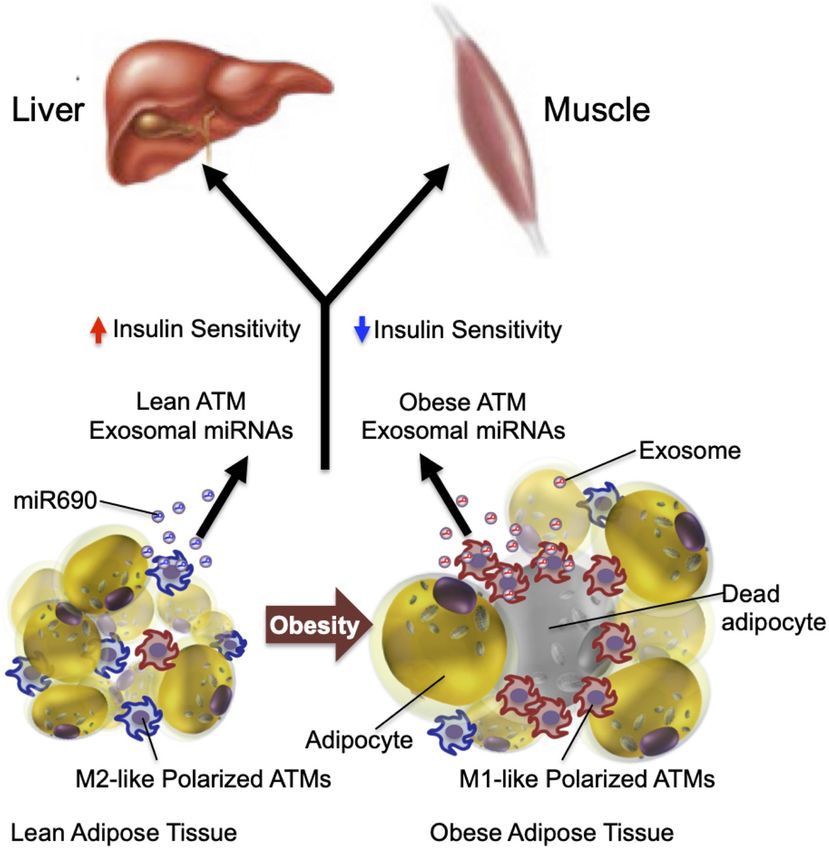
Downloaded from genesdev.cshlp.org on September 30, 2021 - Published by Cold Spring Harbor Laboratory Press

Lee and Olefsky

Table 1. The metabolic effects of obesity-associated tissue             validated in humans, recent advances in genome-wide as-
inflammation                                                            sociation studies (GWAS) suggest that inflammation
                                                                        might be causally related to the incidence of T2DM in hu-
                                                                        mans. While early GWASs showed that T2DM is associat-
                                                                        ed with genetic variations in genes associated with insulin
                                                                        secretion and β-cell function (McCarthy et al. 2010; Di-
                                                                        mas et al. 2014), later studies found that genes involved
                                                                        in peripheral insulin sensitivity and adipose tissue func-
                                                                        tion such as PPARG and KLF14 are also associated with
                                                                        the incidence of T2DM (Voight et al. 2010). Moreover, var-
                                                                        iations in genes regulating T-cell (e.g., PTPRJ and CMIP)
                                                                        or macrophage (e.g., MAEA) function or inflammatory sig-
                                                                        naling pathways (e.g., WWOX, MAP8IP1, IFNGR1,
                                                                        ST6GAL1, JAZF1, MAP3K1, MACROD1, NFE2L3, and
                                                                        TLR4) are also associated with the incidence of T2DM
                                                                        (Waeber et al. 2000; Kooner et al. 2011; Cho et al. 2012;
                                                                        Manning et al. 2012; Locke et al. 2015; Shungin et al.
                                                                        2015; Flannick et al. 2019; Liao et al. 2019; Diedisheim
                                                                        et al. 2020). While more inclusive GWAS and whole-
                                                                        exome sequencing studies are being updated (Mahajan
of TNFα ameliorates insulin resistance (Hotamisligil et al.             et al. 2018; Flannick et al. 2019), many genetic variations
1993). Numerous studies have identified various molecules               associated with the incidence of T2DM are found in non-
within the inflammatory signaling cascade that can impair               coding DNA regions. Moreover, heterogeneity in the eti-
insulin signaling, such as JNK, IKKβ, and various cytokines             ology of T2DM (Philipson 2020) may dilute the
(Xia et al. 2015; Goldfine and Shoelson 2017; Hotamisligil              contribution of specific genetic variations to disease pa-
2017; Zhao et al. 2018; Donath et al. 2019; Hall et al. 2020).          thology. One should take these factors into consideration
Many reports have identified a mechanistic link between                 when interpreting GWAS results.
chronic inflammation and insulin resistance in rodent
studies by use of various genetic gain- and loss-of-function
studies (Lee et al. 2018). For example, mice engineered to              Inflammation in obesity
globally deplete IKKβ, JNK, CCR2, or CCL2/MCP-1 show
improved insulin sensitivity in obesity (Hirosumi et al.                Adipose tissue
2002; Arkan et al. 2005; Weisberg et al. 2006). Adipocyte               Chronic adipose tissue inflammation is a characteristic
or hepatocyte KO of JNK, HIF-1α, NEMO, or TLR4 also pro-                feature of obesity, and two early studies published simul-
duce enhanced insulin sensitivity (Sabio et al. 2008; Wun-              taneously in 2003 (Weisberg et al. 2003; Xu et al. 2003)
derlich et al. 2008; Jia et al. 2014; Lee et al. 2014). In              showed that this was largely due to the increased accumu-
contrast, adipocyte-specific overexpression of Ccl2 or                  lation of proinflammatory macrophages in both human
Hif1a induces insulin resistance and glucose intolerance                and mouse adipose tissue. Many subsequent reports
on a normal chow diet (Kanda et al. 2006; Halberg et al.                have documented this accumulation of adipose tissue
2009). Most importantly, a large number of macrophage                   macrophages (ATMs) in obese adipose tissue and have fur-
(or hematopoietic cell)-specific KOs of inflammatory mol-               ther characterized these macrophages with respect to sur-
ecules have been reported that all lead to protection from              face markers, biological effects, and transcriptomic
insulin resistance and glucose intolerance (Saberi et al.               phenotypes. In addition to macrophages, other innate
2009; Olefsky and Glass 2010; Han et al. 2013; Hill et al.              and adaptive immune cell types participate in obese adi-
2014; Takikawa et al. 2016; Desai et al. 2017). However,                pose tissue inflammation (summarized in Fig. 1). Howev-
while there are a number of studies in human adipose tis-               er, macrophages are the dominant cell type mediating
sue (Lofgren et al. 2000; Fabbrini et al. 2013; Hill et al.             insulin resistance and metabolic dysfunction in obese ad-
2018), the liver (Senn et al. 2002; Ghazarian et al. 2017), pri-        ipose tissue. Thus, macrophages are the most abundant
mary adipocytes (Liu et al. 1998), skeletal muscle (Austin              immune cell type accumulating in obese adipose tissue
et al. 2008), and pancreatic islets (Maedler et al. 2002)               and liver. Moreover, these cells are the main immune cells
that are supportive of this immunometabolism process                    secreting most of the inflammatory cytokines, galectin-3,
(Pickup et al. 1997), the concept that chronic inflammation             and exosomes (Weisberg et al. 2003; Zeyda et al. 2007; Li
causes insulin resistance and β-cell dysfunction still re-              et al. 2016; Ying et al. 2017a), and depletion of macrophag-
mains to be validated in humans.                                        es corrects insulin resistance and glucose tolerance in
                                                                        obese mice (Huang et al. 2010; Chatenoud et al. 2011;
Association between T2DM and genetic variations                         Lee et al. 2011b; Bu et al. 2013).
associated with inflammation
                                                                        Adipose tissue macrophages In lean mice, macrophages
While the contribution of chronic inflammation to the de-               account for 5%–10% of stromal vascular cells in visceral
velopment of insulin resistance and T2DM remains to be                  adipose tissue. As early as 3 d after feeding a high fat diet

308      GENES & DEVELOPMENT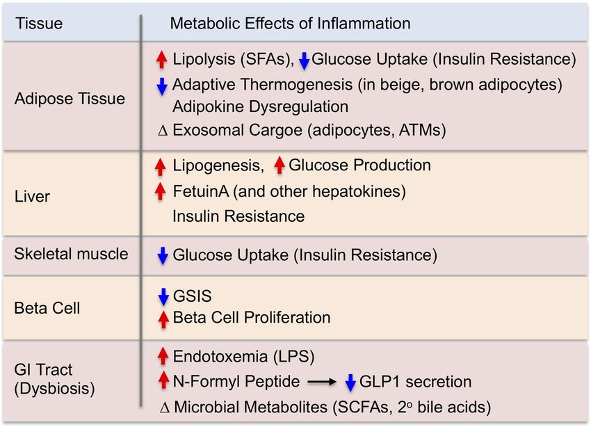
Downloaded from genesdev.cshlp.org on September 30, 2021 - Published by Cold Spring Harbor Laboratory Press
Immunometabolism in obesity
ward a tissue proinflammatory state. The majority of
M1-like polarized ATMs are derived from circulating
monocytes and express CD11c on the surface, whereas
the majority of M2-like polarized ATMs are resident and
do not display CD11c expression (Lumeng et al. 2007b).
The inflammatory state of ATMs can also be modified by
various lipid species. Saturated fatty acids (SFAs) derived
from either dietary sources or adipocyte triglyceride hy-
drolysis can drive M1-like ATM polarization through a
toll-like receptor 4 (TLR4)-dependent mechanism (Saberi
et al. 2009; Holland et al. 2011; Orr et al. 2012; Tao et al.
2017). On the other hand, omega-3 FAs inhibit proinflam-
matory activation of ATMs through a GPR120-dependent
mechanism (Oh et al. 2010).
The terms M1- and M2-like are often used to describe
ATMs in lean and obese states as a matter of convenience.
However, the concept of M1 and M2 macrophages was first
introduced based on the immunologic differences between
mouse strains that preferentially promote Th1 or Th2 re-
sponses. They are specifically defined as bone marrow pro-
genitor cells polarized to an M1 or M2 state in culture by
incubating with LPS/IFNγ or IL-4/IL-13, respectively
(Mills et al. 2000). In the in vivo setting, ATMs do not exist
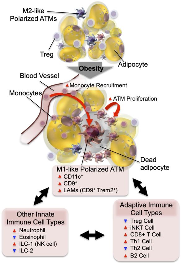
Figure 1. Adipose tissue inflammation in obesity. In the normal
within these narrow categories but actually span across a
state, resident ATMs mostly show an M2-like polarized pheno- polarization spectrum similar to, but distinct from, M1
type. Factors released from Tregs and eosinophils support or M2 macrophages (Li et al. 2019). Thus, studies using sin-
ATMs to maintain this anti-inflammatory state. In obesity, in- gle-cell RNA sequencing (scRNA-seq) analyses revealed
creased adipocyte chemokine production induces increased blood that the transcriptomic profiles of ATMs are highly hetero-
monocyte recruitment, as well as ATM proliferation. The major- geneous in obese adipose tissue. ATMs in obesity display a
ity of monocyte-derived ATMs express CD11c and/or CD9 and proinflammatory profile that is overlapping but different
the M1-like polarized phenotype. The decreased number of eosin- compared with in vitro generated M1 cells (Kratz et al.
ophils and Tregs and the increased number of neutrophils, ILC1, 2014; Hill et al. 2018; Jaitin et al. 2019). Moreover, they
CD8+ T cells, Th1 cells, and B2 cells enhance M1-like ATM po-
show unique metabolic gene expression profiles, including
larization and adipose tissue inflammation.
activation of lysosome-dependent lipid metabolism, col-
lectively termed the “metabolic activation state” (Xu
et al. 2013; Kratz et al. 2014; Coats et al. 2017). Hill et al.
(HFD), increased proinflammatory cytokine expression (2018) reported two subpopulations of ATMs depending
and macrophage infiltration can be observed in visceral ad- on CD9 expression, and the number of CD9+ ATMs in-
ipose tissue (Lee et al. 2011b). These changes gradually in- creases during the development of obesity. These CD9+
crease during the development of obesity, along with ATMs show unique functional, morphological, and loca-
progressive deterioration of insulin sensitivity and glucose tional features: They express high levels of proinflamma-
tolerance until obesity is fully stabilized and these changes tory cytokines, contain intracellular lipid droplets,
start to plateau. In obesity, ATMs accumulate, becoming secrete exosomes, and are located within crownlike struc-
the most abundant immune cell type in adipose tissue, tures surrounding dying adipocytes. In addition, a subset of
and can comprise up to 40% of all stromal vascular cells. ATMs expresses the monocyte marker Ly6C and also ac-
Along with the obesity-induced quantitative changes in cumulates in obese adipose tissue. Since Ly6C is a marker
ATM content, ATMs also undergo phenotypic switching for monocytes, the Ly6C+ ATMs most likely represent
from M2-like polarized anti-inflammatory cells to the newly recruited monocyte-derived macrophages undergo-
M1-like polarized proinflammatory state (Lumeng et al. ing differentiation. As found in aortic wall macrophages,
2007a). M1 macrophages are considered to represent the these Ly6C+ cells lose expression of Ly6C as they fully dif-
“classically activated” proinflammatory phenotype of ferentiate into mature macrophages (Jakubzick et al.
macrophages, and these macrophages produce proinflam- 2017). The Ly6C+ ATMs are found outside of crownlike
matory cytokines, reactive oxygen species, and nitric ox- structures and can stimulate adipocyte differentiation
ide that eliminate pathogens. M2 cells represent the (Hill et al. 2018). Many of the transcriptomic signature
“alternatively activated” anti-inflammatory phenotype genes of CD9+ or Ly6C+ ATMs are distinct from in vitro
of macrophages, which generally promote tissue repair differentiated M1 or M2 macrophages or from each other.
and remodeling (Jakubzick et al. 2017; Koelwyn et al. Jaitin et al. (2019) conducted unbiased scRNA-seq analy-
2018). In obesity, while the number of both M1- and M2- ses and found that ATMs can be divided into three subpop-
like polarized ATMs is increased, the increase in M1-like ulations: one showing transcriptomic signatures of
polarized ATMs is much greater, skewing the balance to- perivascular macrophages and another two subsets with
GENES & DEVELOPMENT 309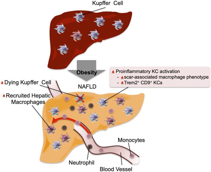
Downloaded from genesdev.cshlp.org on September 30, 2021 - Published by Cold Spring Harbor Laboratory Press

Lee and Olefsky

CD9 expression. In line with the findings of Hill et al.                higher rates of resident ATM proliferation. In vivo label-
(2018), they reported that the number of CD9+ ATMs ex-                  ing of blood monocytes or resident ATMs have shown
pands in obesity and can be found within crownlike struc-               that blood monocyte migration into adipose tissue in-
tures. They further characterized CD9+ ATMs using                       creases in obesity (Lumeng et al. 2007a; Oh et al. 2012).
lineage tracing and found that ∼80% of the CD9+ ATMs                    This increase is associated with monocytosis (Nagareddy
are bone marrow derived. Interestingly, CD9+ ATMs can                   et al. 2014) and increased production of chemokines such
be divided into two subpopulations, depending on the ex-                as CCL2/MCP-1 and leukotriene B4 (LTB4) in visceral ad-
pression of the lipid receptor Trem2. Trem2+ CD9+                       ipose tissue (Li et al. 2015). Adipocyte-specific depletion
ATMs abundantly express genes associated with phagocy-                  of CCL2 reduces obesity-induced ATM accumulation
tosis and lipid metabolism. They termed these cells lipid-              (Kanda et al. 2006). Moreover, genetic deletion or pharma-
associated macrophages (LAMs), and showed that obesity-                 cological inhibition of the receptors for the chemokines
induced LAM expansion is TREM2 dependent and that                       CCL2 (CCR2) or LTB4 (LTB4R) blocks obesity-induced
Trem2 KO mice show increased weight gain, glucose intol-                ATM accumulation (Lumeng et al. 2007a; Li et al. 2015).
erance, and dyslipidemia on a HFD, suggesting that                      This shows that monocyte chemotaxis into adipose tissue
TREM2-dependent LAM ATMs can mitigate obesity and                       is largely mediated by the CCL2/CCR2 and LTB4/LTB4R
metabolic dysregulation.                                                systems. In addition to increased chemotaxis, the levels of
   These CD9+ Trem2+ ATMs display functional similari-                  factors associated with macrophage retention, such as
ties to CD11c+, M1-like polarized ATMs, and CD9 expres-                 Netrin1 and Sema3E, are increased in obese adipose tis-
sion correlates with CD11c expression, although CD11c+                  sue, inhibiting emigration of ATMs (Shimizu et al. 2013;
and CD9+ populations are not identical (Hill et al. 2018).              Wanschel et al. 2013; Ramkhelawon et al. 2014).
This suggests that both the CD9+ and CD11c+ populations                    In addition to chemotaxis into adipose tissue, ATM ac-
might be functionally heterogeneous. Additional studies                 cumulation is also due to increased resident ATM prolif-
are required to further define CD11c and CD9 double                     eration, which can be stimulated by CCL2 (Amano et al.
and single positive and negative populations.                           2014). In subsequent studies, increased ATM proliferation
                                                                        in obese adipose tissue was confirmed by separate groups
ATMs and the control of local catecholamine levels                      (Zheng et al. 2016).
Chawla and his colleagues suggested that M2-like polar-
ized ATMs synthesize catecholamines (Nguyen et al.                      Other innate immune cells Although relatively small in
2011; Qiu et al. 2014). However, this concept was chal-                 number, changes in other innate immune cell types also
lenged by other groups who reported that ATMs do not                    contribute to the development of adipose tissue inflam-
produce physiologically relevant levels of catecholamines               mation, mainly by regulating ATM polarization and func-
and do not express the enzyme tyrosine hydroxylase,                     tion. For example, innate lymphoid cell type 1 (ILC1) cells,
which is necessary for catecholamine synthesis. More-                   including natural killer cells, reside in normal/lean adi-
over, these groups found IL-4 treatment does not alter en-              pose tissue and contribute to adipose tissue homeostasis
ergy expenditure and body weight either in cold, ambient,               (Lee et al. 2016; Boulenouar et al. 2017; Theurich et al.
or thermoneutral temperatures (Fischer et al. 2017). As an              2017). In obesity, the number of ILC1 cells increases and
alternative mechanism for how macrophages might mod-                    ILC1-derived cytokines, including IFNγ, stimulate proin-
ulate local catecholamine levels, a subset of macrophages,              flammatory activation of ATMs. On the other hand, the
termed sympathetic neuron-associated macrophages                        number of ILC2 cells producing Th2 cytokines decreases
(SAMs), are capable of extracellular catecholamine uptake               in obesity (Molofsky et al. 2013; Nussbaum et al. 2013).
and catabolism (Pirzgalska et al. 2017). SAMs represent an-             ILC2-derived cytokines support immigration and matura-
other subset of ATMs found in proximity to sympathetic                  tion of eosinophils in adipose tissue, which produce IL-4
nerves in adipose tissue. In obesity, the number of SAMs                and IL-13 that stimulate M2-like macrophage polarization
increases, contributing to reduced adaptive thermogenic                 (Wu et al. 2011). In obese adipose tissue, decreased ILC2
activity. The concept of macrophage-mediated catechol-                  cells are associated with decreased local eosinophil num-
amine catabolism in adipose tissue was also reported by                 bers. Finally, the recruitment of neutrophils into adipose
Dixit and his colleagues (Camell et al. 2017), who showed               tissue increases in obesity, and factors released from neu-
that aging-induced catecholamine resistance is associated               trophil granules, such as neutrophil elastase and myelo-
with increased ATM catecholamine catabolism. In sup-                    peroxidase, can contribute to the development of insulin
port of the concept that ATMs regulate local catechol-                  resistance (Talukdar et al. 2012; Tam et al. 2020).
amine levels, systemic depletion of tissue macrophages
using clodronate liposomes, but not sympathetic denerva-                Adaptive immune cells Several types of adaptive im-
tion, blocks inguinal WAT beiging in adipocyte Fasn KO                  mune cells are also involved in adipose tissue inflamma-
mice (Henriques et al. 2020).                                           tion. In obesity, the number of adipose tissue CD8+
                                                                        T cells increases, and factors released from these cells
Mechanisms of macrophage accumulation The mecha-                        can facilitate ATM differentiation and chemotaxis (Nish-
nism by which macrophages accumulate in obese adipose                   imura et al. 2009). Among CD4+ T cells, the number of
tissue involves (1) increased monocyte/macrophage che-                  proinflammatory T helper (Th) 1 cells increases (Nishi-
motaxis (increased blood monocyte immigration) and de-                  mura et al. 2009), whereas the adipose tissue content of
creased ATM emigration (or increased retention), and (2)                anti-inflammatory Th2 cells decreases in obesity,

310      GENES & DEVELOPMENT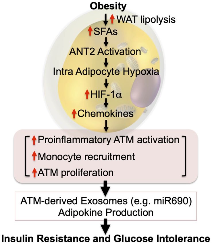
Downloaded from genesdev.cshlp.org on September 30, 2021 - Published by Cold Spring Harbor Laboratory Press

                                                                                                       Immunometabolism in obesity

contributing to increased adipose tissue interferon γ (IFNγ)       pose tissue inflammation on the development of systemic
levels and inflammation (Winer et al. 2009). Tregs                 insulin resistance in obesity, inflammation is a physiolog-
(CD3+CD4+FOXP3+) are another CD4+ T-cell subset well               ical process necessary for defense against pathogenic inva-
studied in adipose tissue (Feuerer et al. 2009; Li et al.          sion or tissue damage and repair. Acute inflammation is
2020). There are only a small number of these cells in             necessary to maintain normal adipose tissue function by
young mice, but they start to accumulate in visceral adi-          mediating healthy adipose tissue remodeling to safely ac-
pose tissue at the age of 10–15 wk, due to increased prolif-       commodate excess lipids into adipose tissue (Wernstedt
eration and decreased turnover. Tregs represent the major          Asterholm et al. 2014). For example, Zhu et al. (2020) re-
CD4+ T-cell subset (40%–80% of total CD4+ T cells) by              ported that inhibition of inflammatory pathways by induc-
20–30 wk of age in normal mice (Bapat et al. 2015). Tregs          ible overexpression of an adenoviral protein, called RIDα/β
control the activity of other T cells and restrain the innate      (which blocks several inflammatory signaling pathways)
immune system by inhibiting monocyte immigration and               reduces weight gain and adiposity and exacerbates glucose
proinflammatory polarization. In obesity, the number, or           and insulin intolerance in HFD/obese mice. Therefore, it is
proportion, of adipose tissue Treg cells decreases, contrib-       likely that acute adipose tissue inflammation is necessary
uting to increased adipose tissue inflammation (Feuerer            for adipose tissue homeostasis, whereas chronic inflam-
et al. 2009). Adipose tissue Tregs show a unique transcrip-        mation contributes to the development of insulin resis-
tomic profile distinct from Tregs in other sites, which is         tance and adipose tissue dysfunction.
largely regulated by peroxisome proliferator-activated re-
ceptor γ (PPARγ). Indeed, >80% of adipose tissue Tregs ex-
press PPARγ, which is unique to adipose Tregs, and Treg            Liver
PPARγ mediates a component of the anti-inflammatory ef-
fects of thiazolidinediones (TZDs) in obese mice (Cipol-           Hepatic macrophages Obesity, or a HFD, leads to hepat-
letta et al. 2012). γδ T cells abundant in adipose tissue of       ic steatosis commonly referred to as nonalcoholic fatty liv-
normal mice support sympathetic innervation and Treg               er disease (NAFLD), which can proceed to a condition
expansion by releasing IL-17 (Kohlgruber et al. 2018; Hu           termed nonalcoholic steatohepatitis (NASH). Obesity is
et al. 2020).                                                      also associated with increased hepatic inflammation, and
   Invariant natural killer T (iNKT) cells account for 10%–        macrophages are the major source of the proinflammatory
30% of all adipose tissue-resident T cells (Huh et al. 2013).      cytokines (Stienstra et al. 2010; Xu et al. 2014). Injection of
These are innate-like αβ T cells that are specifically stim-       gadolinium or intravenous clodronate liposomes selec-
ulated by lipid antigens through the major histocompati-           tively depletes hepatic macrophages owing to the fenes-
bility complex (MHC)-like molecule CD1d (Huh et al.                trated hepatic sinusoidal structure. Selective depletion of
2013; Lynch 2014). iNKT cells release IL-10, IL-4, IL13,           hepatic macrophages improves insulin resistance and liver
and IL-2, potentiating M2-like polarization of ATMs.               steatosis in obese mice or rats (Huang et al. 2010; Lanthier
The number of adipose tissue iNKT cells decreases in obe-          et al. 2010; Lee et al. 2011b; Reid et al. 2016). Liver macro-
sity, and this could contribute to the development of in-          phages can be divided into two major subsets based on
flammation. However, other reports cast doubt on the               their developmental origin: These include yolk-sac-de-
beneficial effects of iNKT cells. For example, Satoh et al.        rived resident Kupffer cells (KCs) and bone marrow-de-
(2016) and Wu et al. (2012b) have shown proinflammatory            rived recruited hepatic macrophages (RHMs). In normal
and negative metabolic effects of iNKT cells in obese              liver, hepatic macrophages account for ∼10% of all hepatic
mice. As a possible explanation for these contradicting re-        cells, and the majority of them are KCs (Bouwens et al.
sults, scRNA-seq analysis found that adipose tissue iNKT           1986). In obesity, the number of KCs is unchanged, where-
cells can be functionally divided into two subsets, depend-        as circulating monocyte chemotaxis and differentiation
ing on NK1.1 expression: NK1.1+ iNKTs are proinflamma-             into macrophages (RHMs) increase, largely through the
tory and produce IFNγ, whereas NK1.1− iNKTs are anti-              CCL2/CCR2 system (Obstfeld et al. 2010; Morinaga
inflammatory and produce IL-10 (LaMarche et al. 2020).             et al. 2015). While both KCs and RHMs are highly hetero-
   B lymphocytes account for 3%–10% of SVCs in normal              geneous, RHMs express higher levels of M1-like polarized
adipose tissue and increase up to twofold in obesity or 10-        macrophage markers and proinflammatory gene expres-
fold in aged mice (Camell et al. 2019). Compared with the          sion, which is exacerbated in obesity (Arkan et al. 2005;
spleen and bone marrow, adipose tissue B cells show a              Morinaga et al. 2015; Seidman et al. 2020). These results
higher B1:B2 ratio in normal mice. However, in obesity,            suggest that a major component of obesity-induced liver
the B1:B2 ratio decreases with increased recruitment of            inflammation can be attributed to increased monocyte in-
proinflammatory B2 cells (Ying et al. 2017b). B2 recruit-          filtration and macrophage differentiation (summarized in
ment into obese visceral adipose tissue is mainly mediat-          Fig. 2).
ed by the LTB4/LTB4R1 system (Ying et al. 2017b;                      Recently, single-cell analyses of transcriptomic and epi-
Srikakulapu and McNamara 2020), and these cells can                genetic changes in hepatic macrophages (e.g., scRNA-seq
contribute to adipose tissue inflammation and metabolic            and scATAC-seq) combined with monocyte tracking and
dysfunction in obesity.                                            KC depletion studies have further elucidated hepatic mac-
                                                                   rophage phenotypes (Seidman et al. 2020). Thus, in mice
Acute versus chronic inflammation While numerous                   fed a NASH-prone HFD, KCs undergo partial loss of their
studies have shown the detrimental effects of chronic adi-         identifying gene expression patterns while gaining a scar-

                                                                                                 GENES & DEVELOPMENT          311
Downloaded from genesdev.cshlp.org on September 30, 2021 - Published by Cold Spring Harbor Laboratory Press

Lee and Olefsky

                                                                        tween intrahepatic cell types such as hepatocytes, various
                                                                        immunocytes, and hepatic stellate cells (Koyama and
                                                                        Brenner 2017). For example, activation of hepatocyte
                                                                        JNK, IKKβ, or IKKε or the unfolded protein response (ER
                                                                        stress) increases production of chemokines and other hep-
                                                                        atokines, stimulating KC proinflammatory activation and
                                                                        circulating monocyte and neutrophil recruitment (Arkan
                                                                        et al. 2005; Seki et al. 2012; Reilly et al. 2013; Lee et al.
                                                                        2018). The incoming immune cells are highly proinflam-
                                                                        matory (Morinaga et al. 2015; Seidman et al. 2020),
                                                                        enhancing liver inflammation. Hepatocyte and immuno-
                                                                        cyte-derived factors such as TGFβ can activate quiescent
                                                                        stellate cells and induce secretion of collagen, leading to
                                                                        liver fibrosis (Hellerbrand et al. 1999; Koyama and Brenner
                                                                        2017). Moreover, activated stellate cell-derived cytokines
                                                                        and chemokines can enhance liver inflammation, exacer-
                                                                        bating liver steatosis and fibrosis (Weiskirchen and Tacke
                                                                        2014). Please see Koyama and Brenner (2017) and Arab
Figure 2. Inflammation in the liver. In the normal physiological        et al. (2018) for excellent reviews on the pathophysiology
state, KCs account for ∼10% of all liver cells. In addition, they       of NAFLD and NASH.
scavenge pathogens and show M2-like polarized anti-inflamma-
tory phenotype. In obese steatotic livers, KCs can show increased
                                                                        Muscle
expression of genes associated with tissue repair and inflamma-
tion. These genes include Cd9 and Trem2. This is accompanied            Skeletal muscle accounts for the majority of in vivo insu-
by increased KC apoptosis, and a component of KC death is com-          lin-stimulated glucose disposal, and this is decreased in
pensated for by increased recruitment of blood monocytes, which         obesity/T2DM, contributing to the development of com-
differentiate into KC-like macrophages. Chemokines released by
                                                                        pensatory hyperinsulinemia and glucose intolerance.
steatotic hepatocytes cause increased recruitment of blood
                                                                        Several mechanisms such as mitochondrial dysfunction,
monocytes into the liver, which differentiate into proinflamma-
tory macrophages (RHMs) and produce factors that can cause in-          microvascular dysfunction, and ectopic fat accumulation
sulin resistance. The recruitment of neutrophils also increases,        (or lipotoxicity) have been proposed as causes of this
and the molecules released from neutrophil granules such as neu-        defect and are well reviewed elsewhere (Serné et al.
trophil elastase and myeloperoxidase can induce insulin resis-          2007; Roden and Shulman 2019; Sergi et al. 2019). While
tance in hepatocytes.                                                   relatively less studied compared with adipose tissue and
                                                                        liver, proinflammatory pathways are enhanced with in-
                                                                        creased macrophage infiltration in skeletal muscle of
associated macrophage phenotype with Trem2 and Cd9                      obese mice and T2DM patients compared with normal
expression. These changes are accompanied by increased                  mice or human subjects (Fink et al. 2014). Systemic
KC apoptosis. As these KC numbers decline, they are re-                 depletion of macrophages or genetic modifications sup-
placed by monocyte-derived macrophages that undergo                     pressing proinflammatory activation of macrophages im-
convergent epigenomic and transcriptomic changes and                    proves insulin sensitivity in skeletal muscle in mice
become new KC-like cells.                                               (Patsouris et al. 2008; Han et al. 2013), suggesting that in-
                                                                        flammation plays a causative role in the development of
Other hepatic immune cells The number of neutrophils                    muscle insulin resistance. Consistent with this, treat-
is also increased in the liver of obese mice compared with              ment with proinflammatory cytokines induces insulin
lean/normal mice (Talukdar et al. 2012), and neutrophil-                resistance in cultured myocytes or skeletal muscle in
derived cytokines and chemokines contribute to the de-                  vivo (de Alvaro et al. 2004; Plomgaard et al. 2005). On
velopment of obesity-induced liver inflammation. More-                  the other hand, the anti-inflammatory cytokine, IL-10,
over, NE released from neutrophils can be taken up by                   can play a protective role. For example, muscle-specific
hepatocytes and cause degradation of IRS-2, promoting de-               overexpression of IL-10 mitigates and muscle-specific
creased insulin sensitivity.                                            deletion of Il10 exacerbates obesity-induced muscle in-
   Among adaptive immune components, the number of                      flammation and insulin resistance (Hong et al. 2009;
CD8+ cytotoxic T cells increases in the liver of obese                  Dagdeviren et al. 2016).
mice compared with lean mice through an IFNγ-depen-                        With respect to mechanisms of muscle inflammation,
dent signaling pathway (Ghazarian et al. 2017). On other                intermyocellular adipose tissue (IMAT) or perimuscular
hand, the numbers of CD4+ T cells, Tregs, and γδ T cells                adipose tissue (PMAT) have been implicated (Khan et al.
decrease or remain unchanged in obesity.                                2015). Thus, in obesity, IMAT and PMAT expand and
                                                                        show macrophage accumulation, similar to visceral adi-
Communication between liver cell types Obesity-in-                      pose tissue. As in visceral adipose tissue, the majority of
duced changes in the liver leading to the development of                macrophages infiltrated into obese skeletal muscle are
insulin resistance and NASH involve interactions be-                    CD11c+ M1-like polarized macrophages. Moreover, the

312      GENES & DEVELOPMENT
Downloaded from genesdev.cshlp.org on September 30, 2021 - Published by Cold Spring Harbor Laboratory Press
Immunometabolism in obesity
changes in the proportion of different T-cell subsets (e.g., erties (Ying et al. 2019). These islet macrophages show a
increased Th1 cells and decreased Tregs) show similar pat- unique transcriptomic profile distinct from M1- or M2-
terns as seen in visceral adipose tissue (Wu and Ballantyne like polarized macrophages or other tissue resident mac-
2017). rophages. Intraislet macrophages substantially proliferate
in obesity, whereas peri-islet macrophages lining the islet
capsule do not. Moreover, intraislet macrophages, but not
Pancreatic islets
peri-islet macrophages, inhibit β-cell insulin secretion.
β-Cell dysfunction is a critical etiologic component of This effect of intraislet macrophages is mediated by a
T2DM, and a variety of mechanisms for this defect have cell–cell contact-dependent mechanism, involving the
been identified (for reviews, see Halban et al. 2014; Hud- engulfment of β-cell insulin secretory vesicles. In addi-
ish et al. 2019). Several reports have shown islet inflam- tion, proinflammatory cytokines produced from islet
mation can also be etiologically important in causing β- macrophages also suppress β-cell GSIS (Maedler et al.
cell dysfunction in T2DM. The expression of proinflam- 2002; Eguchi et al. 2012). For example, IL-1β produced
matory cytokines (such as IL-1β, IL-33, IL-23, and IL-24) from islet macrophages binds to the IL-1 receptor (IL-
and macrophage infiltration is increased in the islets of 1R), which is abundantly expressed on β cells, leading to
obese/T2DM patients compared with normal subjects decreased GSIS through stimulation of the JNK and NF-
(Ehses et al. 2007 ; Böni-Schnetzler et al. 2008; Richardson κB signaling pathways (Kwon et al. 1998; Larsen et al.
et al. 2009; Mahdi et al. 2012; Butcher et al. 2014; Hasnain 2005; Ortis et al. 2006; Wang et al. 2009). Furthermore, is-
et al. 2014; Kamata et al. 2014; Schludi et al. 2017). Treat- let macrophages (both intraislet and peri-islet) release
ment of isolated human islets with proinflammatory cyto- growth factors such as PDGF and IGF-1, simulating β-
kines reduces glucose-induced intracellular Ca2+ levels cell proliferation (Ying et al. 2019; Nackiewicz et al.
and GSIS (Corbett et al. 1993; O’Neill et al. 2013), and in- 2020). Therefore, it is likely that obesity-associated islet
duces β-cell death (Brozzi et al. 2015). Unlike T1DM islets, inflammation induces decreased β-cell GSIS and promotes
obesity- or T2DM-associated islet inflammation is domi- the β-cell proliferation that exists in obese islets. This re-
nated by macrophages and does not involve T-cell infiltra- sults in changes to islet function favoring increased basal
tion (Ehses et al. 2007). The concept that islet insulin secretion and decreased GSIS.
inflammation induces β-cell dysfunction was more exten- One can question whether increased β-cell proliferation
sively tested in rodent studies. Systemic depletion of mac- by islet macrophages is necessarily detrimental to meta-
rophages by intraperitoneal injection of clodronate bolic homeostasis. Indeed, although adverse effects of is-
liposomes potentiates glucose-stimulated insulin secre- let inflammation on GSIS and β function were reported
tion (GSIS), as well as insulin sensitivity in obese mice in chronic obese inflammatory states, inflammation is a
(Eguchi et al. 2012). Ex vivo depletion of macrophages physiological process, and acute islet inflammation may
within primary islets isolated from HFD/obese mice en- be beneficial for the maintenance of islet function in the
hances GSIS (Eguchi et al. 2012; Ying et al. 2019). Treat- normal state (Banaei-Bouchareb et al. 2004; Geutskens
ment of primary mouse islets with proinflammatory et al. 2005; Yano et al. 2007; Kayali et al. 2012; Lee et al.
cytokines (e.g., IL-1β, IL-23, IL-24) reduces GSIS (Mahdi 2013; Dror et al. 2017; Burke et al. 2018; Riopel et al.
et al. 2012; Jourdan et al. 2013; Hasnain et al. 2014). More- 2018; Ying et al. 2019). Therefore, it is possible that acute
over, Myd88 or TLR4 KO mice are protected from β-cell inflammation is necessary to maintain islet function,
dysfunction induced by subchronic lipid infusion (lipotox- whereas chronic inflammation contributes to the devel-
icity) (Eguchi et al. 2012), while β-cell-specific IL-1 recep- opment of β-cell dysfunction.
tor antagonist (IL-1Ra) KO mice develop β-cell
dysfunction (Böni-Schnetzler et al. 2018).
GI tract
While similar to adipose tissue and liver inflammation
in causing functional defects in parenchymal cells (in Recent advances in our understanding of the gut micro-
this case, β cells), there is a sharp difference in the mech- biota have provided potential new insights as to how obe-
anism for macrophage accumulation in obese islets com- sity affects tissue inflammation and metabolic defects. In
pared with liver and adipose tissue. Thus, in the liver and both human and rodent obesity, the composition of the
adipose tissue, macrophage expansion largely involves gut microbiota changes (dysbiosis), which can modify
recruitment of circulating monocytes (Weisberg et al. the host immune system and metabolism. HFD/obese
2003; Morinaga et al. 2015; Jaitin et al. 2019; Seidman WT mice display increased proinflammatory cytokine ex-
et al. 2020a), with a smaller component due to resident pression and IL17-producing γδ T cells, Th1 cells, CD8+
macrophage proliferation (Amano et al. 2014). In islets, T cells, macrophages, and dendritic cells, along with de-
macrophage accumulation is largely mediated by resi- creased Tregs, ILC3 cells, and eosinophils in the intestinal
dent islet macrophage proliferation (Ying et al. 2019). lamina propria (Johnson et al. 2015; Monteiro-Sepulveda
Blood monocytes are recruited to the peri-islet region et al. 2015; Winer et al. 2016). Fecal transplantation exper-
but do not enter the islet capsule (Eguchi et al. 2012; iments and clinical trials further showed that dysbiosis
Ying et al. 2019). contributes to the development of inflammation and insu-
In islets, there are two resident macrophage populations lin resistance in obesity. For example, transplantation of
(intraislet and peri-islet macrophages) showing distinct germ-free mice with fecal preparations from human twins
anatomical distribution, phenotypes, and functional prop- discordant for obesity showed that the obese gut
GENES & DEVELOPMENT 313Downloaded from genesdev.cshlp.org on September 30, 2021 - Published by Cold Spring Harbor Laboratory Press Lee and Olefsky microbiota can induce increased weight gain, adipose tis- In addition, the dysbiosis of obesity provides another po- sue mass, and glucose intolerance (Ridaura et al. 2013). tential mechanism in which specific GI bacterial species, Moreover, a clinical study found that administration of in- or combinations of species, can contribute to metainflam- testinal microbiota from lean donors to recipients with mation. The latter is clear in mice but will require further metabolic syndrome improved insulin sensitivity (Vrieze translational validation in humans. This is a promising et al. 2012). These studies suggest that a component of the field, since further studies could identify specific factors obesity-associated metabolic phenotype is due to dysbio- that might be useful in drug discovery. sis. Nonetheless, it should be noted that the effects of fe- cal transplantation were small, and healthy gut microbiome transfer does not normalize the metabolic de- Hypoxia as initiating event fects induced by obesity. In this context, several gut microbiota-derived mole- As noted above, it is well known that obesity leads to an cules/mechanisms that can modify host immune tone accumulation of proinflammatory macrophages in adi- and metabolism have been reported. For example, in obesi- pose tissue in both mice and humans. These ATMs are ty or shortly after a HFD, gut permeability increases, al- key drivers of the development of insulin resistance and lowing bacterial translocation across the intestinal glucose intolerance. Areas of current active interest focus barrier (Neal et al. 2006; Amar et al. 2011; Ha et al. on how macrophage accumulation is initiated at the onset 2020). Moreover, increased gut permeability also allows of obesity and how do proinflammatory ATMs cause sys- gut microbiota-derived molecules, such as lipopolysaccha- temic metabolic dysfunction. Several studies have been ride (LPS), to leak into the blood circulation (Erridge et al. published, providing important insights into these ques- 2007; Kumar et al. 2011). Increased LPS-stimulated TLR4 tions. In this section, we attempt to integrate these new induces proinflammatory responses in various immuno- insights into an overall sequential hypothesis. cytes, gut epithelial cells, and insulin target cells, resulting For years, it has been known that adipose tissue oxygen in insulin resistance and glucose intolerance (Shi et al. tension is decreased in obesity in both mice and humans 2006; Saberi et al. 2009; Guo et al. 2013; Tao et al. 2017). (Hosogai et al. 2007; Halberg et al. 2009; Pasarica et al. Plasma levels of another gut microbiota-derived molecule, 2009, 2010; Lawler et al. 2016; Seo et al. 2019; Smith formyl-methionyl-leucyl-phenylalanine (N-formyl pep- et al. 2019). The initial view was that this adipose tissue tides), are also elevated in HFD/obese mice. Genetic dele- hypoxic state was due to an imbalance between expanding tion or pharmacological inhibition of the N-formyl peptide adipose tissue mass and the available oxygen delivery. receptor Fpr1 improves glucose tolerance by increasing However, tissue oxygen tension is maintained by the bal- glucagonlike peptide 1 section from enteroendocrine L ance between oxygen supply and demand, and more re- cells with a subsequent increase in β-cell GSIS. Of the bio- cent data show that obesity leads to increased adipocyte active metabolites produced by the gut microbiota, short oxygen consumption (Lee et al. 2014). This increased con- chain fatty acids (SCFAs) are among the most abundant sumption accounts for ∼40% of the decrease in interstitial (Stevens and Hume 1998). SCFAs activate G-protein-cou- oxygen tension in obesity (Seo et al. 2019). The remaining pled receptors (GPCRs), including GPR41 and GPR43, component of the decreased interstitial oxygen tension is which are expressed in adipocytes, enterocytes, immuno- not due to decreased arterial oxygen delivery (blood flow cytes, and pancreatic β cells. Several reports showed ben- into adipose depot and blood O2 saturation) but is ex- eficial effects of GPR43 activation to reduce adiposity plained by decreased functional capillary density in obese and improve insulin sensitivity, as well as β-cell GSIS adipose tissue (Lee et al. 2014). Therefore, there are two in obese mice (Tolhurst et al. 2012; Kimura et al. 2013; components, decreased capillary density and increased ad- McNelis et al. 2015). However, conflicting results were ipocyte oxygen consumption, that contribute to the rela- also reported (for review, see Ang and Ding 2016): For ex- tive hypoxia of adipose tissue in obesity. As a general ample, β-cell-specific depletion of both GPR41 and matter, hypoxic conditions lead to greater expression of GPR43 increases GSIS and improves glucose tolerance the adipocyte HIF-1α transcription factor, triggering the in mice (Tang et al. 2015). In addition to SFAs, secondary downstream hypoxia transcriptome response. However, and tertiary bile acids produced by gut microbes alter the the induction of HIF-1α does not incur until oxygen ten- composition of the bile acid pool (Brufau et al. 2010; sions fall to ≤1% (Seo et al. 2019). Therefore, the decrease Suhre et al. 2010; Haeusler et al. 2013; Ridaura et al. in extracellular adipose tissue oxygen tension described in 2013; Yoshimoto et al. 2013) and modulate inflamma- obesity is probably not enough to trigger the HIF-1α hyp- tion and metabolism by stimulating the bile acid recep- oxic response. On the other hand, the increase in adipo- tor 1 (TGR5) and farnesoid X receptor (FXR) (Pols et al. cyte oxygen consumption is quite marked in obesity, 2011; Wollam and Antebi 2011). bringing the intra-adipocyte oxygen concentration down Taken together, the GI tract plays a role in obesity- to levels below ∼1.4%. This is adequate to induce adipo- associated chronic tissue inflammation and metabolic cyte HIF-1α expression and the downstream effects medi- dysregulation. One obvious mechanism is that the devel- ated by this transcription factor (Hosogai et al. 2007; opment of obesity leads to increased gastrointestinal per- Halberg et al. 2009; Lee et al. 2014). This view is supported meability, such that proinflammatory bacterial products by the finding that hypoxia-induced pimonidazole ad- (e.g., LPS), and even bacteria themselves, gain access to ducts in obese adipose tissue are confined to adipocytes surrounding lymph nodes and the systemic circulation. (Lee et al. 2014). Taken together, obesity-induced 314 GENES & DEVELOPMENT
Downloaded from genesdev.cshlp.org on September 30, 2021 - Published by Cold Spring Harbor Laboratory Press

                                                                                                        Immunometabolism in obesity

increased adipocyte oxygen consumption is likely the ma-
jor contributor to the intracellular adipocyte hypoxic re-
sponse, whereas the decreased interstitial oxygen
tension probably plays a contributory or permissive role
to exaggerate the impact of increased adipocyte oxygen
consumption.
   The mechanisms by which intracellular oxygen levels
regulate HIF-1α have been well described and occur main-
ly at the protein level with smaller effects on mRNA ex-
pression (Ivan and Kaelin 2017; Schödel and Ratcliffe
2019; Semenza 2019). In normoxic conditions, prolyl hy-
droxylase domain proteins (PHDs) bind to HIF-1α, and
PHD-dependent hydroxylation of HIF-1α leads to ubiqui-
tin-dependent proteasomal degradation of the protein.
Under hypoxia, PHDs are inactivated, which results in
HIF-1α protein stabilization, causing increased HIF-1α
protein expression. Although this is the major driver for          Figure 3. Intracellular adipocyte hypoxia triggers inflammation
regulation of HIF-1α expression, in obesity, modest in-            and insulin resistance. In obesity, increased intracellular FFAs
creases in HIF-1α mRNA have also been reported as a con-           stimulate ANT2-dependent increased mitochondrial uncoupled
tributing factor (Lee et al. 2014).                                respiration in adipocytes. Combined with decreased functional
   This induction of adipocyte HIF-1α plays an important           capillary density, this leads to intracellular hypoxia and HIF-1α
role in mediating overall adipose tissue inflammation              stabilization. HIF-1α induces increased chemokine production,
(Lee et al. 2014). This can occur through multiple mecha-          leading to increased immune cell infiltration, including mono-
                                                                   cytes, and increased ATM proliferation. These changes critically
nisms. For example, several chemokines are downstream
                                                                   affect ATM exosome secretion, as well as adipocyte exosome and
transcription targets of HIF-1α, and expression of these
                                                                   adipokine production, causing systemic insulin resistance. Most
chemokines is increased in obesity and other hypoxic con-          of these sequential events have been shown in mouse models.
ditions. These chemokines serve to recruit monocytes               This hypothesis remains to be validated in humans.
into the hypoxic adipose tissue, which then predominant-
ly differentiate into proinflammatory, M1-like ATMs. As
discussed in more detail above, the increase in M1-like
                                                                   space, leading to uncoupled mitochondrial respiration
ATMs is a key mechanism for the metabolic dysfunction
                                                                   with enhanced O2 consumption. It should be noted that
characterized by insulin resistance and glucose intoler-
                                                                   another important uncoupling protein, UCP1, is not ex-
ance in obesity (Fig. 3). There are many studies in which
                                                                   pressed in white adipocytes (Cousin et al. 1992; Wu
various proinflammatory genes have been deleted in mac-
                                                                   et al. 2012a). Adipose tissue SFA levels are elevated in obe-
rophages, and all of these studies consistently show ame-
                                                                   sity, and SFAs can activate ANT2-dependent uncoupling
lioration of obesity-induced metabolic dysfunction
                                                                   of oxidative metabolism, leading to increased adipocyte
(Arkan et al. 2005; Weisberg et al. 2006; Saberi et al.
                                                                   oxygen consumption (Lee et al. 2014). In theory, increased
2009; Han et al. 2013; Li et al. 2015). Consistent with
                                                                   fatty acid oxidation could also lead to greater oxygen con-
this formulation, the central role of HIF-1α in the proin-
                                                                   sumption, but more detailed studies have shown that this
flammatory responses leading to insulin resistance has
                                                                   component is rather negligible compared with the direct
been shown through cell type-specific knockout studies
                                                                   effect of SFAs to activate ANT2 protein. To further
(Jiang et al. 2011; Lee et al. 2011a, 2014; Krishnan et al.
                                                                   solidify this overall concept, deletion of ANT2 in adipo-
2012; Sun et al. 2013). Thus, adipocyte-specific HIF-1α
                                                                   cytes can reverse all of these hypoxic responses. Thus,
knockout largely prevents obesity-induced ATM accumu-
                                                                   adipocyte-specific ANT2 KO is sufficient to block the
lation, adipose tissue inflammation, glucose intolerance,
                                                                   obesity-induced increased adipocyte O2 consumption,
and insulin resistance but does not cause any change in
                                                                   ameliorating the adipocyte hypoxia response and prevent-
overall body weight. This highlights the importance of
                                                                   ing HIF-1α induction (Seo et al. 2019). This mitigates
HIF-1α in the overall adipose tissue inflammatory re-
                                                                   the development of adipose tissue inflammation with
sponse, consistent with the idea that obesity-induced in-
                                                                   reduced proinflammatory ATM content and decreased ex-
tracellular adipocyte hypoxia leads to HIF-1α induction,
                                                                   pression of inflammatory pathway genes. This is accom-
which in turn promotes ATM accumulation, inflamma-
                                                                   panied by a robust improvement in glucose tolerance
tion, and subsequent glucose intolerance and insulin
                                                                   and insulin sensitivity in obese mice without any changes
resistance.
                                                                   in body weight.
   The mechanisms underlying obesity-induced increased
adipocyte oxygen consumption are also of interest. Ade-
nine nucleotide translocase (ANT) 2 is a mitochondrial in-         How does adipose tissue inflammation cause insulin
ner-membrane protein that pumps protons from the                   resistance?
intermembrane space into the mitochondrial matrix (Ber-
tholet et al. 2019). Therefore, an increase in ANT2 activ-         The fact that increased obesity-induced proinflammatory
ity results in proton leakage from the intermembrane               ATM content can directly cause insulin resistance is well

                                                                                                 GENES & DEVELOPMENT            315
Downloaded from genesdev.cshlp.org on September 30, 2021 - Published by Cold Spring Harbor Laboratory Press Lee and Olefsky established, at least in mice, but precisely how M1-like studies of IL-6’s metabolic activities are mixed with macrophages lead to decreased insulin sensitivity is not some studies showing insulin-like actions to improve in- entirely clear and has been the subject of intense investi- sulin sensitivity, while others report that it inhibits insu- gation. A logical presumption is that proinflammatory lin action (Carey et al. 2006; Franckhauser et al. 2008). macrophages release factors that can cause paracrine or Given the above discussion, many laboratories have systemic effects on insulin target cells to impair insulin been trying to identify other factors that are elaborated signaling. Classically, M1-like polarized macrophages re- from M1-like macrophages that could cause metabolic lease a variety of cytokines and chemokines. Chemokines dysfunction. One such factor is Galectin-3. Galectin-3 is act by providing a concentration gradient in which mono- produced almost exclusively from macrophages, and in cytes and other types of immune cells can migrate down both human and mouse obesity, blood levels are quite that gradient toward the chemokine source; this is the op- high compared with normals and are in the range where erational definition of chemotaxis. In obese states, some they exert biologic effects (Li et al. 2016). Indeed, in hu- of these chemokines produced in adipose tissue can leak mans, circulating levels of Galectin-3 are elevated com- into the circulation. However, circulating chemokines pared with normal subjects, and the degree of the are not able to generate a differential concentration gradi- increase in Galectin-3 levels is correlated with the magni- ent to promote chemotaxis of monocytes out of the circu- tude of obesity and the degree of insulin resistance. In vi- lation into inflamed tissues. In addition, the circulating tro studies show that Galectin-3 treatment of insulin levels of chemokines due to this “spillover” are generally target cells can directly cause decreased insulin signaling much lower than the biologically effective concentra- by interfering with the ability of insulin to properly acti- tions, so they will not have meaningful systemic physio- vate the insulin receptor at the cell surface. Loss-of-func- logic effects. Even if a particular chemokine can cause tion studies have been performed by analyzing decreased insulin signaling in vitro (Sartipy and Loskutoff macrophage-specific Galectin-3 KO mice. Depletion of 2003), it would still be a challenge to conclude that such a macrophage Galectin-3 protects HFD mice from the de- chemokine could act systemically, since the circulating velopment of glucose intolerance and insulin resistance, systemic concentrations are so low. while weight gain is entirely normal. Additional gain- More attention has been placed on secreted cytokines, and loss-of-function studies with Galectin-3 are consis- since it is well known that certain cytokines (e.g., tent with this formulation, suggesting that macrophage- TNFα) have potent effects to cause decreased insulin sig- derived circulating Galectin-3 can contribute to the devel- naling. TNFα can cause local tissue effects to decrease in- opment of insulin resistance in obesity. sulin action (Hotamisligil et al. 1994) by several The above considerations have led to a search for addi- mechanisms. TNFα can reduce Irs2 and Glut4 expression, tional factors that can enter the circulation from obese ad- promote inhibitory phosphorylation of insulin receptor ipose tissue and cause systemic insulin resistance. Indeed, substrate (IRS) proteins, enhance adipocyte lipolysis recent studies suggest that ATM-derived exosomes might (FFA release) and ceramide synthesis, and inhibit PPARγ fulfill this role. Exosomes are small nanoparticles (∼150 expression (Stephens et al. 1997; Guilherme et al. 2008). nm) produced by most cell types (Pegtel and Gould This phenomenon could occur in the various adipose tis- 2019). They can be secreted into the interstitial space sue depots, which become enlarged and inflamed during where they then enter the circulation leading to both para- the course of obesity (TNFα is used as an example, since crine and endocrine-like effects. Exosomes contain a vari- it is the most well studied immune cell-derived cytokine). ety of cargo constituents including proteins, lipids, However, systemic insulin resistance requires decreased microRNAs (miRNAs), and mRNAs, as well as a number insulin sensitivity in muscle and the liver, as well as adi- of other RNA species. Recent studies have shown that pose tissue, and unless muscle and the liver are generating exosomes derived from ATMs in obese mice can directly high levels of tissue cytokines, it is unlikely that TNFα or cause insulin resistance in vitro when applied to adipo- other cytokines that leak out of obese adipose tissue could cytes, primary hepatocytes, or myocytes (Ying et al. cause systemic effects. Thus, the circulating concentra- 2017a). These “obese” ATM-derived exosomes can be har- tions of TNFα and other cytokines are substantially below vested and administered intravenously to lean mice. the levels needed to exert biologic effects in tissues (Ste- When this was done, the lean recipient mice developed phens et al. 1997; Amar et al. 2007; McGillicuddy et al. glucose intolerance, hyperinsulinemia, and insulin resis- 2011). In contrast, adipose tissue levels of cytokines can tance comparable with that seen in the obese state. Impor- be quite elevated in obesity, and this could cause local ef- tantly, eating behavior and body weight do not change as a fects. If one administers large amounts of TNFα in vivo, result of in vivo exosome treatment. On the other side of raising circulating levels far higher than they normally ex- this coin, ATM exosomes harvested from chow-fed lean ist in obesity, then an insulin-resistant state ensues (Lang mice produce the opposite phenotype. Treatment of adi- et al. 1992). However, this does not mirror the actual path- pocytes, myocytes, or primary hepatocytes with “lean” ophysiologic state in obesity in which cytokine levels in ATM exosomes directly leads to increased insulin signal- the blood, while higher than in normals, are not elevated ing. More importantly, when obese mice were treated to levels at which they exert substantial biologic effects. with “lean” ATM-derived exosomes for a period of 3 IL-6 may be an exception to this general principle, since wk, these mice showed markedly improved glucose toler- the levels of IL-6 that enter the circulation in obesity ance with an increase in insulin sensitivity. In a very real have been reported to be biologically active. However, sense, these “lean” ATM exosomes produce a therapeutic 316 GENES & DEVELOPMENT
You can also read