Pattern recognition receptors in health and diseases - Nature
←
→
Page content transcription
If your browser does not render page correctly, please read the page content below
Signal Transduction and Targeted Therapy                                                                                                    www.nature.com/sigtrans

                  REVIEW ARTICLE                         OPEN

                  Pattern recognition receptors in health and diseases
                  Danyang Li1,2 and Minghua Wu1,2

                     Pattern recognition receptors (PRRs) are a class of receptors that can directly recognize the specific molecular structures on the surface
                     of pathogens, apoptotic host cells, and damaged senescent cells. PRRs bridge nonspecific immunity and specific immunity. Through
                     the recognition and binding of ligands, PRRs can produce nonspecific anti-infection, antitumor, and other immunoprotective effects.
                     Most PRRs in the innate immune system of vertebrates can be classified into the following five types based on protein domain
                     homology: Toll-like receptors (TLRs), nucleotide oligomerization domain (NOD)-like receptors (NLRs), retinoic acid-inducible gene-I
                     (RIG-I)-like receptors (RLRs), C-type lectin receptors (CLRs), and absent in melanoma-2 (AIM2)-like receptors (ALRs). PRRs are basically
                     composed of ligand recognition domains, intermediate domains, and effector domains. PRRs recognize and bind their respective
                     ligands and recruit adaptor molecules with the same structure through their effector domains, initiating downstream signaling
                     pathways to exert effects. In recent years, the increased researches on the recognition and binding of PRRs and their ligands have
                     greatly promoted the understanding of different PRRs signaling pathways and provided ideas for the treatment of immune-related
                     diseases and even tumors. This review describes in detail the history, the structural characteristics, ligand recognition mechanism, the
                     signaling pathway, the related disease, new drugs in clinical trials and clinical therapy of different types of PRRs, and discusses the
                     significance of the research on pattern recognition mechanism for the treatment of PRR-related diseases.
1234567890();,:

                     Signal Transduction and Targeted Therapy (2021)6:291                                    ; https://doi.org/10.1038/s41392-021-00687-0

                  INTRODUCTION                                                                            host that can recognize pathogenic microorganisms and activate
                  The first line of defense against pathogens that gradually evolved                       the second signal in time, which are independent of gene
                  in organisms is innate immunity,1 which is divided into two levels:                     rearrangement. In vertebrates, innate immunity recognizes
                  first, the skin, mucosal tissue, blood–brain barrier, and chemical                       pathogenic microorganisms and assists in the activation and
                  barrier (e.g. fatty acid, pH, enzyme, and complement system) of                         expression of second signals that activate the adaptive
                  the host can effectively resist the invasion of general pathogenic                      immunity.13
                  microorganisms;2–4 second, the innate immune system of                                     Toll-like receptors (TLRs) are one of the earliest PRRs discovered
                  vertebrates protects the organism through nonspecific immune                             in the innate immune system, which plays an important role in
                  defense and surveillance by innate immune cells. Innate immune                          inflammatory responses.14,15 Therefore, here is a brief description
                  cells mainly include monocytes, neutrophils, macrophages,                               of the development history of PRRs with TLRs as a representative.
                  dendritic cells, natural killer (NK) cells, mast cells, eosinophils,                    TLRs were first found in Drosophila in the form of genes in 1994.
                  and basophils.5,6 Unlike T cells and B cells, which have high                           Studies have shown that the function of this gene is related to the
                  specificity, innate immune cells do not express specific antigen                          formation of the dorsal–ventral axis during the embryonic
                  recognition receptors. Through the recognition and binding of                           development of Drosophila.16 In 1988, Hashimoto et al. discovered
                  some common molecules on the surface of pathogens, apoptotic                            that the Toll gene encodes a transmembrane protein and clarified
                  host cells, and damaged senescent cells, pattern recognition                            the structure of the Toll protein.17 In 1991, Gay et al. found that
                  receptors (PRRs) induce immunoprotective effects, such as anti-                         Toll protein had structural homology with interleukin-1 (IL-1), a
                  infection and antitumor effects, and participate in the initiation                      natural immune molecule in mammals, suggesting that the
                  and effect process of specific immune response.7–9                                       function of Toll may be related to immunity.18 In 1996, Hoffmann
                     In the 1990s, the hypothesis of pathogen-associated molecular                        team found that Toll plays a role in the resistance of Drosophila to
                  patterns (PAMPs) and PRRs that recognize PAMPs was proposed                             fungal infection. Toll-activated mutants persistently express
                  by Janeway, which was of epoch-making significance and                                   antifungal peptides, while Toll-deletion mutants, on the contrary,
                  changed research on innate immunity.10 The main point of this                           lose their ability to arrest fungal infection. It has been found that
                  hypothesis is the connection between the innate immune signal                           Toll can recognize spatzle (an important protein in the develop-
                  and the initiation of the adaptive immune response. Some unique                         ment of the dorsal and abdomen of Drosophila) and initiate a
                  and conserved components of pathogenic microorganisms can                               series of signal transduction to activate the expression of
                  induce the second signal required to activate T cells, so as to                         antifungal peptide.19 In 1997, Janeway et al. cloned human
                  control the adaptive immunity from being activated under normal                         TLR4. TLR4 can induce the activation of nuclear factor (NF)-κB and
                  conditions.11,12 In addition, there are a class of receptors in the                     the expression of the co-stimulatory molecule CD80. This proves

                  1
                   Hunan Provincial Tumor Hospital and the Affiliated Tumor Hospital of Xiangya Medical School, Central South University, Changsha, Hunan, China and 2The Key Laboratory of
                  Carcinogenesis of the Chinese Ministry of Health, The Key Laboratory of Carcinogenesis and Cancer Invasion of the Chinese Ministry of Education, Cancer Research Institute,
                  Central South University, Changsha, Hunan, China
                  Correspondence: Minghua Wu (wuminghua554@aliyun.com)

                  Received: 1 September 2020 Revised: 23 May 2021 Accepted: 22 June 2021

                  © The Author(s) 2021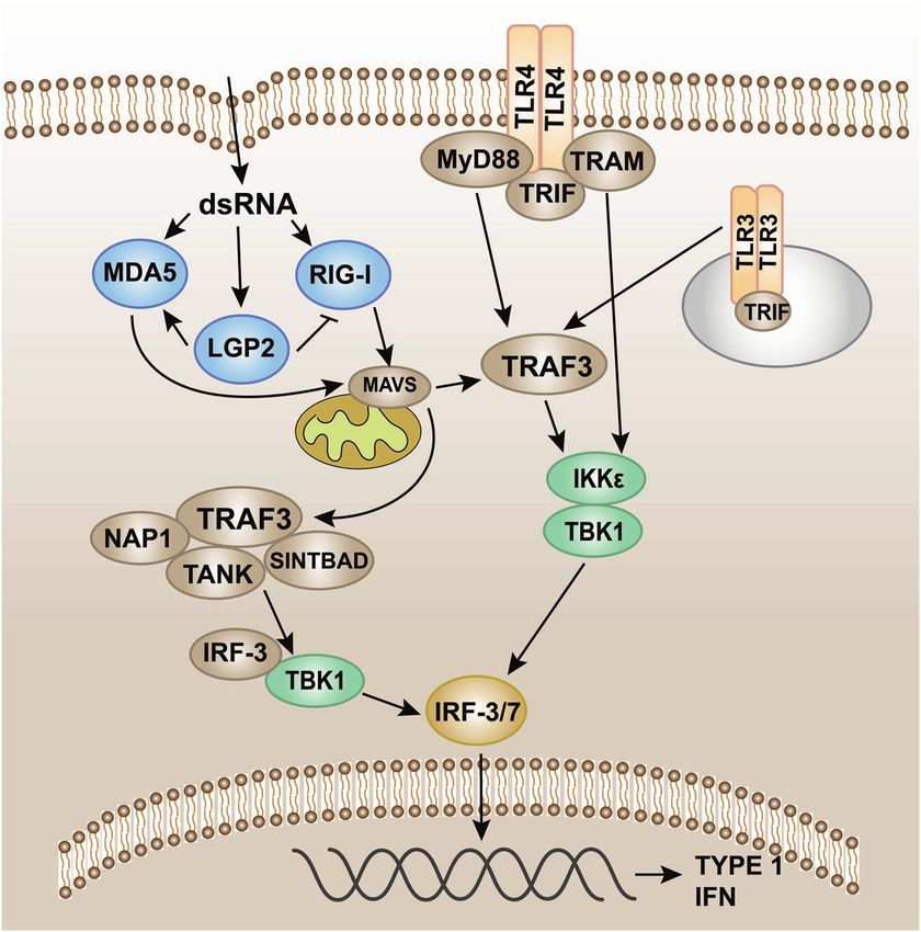
Pattern recognition receptors in health and diseases
                                                                    Li and Wu
2
     Table 1.   Common PRRs in innate immunity

    Items                 PRR             Domains                         Cellular     PAMP                       Sources             Signaling pathways
                                                                          distribution

    Toll-like             TLR1            LRR                             Mo, DC,        Triacyl lipopeptide      Bacteria            Most TLRs: MyD88-
    receptors (TLRs)      (TLR1–TLR2)     domain–transmembrane            Ma, Eo, Ba                                                  dependent pathways;
                          TLR2            domain–TIR domain               Mo, DC,        Lipoteichoic acid        Bacteria            TLR3: TRIF-dependent
                          (TLR1–TLR2,     (extracellular to               Ma, Eo, Ba                                                  pathways; TLR4: MyD88-
                                          intracellular)                                 Arabinomannan            Mycobacterium       dependent pathways and
                          TLR2–TLR6)
                                                                                         Peptidoglycan            Bacteria            TRIF-dependent pathways
                                                                                         Zymosan                  Fungi
                                                                                         Lipoprotein              Mycoplasma
                                                                                         Pore protein             Neisseria
                          TLR3                                            Mφ, DC, IEC dsRNA                       Virus
                          TLR4 (MD-2/                                     Mφ,         Lipopolysaccharides         Bacteria
                          CD14)                                           DC, Ma, Eo Heat-shock proteins          Host
                          TLR5                                            IEC            Flagellin                Bacteria
                          TLR6                                            Mo, DC,        Lipoteichoic acid        Bacteria
                          (TLR2–TLR6)                                     Ma, Eo, Ba     Peptidoglycan            Bacteria
                          TLR7                                            pDC,           ssRNA                    Virus
                                                                          Mφ, Eo         Imidazoquinoline         Artificially
                                                                                                                  synthesized
                          TLR8                                            Mφ, N          ssRNA                    Virus
                          TLR9                                            pDC, Eo, Ba Non-methylated              Bacteria, Virus
                                                                                      CpG DNA
                          TLR10                                           pDC, Eo, Ba dsRNA                       Virus
                          (human)
                          TLR11                                           Mφ, DC         Profilin and related      Toxoplasma
                          (mouse)                                                        proteins                 gondii
                          TLR12                                           DC             Profilin and related      Toxoplasma
                          (mouse)                                                        proteins                 gondii
                          TLR13                                           Unknown        23s ribosomal RNA        Bacteria
                          (mouse)
    Nucleotide-           NOD1            LRR domain–NBD–effector         IEC, cytosol   iE-DAP                   Gram negative       RIP2-TAK1-NF-κB pathways
    binding                               domains                         of Mφ                                   bacteria
    oligomerization       NOD2                                                           MDP                      Gram-negative
    domain-like                                                                                                   bacteria, Gram-
    receptors (NLRs)                                                                                              positive bacteria
    RIG-I-like            RIG-I           (RD)-CTD-DexD/H helicase        Cytosol        5’-triphosphorylated     Virus               MAVS-TRAF6-NF-κB/TBK1
    receptors (RLRs)                      domain–CARD                                    RNA, short-                                  pathways
                                                                                         chain dsRNA
                          MDA5                                                           poly IC, long-           Virus
                                                                                         chain dsRNA
                          LGP2                                                           dsRNA                    Virus
    C-type lectin         Dectin-1        CTLD–ITAM                       DC, Mφ         β-Glucan                 Fungus              Tyrosine kinase-dependent
    receptors (CLRs)      Dectin-2                                                       α-Mannan                 Fungus              and non-tyrosine kinase-
                                                                                                                                      dependent pathways
    Absent in             ALRs            HIN-200-PYD                     Cytosol        dsDNA                    Bacteria            Inflammasome–pyroptosis
    melanoma-2-like
    receptors (ALRs)
     LRR leucine-rich repeat, TIR Toll/IL-1R domain, NBD nucleotide-binding domain, RD repressor domain, CTD C-terminal domain, CARD caspase activation and
     recruitment domain, CTLD C-type lectin-like domains, ITAM immunoreceptor tyrosine-based activation motif, PYD pyrin domain, Mo monocyte, DC dendritic
     cell, Ma mastocyte, Eo eosinophils, Ba basophils, pDC plasmacytoid dendritic cell, IEC intestinal epithelial cell, N neutrophil, dsRNA double-stranded RNA, ssRNA
     single-stranded RNA, iE-DAP γ-D-glu-meso-diaminopimelic acid, MDP muramyl dipeptide, MyD88 myeloid differentiation factor 88, TRIF TIR domain-containing
     adaptor protein-inducing interferon β, RIP2 receptor-interacting serine–threonine protein 2, TAK1 transforming growth factor-β-activated kinase 1, NF-κB
     nuclear factor κB, MAVS mitochondrial antiviral signaling protein, TRAF6 tumor necrosis factor receptor-associated factor, TBK1 TANK-binding kinase 1

    that innate immunity recognizes pathogenic microorganisms and                        oligomerization domain (NOD)-like receptors (NLRs), retinoic acid-
    activates the expression of the second signal, which is indis-                       inducible gene-I (RIG-I)-like receptors (RLRs), C-type lectin recep-
    pensable for the activation of adaptive immunity.20 Since the                        tors (CLRs), and absent in melanoma-2 (AIM2)-like receptors (ALRs)
    discovery of TLR4, many PRRs and their corresponding ligands                         (Table 1).21 PRRs are representative of immune receptors in innate
    have been discovered. PRRs can be divided into the following five                     immunity and exist in various forms. PRRs are not only expressed
    types based on protein domain homology: TLRs, nucleotide                             on the cell membrane but also widely distributed in intracellular

                                                                                           Signal Transduction and Targeted Therapy (2021)6:291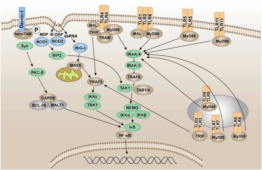
Pattern recognition receptors in health and diseases
                                                                         Li and Wu
                                                                                                                                                  3

Fig. 1 The signal transduction pathways and structure of TLR-binding ligand complex. TLRs can recognize one or more PAMPs through LRR
domain. They usually dimerize themselves and recruit adaptor molecules with the same TIR domain to transmit signals

compartment membranes and the cytoplasm.22 Membrane-                     PRRS AND LIGAND-RECOGNITION MECHANISMS
bound PRRs and PRRs in the cytoplasm are basically composed              Toll-like receptors
of ligand recognition domains, intermediate domains, and effector        TLRs are membrane-bound signal receptors and are important
domains.23,24 PRRs activate downstream signaling pathways                PRRs in the innate immune system of vertebrates.15,33 Such
through recognition of their ligands. The activation of down-            receptor molecules usually have two functions, one is to bind
stream signaling pathways can produce many effects: recruiting           specifically to the ligand, and the other is to transmit signals. The
and releasing cytokines, chemokines, hormones, and growth                corresponding signal transduction will amplify the effect of anti-
factors; inducing chronic inflammation; forming an inflammatory            pathogen infection, so that the immune cells active in the
microenvironment; initiating innate immune killing and subse-            inflammatory response can be activated through the transcription
quent acquired immune response,9 maintaining the balance of              of genes, and produce and secrete a variety of pro-inflammatory
host microecology; and eliminating dead or mutated cells.                and antiviral factors.34–36 Up to now, 10 functional TLRs (TLR1–10)
   PAMPs are the specific and highly conserved molecular                  have been found in humans and 12 (TLR1–9 and TLR11–13) in
structures shared by the same kind of pathogenic microorgan-             mice.37–41 TLR10 in mice is not functional due to the insertion of
isms,25,26 including lipids, proteins, and nucleic acids, such as        reverse transcriptase.42 TLRs recognize PAMPs in different
lipopolysaccharides (LPS), lipoteichoic acid (LTA), and bacterial        subcellular structures. The cellular localization of TLRs determines
DNA.27,28 PAMPs are essential for pathogen survival and usually          the types of ligands and the recognition mechanism. Some TLRs
have unique molecular or subcellular characteristics that are not        (TLR1, 2, 4, 5, 6, 10) are expressed on the surface of immune cells
found in host cells. Therefore, innate immune cells can recognize        in the form of heterodimers or homodimers, mainly recognizing
PAMPs via PRRs, distinguish “self” and “non-self,”29 and respond to      the membrane components of pathogenic microorganisms, such
pathogens and their products. However, the host will produce             as lipids, lipoproteins, and proteins; others (TLR3, 7, 8, 9) are
some proteins and metabolites after being stimulated by its own          expressed in the form of homodimers, which mainly recognize the
tissue damage, cell necrosis, and other factors.30 These molecules       nucleic acids of microorganisms (Fig. 1).43
are called damage-associated molecular pattern (DAMP).31 PRRs               TLRs are type I transmembrane glycoproteins and are com-
can also recognize such molecules, activate natural immunity, and        posed of an extracellular region, a transmembrane region, and an
cause inflammation.32                                                     intracellular region.44 The extracellular region contains leucine-rich
   With advances in research on PRRs structure and distribution at       repeats (LRRs), which are responsible for the recognition of
different levels, the role of PRRs in the innate immune regulatory       specific ligands and perform extracellular pattern recognition. The
network has become clearer. The recognition and binding of PRRs          intracellular domain contains the same Toll/IL-1R (TIR) domain as
to their ligands is critical in initiating the innate immune response.   IL-1R, which plays a role in signal transduction. The extracellular
Therefore, the study of PRR-mediated pattern recognition                 region of TLRs contains LRRs, which mediate the pattern
mechanisms will help to elucidate the signaling pathways and             recognition of TLRs (Fig. 1).45 In 2007, researchers used X-ray
mechanisms of disease and provide new targets and methods for            crystal diffraction to analyze and determine the structure of the
the treatment of diseases. In this review, we describe the structural    TLR–ligand complex,46 which provided a deeper understanding of
characteristics, ligand recognition mechanism, the signaling             the LRR domain. The LRR domain is shaped like a horseshoe, and
pathway, the related disease, new drugs in clinical trials, and          each module consists of a conserved leucine motif and a variable
clinical therapy of different types of PRRs in detail. We focus on the   region. The “LxxLxLxxN (L leucine, x any amino acid, N
different domains and ligand recognition mechanisms between              asparagine)” motif is composed of 20–30 amino acids and is on
PRRs, which can not only provide new ideas for the definition, role,      the concave surface of the horseshoe-like structure.47–50 The
and clinical application of PRRs but also promote the study of the       horseshoe-shaped N-terminus and C-terminus contain disulfide
role of the innate immune system in related diseases and even            bridges formed by cysteine clusters51,52 to protect the hydro-
tumors.                                                                  phobic core. After TLRs recognize and bind the corresponding

Signal Transduction and Targeted Therapy (2021)6:291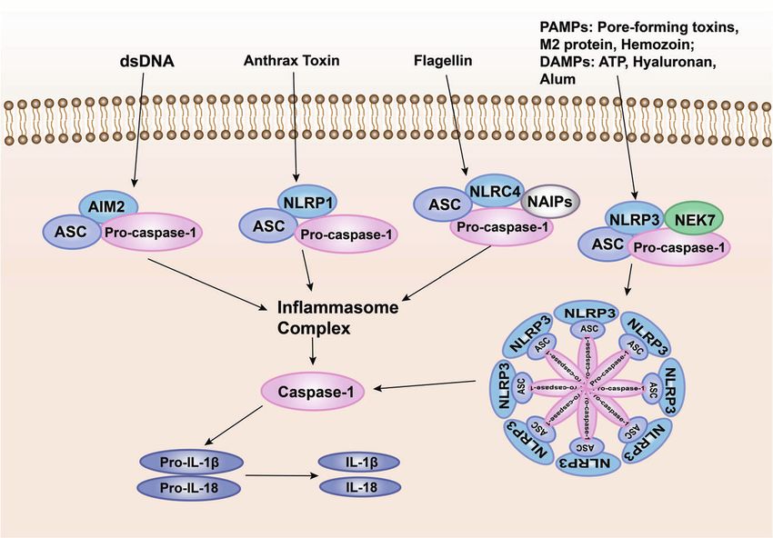
Pattern recognition receptors in health and diseases
Li and Wu
4
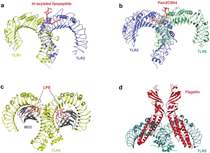
Fig. 2 Crystal structure of TLRs with ligands. a Crystal structure of the TLR1–TLR2 heterodimer induced by binding of a tri-acylated
lipopeptide (PDB 2Z7X). TLR2 initiates immune responses by recognizing di-acylated and tri-acylated lipopeptides. The ligand specificity of
TLR2 is controlled by whether it heterodimerizes with TLR1 or TLR6. Binding of the tri-acylated lipopeptide (red) induced the formation of M-
type crystal structures of the TLR1 (pale yellow) and TLR2 (slate) ectodomains. b Crystal structure of TLR2–TLR6–Pam2CSK4 complex (PDB
3A79). Binding of the di-acylated lipopeptide, Pam2CSK4 (red), induced the formation of M-type crystal structures of the TLR2 (slate) and TLR6
(pale green) ectodomains. c Crystal structure of mouse TLR4/MD2/LPS complex (PDB 3VQ2). After LPS (red) binds with the TLR4 (yellow)/MD2
(gray) complex, the hydrophobic pocket of MD2 is used to bridge the two TLR4–MD2–LPS complexes to form a spatially symmetrical M-type
structure. Mouse TLR4/MD2/LPS exhibited an complex similar to the human TLR4/MD2/LPS complex. d Crystal structure of the N-terminal
fragment of zebrafish TLR5 in complex with Salmonella flagellin (PDB 3V47). Two TLR5 (cyan)–flagellin (firebrick) 1:1 heterodimers assemble
into a 2:2 tail-to-tail signaling complex to function
PAMPs and endogenous ligands, the TIR domains conduct signals module (Fig. 2a, b).63 Researches on the structure of ligand
by binding to different receptor adaptor proteins in the complexes can significantly promote the discovery of small
cytoplasmic region.53,54 The TIR domain has three conserved molecule agonists/antagonists targeting PRRs. A recent study
amino acid sequences, which are called 1,2,3 cassettes. Depending revealed the activation mechanism of atypical agonists for
on the different adaptor proteins, TLRs signaling can be divided TLR1–TLR2. Diprovocim is a recently found small molecule
into myeloid differentiation factor 88 (MyD88)-dependent and activator for TLR1–TLR2, but it has no structural similarity with
MyD88-independent pathways (Fig. 1).55,56 the tri-acylated lipopeptide complex. It also interacts with
Exploring the pattern recognition mechanisms of TLRs is very TLR1–TLR2 in the same binding pocket as typical lipopeptide
valuable for understanding innate immunity and some tumor- ligand.64 Crystal structure analysis revealed that double-stranded
igenesis mechanisms. Therefore, researchers used X-ray crystal RNA (dsRNA) binds to the LRR domains of the N-terminus and C-
diffraction to determine the crystallographic structure of the terminus of TLR3.65,66 Different from the way that other TLRs
extracellular domain of TLRs and the ligand complex. Although directly recognize ligands,67–69 TLR4 specifically recognizes LPS in
the ligand complexes have different structures, all these combination with two auxiliary molecules, myeloid differentiation
complexes have similar M-type crystal structures (Fig. 2).50,51 factor 2 (MD2) and the LRR structural protein CD14. LPS is
TLR1 or TLR6 can form TLR1/TLR2 and TLR6/TLR2 heterodimers transported by LPS-binding protein to CD14 on the cell membrane
with TLR2 to recognize tri-acylated lipopeptide and di-acylated of monocytes and macrophages to form a complex and then
lipopeptide,57 respectively. After recognizing the appropriate interacts with TLR4/MD2.70 After LPS binds with the TLR4/MD2
ligands, TLR2 can form an M-type structure with the extracellular complex, the hydrophobic pocket of MD2 is used to bridge the
region of TLR1 and TLR6, and the pocket structure formed binds to two TLR4–MD2–LPS complexes to form a spatially symmetrical M-
the ligand.58–61 The crystal structure of TLR1–TLR2–tri-acylated type TLR4–MD2–LPS dimer,71 and then conformational changes
lipopeptide complex is similar to that of TLR2–TLR6–di-acylated affect their respective functional domains and transmit signals
lipopeptide complex, but there are important structural differ- (Fig. 2c). In addition to binding to LPS, TLR4 is also involved in the
ences between TLR1 and TLR6 in the ligand-binding site and recognition of natural products (carnosic acid, paclitaxel) and
dimerization surface. The ligand-binding pocket of TLR1–TLR2 is pneumolysin.72–74 TLR5 is the most conserved and important PRR,
located in the interface between the central and C-terminal which is usually stimulated by bacterial flagellin. In the form of
domain, and TLR1–TLR2–tri-acylated lipopeptide is stabilized by homodimer, TLR5 plays a major role in the primary defense of
non-covalent bonds such as hydrogen bonds, hydrophobic invasive pathogens and immune homeostasis regulation.75
interactions, and ionic interactions near the ligand-binding Although the heterodimeric structure of TLR5a–TLR5b in zebrafish
pocket.62 In TLR6, the side chains of amino acid residues block and the crystallographic structure of TLR5–flagellin complex has
the ligand-binding pocket, resulting in a pocket less than half the been clearly reported, the lack of biochemical and structural
length of TLR1. In addition, the TLR2–TLR6 heterodimer is mainly information of fish TLR hinders the understanding of flagellin-
regulated by the surface exposed residues of the LRR11–14 based therapies. In the future, experimental TLR5–flagellin
Signal Transduction and Targeted Therapy (2021)6:291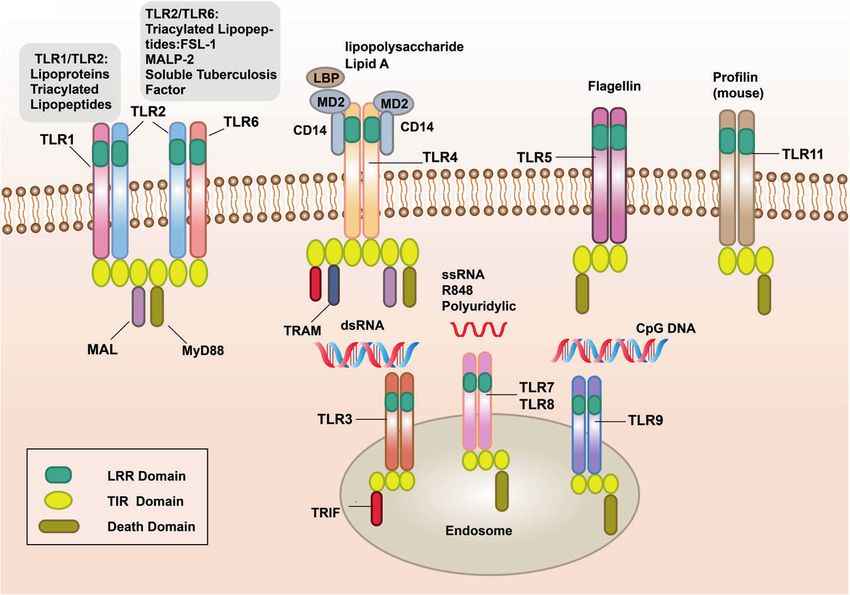
Pattern recognition receptors in health and diseases
                                                                       Li and Wu
                                                                                                                                               5
                                                                       but it must be a complete viral ssRNA.92 The basic process of
                                                                       NOD2 activation and signal transduction is as follows: after
                                                                       pathogenic bacteria are phagocytosed by macrophages, they first
                                                                       form phagosomes, and then fuse with lysosomes to become
                                                                       phagolysosomes. Under the action of lysosomal enzymes,
                                                                       bacterial cell wall components are decomposed into peptidogly-
                                                                       can, which can be degraded into a cell wall peptide with
                                                                       immunomodulatory activity and enter the cytosol, thereby
                                                                       activating NOD2.93 In general, the LRR domain of the NLR
                                                                       molecule folds to form a U-shaped configuration with the central
                                                                       NACHT domain, which inhibits its multimerization and makes the
                                                                       NLRs inactive.94 Once PAMPs directly or indirectly bind to
                                                                       the LRRs, the NLR molecule change their conformation, exposing
                                                                       the NACHT oligomerization domain, which triggers oligomeriza-
                                                                       tion, and the NLR molecule is activated.95 At the same time, the N-
Fig. 3 The ligand recognition mechanism of NLRs. The combination       terminal effector domain is exposed, and through homotypic
of PAMP and LRR changes the conformation of NLRs from self-
inhibition to activation
                                                                       interactions, downstream adaptor molecules and signaling
                                                                       proteins with the same structure are recruited to initiate the
                                                                       corresponding signal transduction (Fig. 3).96 Although NOD1 and
complex structure modeling and computational simulation should
                                                                       NOD2 do not have transmembrane domains, studies have shown
be used to study flagellin-mediated interactions between various
                                                                       that they are recruited into the plasma membrane and endosomal
pathogens and host immune receptors (Fig. 2d).76 It has been
                                                                       membrane, which is necessary for signal transduction.97 In this
reported that TLR1–6 each exist as monomers in solution, and
                                                                       process, palmitoylation plays a vital role. The modification of
dimerization occurs only when the ligand is bound; in contrast,
                                                                       NOD1/2 protein under the action of palmitoyltransferase ZDHHC5,
TLR8 and TLR9 exist as preformed dimers, and the binding of
                                                                       which makes NOD1/2, possess the characteristics of rapid and
ligands induces conformational changes in preformed dimers (Fig.
                                                                       reversible localization changes, which is necessary for membrane
1).49,77,78 Lee revealed that TLR10 binds dsRNA in vitro at
                                                                       recruitment and inflammatory signal transduction.98 This study
endosomal pH, indicating that dsRNA is a ligand of TLR10. The
                                                                       gives us a good enlightenment that the modification of PRRs may
recognition of dsRNA by TLR10 recruits MyD88, thereby transdu-
                                                                       play a key role in the regulation of host innate immune signal.
cing signals and inhibiting interferon regulatory factor 7 (IRF7)-
dependent type I interferon (IFN) production.79 In mice, TLR11 and
                                                                       RIG-I-like receptors
TLR12 are the main effector molecules to recognize Toxoplasma
                                                                       RLRs are also intracellular PRRs. In innate antiviral immunity, in
gondii. The recognition of T. gondii profilin by TLR11 depends on
                                                                       addition to the recognition of viral nucleic acids by TLR7 and TLR9,
the parasite-specific, surface-exposed motif in TgPRF consisting of
                                                                       most other types of cells recognize viral nucleic acids through
an acidic loop and a β-hairpin.80–82
                                                                       RLRs to induce antiviral immune responses.99,100 The currently
                                                                       discovered RLR family members mainly include three: RIG-I,
NOD-like receptors
                                                                       melanoma differentiation-associated gene 5 (MDA5), and labora-
The growth cycle of some pathogenic microorganisms involves
                                                                       tory of genetics and physiology 2 (LGP2) (Fig. 4).101
infection of the cytoplasm. For example, viral genes are often
                                                                          RIG-I was first discovered in acute promyelocytic leukemia cells
transcribed and translated in the cytoplasm, and virus particles are
                                                                       induced by retinoic acid. In 2004, it was found that RIG-I could
assembled. In addition, some bacteria and parasites have a series
                                                                       induce the expression of a reporter gene in the IFN-β promoter
of escape mechanisms, such as making holes in the phagosome
                                                                       region, which confirmed its antiviral activity.102 The structure of
membrane and entering the cytoplasm. Therefore, pathogens and
                                                                       the RIG-I protein consists of three parts.103–105 The middle part is
their components, as well as other components produced by
                                                                       the DexD/H helicase domain, which is the common domain of the
infection and injury, will appear in the cytoplasm,83 which requires
                                                                       RLR family, and has ATPase and helicase activities.106–108 The N-
the recognition of PRRs in the body. NLRs are intracellular PRRs,
                                                                       terminus of the RIG-I protein is composed of two caspase
composed of three domains:84,85 one is the central nucleotide-
                                                                       activation and recruitment domains in series,109 which are
binding domain (NBD), also known as the NACHT domain
                                                                       responsible for transmitting signals downstream.110 The C-
(synthesized by the abbreviations of the following four kinds of
                                                                       terminus is composed of the repressor domain (RD) and the C-
NLR members: NAIP, CIITA, HETE, TP1), which is shared by the NLR
                                                                       terminal domain (CTD), which can regulate its own state.106,111 The
family and is very important for nucleic acid binding and
                                                                       former can inhibit the activation of the receptor, and the latter is
oligomerization of NLRs (Fig. 3); LRRs at the C-terminus, which
                                                                       responsible for the recognition of viral RNA.112,113 In the resting
are used to identify ligands; and the N-terminal effector domain,
                                                                       state, CARD, CTD, and the helicase domain are folded, and RIG-I is
which is the protein interaction domain, such as the caspase
                                                                       in a self-inhibited state. During viral infection, the CTD of RIG-I
activation and recruitment domain (CARD) or the pyrin domain
                                                                       recognizes viral RNA and undergoes a conformational change.114
(PYD).86–89 According to the different N-terminal effector domains,
                                                                       RIG-I uses ATP hydrolase activity to expose and activate the CARD
the NLRs family can be divided into five subfamilies: the NLRC
                                                                       and multimerize, thereby recruiting downstream signaling linker
subfamily, which contain CARDs; the NLRP subfamily, which
                                                                       molecules (Fig. 4).115–117
contain PYDs; the NLRB subfamily, which contain baculovirus
                                                                          The structure and functions of MDA5 are similar to those of RIG-I,
inhibitor of apoptosis protein repeats; the NLRA subfamily, which
                                                                       with the DexD/H helicase domain in the middle, two CARD at the
contain acidic activation domains; and the NLRX subfamily
                                                                       N-terminus, and a CTD at the C-terminus; however, MDA5 lacks the
containing other NLR effector domains.85
                                                                       RD, and so it does not have self-inhibitory functions. In contrast to
   Among the NLRs family, the most in-depth study has focused
                                                                       other RLRs, LGP2 does not have CARD,118,119 and so it cannot
on NOD1 and NOD2 proteins. NOD1 mainly recognizes the
                                                                       recruit molecules of the same structure to transmit signals, but it
diaminopimelic acid (γ-D-glu-meso-diaminopimelic acid (iE-DAP))
                                                                       can regulate the recognition of viral nucleic acids by RIG-I and
of the cell wall of Gram-negative bacteria.90,91 In addition to
                                                                       MDA5, thereby preventing RLR-mediated resistance.120–123 LGP2
recognizing muramyl dipeptide (MDP) in all bacterial cell walls,
                                                                       can negatively regulate RIG-I-mediated recognition of viral dsRNA,
NOD2 can also recognize single-stranded RNA (ssRNA) of the virus,
                                                                       reduce the production of IFNs and inflammatory factors, and

Signal Transduction and Targeted Therapy (2021)6:291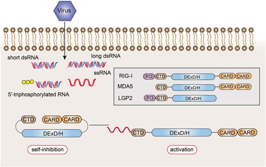
Pattern recognition receptors in health and diseases
                                                                 Li and Wu
6

    Fig. 4 Structural features and ligand recognition mechanism of RLRs. The structure and functions of MDA5 are similar to those of RIG-I.
    However, MDA5 lacks the repressor domain, so it does not have self-inhibitory functions. LGP2 does not have CARD, and so it cannot transmit
    signals. The combination of viral RNA and CTD changes the conformation of RLRs

    ultimately inhibit the antiviral innate immune response.124 LGP2 is       C-type lectin receptors
    also critical in the antiviral response mediated by MDA5.125 LGP2         CLRs, which belong to phagocytic PRRs, are also a popular type of
    exhibits a concentration-dependent conversion between MDA5-               receptor under study.143 The function of phagocytic receptor is
    specific enhancement and interference.126 The latest research              different from the receptor that activates cells by signal
    revealed a mechanistic basis for LGP2-mediated regulation of              transduction. It recognizes and binds to PAMPs through PRRs
    MDA5 antiviral innate immune responses. LGP2 facilitates MDA5             and places pathogens in cytoplasmic vesicles for direct digestion
    fiber assembly and is incorporated into the fibers, forming hetero-         and elimination to control infection.144 CLRs are a class of
    oligomers with MDA5.127 In addition, LGP2 can significantly induce         receptors that recognize carbohydrates on the surface of
    the exposure of the CARD domain of MDA5.128 Under bacterial               pathogenic microorganisms with the participation of Ca+.145 It is
    infection of the Indian major carp Labeo rohita, LGP2 gene                expressed on macrophages, dendritic cells (DCs), and certain
    expression was significantly increased after dsRNA and various             tissue cells. The ability of CLRs to recognize carbohydrates existing
    PAMPs were stimulated, indicating that LGP2 can act as an antiviral       on self and non-self structures is mediated by carbohydrate
    and antibacterial cytoplasmic receptor.129                                recognition domain (CRD).146 The CRD of CLRs is a compact
       Although the RLR family members have similar structures, they          spherical structure, and this region is called C-type lectin-like
    recognize the RNA of different viruses through ligand-recognition         domain (CTLD).147,148 Depending on the location of the protein on
    domains.130 Both RIG-I and MDA5 can recognize viral dsRNA, but            the cell membrane, CLRs are divided into transmembrane
    their recognition depends on the length of the dsRNA.131 RIG-I            receptors and secretory receptors.146,149,150 The main representa-
    mainly recognizes viruses with relatively short dsRNA (1000 bp).132             “Extracellular pattern recognition molecules”).151 Transmembrane
    Additionally, RIG-I mediates the antiviral response by recognizing        receptors can be divided into type I and type II according to their
    the 5’-triphosphate RNA of viruses.133 The 5’-terminal triphosphate       topological structure.152,153 The N-terminal of type I receptors
    group can be recognized by RIG-I as a non-self component, but             points to extracellular and contains multiple CRDs, while the N-
    after posttranslational modification, this molecule cannot be              terminal of type II receptors points to intracellular and contains
    recognized by RIG-I.134 Because host cell RNA needs to undergo            only one CRD.154,155 It has been shown that the vast majority of
    different degrees of processing and modification after synthesis in        CLRs are involved in the presentation of antigens as active
    the nucleus, these results indicate that RIG-I can distinguish viral      membrane-associated receptors, and CLRs are mainly expressed
    dsRNA from endogenous RNA. In the cell, RIG-I mainly recognizes           on antigen-presenting cells such as DCs and macrophages.145
    influenza virus,135 vesicular stomatitis virus,136 Sendai virus, and       CLRs are circular structures connected by two disulfide bonds.156
    Japanese encephalitis virus,137,138 while MDA5 mainly recognizes          CLRs contain at least one CTLD outside the cell, while the
    small RNA viruses, such as poliovirus.139,140 MDA5 also participates      intracellular domain is different.
    in the synthesis of the dsRNA analog polycytidylic acid (poly I:C).          Mannose receptors (MRs) belong to membrane CLRs, which are
    Previous studies have shown that filamentous fibers are formed              single-chain transmembrane molecules.157–159 The extracellular
    during the recognition of ligands by RIG-I and MDA5, and                  segment of MR consists of two parts: one is the proximal membrane
    signaling pathways are initiated from the tail and inside of the          end with eight consecutive CTLDs, which is responsible for the
    viral dsRNA, respectively.141                                             endocytosis and transport of the ligand; the other is the distal
       Although it is mentioned in the “Toll-like receptors” section that     membrane end of the cysteine-rich lectin domain, which recognizes
    TLR3, TLR7, TLR8, and TLR9 specifically recognize virus-derived            sulfation of carbohydrate conjugates.160 The endogenous ligands of
    nucleic acid molecules and bacterial nuclear components, they             MR are lysosomal hydrolase and myeloperoxidase, as well as the
    mainly appear in the endosomal membrane. RLRs can not only be             mannan-rich structure expressed by pathogens.161,162
    expressed in cells infected by various viruses but also can directly         Dendritic cell-associated C-type lectin (Dectin)-1 and Dectin-2
    recognize and perceive the virus products and virus particles that        are typical representatives of the CLR family.163,164 Dectin-1 is a
    exist in the cytosol. Its antiviral significance cannot be ignored.142     type II transmembrane protein expressed in DCs, macrophages,

                                                                                Signal Transduction and Targeted Therapy (2021)6:291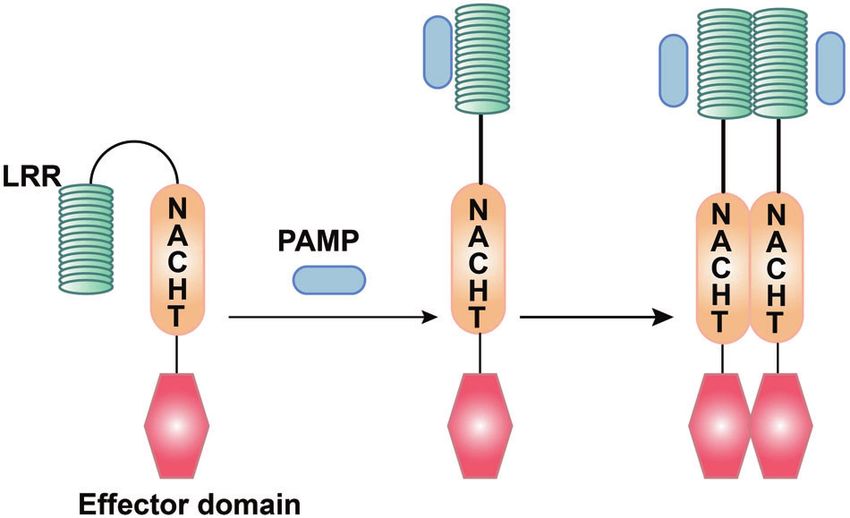
Pattern recognition receptors in health and diseases
                                                                          Li and Wu
                                                                                                                                                  7
neutrophils, and monocytes.165 The extracellular region is a CTLD.        collectin, and ficolin.203 They generally function in two ways:
The intracellular tail is connected to an immunoreceptor tyrosine-        one is that they recognize various pathogenic factors and
based activation motif (ITAM),166 indicating that the receptor also       eliminate them through complement activation,204,205 opsoniza-
has a signal transduction function. Dectin-1 can identify a variety       tion,206 aggregation, and neutralization of inflammatory regula-
of fungi,167 including yeast,168 Candida albicans,169,170 Pneumo-         tion; the other is that they interact with cell-related PRRs and
cystis carinii,171,172 Cryptococcus,173,174 and Aspergillus.175,176 The   regulate their functions to jointly regulate innate immune
ligand of Dectin-1 is β-1,3-glucan, which can activate downstream         response.207
signals through tyrosine kinase-dependent and tyrosine kinase-               Pentraxin is characterized by the aggregation of five molecules
independent pathways after recognition and binding of the                 and is highly conserved in evolution, including two families of
ligand.177,178 Glycosylation is an important modification of the           short molecules and long molecules.208–211 The family of short
posttranslational modification of proteins (including antibo-              molecules is called acute phase proteins, which is represented by
dies),179 which can significantly change the structure and function        C-reactive protein (CRP)212–214 and serum amyloid P compo-
of proteins or antibodies, so it is also a key mechanism for the          nent215,216 in humans and mice, respectively. These molecules are
immune system to regulate biological activity.180 Abnormal                mainly produced by the liver under the stimulation of inflamma-
glycosylation is usually associated with malignant tumors.179             tory signals and interleukins. They are non-specific proteins that
Therefore, the identification of molecules that bind glycosylated          reflect the systemic inflammatory response. Serum levels increase
glycans can provide a new way for the treatment of human                  rapidly after the body is infected or injured. CRP generally binds to
infectious and malignant diseases. Studies have found that Dectin-        phosphocholine expressed on the surface of pathogenic micro-
1 can recognize aromatic amino acids adjacent to the N-terminal           organisms in a Ca+-dependent manner.217 SAA can bind to the
asparagine at the glycosylation site as well as the core fucose on        outer membrane protein A of bacteria and interact with
IgG antibodies, which do not compete for the same protein                 TLRs.218,219 In clinic, SAA and CRP are usually used as auxiliary
binding site for β-glucan, so Dectin-1 can regulate the immune            diagnostic indicators for infectious diseases, but studies have
response induced by IgG by combining with core fucose.181                 shown that they also have diagnostic value in non-infectious
   Dectin-2, which is different from Dectin-1, does not contain the       diseases and can be used as disease classification markers.220,221
ITAM sequence and has no signal transduction function.182 Dectin-         The representative of the pentraxin long molecule family is
2 mainly recognizes α-mannan in the fungal cell wall and                  PTX3,222 which is unique in that it has a long N-terminal domain.
recognizes the Schistosoma mansoni egg antigen.183,184 The                PTX3 is produced by dendritic cells, monocyte macrophages,
molecular mechanism by which Dectin-2 recognizes the binding              epithelial cells, smooth muscle cells (SMCs), and endothelial cells
ligand has always been the focus of research. Decout et al.185            under the regulation of a variety of inflammatory factors.223 PTX3
found that the stimulation of Dectin-2 by purified Mycobacterium           is involved in the defense of selected pathogens and the
tuberculosis mannose-capped lipoarabinomannan requires the                regulation of inflammation.224–226 Due to its expression increases
(α1 → 2)-linked mannosides forming the cap. Besides, Dectin-2             sharply under the conditions of inflammatory stimulation, PTX3
can also recognize lipoglycans from other bacterial species.185,186       can become a biomarker of general acute inflammation and a
From the perspective of the relationship between the structure            variety of tumors.227 In coronavirus disease 2019 (COVID-19)
and function of the above two ligands, dimannoside caps and               patients, circulating and lung bone marrow monocytes and
multivalent interaction are necessary for Dectin-2 to recognize           endothelial cells express high levels of PTX3, and PTX3 plasma
binding ligands and conduct signals.187                                   concentration can serve as an independent strong prognostic
                                                                          indicator of short-term mortality in COVID-19.228,229
AIM2-like receptors                                                          Collectin mainly includes mannose-binding lectin (MBL) and
ALRs are a new type of PRRs that can recognize intracellular              surfactant protein (SP).151,230 MBL is formed by connecting
DNA.188,189 The C-terminus is the DNA-binding domain HIN-200,             multiple homotrimers. Each component of the trimer includes a
and the N-terminus is the PYD.189–192 The HIN-200 domain                  CRD, an alpha helix, and a main stem formed by spirals of
recognizes double-stranded DNA and binds to it. The N-terminal            collagen.231,232 The main stem of collagen gathers each trimer into
PYD binds to the PYD of apoptosis-associated speck-like protein           bundles. MBL is composed of six CRDs.151 The end of CRD can
containing CARD (ASC),193,194 thereby promoting the formation of          identify the sugar structure on the surface of various pathogens,
inflammasomes and the maturation and release of IL-1β and IL-              such as mannose, fucose, glucose, etc.233–235 The pathogens
18.195 Both the DNA-binding affinity of AIM2 and the activity of its       involved include yeast, parasites, Gram bacteria, and so on.236–240
inflammasome depend on dsDNA, and it can assemble into                     When the distance of each CRD between the same trimer or
filamentous structures along dsDNA. However, without dsDNA, it             adjacent trimers is 45 Å, it is most conducive to ligand binding.241
can also form filaments at high protein concentrations.196–198 ALRs        The other family members include A and D,242 which exist on the
can not only participate in the innate immune response but also           surface of the alveoli and are important innate immune defense
regulate apoptosis, which is related to the occurrence and                molecules in the lungs. Both of them are composed of N-terminal
development of tumors.199                                                 region, CRD, neck region, collagen-like region, and other parts.243
                                                                          CRD recognizes and binds glycosyl groups. The biological
Extracellular soluble pattern recognition molecules                       significance is that they can selectively identify microbial
The initiation of the innate immune response depends on the               carbohydrate structures that are harmful to themselves.244,245
recognition of PAMPs by pattern recognition molecules (PRMs),                The domain of ficolin is similar to collectin, but it recognizes a
including cell PRRs and extracellular soluble PRMs. They are a class      variety of bacteria with a fibrinogen-type carbohydrate recogni-
of free receptors that can play an antibacterial effect in serum.200      tion structure.246,247 Its ligands are N-acetylglucosamine and LTA, a
Although the pattern recognition of innate immunity does not              cell wall component of Gram-positive bacteria.248,249
have the antigen specificity of the adaptive immune response,
some PRMs produced by the body after infection by pathogenic
microorganisms will exist in the serum. Once the new pathogens            SIGNALING PATHWAYS OF PRRS
invade, they can also bind to the pathogen like an antibacterial          There are three main types of molecules involved in signal
molecule and play an effective function. Unlike cell-related PRRs,        transduction: protein kinases, adaptor proteins, and transcription
extracellular soluble PRMs are an important part of non-specific           factors. Although PRRs are activated by their respective ligands in
humoral immunity.201 Extracellular soluble PRMs are composed of           different subcellular structures with different mechanisms, the
different molecular families, mainly including pentraxin,202              three main types of molecules involved in signal transduction

Signal Transduction and Targeted Therapy (2021)6:291
Pattern recognition receptors in health and diseases
Li and Wu
8
Fig. 5 Pattern recognition receptor-mediated NF-κB signaling. The NF-κB protein can regulate gene expression and affect various biological
processes, including innate and adaptive immunity, inflammation, stress response, B cell development, and lymphoid organ formation. TLRs,
NLRs, RLRs, and CLRs can generally phosphorylate IκB protein, which inhibits the activation of NF-κB protein, thereby promoting the
transcription and activation of inflammatory genes
have similar structures and functions, and the signals they receptors are activated, self-dimerize, and recruit downstream
transmit are cross-talking, which can converge into several receptor-interacting serine–threonine protein 2 (RIP2) through its
common signaling pathways. CARD.268 Activated RIP2 gathers downstream TAK1, TAK1-binding
protein 1, and the NF-κB essential modulator/IKKα/IKKβ complex,
The NF-κB signaling and the former activates IKKα/IKKβ,269 thereby activating the
The transcription factor NF-κB is named after it was first transcription of NF-κB and promoting the release of pro-
discovered to be involved in the transcription of B cell κ chain inflammatory factors.
genes.250 NF-κB is a heterodimer composed of two molecules, p50 When virus invades cells, RIG-I and MDA5 recognize the
and p65, and is inactive due to binding to the inhibitory protein corresponding viral RNA through the CTD and undergo con-
IκB under normal conditions. NF-κB plays a key role in the process formation changes.270 Activated RIG-I and MDA-5 induce down-
of cellular inflammation and immune response,251,252 and its stream signal transduction by binding with mitochondrial antiviral
mediated signal pathways are commonly seen in the activation of signaling protein (MAVS). MAVS is an important adaptor protein
various immune cells, including signal transduction initiated by for downstream signal transduction. The N-terminus contains a
PRRs in innate immunity (Fig. 5).253 CARD-like domain, which binds to RIG-I and MDA-5 through the
In the signal transduction initiated by TLRs,45 after TLRs CARD–CARD interaction.271,272 The proline-enriched domain in
recognize and bind the corresponding PAMPs and DAMPs, the MAVS can interact with a series of downstream signaling
TIR domains conduct signals by binding to different receptor molecules, such as TRAF3 and 6,273 and activate the protein
adaptor proteins in the cytoplasmic region.254,255 Depending on kinase IKK, which causes phosphorylation of IκB,265 and then IκB is
the different adaptor proteins, TLR signaling can be divided into ubiquitinated and degraded by proteases, activating the NF-κB
MyD88-dependent and MyD88-independent pathways.256 MyD88 pathway.274
has a TIR domain at the C-terminus and a death domain at the N- Different from other typical PRR-mediated signaling pathways,
terminus and is the linker molecule in most TLR signal transduction spleen tyrosine kinase (Syk) can be activated by associating with
pathways.257 The current research indicated that, in the MyD88- the phosphorylated ITAM motif of CLRs.275 In the Dectin-1/Syk
dependent pathway, MyD88 signaling mainly leads to the pathway, Syk activates protein kinase C-δ, which mediates the
production of pro-inflammatory cytokines, such as tumor necrosis phosphorylation of CARD9.276 This allows CARD9 to bind to B cell
factor (TNF), IL-6, IL-1, and chemokines.258–260 The C-terminus of lymphoma 10277 and para-aspase mucosa-associated lymphoid
MyD88 binds to the intracellular TIR domain of TLRs, and the N- tissue lymphoma translocation protein 1, forming a three
terminus of MyD88 recruits IL-1R-related kinase 4 (IRAK4)261 and molecular structure that can typically activate NF-κB.278
activates IRAK1 and IRAK2 through autophosphorylation of its
central kinase domain. Then ubiquitin ligase TNF receptor- The mitogen-activated protein kinase (MAPK) signaling
associated factor 6 (TRAF6) is recruited to form a complex with MAPK is a group of serine–threonine protein kinases that can be
transforming growth factor (TGF)-β-activated kinase 1 (TAK1) and activated by different extracellular stimuli,279 such as cytokines,
two TAK-binding proteins (TAB1 and TAB4). TRAF6 is degraded due neurotransmitters, hormones, cell stress, and cell adhesion. The
to its own ubiquitination.262,263 The TAK1–TAB1–TAB4 complex MAPK pathway is one of the common intersections of signal
activates the IκB kinase (IKK) complex through phosphorylation. transduction pathways, such as cell proliferation, stress, inflamma-
The latter phosphorylates IκB and degrades itself by ubiquitination. tion, differentiation, functional synchronization, transformation,
NF-κB is released and translocated to the nucleus, thereby and apoptosis.280,281 It is an important transmitter of signals from
regulating the transcription of inflammatory genes.264,265 the cell surface to the inside of the nucleus.
In the signal pathway mediated by NLRs, when the bacterial In the MyD88-dependent pathway of TLRs, IRAK-1 is activated
component invades the cell, NOD1 and NOD2 recognize the by phosphorylation and interacts with TRAF6. In addition to
bacterial iE-DAP and MDP, respectively.266,267 And then NOD-like activating the IKK complex, it can also cause the activation of
Signal Transduction and Targeted Therapy (2021)6:291Pattern recognition receptors in health and diseases
Li and Wu
9
Fig. 6 Pattern recognition receptor-mediated TBK1-IRF-3 signaling. Intracellular induction of pathogens is carried out through the detection
of foreign molecular components (including cytoplasmic viral and bacterial nucleic acids). Once detected, the innate immune system induces
type I interferon (IFN) production through the TANK-binding kinase 1 (TBK1)-interferon regulatory factor-3/7 (IRF-3/7) pathway. IRF-3/7 can be
activated through two innate immune antiviral signal pathways, TLR3/TLR4-TIR domain-containing adaptor protein-inducing interferon β
(TRIF) and RIG-I-MAVS, and then dimerize and merge into the nucleus to work
MAPKs (c-Jun N-terminal kinase (JNK), p38 MAPK).282 In addition, IRF-3 phosphorylation and subsequent dimerization induce IRF-
when bacterial components invade cells, NLRs are activated, 3 nuclear translocation, leading to type I IFN gene expression
recruiting downstream CARD9, thereby activating p38, JNK, and (Fig. 6).192,293,294
finally activating the MAPK pathway283 to promote the release of
pro-inflammatory factors. The inflammasome signaling
Inflammasome is the multi-protein complex assembled by PRRs in
The TBK1–IRF-3 signaling the cytoplasm and is an important part of the innate immune
IRF-3 is a key transcription factor that promotes the synthesis of system.295 The inflammasome can recognize PAMPs or DAMPs
type I IFN and plays an important role in the antiviral innate and recruit and activate Caspase-1. The activated Caspase-1
immune response.284 IRF-3 can be activated through two innate spliced proIL-1β/proIL-18 into the corresponding mature cyto-
immune antiviral signal pathways, TLR3/TLR4-TIR domain- kine.193,296 There are five main types of inflammasomes that have
containing adaptor protein-inducing interferon β (TRIF) and RIG- been discovered, namely, NLRP1 inflammasome,297 NLRP3 inflam-
I-MAVS,285 and then dimerize and merge into the nucleus to work masome,298 NLRC4 inflammasome,299,300 IPAF inflammasome, and
(Fig. 6).286 AIM2 inflammasome.301 Known inflammasomes generally contain
The adaptor protein in the MyD88-independent pathway is TRIF. ASC, caspase protease, and a protein of the NLR family (e.g.,
The TRIF axis mainly induces the expression of type I IFNs.287 After NLRP3) or HIN-200 family protein (e.g., AIM2). Taking NLRP3 as an
the receptor is recognized and combined with the ligand, the example,302 the dimerization of NLRP3 under the action of
pathway is activated by TRIF and TRAF3, leading to the intracellular PAMPs or DAMPs makes the two PYDs to polymerize.
recruitment of IKKε/TANK-binding kinase 1 (TBK1),288 phosphor- With the help of homotype interaction, NLRP3 binds and activates
ylation of IRF3, and the activation of type I IFN genes, which the ASC complex with both PYD and CARD domains, which
promotes the expression of IFN-α and IFN-β, and exerts antiviral reactivates the effector complex composed of CARD and caspase-
effects (Fig. 6).289–291 1. In this way, NLRP3 (LRR + NACHT + PYD), ASC (PYD + CARD),
RLRs such as RIG-I and MDA5 can detect viral nucleic acid. and the effector complex (CARD + Caspase-1) together constitute
MDA5 and RIG-I will interact with the shared caspase recruit- the inflammasome, which produces important pro-inflammatory
ment domain to induce MAVS to dimerize and bind to factors.303–305 After AIM2 recognizes cytoplasmic dsDNA, it also
TRAF3.134,140,292 In turn, TRAF3 recruits the adaptor proteins uses inflammasomes to produce IL-1β and IL-18. After AIM2
TANK, NAP1, and SINTBAD. TANK connects upstream RLR signal recognizes cytoplasmic dsDNA, it also produces IL-1β and IL-18
transduction to TBK1, which induces phosphorylation of IRF-3. through the inflammasome pathway (Fig. 7).306
Signal Transduction and Targeted Therapy (2021)6:291Pattern recognition receptors in health and diseases
Li and Wu
10
Fig. 7 Pattern recognition receptor-mediated inflammasome signaling. One way for pathogenic microorganisms to induce inflammation is by
activating inflammasomes, which are multi-protein complexes assembled by PRRs in the cytoplasm and activate caspase-1 and subsequent
activation of pro-inflammatory cytokines IL-1β and IL-18. The inflammasome complex usually contains cytoplasmic PRRs, adaptor protein
(ASC), and pro-caspase-1. Many different inflammasome complexes have been detected, each with unique PRRs and activation triggers
Innate immunity not only plays a role in controlling the We can see from the data in GEPIA316 (http://gepia.cancer-pku.
infection and spread of pathogens in the early stage of infection cn/) that TLR level is significantly increased in tumors, including
but also plays an important role in initiating and regulating glioblastoma multiforme, brain lower-grade glioma, kidney renal
adaptive immunity.12,307 Innate immune cells produce different clear cell carcinoma, acute myeloid leukemia (AML), and
types of cytokines through signal transduction initiated after PRRs pancreatic adenocarcinoma (PAAD). NOD1/2 are highly expressed
recognize PAMPs, which directly affect the differentiation of T in AML and PAAD, while RLRs are highly expressed in AML, PAAD,
helper type 1 (Th1), Th2, Th17, and other subgroups in adaptive diffuse large B cell lymphoma, head and neck squamous cell
immunity.15 For example, pathogenic microorganisms activate carcinoma, and thymoma.
macrophages to secrete IL-6, TGF-β, IL-23, and other cytokines,
which promote Th17 response, leading to excessive immune- Colorectal cancer (CRC). CRC, including colon cancer and rectal
inflammatory effects and tissue damage, or activate NK cells to cancer, is one of the most common gastrointestinal malignancies
secrete IFN-γ, and then activate macrophages to secrete IL-12, in clinical practice, and it is also one of the cancers that seriously
promote the differentiation of Th0 into Th1, promote cellular endanger human health.317,318 Intestinal mucosal epithelial cells
immune response, and effectively eliminate viral infections.13,308 and immune cells recognize intestinal microorganisms and their
Therefore, the immune system is a system of mutual influence. products through TLRs.319–322 TLR2 can recognize peptidoglycans
Any anti-infection process is completed by mutually activating or and lipopeptides that infect intestinal epithelial bacteria and
inhibiting of different components. These components are as produce anti-infection and other immune-protective effects.323
small as each cytokine and as large as the immune system. Studies have shown that the expression level of TLR2 protein in
colon cancer is significantly upregulated compared with normal
epithelial tissues,324 and the use of TLR2 agonists significantly
PRR-RELATED DISEASES enhances the proliferation, migration, and invasion capabilities of
PRRs and cancers colon cancer cells.325 TLR4 is highly expressed on the surface of
The inflammatory microenvironment of tumor constitutes the colon cancer cells. After stimulation and activation, it can induce a
barrier for tumor growth, which is conducive to tumor formation variety of immunosuppressive factors, thus promoting the
and development.309 PRRs are widely expressed in a variety of proliferation and immune escape of colon cancer cells.326 First,
tumor tissues, such as colon cancer, lung cancer, breast cancer, TLR4 can produce trophic factors and vascular growth factors
gastric cancer, melanoma, and so on.310,311 The activation of through the TLR4/MyD88/NF-κB signaling pathway, thereby
PRRs on the surface of tumor cells can induce the expression of promoting tumor cell invasion.327,328 Second, TLR4 can promote
a large number of cytokines, chemokines, hormones, and tumor proliferation through TLR4/Cyclooxygenase 2 (COX2)/
vascular-promoting factors, which is one of the important prostaglandin E2 (PGE2). PGE2 is an important cell growth and
factors to induce the formation of tumor inflammatory micro- regulatory factor. After binding to specific receptors, it plays a key
environment and promote the development of tumor.312,313 At role in mediating a series of cell activities, such as cell proliferation,
the same time, the activation of PRRs on immune cells can differentiation, and apoptosis, and has immunosuppressive and
induce antigen-presenting cells including DCs, tumor-associated anti-inflammatory effects. COX2 is the rate-limiting enzyme of
macrophages, and B cells to activate tumor-specific T cell prostaglandin synthesis, and it is also highly expressed in
responses or enhance the antitumor effects of phagocytes. inflammation, tumor, and other pathological states.329 Hsu
These also indicate that the role of PRRs in immunotherapy et al.330 found that knocking out the mouse TLR4 gene
against tumors is very important and might represent a new significantly reduced the expression of COX2 and PGE2 in the
strategy for patients with tumors.314,315 intestinal mucosa; after administration of PGE2, the expression of
Signal Transduction and Targeted Therapy (2021)6:291Pattern recognition receptors in health and diseases
Li and Wu
11
COX2 in the intestinal mucosa increased significantly and of HCC remains uncertain. Song et al.347 found that the deficiency
promoted the occurrence of intestinal tumors. After administra- of TLR4, TLR9, and their downstream molecule MyD88 in a mouse
tion of PGE2, the expression of COX2 in the intestinal mucosa model characterized by hepatic deletion of TAK1 could block the
increased significantly and promoted the occurrence of intestinal liver inflammation–fibrosis–cancer axis and reduce liver injury and
tumors. At the same time, it has also been found to promote the tumor growth. For TLR3, the downregulation of TLR3 in HCC
expression of amphiregulin and epidermal growth factor receptor patients leads to poor prognosis (e.g., defective immune cell
(EGFR) in the intestinal mucosa. Finally, the study showed that the recruitment and lack of killing of transformed hepatocytes), leading
abnormal expression of TLR4 in CRC caused by chronic inflamma- to protection of transformed hepatocytes from apoptosis, thereby
tion of the intestine can significantly enhance the expression of promoting the occurrence of liver cancer.348 Therefore, the
PGE2, the upregulation of COX2, and the phosphorylation of EGFR expression of TLR3 may become a useful clinical treatment
in intestinal mucosal cells, thereby positive feedback promotes the monitoring marker.
proliferation of tumor cells. TLR5 also plays an important role in In addition to the above reasons, there is now more and more
tumor immunotherapy.331 In mouse xenograft models of human evidence that the imbalance of the gut–hepatic axis may also play
colon cancer, flagellin around the tumor activates TLR5 to inhibit a role in the occurrence of HCC.349 Zhou et al.350 discovered that
tumor growth and promote tumor apoptosis.332 In addition, NOD2 acts as a bacterial sensor, linking gut-derived microorgan-
TLR9 is expressed on the surface of the mesentery, which isms to the occurrence of HCC through a known mechanism and a
maintains intestinal homeostasis and repairs intestinal damage newly discovered mechanism. The known mechanism is that
by generating an immune response.333 TLR9 relies on the MyD88 NOD2 activates NF-κB, JAK2/STAT3, and MAPK pathways in a RIP2-
pathway to induce downstream signals to recruit many inflam- dependent manner, leading to liver inflammation.351 It is worth
matory factors, such as IL-8, TGF-β, PGE2, and other immunosup- noting that activated NOD2 can also act as the initiator of the
pressive molecules,334 leading to the continuous development of nuclear autophagy pathway that does not depend on RIP2, thereby
inflammation, resulting in immune escape, and promoting the promoting the degradation of the nuclear component lamin A/C,
unlimited proliferation of tumor cells. After TLR9 recognizes the leading to damage to DNA damage repair mechanisms and
exogenous ligand, it upregulates the expression of NF-κB signaling increased genomic instability, which eventually leads to the
factor. Once this pathway is opened, it may induce the secretion occurrence of HCC.350 Meanwhile, the study found that the
of matrix metalloproteinase-13 (MMP-13) and the activation of expression of ALRs was negatively correlated with tumor volume,
intercellular adhesion molecule-1, thus promoting the metastasis stage, and metastasis of HCC patients. The researchers proposed
of tumor cells.335,336 At the same time, the metastatic tumor cells that overexpression of ALRs in HCC cells could increase the
are better adapted and combined with the cell matrix at the expression of caspase-1 and IL-1a, and the release of lactate
metastasis, and the stability of tumor cell metastasis is enhanced. dehydrogenase was also observed,352 which was a marker of the
Although TLRs have been shown to enhance colon cancer initiation of apoptosis. Thus, ALRs may play an antitumor role by
metastasis, inhibiting these receptors cannot completely hinder promoting tumor cell apoptosis.
tumor progression. Surprisingly, NOD1 is highly expressed in
human CRC and its cell lines. After being activated by C12-iE-DAP, Breast cancer. Breast cancer is one of the most common
it mainly enhances the adhesion, migration, and metastasis of CRC malignant tumors in the female population. It has a strong ability
cells through the p38 MAPK pathway.337 to invade and metastasize.353 It can metastasize to the liver, lung,
brain, bone, and other organs, forming complications and
Hepatocellular carcinoma (HCC). HCC is the most common type of increasing the difficulty of treatment. Studies have shown that
primary liver cancer. Among its many influencing factors, the promotion of the TLR2 signaling pathway on the metastasis
inflammation is one of the main reasons that induce liver and invasion ability of human breast cancer cells is achieved by
cancer.338 The expression of TLR2 in liver cancer tissues is upregulating the secretion of inflammatory cytokines.354 LTA, a
significantly higher than that in normal liver tissues, and the TLR2 specific ligand, can significantly promote the secretion of
expression of TLR2 protein is related to some mutant genes that tumor metastasis-related factors IL-6, TGF-β, and vascular
lead to the occurrence of HCC, such as p53, PIK3CA, and endothelial growth factor (VEGF) in breast cancer cells,355 thereby
β-catenin.339–342 In addition, Chew et al. revealed that the promoting the proliferation and metastatic invasion of breast
expression of TLR3 has independent effects on tumor parenchyma cancer cells, and this promotion is related to the level of TLR2
and infiltrating NK cells, and the expression of these two parts is expression.356,357 In addition, the activation of TLR4 can increase
related to inhibiting tumor cell proliferation, promoting tumor cell the secretion of IL-6 and IL-10 of cancer cells and induce the
death, and prolonging the survival rate of patients.343 This production of more MMP-2, MMP-9, and VEGF,358,359 which can
indicates that TLR3 may directly act on tumor parenchymal cells, significantly enhance the invasion ability of breast cancer. It has
promote the recruitment and activation of NK cells, and exert been reported that activation of TLR4 on metastatic breast cancer
antitumor effects. More and more evidences show that LPS plays a cells can regulate the expression of integrin, which can promote
role in the development of HCC. Zhou et al.344 found that LPS its adhesion and invasion.360
activates the TLR4–AKT–SOX2 signaling pathway of liver cancer cell
lines to improve the ability of cancer stem cells; Lin et al.345 found Head and neck squamous cell carcinoma. Squamous cell carci-
that there is a positive feedback loop of COX-2/PGE2/signal noma, also known as epidermal carcinoma, is a malignant tumor
transducer and activator of transcription factor 3 (STAT3) activated occurring in the epidermis or adnexal cells.361,362 It is more
by LPS in liver cancer cells, which regulates the expression of genes common in the parts covered by squamous epithelium, such as
related to tumor proliferation, differentiation, and apoptosis. In the skin, mouth, lip, esophagus, cervix, vagina, etc.363,364 TLR2, TLR4,
latest research on the treatment of liver cancer, it is found that the and TLR9 are expressed in primary tumors, neck metastases, and
antitumor effect of TLR9 agonist combined with anti-PD-1 antibody recurrent tumors of oral tongue squamous cell carcinoma
or anti-PD-L1 is significantly better than single-agent therapy.346 (OTSCC), and their expression varies from the tumor surface to
The activation of TLR9 inherent in liver cancer cells regulates the the invasive front, which may be one of the important factors to
autoarylation and ubiquitination of poly(ADP-ribose) polymerase-1 promote the invasion of OTSCC.365,366 NOD1 and NOD2 genes are
and the phosphorylation of STAT3, which together upregulate the expressed in the human oral squamous cell carcinoma (OSCC) cell
expression of PD-L1 and eventually induce immune escape. line YD-10B, and they may trigger immune responses through the
Although TLRs have been reported to be associated with chronic MAPK pathway. Surprisingly, the study revealed that stimulation
inflammation of the liver, whether they promote the development by the NOD2 agonist MDP can inhibit cell growth by inducing
Signal Transduction and Targeted Therapy (2021)6:291You can also read