Metalloproteinases and Their Inhibitors: Potential for the Development of New Therapeutics - MDPI
←
→
Page content transcription
If your browser does not render page correctly, please read the page content below
cells
Review
Metalloproteinases and Their Inhibitors: Potential
for the Development of New Therapeutics
Maryam Raeeszadeh-Sarmazdeh * , Linh D. Do              and Brianne G. Hritz
 Chemical and Materials Engineering Department, University of Nevada, Reno, NV 89557, USA
 * Correspondence: maryamr@unr.edu; Tel.: +1-(775)-682-8494

 Received: 23 April 2020; Accepted: 19 May 2020; Published: 25 May 2020

 Abstract: The metalloproteinase (MP) family of zinc-dependent proteases, including matrix
 metalloproteinases (MMPs), a disintegrin and metalloproteases (ADAMs), and a disintegrin and
 metalloproteinase with thrombospondin motifs (ADAMTSs) plays a crucial role in the extracellular
 matrix (ECM) remodeling and degradation activities. A wide range of substrates of the MP family
 includes ECM components, chemokines, cell receptors, and growth factors. Metalloproteinases
 activities are tightly regulated by proteolytic activation and inhibition via their natural inhibitors,
 tissue inhibitors of metalloproteinases (TIMPs), and the imbalance of the activation and inhibition
 is responsible in progression or inhibition of several diseases, e.g., cancer, neurological disorders,
 and cardiovascular diseases. We provide an overview of the structure, function, and the multifaceted
 role of MMPs, ADAMs, and TIMPs in several diseases via their cellular functions such as proteolysis
 of other cell signaling factors, degradation and remodeling of the ECM, and other essential
 protease-independent interactions in the ECM. The significance of MP inhibitors targeting specific
 MMP or ADAMs with high selectivity is also discussed. Recent advances and techniques used in
 developing novel MP inhibitors and MP responsive drug delivery tools are also reviewed.

 Keywords: metalloproteinases; metzincins; matrix metalloproteinases; MMPs; a disintegrin
 and metalloproteases; ADAMs; tissue inhibitors of metalloproteinases; TIMPs; MMP inhibitors;
 MMP-responsive therapeutics

1. Introduction
     Metzincins consist of a large heterogeneous superfamily of zinc-dependent endopeptidases present
in the extracellular matrix (ECM). The metzincin family of metalloproteinase (MP) includes matrix
metalloproteinase (MMP), ADAM (a-disintegrin and metalloproteinase), and ADAMTS (a-disintegrin
and metalloproteinase with thrombospondin motifs) [1]. The metalloproteinases (which we refer
to MMPs, ADAMs, and ADAMTSs) play a critical role in remodeling of the ECM by proteolytic
degradation of ECM components, activation of cell surface proteins, and shedding of membrane-bound
receptor molecules. They regulate activity of other proteinases, growth factors, chemokines, and cell
receptors, and mediate several biological activities such as cell migration, differentiation, proliferation,
and survival [2] in various forms of cellular function. There are 23 different members of MMPs, 21 of
ADAMs, and 19 of ADAMTSs known to date in humans [3].
     These proteases are classified based on various criteria, such as their substrate preferences,
mechanism of enzymatic reaction, soluble or transmembrane domains, and structural homology.
The major structural homology which was found in all proteins of this superfamily is highly conservative
motif HEXXHXXGXXH present within the active site of the enzyme [4]. The majority of differences
between zinc-dependent metalloproteases are associated with the occurrence of additional domains
within the C-terminus of these proteins [5] (Figure 1).

Cells 2020, 9, 1313; doi:10.3390/cells9051313                                     www.mdpi.com/journal/cells
Cells 2020, 9, 1313                                                                                     2 of 34
Cells 2020, 9, x FOR PEER REVIEW                                                                       2 of 38

        Figure
     Figure  1. 1.  Schematic
                Schematic      representation
                           representation       of matrix
                                            of matrix       metalloproteinases
                                                        metalloproteinases      (MMPs),
                                                                            (MMPs),       a-disintegrin
                                                                                       a-disintegrin andand
        metalloproteinases
     metalloproteinases    (ADAMs),
                        (ADAMs),  andand  a-disintegrin
                                       a-disintegrin andand  metalloproteinase
                                                          metalloproteinase withwith thrombospondin
                                                                                 thrombospondin       motifs
                                                                                                   motifs
     (ADAMTSs). The structural domains of different metalloproteinases (MPs) are displayed. GPI,GPI,
        (ADAMTSs).     The structural domains   of different metalloproteinases  (MPs)  are displayed.
        Glycosylphosphatidylinositol-anchoring sequence; EGF, epidermal growth factor-like domain.
     Glycosylphosphatidylinositol-anchoring sequence; EGF, epidermal growth factor-like domain.
          Metalloproteinase (MP) activity is tightly regulated by proteolytic activation of the zymogen form
      Metalloproteinase (MP) activity is tightly regulated by proteolytic activation of the zymogen
    and its natural inhibitor, tissue inhibitor of metalloproteinases (TIMPs). Under pathologic conditions,
form and its natural inhibitor, tissue inhibitor of metalloproteinases (TIMPs). Under pathologic
    overexpression of metalloproteinases or insufficient control of TIMPs results in the dysregulation of
conditions, overexpression of metalloproteinases or insufficient control of TIMPs results in the
    tissue remodeling, causing a variety of diseases such as cancer [6,7], neurodegenerative disease [8,9],
dysregulation of tissue remodeling, causing a variety of diseases such as cancer [6,7],
    arthritis, cardiovascular diseases [10,11], and fibrotic disorders [12,13]. Although early efforts of
neurodegenerative disease [8,9], arthritis, cardiovascular diseases [10,11], and fibrotic disorders
    targeting MMPs largely failed in later stages of clinical trials, metalloproteinases remain a highly
[12,13]. Although early efforts of targeting MMPs largely failed in later stages of clinical trials,
    desirable therapeutic target based on their key role in progression of several diseases [6].
metalloproteinases remain a highly desirable therapeutic target based on their key role in progression
          Different classes of MP inhibitors were developed and tested including small molecules,
of several diseases [6].
    peptides, and protein-based binders such as antibodies and TIMPs. With recent advances in protein
      Different classes of MP inhibitors were developed and tested including small molecules,
    engineering and design, from recruiting better understanding of the structure and function of these
peptides, and protein-based binders such as antibodies and TIMPs. With recent advances in protein
    metalloproteinases to state-of-the-art techniques such as directed evolution and high throughput
engineering and design, from recruiting better understanding of the structure and function of these
    screening, new classes of therapeutics targeting MMPs with high affinity and selectivity are on the
metalloproteinases to state-of-the-art techniques such as directed evolution and high throughput
    rise [6]. Design of “smart,” MMP-responsive therapeutics and drug delivery vehicles also enhanced
screening, new classes of therapeutics targeting MMPs with high affinity and selectivity are on the
    site-specific drug delivery to tumor sites, where MMPs are upregulated [14]. Among all MPs, the role
rise [6]. Design of “smart,” MMP-responsive therapeutics and drug delivery vehicles also enhanced
    of MMPs and their inhibitors were studied more extensively [15]; however, we also included the role
site-specific drug delivery to tumor sites, where MMPs are upregulated [14]. Among all MPs, the role
    of ADAMs and ADAMTSs in developing several diseases in this review. This review focuses on
of MMPs and their inhibitors were studied more extensively [15]; however, we also included the role
    therapeutic applications for metalloproteases as targets for inhibition and as tools for drug activation.
of ADAMs and ADAMTSs in developing several diseases in this review. This review focuses on
    It has the following sections:
therapeutic applications for metalloproteases as targets for inhibition and as tools for drug activation.
    • the
It has    MP  structuresections:
            following    and function in ECM
    •     MPs in cell signaling
•     MP structure and function in ECM
•   • MPs MPs   in cancer
            in cell signaling
•   •     MPs   in
      MPs in cancercentral nervous system and neurodegenerative diseases
•    MPs in central nervous system and neurodegenerative diseases
•    MPs in cardiovascular diseases
Cells 2020, 9, 1313                                                                                 3 of 34

•     MPs in cardiovascular diseases
•     MPs in fibrosis and other diseases
•     MMP inhibition for developing therapeutics
•     MMP-responsive drugs and drug delivery tools
•     Conclusion and future directions

2. MP Structure and Function in ECM
     The structure of MPs contains a propeptide sequence and a catalytic domain. MMP structure also
includes a hinge region and a hemopexin (PEX) domain [4,16]. Based on their structural domains, MMPs
have been classified into collagenase, gelatinase, stromelysin, matrilysin, and membrane-bound MMPs
(MT-MMPs) [6,17] (Figure 1). MT-MMPs contain a transmembrane or Glycosylphosphatidylinosotol
(GPI)-anchored domain at their C-terminus. MT-MMPs are anchored to the cell membrane via a
covalent bond. The secreted MMPs can localize to the cell surface by binding interactions to cell-surface
associated proteins such as CD44. Other binding interactions include heparan sulfate proteoglycans,
collagen type IV, or extracellular matrix metalloproteinase inducer (EMMPRIN) [18]. Both soluble and
MT-MMPs are essential for diverse physiological pathological processes that are involved with both
extracellular matrix remodeling and pericellular proteolysis [19].
     ADAMs are membrane-anchored metalloproteinases. They have similar catalytic domains to
MMPs; however, they do not have a PEX domain, and instead possess three additional epidermal
growth factor (EGF)-like domains along with the disinterring domain. ADAMTS family members
contain a variable number of type-1 thrombospondin (TSP-1) domains at the C-terminal region [3]
(Figure 1).
     The propeptide domain of MMPs is highly conserved, and it is the switch sequence that interacts
with Zn2+ . The cystine within this region is what allows the MMP to either be in the active or inactive
state [20]. The catalytic domain is characterized as being the zinc-binding motif which, in the active
state, will disassociate from the propeptide domain [16]. Hinge regions are mainly used to allow
movement between the catalytic and hemopexin domain. MMPs are synthesized as inactive zymogens.
In the zymogen (pro-MMP) form, the pro-domain interacts through the sulfhydryl group of the
cysteine with the catalytic zinc ion and inhibits MMP proteolytic activities. MMP activation requires
proteolytic removal of the pro-domain, usually by other MMPs or serine proteases outside the cell
with the exception of MMP-11 and MMP-28, as well as some of the MT-MMPs, which are activated by
intracellular furin-like serine proteinases before reaching the cell surface [6].
     MMPs with gelatinase activity hydrolyze gelatin into polypeptides, peptides, and amino acids
that can then be secreted through the cellular membrane. MMP-2 and MMP-9 belong to gelatinases
and facilitate both gelatin and collagen-binding through three fibronectin type-II-like repeat domains
inserted in the catalytic domain of the structure [21]. MMP-9 also contributes to wound healing
suppression, where overexpression of MMP-9 results in a leakage in the healing process attributing
to the continuous break down of collagen [22]. The family of collagenases, consisting of MMP-1,
MMP-8, and MMP-13, cleave collagen, resulting in breakdown of joint cartilage, which contributes
to inflammation [23]. The MMP members of the stromelysin family, including MMP-3 and MMP-10,
directly degrade non-collagen connective tissues, including but not limited to proteoglycans, fibronectin,
and laminin, while also interacting with other MMPs to perform other degradation mechanisms [24].
The matrilysin class of MMPs, including MMP-7, is found to directly cleave elastin, type II collagen,
fibronectin, vitronectin, aggrecan, and proteoglycan [25].
     Similar to MMPs, ADAMs carry proteolytic function by degrading ECM components and regulate
intermolecular interactions by interacting with cell receptors and cell signaling components. On the
other hand, ADAMTS proteases have a conserved role in the human organism as proteases, with some
differentiation in terms of substrate specificity [26].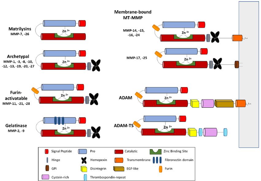
Cells 2020, 9, 1313                                                                                4 of 34

3. MMP Regulation in the ECM
      Metalloproteinase activity is tightly regulated by activation of inactive zymogens and natural
inhibition by endogenous inhibitors. Zymogen activation occurs by proteolytic cleavage of the
pro-domain protecting the catalytic site. Several MMPs participate in activation of other pro-MMPs,
usually leading to a cascade of activations. For example, MMP-3 and MMP-10 activate MMP-1, MMP-7,
MMP-8, and MMP-9, enhancing ECM degradation [21], whereas MMP-14 activates both MMP-2 and
MMP-13 in the presence of TIMP-2. MMP-2 and MMP-13 were shown to aid the cleavage of the pro
domain in pro-MMP-9 [24], which was shown to affect tumor invasion and metastasis by promoting
cell migration [22]. This complex network of MMP cross-activation is able to destruct the ECM if the
tight regulation of MMPs is compromised [26].
      Tissue inhibitors of metalloproteinases (TIMPs), a family of four members in humans,
are endogenous inhibitors of MPs and regulators of MMPs and ADAMs’s function, forming a
tight 1:1 stoichiometric inhibitory complex [27]. TIMPs consist of two domains that are packed
side-by-side, each domain is stabilized by three internal disulfide bonds [28,29]. The N-terminal
domain of TIMPs is known as the “inhibitory domain” since the isolated N-terminal domain of TIMPs
were found to independently inhibit MMP. The conserved core epitope centered around the N-terminal
strand, including the ‘Cys-X-Cys’ motif, is the major interaction site of TIMP with an MMP catalytic
domain, which coordinates to the active-site divalent catalytic zinc ion of the MMP, thus blocking
the substrate-binding cleft [30]. However, full-length TIMP-1 was shown to provide stronger MMP
inhibition [31]. The cooperation between two domains of TIMP-1 in improving inhibition of MMP-3 is
another indication for the importance of the TIMP-1 C-terminal domain in TIMP/MMP interactions
and inhibition of MMPs [32].
      TIMPs have broad, overlapping specificity in binding and inhibition of MMPs, ADAMs,
and ADAMTSs ranging from 0.6 fM for the association of TIMP-2 with MMP-2 [33], to the
high nanomolar range for the relatively poor inhibition of MMP-14 and MMP-15 by TIMP-1.
The non-inhibitory interactions can also occur between select TIMPs and MMPs. TIMP-1 and
TIMP-2 interact with the PEX domains of MMP-2 and MMP-9, resulting in some of the strongest
interaction of TIMP/MMP pairs [34–36]. The C-terminal domains of TIMP-2, TIMP-3, or TIMP-4 can
bind to the hemopexin domain of MMP-2 or pro-MMP-2, while the C-terminal domains of TIMP-1 or
TIMP-3 can associate with MMP-9 or pro-MMP-9 [37]. TIMP-2 uniquely interacts with pro-MMP-2 [38],
which is shown to be important in MMP-2 activation by MMP-14 [39]. The TIMP-1 C-terminal domain
is also shown to interact with pro-MMP-9 hemopexin domain and the protect the secreted proenzyme
from activation by other MMPs both in vitro and in vivo [30,36,40–43]. TIMPs also inhibit some of the
ADAM and ADAMTS family of MP through similar interactions with MMPs. Among all four TIMPs,
TIMP-3 was found to inhibit ADAM-17 and ADAM-12 [44,45], as well as ADAMTS-1, ADAMTS-4,
and ADAMTS-5 [46,47], both TIMP-1 and TIMP-3 could inhibit ADAM-10 [44].
      Dysregulation of MMPs can affect many physiological processes such as morphogenesis,
angiogenesis, tissue remodeling (e.g., cardiovascular remodeling), embryonic development, regulation
of cell growth and death, and wound healing [11,48]. In most typical adult tissues, MMPs remain at a
rather low expression level, and a balance between MMPs and Tissue Inhibitors of Metalloproteinases
(TIMPs) maintain the ECM equilibrium [37]. However, with inflammation and certain demand in
remodeling activities, MMP expression increases causing an imbalance of MMP and TIMP levels [48,49].
      MMP expression and activity is strictly controlled by several metabolic pathways and cell signaling
molecules. Tumor necrosis factor (TNF)-α was shown to upregulate the collagenase class of MMPs
and downregulate TIMP-1 expression [50]. The ERK 12 pathway is also known to impact transcription
of several MMPs and when the ERK 12 pathway is inhibited, MMP transcription is downregulated [51].
KISS1R, a G protein coupled receptor (GPCR), was identified to regulate the phosphorylation of
MT1-MMP and interacts with the ERK 12 pathway which is known to play a role in several stages of
cancer [52]. Hypoxia, lack of oxygen in lungs caused by Mycobacterium tuberculosis, was also shown
to upregulate the transcription of MMP-1 due to the lack of regulation that dimethyl oxalyl glycerin
Cells 2020, 9, 1313 5 of 34
has on the hypoxia-inducible factor (HIF)-1 [53]. Relaxin-2, a peptide hormone, has been attributed to
greater anterior cruciate ligament (ACL) injuries in females due to its role in upregulation of MMP
transcription in the MMP pathway and downregulation of the expression of TIMP-1 [24].
RD1 genes, encoding early secretory antigenic target (ESAT-6), were also found to be involved
in the upregulation of MMP-10, which interacts with MMP-1 causing higher collagenase activity
in patients diagnosed with tuberculosis [54]. Along with several signaling pathways that regulate
various MMPs, there are also various transcription factors such as nuclear factor kB (NF-kB) and
signal transducer and activator of transcription-3 (STAT-3), that transcribe various MMPs with a role
in the metastasis in cancer [55]. NF-kB contains regions of DNA-binding and dimerization domains,
nuclear translocation signal (NLS), and binding site for the inhibitor of kappa B (IkB). In spontaneously
hypertensive rate (SHR), higher levels of NF-kB were found, which are responsible for inducing MMP
expression [56]. It was also identified that NF-kB is a key factor in macrophage-derived MMP-1 and
MMP-3 secretion; however, if the NF-kB pathway is inhibited, the MMP expression is negatively
affected. This finding clarified the role of NF-kB inhibition through drug treatment, and it was also
identified that the NF-kB knockout leads to severe phenotypes or lethality [57]. It has also been
identified that when NF-kB affects MMP-9 transcription; MMP-9 transcription is improved by two-fold
when NF-kB is present [58]. Overall, although the inhibition of NF-kB is known to reduce transcription
of various MMPs, it usually did completely knockdown the MMPs expression.
EGFR, integrin-β1, integrin-α2, myosin regulatory light chain (MRLC), and YAP (yes-associated
protein) were also found to enhance MMP-7 expression through the generation of a positive
feedback loop. Upregulation of integrin-β1, integrin-α2, and EGFR phosphorylation led to MRLC
phosphorylation, causing YAP activation which in turn led to enhancement of MMP-7 expression [25].
Syndecan-2, a heparan sulfate proteoglycan, is located on the cell surface, attributes to cell adhesion,
and activates membrane localization of PKC-γ. Activating PKC-γ will then cause activation of
FAC/ERK signaling and therefore result in upregulation of MMP-7 [59].
4. MPs and Their Inhibitors in Cell Signaling
Matrix Metalloproteinases (MMPs) have been identified to contain a central role in various
types of cells. MMPs are well-known for degrading structural components of the ECM. However,
cleavage of ECM proteins may trigger cellular receptors for structural ECM components that affects cell
signaling and behavior [18]. MMPs and ADAMs also accept substrates other than ECM components
such as growth-factor receptors, cell-adhesion molecules, and chemokines. The cross-reactivity and
interactions of the metalloproteinase network as well as overlapping substrate recognition make it
difficult to understand the individual role of each MMP in the ECM.
Beyond the degradation of ECM components, several types of MMPs and ADAMs are known for
proteolysis of growth factors such as epidermal growth-factor (EGFR), HER2/neu (or ERBB2), HER4
(or ERBB4), and the hepatocyte-growth-factor receptor (HGFR) in the c-MET signaling pathway [49].
For instance, MMP-2 plays a role in cleavage of fibroblast growth factor receptor-1 (FGF). MMPs are
also known to target chemokines which affect infiltration and migration of leukocytes. MMP-9 is
known to cleave CXCL8 (also known as IL-8) as a substrate [49]. Thus, MPs are known in regulating of
inflammatory and immune responses.
MMPs are also known to interact with ADAMs which are capable of degrading aggrecan,
a cartilage-specific proteoglycan core protein [60]. MMPs and ADAMs are both involved with the
c-MET signaling pathway that involves the binding of HGF, causing tyrosine residues Y1234 and Y1235
to phosphorylate, which in turn allows them to recruit signaling effectors with important functions in
cell viability and motility [61]. ADAMs, such as ADAM-10 and ADAM-17, are also involved with
cell-cell communication through the Notch signaling pathway and help to determine cell fate during
development [62]. The ADAM cleavage site of Notch receptor in a protease-resistant state is protected
by a negative regulatory region (NRR). The ligand-bearing cells induce global conformational changes
in Notch that unfold the NRR structure and expose the ADAM cleavage site, initiating proteolyticCells 2020, 9, 1313 6 of 34
activation of the Notch receptor. Both ADAM10 and ADAM17 were found to accept Notch1 (N1) as a
substrate, however, the particular ADAM required for receptor activation is dependent on signaling
induced by ligands [63]. Overexpression of ADAM will lead to a higher abundance of Notch cleavages
which can result in the production of cancerous cells. MMPs and ADAMs interact with many other
substrates that can cause a pyramid effect in cellular signaling [63,64].
Tissue inhibitors of metalloproteinases (TIMPs) also hold an important role in cell signaling as
they inhibit MMPs and ADAMs which restrict the formation of cancer, inflammatory, and degenerative
diseases. TIMPs are necessary to maintain cellular hemostasis since many MMPs interact with varying
substrates, which necessitates the four groups of TIMPs for higher-affinity binding to the various
MMPs and ADAMs [27]. Non-selective inhibition of MMPs could also be detrimental to the cell
since MMPs are regulators for the ECM and the degradation of collagen is necessary for adequate
survival [65]. In retrospect, if the opposite occurs and TIMP is unable to inhibit MMPs efficiently,
or if there is overexpression of the mRNA of MMPs, the excessive degradation of the extracellular
membrane leads to inconsistent regulation and initiation and progression of cancer or various diseases.
Research recently has been leading towards either identifying chemicals or different pathways that
can inhibit MMPs without adding TIMPs, such as hispidulin which induces TIMP expression [66].
Overall, a perfect balance between the various MMPs and TIMPs are necessary for proper hemostasis
of every cell to limit the creation of various cancers and diseases. MMPs are also known to regulate
angiogenesis, a critical step in tumor growth. MMP-2, -9, and -14 directly regulate angiogenesis [67–69].
MMPs are also known to interact with ADAMs, which are capable of degrading aggrecan,
a cartilage-specific proteoglycan core protein [61]. MMPs and ADAMs are both involved with the
c-MET signaling pathway that involves the binding of HGF, causing tyrosine residues Y1234 and Y1235
to phosphorylate, which in turn allows them to recruit signaling effectors with important functions in
cell viability and motility [62]. ADAMs, such as ADAM-10 and ADAM-17, are also involved with
cell–cell communication through the Notch signaling pathway and help to determine cell fate during
development [63]. The ADAM cleavage site of Notch receptor in a protease-resistant state is protected
by a negative regulatory region (NRR). The ligand-bearing cells induce global conformational changes
in Notch that unfold the NRR structure and expose the ADAM cleavage site, initiating proteolytic
activation of the Notch receptor. Both ADAM10 and ADAM17 were found to accept Notch1 (N1) as a
substrate; however, the particular ADAM required for receptor activation is dependent on signaling
induced by ligands [64]. Overexpression of ADAM will lead to a higher abundance of Notch cleavages,
which can result in the production of cancerous cells. MMPs and ADAMs interact with many other
substrates that can cause a pyramid effect in cellular signaling [64,65].
Tissue inhibitors of metalloproteinases (TIMPs) also hold an important role in cell signaling as they
inhibit MMPs and ADAMs, which restrict the formation of cancer, inflammatory, and degenerative
diseases. TIMPs are necessary to maintain cellular hemostasis since many MMPs interact with varying
substrates, which necessitates the four groups of TIMPs for higher-affinity binding to the various
MMPs and ADAMs [27]. Non-selective inhibition of MMPs could also be detrimental to the cell
since MMPs are regulators for the ECM and the degradation of collagen is necessary for adequate
survival [66]. In retrospect, if the opposite occurs and TIMP is unable to inhibit MMPs efficiently,
or if there is overexpression of the mRNA of MMPs, the excessive degradation of the extracellular
membrane leads to inconsistent regulation and initiation and progression of cancer or various diseases.
Research recently has been leading towards either identifying chemicals or different pathways that
can inhibit MMPs without adding TIMPs, such as hispidulin which induces TIMP expression [67].
Overall, a perfect balance between the various MMPs and TIMPs are necessary for proper hemostasis
of every cell to limit the creation of various cancers and diseases. MMPs are also known to regulate
angiogenesis, a critical step in tumor growth. MMP-2, -9, and -14 directly regulate angiogenesis [68–70].Cells 2020, 9, 1313 7 of 34
5. MPs in Cancer
MPs have long been held responsible for cancer cell invasion and metastasis because of the great
impact of metalloproteinases have in remodeling the ECM of tumors. MPs and their proteolytic function
are responsible in several aspects of cancer such as cancer-cell growth, differentiation, apoptosis,
migration, invasion, and tumor angiogenesis [49]. They have significant roles in initial stages of cancer
progression via inducing genomic instability and DNA damage [70] as well as later stages of cancer
such as invasion and metastasis [7] by facilitating pathway clearance for invasion of cancer cells caused
by ECM degradation [6]. Epithelial-mesenchymal transition (EMT) is also a critical part of tumor
invasion and metastasis, where the epithelial cell features are lost in favor of adopting mesenchymal
traits, and it involves loss of the apicobasal cell polarity, through intracellular adhesion alteration [71].
MMPs can directly induce the EMT in target epithelial cells which leads to cancer progression [6].
The role of specific MMPs and ADAMs in breast [6,72,73], lung [74–77], prostate [78–80],
and colorecta [81] cancer progression, invasion, and metastasis has been studied extensively [18,49,82].
MPs carry a multifaceted role in cancer and they can act as a pro-tumorigenic, anti-tumorigenic, or null
factor [6], making it difficult to draw a general conclusion on the role of all MPs without considering
all the parameters involved in the model of study such as the individual genomic profile, proteomic
profile, and experimental conditions. MMP-9, MMP-14, ADAM-12 and ADAM-17 are some of the MPs
that were found to have a significant role in several types of cancer.
MMP-9 is the only MMP found in the 70 genes of the Rosetta poor prognosis for breast cancer [83],
and is strongly related to the prognosis and poor survival of breast cancer patients [6]. High levels
of MMP-9 in serum in breast cancer patients were also correlated with poor outcomes [16]. High
expression levels of MMP-9 are also associated with prostate and lung cancer [82]. MMP-9 was shown
to be upregulated in the basal-like triple negative and HER2 positive breast cancer cell lines, and MMP-9
knockdown was shown to block metastasis in both triple negative breast cancer cell models and an
orthotopic mouse model of human breast cancer [73]. These findings suggest that MMP-9 is a potential
therapeutic target for breast cancer as well as other type of cancer and MMP-9-driven diseases.
MMP-14 is known as one of the key drivers of cancer invasion and progression. MMP-14 was
originally found to contribute to cancer progression by activation of proMMP-2 and degradation of
ECM to facilitate cancer metastasis. However, MMP-14 activity was later shown to also have a negative
effect on immune responses to tumors [84]. MMP-14 also plays a significant role in angiogenesis [84].
Similar to MMP-9, the PEX domain of MMP-14 was shown to be responsible for facilitating cell
migration through the molecular cross-talk resulted from the homodimerization of MMP-14 and
heterodimerization of MMP-14 with the cell surface adhesion molecule CD44 [85]. Mutagenesis studies
in the PEX domain of MMP-14 which is a four-blade β-propeller, showed that blade IV is necessary
for MMP-14 homodimerization and that blade I is required for CD44 MMP-14 heterodimerization.
The interaction between MMP-14 and CD44 leads to activation of the MAPK and PI3K signaling
pathways through phosphorylation of EGFR which will further lead to cell migration [85,86].
Other MMPs, such as MMP-1 [87,88], MMP-7 [89], and MMP-11 [90] were also shown to contribute
to cancer progression and metastasis [59]. Ovarian cancer cells are found to be leptin-induced and
conduct invasion through the MAPK signaling pathway. MMP-7 was found to have a direct role in the
activation of the ERK 12 and JNK 12 signaling pathways, which attribute to leptin-induced cell migration
and therefore play a pivotal role in these cells [91].
Similar to different MMPs, members of the ADAM and ADAMTS families are known to play
a multifaceted role in various aspects of cancer. Many ADAMs are found to play a role in growth
factor and cell receptor shedding. For instance, ADAM-12, a multi-domain MP that regulates cell
proliferation and movement, is known for shedding of heparin-binding, and EGF-like growth factor
(HB-EGFR) to activate the EGFR signal pathway [92]. EGFR, one of the key factors in triple-negative
breast cancer (TNBC), is normally activated following the release of ligands, such as TGFα, mediated
by ADAM-10 and ADAM-17. ADAM-17, or TACE, is processing membrane-bound TNFa precursor to
its soluble form as well as other membrane proteins [93]. A humanized monoclonal antibody targetingCells 2020, 9, 1313 8 of 34
both the catalytic domain and the cysteine-rich domain of ADAM-17 was found to significantly inhibit
the release of TGFα and decrease downstream EGFR-dependent cell signaling, resulting in reduced
proliferation in two-dimensional clonogenic assays as well as growth in a three-dimensional culture
of breast cancer cells. Furthermore, the antibody provides a potential anti-cancer therapeutic based
on the inhibition of ADAM-17 which resulted in reduced invasion and migration of HCC1143 TNBC
mammary gland epithelial cells in vitro [94].
ADAM-12 was also found in high levels for both mRNA transcriptional and protein level in
tumor tissues, while ADAMTS-1 mRNA and protein levels were significantly lower in non-small-cell
lung cancer cells, suggesting distant roles of different metzincin family members [95]. ADAM-12
was also shown to be highly expressed in breast [92], liver [96], and lung cancer [92]. This might be
due to ADAM-12’s role in several cell signaling functions such as receptor shedding and binding
to activate the Notch signal pathway, the TGF-β signaling pathway, regulation of cell migration,
and promotion of angiogenesis [92]. ADAM-12 is upregulated in human breast cancers and is a
predictor of chemoresistance in estrogen receptor (ER)-negative tumors. ADAM-12 is induced during
the EMT transition, a feature associated with claudin-low breast tumors, which are enriched in cancer
stem cell (CSC) markers [92,97].
TIMPs are also involved in cell signaling through regulation of MPs and MMP-independent
activities, thus, contributing to different aspects of cancer [27]. TIMP-1 has been also found to
support pro-tumorigenic signaling by activating hypoxia-inducible factor (HIF-1), which in turn
results in upregulation of microRNA-210 (miR-210) in lung cancer progression [98]. It has also been
suggested that TIMP-1 interacts with CD63 to activate ERK 12 kinase which promotes accumulation of
cancer-associated fibroblast (CAF) in prostate and colon cancer [99].
TIMP-1 overexpression was shown to facilitate the EMT of hepatocellular carcinoma (HCC)
cells via MMP-independent activities such as modulating apoptosis, mitogenic activity, and cellular
proliferation and morphology [100]. TIMP-1 upregulation was also found as a prognostic marker for
lung metastasis in HCC, since TIMP transcripts were clearly demonstrated in the metastatic HCC
nodules in the lung. Similarly, TIMP-2 levels in serum and tissue were decreased in HCC patients
with metastasis compared to those without [101], and patients with high levels of TIMP-2 have were
shown higher survival rates [102,103]. On the other hand, TIMP-3 was shown to have a potential
role in alleviating invasion of HCC, likely through suppressing tumorigenesis and angiogenesis by
interacting with integrin α7 and angiotensin II type 2 receptor [103]. TIMP-3 was also shown to inhibit
angiogenesis via blocking the binding of vascular endothelial growth factor (VEGF) to its receptor,
vascular endothelial growth factor-2 (VEGFR-2) [104,105]. The C-terminal domain of TIMP-3 was
found to be responsible for the MMP-independent angiogenesis inhibitory function of TIMP-3 [104].
This VEGF inhibitory property is unique to TIMP-3, and TIMP-1 and TIMP-2 could not inhibit the
binding of VEGF to KDR in intact cells. This inhibition appeared to be specific for VEGF, because
signaling by PDGF and bFGF through their receptors was unaffected [106]. TIMP-3 downregulation is
related to cancer invasion and metastasis in various cancers; however, higher serum level of TIMP-1
is associated with lower response to therapy in breast cancer patients [107], and TIMP-1 has been
used as a biomarker along with MMP-2 and ICAM-1 for pancreatic cancer [108,109]. It needs to be
considered that higher levels of TIMP-1 expressed in tumor or stroma might be a result of MMP
expression in cancer patients [27]. More importantly, TIMP-1 is structurally distant from other TIMPs,
and its MMP-independent roles in cell signaling should not be overlooked.
6. MPs in Central Nervous System and Neurodegenerative Diseases
MPs play key physiological and pathological roles in the central nervous system by regulating
signaling pathways during neuroinflammation, blood-brain barrier (BBB) disruption, synaptic
dysfunction, or neuronal death [8,110]. Upregulation of some of the MPs activity and an imbalance
between metalloproteinases and their inhibitors play a key role in the central nervous system, and mightCells 2020, 9, 1313 9 of 34
contribute to neurodegenerative diseases (ND) such as Alzheimer’s disease (AD), Parkinson’s disease
(PD), Huntington’s disease (HD), and multiple sclerosis (MS).
MMPs and TIMPs were shown to be localized in neurotic senile plaques and neurofibrillary
tangles in the postmortem brains of patients with AD [111]. Some MMPs were also shown to induce tau
aggregation and the formation of neurofibrillary tangles in vitro. Moreover, MMPs play a role in ND
pathogenesis via the disruption of the blood-brain barrier and promotion of neurodegeneration [112].
However, MMPs can degrade both soluble and fibrillar forms of amyloid-beta (Aβ). Amyloids are
insoluble and resistant to degradation in organs and tissues. Therefore, the formation of amyloids is
undesirable and can potentially lead to amyloid diseases such as AD and PD by decreasing the Aβ
clearance and providing poor Aβ transport at the blood-brain barrier (BBB) [113,114]. It has also been
shown that Aβ upregulates the expression of MMPs in neuroglial cultures and induces the release
of TIMP-1 by brain cells. Inhibition of Aβ-induced MMP activity resulted in an improvement of
performance tests in mice. Moreover, simultaneous examination of MMP-9, MMP-2, and TIMP-1 in
the cerebrospinal fluid (CSF) contributed to the ability to differentiate between AD and other types
of dementia [115]. MMP-9 and its endogenous activator, MMP-3, were shown to contributes to AD
pathology are associated with synaptopathic neurodegenerative disorders [9,111,116,117]. MMP-9 and
MMP-3 are upregulated in the brain tissue of AD patients [118,119]. Overexpressed MMP-9 has also
been found in the cytoplasm of neurons, neurofibrillary tangles, senile plaques, and vascular walls of
the hippocampus and cerebral cortex of AD patients, and inhibiting MMP-9 improves Aβ-mediated
cognitive impairment and neurotoxicity in mice [112]. MMP-9 also has a pro-aggregatory influence
on tau oligomer formation in select brain regions, which may contribute to tau’s potential neurotoxic
side effects. Further supporting a pathological role of MMP-9 in AD, pharmacologically inhibiting
MMP-9 reversed cognitive decline in AD mice treated with an intracerebral-ventricular injection of a
broad-spectrum inhibitor of MMPs and reduced neurodegeneration in β-amyloid-treated cultures [112].
Unlike MMP-9, ADAM-10 was shown to decrease Aβ aggregation and alleviates AD progression [120].
Aβ precursor protein (AβPP) is the origin of Aβ, found in humans in only two areas: the brain and
cerebrovascular system or skeletal muscle [121]. The creation of Aβ from AβPP is via the processing of
three enzymes: α-, β-, and γ-secretase [121]. ADAM10, an α-secretase, was shown to be responsible
for degradation of Aβ and most of the proteolytic activities in the CNS [122,123].
Among all MMPs, MMP-2, -3, -9, and -14 were the major MMPs found in the brain [124], and high
levels of MMP-9 and MMP-2 were observed in patients with frontotemporal dementia (second
most common form of dementia)—a disorder often causing significant personality and behavior
changes [125]. In some cases, MMPs can be beneficial to the CNS via regulating synaptic plasticity or
even repairing the CNS in adults. In a transgenic AD mouse model (5×FAD), MMP-9 was proposed
to be a promising target in AD treatment, capable of interfering with the development and disease
progression of AD [126,127]. However, many studies have demonstrated MMPs’ adverse role in the
CNS. Lipoprotein receptor-related protein-1 (LRP1) is a key receptor in clearing Aβ [128]. This receptor
was found to closely bind to apolipoprotein E (apoE) ligands via different forms in human such as
apoE2, apoE3, and apoE4, among which apoE4 embodies the highest risk of AD [113]. It was also
found that in human brain microvascular endothelial cells, Aβ concentration and the concentration
of soluble LRP1 increase with increased concentrations of MMP-9, suggesting a potential toxicity of
MMP-9 in the BBB [113]. SB-3CT, an MMP-9 inhibitor, was also shown to contribute to significant
decreases in the level of Aβ and was responsible for about a 40% decrease in soluble LRP1 [113].
The downregulation of MMP-9 has also been shown as a mechanism to regulate neuroinflammation in
AD patients [129]. Another investigation also suggested that MMP activation causes neurotoxicity,
especially MMP-10 and MMP-14 in HD, another type of dementia [130]. MMPs’ responsibility in
advancing neurodegenerative diseases can also be displayed via retinal degeneration, which was
exhibited in an MMP-9 knock out mice model [131]. The result of this examination insinuated that
MMP-90 s activity negatively affects retinal cells and optic functions [131].Cells 2020, 9, 1313 10 of 34
Mutations in microtubule-associated protein tau (MAPT) result in the alteration of protein structure
and the availability of different tau isoforms, which later cause impairments in the microtubule assembly
and axonal transport, and eventually lead to frontotemporal dementia [132]. Upregulation of MMP-2
and -9 was found to be correlated with some specific tau mutants such as tau-A152T and MAPT
IVS10+16 compared to control, which further highlights the roles of MMP-2 and -9 in dementia [125].
The elevated level of MMP-2 and -9 were also contributed to the neuronal cell death of induced
pluripotent stem cell (iPSC) with MAPT mutation [125]. Besides the negative effect of MAPT mutations
on CNS, the deposition of Amyloid-β (Aβ) plaques has also indicated its undesirable correlation with
the severity of dementia, AD, PD, and even HD [121].
MMPs’ involvement in neuroinflammation diseases and neurovascular disorders and
neuroinflammation have also been extensively studied. Multiple sclerosis (MS), one of many
neuroinflammation phenomena, is considered a remyelination failure disease. Around axons in
normal adults’ CNS, new myelin sheaths are created and generated (remyelination) to ensure axons’
conductive properties and to protect axons; however, myelin may be lost or not reconstructed, causing
diseases such as multiple sclerosis [133]. Various studies have established the link between MS and
MMP activities i.e., high levels of MMP-1, -2, -3, -7, and -12 have been detected in MS patients [134].
The irregular activities of MMPs, specifically MMP-2 and MMP-9, allow the excessive migration of
immune cells into the CNS during MS, which are considered undesirable as MS is considered an
autoimmune disease [135]. Remarkably, under conditions where the immune system is fragile or
compromised, MS is a result of progressive multifocal encephalopathy (PML) disease, caused by
John Cunningham (JC) virus or JCV [136]. The correlation between high levels of MMP-9 and the
JCV reactivation in relapsing-remitting MS patients was another indicator for the role of MMP-9 in
progressive MS [135]. Furthermore, the abundance of MMP-7 and MMP-9 in experimental autoimmune
encephalomyelitis, such as MS, suggests the key contribution of specific MMPs in neuroinflammation
disorder [137].
Several MMPs including MMP-2, -3, -7, -9, -10, and -1 have also been shown to be involved in
causing one of the most fatal diseases in the CNS: stroke [138]. The MMP-8 gene in mice was also
found to have a correlation with ischemia risk, and the risk seems to be higher, depending on the
cooperation and cross-signaling pathways of multiple MMPs [139]. In stroke, MMP-2 and MMP-9 are
considered the two most critical enzymes, and therefore, they were among the ones most studied [140].
MMP-2 and MMP-9 were demonstrated having key roles in increasing oxidative stress and brain
matrix remodeling [140]. In mice with hyperhomocysteinemia (HHcy), a vascular dysfunction and
stroke-like condition, the protein levels of MMP-2 and MMP-9 were significantly higher than that of
the control mice [140]. In a trial of acute ischemic stroke, the patients with high levels of TIMP-1 and
MMP-9 also held the highest risks of experiencing major disability or death within 3 months [141].
In another study, high level of active MMP-3 was revealed in the rat’s brain tissue after ischemia [142].
MMP-3 was also detected to be able to cleave and degrade brain agrin, an ECM structure supporting
the microvasculature and maintaining the integrity of the BBB [142]. Furthermore, MMP-9 was found
to play a role in post-stroke depression. In a population of about 600 ischemic stroke patients and in a
period of 3 months after the event, approximately 40% of the patient population were classified to have
post-stroke depression symptoms, and the MMP-9 level in these patients (658.8 ng/mL on average)
were much higher than those identified without post-stroke depression (485.7 ng/mL on average) [143].
Patients’ blood samples were collected after a minimum of 8 h of fasting for MMP-9 measurements;
MMP-9 serum was detected and measured using commercially available ELISA kits [143]. It was
suggested that MMP-9 was also involved in cerebral damage following stroke events [143].
7. MPs in Cardiovascular Diseases
MMPs have been also long associated with atherosclerosis, one of the most common vascular
diseases. Atherosclerosis involves the buildup of major plaque-containing components, such as
fat and cholesterol, within the arterial walls, eventually causing abnormal blood movement andCells 2020, 9, 1313 11 of 34
later progressing to a series of severe health problems such as stroke, hypertension, heart attack,
heart failure, and even death [144,145]. Various members of MMP family were identified to play an
essential role in cardiovascular diseases. For instance, high activation of MMPs has the capability
to distort the structures of arterial plaques and later burst susceptible buildup plaques [11,115,146].
Other cardiovascular diseases that MMPs may impact are aneurysm (the weakening of arterial
walls), diabetes, myocardial infarction, hypertension, cardiomyopathies, periodontitis, hyperglycemia,
and dyslipidemia [11,115]. This mechanism can be described generally as such: MMP-2, -3, -9, and -14
cause damages to the vascular smooth muscle cells and endothelial cells [147]. This harmful mechanism
then causes the release of TGF-β, which will then lead to vascular calcification, arterial stiffening,
and anthogenesis [147].
Among the identified MMPs, MMP-1, -2, -3, -9, and -14 attracted more attention regarding their
relation to cardiovascular disorders. MMP-1, -2, -3, -9, and -12 are majorly expressed by fibroblasts [148].
MMP-1, -2, -3, -9, and -12 are all secreted via endothelial cells, while MMP-1 was also shown to
be secreted by leukocytes [148]. Other cell types that express these aforementioned MMPs include
cardiomyocytes (MMP-2, -9, and -14), macrophages (MMP-2, -3, -9, and -14), vascular smooth muscle
cells (MMP-2 and -12), and neutrophils (MMP-9) [148]. Various studies have shown that the increase
in the level of active MMP-2 and MMP-9 degrades the ECM and stimulates the rupture of vulnerable
arterial plaques [149–151]. Other studies have also shown that MMP-1, -3, -9, and -14 contributed
to the increase in plaque instability [147]. The correlation between the increase in the expression
level of MMP-1, MMP-3, and MMP-12 and plaque instability and cardiovascular disease progression
were demonstrated clearly [149]. In the cells infected with influenza a virus, an increase in MMP-13
within 3 days of infection was found [152]. MMP-13 expression, mediated by influenza a virus
via p38 mitogen activated protein kinase, was found to destabilize the susceptible arterial plaques
triggering cardiovascular events eventually [152]. In patients with carotid stenosis—a narrowing of
the carotid arteries, caused by atherosclerosis—MMP-1, -7, and -10 proteins were seen to circulate
at a much more significant level/frequency than the controls, providing more evidence and further
confirming the hypothesized correlation of MMPs and atherosclerosis [153]. Interestingly, MMP-7
recently showed its promising role as a novel biomarker, in patients with severe carotid stenosis,
for recurrent cardiovascular events [153]. The elevated protein expression of MMP-1 and -7 was
selected as potential candidates for early death prediction in patients with heart failure [154].
Furthermore, the effects of the MMP family specifically on patients with type 2 diabetes mellitus
(T2DM) and advancement of cardiovascular, organ, and tissue damage were shown to correlate with a
high level of MMP-12 in T2DM patients [155]. The observation of increased MMP activity in T2DM
patients was also made in other studies [156]. As an example, uncontrolled glycemic conditions
in T2DM patients were particularly identified with modifications in periodontal tissues; therefore,
the severity in periodontitis is analogous to a measurement of T2DM disease progression [157]. Patients
with severe periodontitis indicated a considerably higher level of proMMP-2 and MMP-2 than healthy
individuals [157]. T2DM is also considered a risk for periodontitis as T2DM elevates inflammation
in periodontal tissues in the presence of high levels of inflammatory mediators, i.e., IL-1β, TNFα,
nitrites, and MMP activities [153]. Patients with severe periodontitis indicated a considerably higher
level of salivary proMMP-2 and MMP-2 than healthy individuals [157]. This result further reinforces
the cause-and-effect relationship between high MMP expression and periodontitis and/or T2DM
disease progression.
8. MPs in Fibrosis and Other Diseases
Epithelial and/or endothelial cells produce MPs in response to an injury, which leads to
the activation of cytokines and other growth factors, such as TGF-β, IL-13, and PDGF [158,159].
The dysregulation and overexpression of MPs, cytokines, and aforementioned growth factors cause
permanent fibrotic scars [158]. Fibrosis is a condition where various tissues become damaged, thickened,
and scarred, mainly due to the excess accumulation of components in the ECM [158]. Fibrosis, whichCells 2020, 9, 1313 12 of 34
can take place in the lung, liver, or kidney, is a chronic inflammatory response to a number of stimuli
such as persistent infections, allergic reactions, chemical and radiation, and tissue injury [158]. MPs
play a complex, dual role in fibrosis similar to other diseases. MPs can facilitate fibrosis clearance with
proteolytic cleavage of ECM components which works in a favorable direction, however, under special
circumstances, upregulation of specific MPs were also found to have an adverse effect which leads to
progression of the fibrosis in liver, lung, and kidney.
Idiopathic pulmonary fibrosis (IPF) has notable consequences, such as excess deposition of
components in the ECM, specifically collagen [13]. MMPs were found to be upregulated in pulmonary
fibrosis, along with inflammatory agents such as TGF-β and INF-γ, which were also considered as
critical fibrosis-inducing factors [160,161]. Inhibition of bleomycin-induced IPF was investigated by
bone marrow-derived mesenchymal stem cells. The elevated expression levels of MMP-9, TIMP-1,
TGF-β, and INF-γ have been observed in facilitating the IPF disease progression [161]. A significant
increase in the levels of MMP-1, ADAM-9, -10, and -17 expression were found by immunohistochemistry
that suggested the significant correlation of the overexpression of specific members of MPs in fibrotic
lung diseases [162,163]. Various other MMPs were also associated with the pathogenesis of IPF,
indicated by the high levels of MMPs in pulmonary cells, including MMP-3, -7, -8, -9, -12, -13, -19,
and -28. Some other studies also suggested that the upregulation of MMP-1, MMP-2, -10, -11, and -14
were responsible for the fibrosis progression [13,164]. The highly expressed MMP-2—a producer of
structural tissue such as fibroblasts and endothelial cells, and MMP-9—responsible for inflammation
in cells related to collagen deposition—were shown to lead to IPF or other fibrosis-related diseases in
the lung i.e., Hermansky-Pudlak syndrome [165,166]. In addition, MMP-10 was found to contribute to
disease progression of emphysema or shortness of breath due to the impaired lung [167]. Although
most studies have indicated the adverse effects of MMPs on IPF, some MMPs could be beneficial in
halting disease progression. For instance, MMP-19 was shown to have adverse effects on fibrosis
depending on the signaling pathway in some studies [12,163,168], and mice lacking MMP-19 showed
more severe symptoms of IPF suggesting MMP-19 could act as a major regulator in pulmonary
fibrosis [169].
In hepatic fibrosis, TGF-β is probably the most prominent effector. Another effector, such as
insulin-like growth factor binding protein related protein 1 (IGFBPrP1), activated hepatic stellate cells
(HSC). These cells then produced α-smooth muscle actin (α-SMA), which contributed greatly to the
ECM secretion and deposition [170], thus, suggesting that IGFBPrP1 regulated the balance between
MMP/TIMP level in liver fibrosis models. It was also found that the level of UGFBPrP1, along with
α-SMA and TGF-β1, increased during fibrogenesis [170]. Moreover, more studies showed that MMP-14,
MMP-2, -3, -8, -10, -12, and -13 among other MMPs were significantly elevated during liver injury,
resultantly speeding up the process of fibrosis formation [171,172]. Mounting evidence has exhibited
that MMP-9 is able to activate TGF-β, accelerating the fibrogenesis process [173]. Interestingly, MMP-9
is also recognized to have the ability to both generate and resolve fibrosis in the liver [173]. MMP-9
resolves fibrosis via decreasing collagen I and assisting in the programming of the activated HSC cell
death pathway [173]. However, more study still needs to be done to understand the indirect fibrosis
resolution properties of MMP-9.
The MMP family also plays a role in chronic kidney disease, a consequence of diabetic kidney
disease (DKD). Similar to the fibrosis development in the lung, heart or liver, the excess deposition of
components in the ECM causes the thickening of the basement membranes. This eventually leads to
sclerosis and tubulointerstitial fibrosis, the hallmark of DKD pathogenesis [174]. Besides the major
players, MMP-2 and MMP-9, TGF-β1 is recognized as a key factor that takes parts in the nephrotic
fibrosis [175]. Considering children with nephrotic syndrome displayed a higher level of MMP-1, -2,
and -9, these MMPs have the potential to act as biomarkers for this disease [176]. However, one recent
study has presented that the diabetic children urinary fibrosis biomarkers (levels of MMP-2, MMP-9,
and TGF-β) versus the control group showed no significant difference [175]. Therefore, the role of
MMPs in DKD is not yet fully understood. There is also evidence that other MMPs play a role in kidneyCells 2020, 9, 1313 13 of 34
fibrosis. For example, MMP-10 upregulation is responsible in early stage of DKD [177]. MMP-7 was
also found to contribute to kidney fibrosis via 3 pathways: EMT, TGF-β, and ECM deposition [173].
The MMP-7 expression level was also evaluated in each pathway, which was found out to be strongly
associated with fibrosis development and complications in renal diseases [178].
MMP, ADAM, and ADAMTS also play a critical role in other diseases, such as rheumatoid
arthritis and osteoarthritis, by the degradation and remodeling of extracellular matrix proteins and
the shedding of cell signaling receptor molecules. Although MPs are responsible for the maintenance
of homeostasis in normal conditions, the pro-inflammatory cytokines may affect the upregulation
of MMP, ADAM, and ADAMTS production in inflamed synovial joints [179], which leads to joint
damage and progression of rheumatoid arthritis and osteoarthritis. A summary of MPs responsible for
developing several diseases is depicted in Figure 2.
Cells 2020, 9, x FOR PEER REVIEW 13 of 38
Figure 2. Metalloproteainses
Figure 2. Metalloproteainses (MPs)
(MPs) in
in developing
developing diseases.
diseases.
9. MMP Inhibition for Developing Therapeutics
9. MMP Inhibition for Developing Therapeutics
Upregulation of specific MMPs have been long associated with several diseases,
Upregulation of specific MMPs have been long associated with several diseases, and
and downregulation and inhibition of specific MMPs were proven to be effective in alleviating several
downregulation and inhibition of specific MMPs were proven to be effective in alleviating several
diseases. Several MMP knockdown studies in mice were conducted to prove this point [73,88,180–183].
diseases. Several MMP knockdown studies in mice were conducted to prove this point [73,88,180–
Thus, inhibition of MMP catalytic activity provides a conventional strategy to inhibit progression of
183]. Thus, inhibition of MMP catalytic activity provides a conventional strategy to inhibit
these diseases, and MMP inhibitors may offer potential therapy. However, MMP inhibitors based on
progression of these diseases, and MMP inhibitors may offer potential therapy. However, MMP
small molecules such as marimastat and rebimastat previously failed in late stages of the clinical trials
inhibitors based on small molecules such as marimastat and rebimastat previously failed in late
due to lack of selectivity in targeting the catalytic domain of proteases. Most of these small molecules
stages of the clinical trials due to lack of selectivity in targeting the catalytic domain of proteases.
were zinc ion chelators and not specific to any MMP. Therefore, there is still a lack of efficient MMP
Most of these small molecules were zinc ion chelators and not specific to any MMP. Therefore, there
inhibitors that selectively target pathological MMPs [184]. Because of the extensive role of MPs in
is still a lack of efficient MMP inhibitors that selectively target pathological MMPs [184]. Because of
cell signaling and immune system, inhibition of MPs may also enhance immunotherapy since it was
the extensive role of MPs in cell signaling and immune system, inhibition of MPs may also enhance
shown that inhibition of MMP-9 and MMP-14 improved T-cell anti-tumor response [185,186].
immunotherapy since it was shown that inhibition of MMP-9 and MMP-14 improved T-cell anti-
Several members of the MMP family have overlapping substrate specificity, which makes it
tumor response [185,186].
challenging to narrow down the specific function of each MMP and their role in ECM. Protein-based
Several members of the MMP family have overlapping substrate specificity, which makes it
challenging to narrow down the specific function of each MMP and their role in ECM. Protein-based
MMP inhibitors provide more selective therapeutics. TIMPs, as natural inhibitors, and antibodies are
two main groups of protein antagonist against MMP targets. TIMPs bind to the MMP catalytic site
with their N-terminal domain, known as the inhibitory domain. TIMPs neutralize activity of MMPs,You can also read