MYC's Fine Line Between B Cell Development and Malignancy - MDPI
←
→
Page content transcription
If your browser does not render page correctly, please read the page content below
cells
Review
MYC’s Fine Line Between B Cell Development
and Malignancy
Oriol de Barrios † , Ainara Meler † and Maribel Parra *
 Lymphocyte Development and Disease Group, Josep Carreras Leukaemia Research Institute, IJC Building,
 Campus ICO-Germans Trias i Pujol, Ctra de Can Ruti, 08916 Barcelona, Spain;
 odebarrios@carrerasresearch.org (O.d.B.); ameler@carrerasresearch.org (A.M.)
 * Correspondence: mparra@carrerasresearch.org
 † These authors contributed equally to this work.

 Received: 31 January 2020; Accepted: 21 February 2020; Published: 24 February 2020

 Abstract: The transcription factor MYC is transiently expressed during B lymphocyte development,
 and its correct modulation is essential in defined developmental transitions. Although temporary
 downregulation of MYC is essential at specific points, basal levels of expression are maintained,
 and its protein levels are not completely silenced until the B cell becomes fully differentiated into
 a plasma cell or a memory B cell. MYC has been described as a proto-oncogene that is closely
 involved in many cancers, including leukemia and lymphoma. Aberrant expression of MYC protein
 in these hematological malignancies results in an uncontrolled rate of proliferation and, thereby,
 a blockade of the differentiation process. MYC is not activated by mutations in the coding sequence,
 and, as reviewed here, its overexpression in leukemia and lymphoma is mainly caused by gene
 amplification, chromosomal translocations, and aberrant regulation of its transcription. This review
 provides a thorough overview of the role of MYC in the developmental steps of B cells, and of how it
 performs its essential function in an oncogenic context, highlighting the importance of appropriate
 MYC regulation circuitry.

 Keywords: MYC; B cell development; leukemia; lymphoma

1. The Role of MYC in B Cell Differentiation
      Hematopoietic stem cells (HSCs) give rise to mature B cells through the sequential differentiation
of lymphoid progenitors. Long-term HSCs (LT-HSCs) have the ability to self-renew and reconstitute
the entire immune system by differentiating into short-term HSCs (ST-HSCs). ST-HSCs differentiate
into multipotent progenitors (MPPs) that branch later into common myeloid progenitors (CMPs)
and lymphoid-primed multipotent progenitors (LMPPs) [1]. LMPPs become common lymphoid
progenitors (CLPs) [2], which have the potential to differentiate into B and T lymphocytes, as well as
natural killer (NK) cells [2]. Once committed to the lymphoid lineage, additional differentiation steps
lead to the formation of pro-B and pre-B cells, which are the early B cell precursors for immature and
germinal center (GC) B cells. Bone marrow-escaping mature naïve B cells receiving T cell-dependent
signals become activated and localize to the GCs. At this point, they undergo massive proliferation
and programmed Ig mutation coupled to antibody affinity-based selection, a process triggered by
somatic hypermutation (SHM) and class switch recombination (CSR). Finally, they differentiate into
memory B cells or plasma cells (PCs) [3,4] (Figure 1).
      The inhibition of erythroid differentiation was the first evidence of MYC activity in vitro, leading to
the suggestion that it could have a role in hematopoietic cell development [5,6]. Moreover, the findings
that some type of retroviruses expressing MYC provoke the formation of hematopoietic tumors, such as
myeloid leukemia [7], and that its expression is deregulated in Burkitt lymphoma [8], reinforced the

Cells 2020, 9, 523; doi:10.3390/cells9020523                                       www.mdpi.com/journal/cells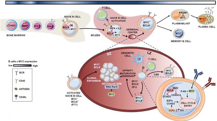
Cells 2020, 9, 523                                                                                   2 of 24

idea of the potential involvement of MYC in hematopoiesis. In the specific case of B lymphocytes,
the use of transgenic mice overexpressing MYC revealed a developmental blockade at the B cell stage,
before the onset of lymphoma [9].
      Given the importance of MYC deregulation in human leukemia and lymphoma, it is not surprising
that its correct modulation is essential throughout the whole B lymphocyte development [10]. At the
LT-HSC stage, there is a combined expression of c-MYC and N-MYC isoforms, but there is a complete
absence of L-MYC family members [11]. Interestingly, MYC expression allows LT-HSCs and MPPs
to be distinguished [10]. On the one hand, LT-HSCs display low levels of MYC to maintain a tight
equilibrium between cell self-renewal capacity and differentiation. On the other hand, the activation
of MYC expression promotes the differentiation of LT-HSCs into MPPs, which present increased
proliferating activity [10,12].
      Despite its role in maintaining the self-renewal capacity of LT-HSCs, MYC is also essential
for controlling proper hematopoiesis. In fact, Myc-deficient murine embryos exhibit impaired
hematopoiesis and die before mid-gestation [13]. At this developmental stage, the role of MYC
proteins is hierarchical. N-MYC and L-MYC cannot be expressed alone and require the concomitant
expression of c-MYC. For instance, the single deletion of N-MYC does not affect the quiescent state of
HSCs or hematopoiesis, whereas the deletion of c-MYC in HSCs alters proliferation and survival [11].
In summary, c-MYC is essential for balancing self-renewal and differentiation at the HSC stage.
Sustained expression of MYC encompasses the transition from HSCs to lymphoid-committed cells
since its extensive ability to bind to promoter and enhancer regions endows it with an extensive gene
transcription role in both developmental stages [10,14].
      N-MYC and c-MYC are both expressed in lymphocyte progenitors, meanwhile only the expression
of c-MYC is maintained during the rest of the differentiation process, despite being reduced in precursor
and mature B cell stages [15]. MYC expression is induced in pre-B cells in response to B-cell receptor
(BCR) stimulation [16,17]. MYC expression peaks coincide with the stages of higher proliferative rates
in B lymphocyte generation [18]. In consequence, MYC has an essential role in the expansion of pro-B
cells and differentiation to the pre-B stage [10]. Conditional knockout of c-MYC or N-MYC using the
Cd19-Cre transgenic mouse model blocks the transition from pro-B to pre-B cells, confirming its role at
this stage of lineage development [19].
      In connection with these data, aberrant expression of MYC in transgenic mice results in a reduction of
mature B lymphocyte numbers relative to those of pre-B cells [9]. In a similar way, the regulation of MYC
expression may be altered by the presence of the antiapoptotic factor BCL2 [20], or the stimulation with
cytokines, such as interleukin 7 (IL-7) [21], resulting in a tumorigenic outcome, given the ability of these
two proteins to enhance cell survival. Conversely, Myc-null B lymphocytes have an impaired proliferation
capacity when treated with stimulatory cytokines, such as the B-cell surface antigen CD40 and IL-4 [22].
      During the complex program that naïve B cells undergo in the GC before they differentiate to
memory B cells or PCs, the expression of MYC is maintained, though being depleted when the B cell
exits de GC reaction [15,21]. In this context, MYC is basically restricted to specific phases of the GC
reaction development and is mainly expressed during naïve B cell expansion and at stages preceding
the light zone (LZ) to dark zone (DZ) transition [23,24].
      B-cell lymphoma 6 (BCL6) is a direct repressor of MYC during the GC reaction [23]. BCL6 binds
to the promoter region of MYC in pre-B and differentiated B cells [25–27]. Therefore, the expression
pattern of both factors is mutually exclusive in most GC B cells, with 91% of those cells expressing
either BCL6 or MYC, and only 8% showing co-expression of both proteins [23]. In GCs, when B
cells interact with antigens and access T-helper (Th) cells, they transiently express MYC due to the
transcriptional inhibition of BCL6 by the repressive machinery comprising BCR, IL-2, and interferon
regulatory factor 4 (IRF4), the latter being induced upon CD40 activation [24,28,29]. In the LZ, the BCR
also synergizes with CD40 to activate MYC and induce p-S6, allowing cell-cycle entry [30,31].
      In these early stages of GC formation, MYC-expressing B cells express cyclin D2 (CCND2) [32,33]
and D3 (CCND3) [34,35], which possibly contributes to their hyperproliferative phenotype during the
Cells 2020, 9, 523 3 of 24
Cells 2020, 9, 523 3 of 24
during the initial rounds of cell division that give rise to the bulk of the GC B cells [36]. As described
initialVictora
by roundsetof al.,
cell B cell clonal
division expansion
that give is restricted
rise to the bulk of thetoGCthe DZ,[36].
B cells and Ascells move to
described bythe LZ in
Victora a
et al.,
bi‐directional process controlled by T cells. Based on the amount of Ag captured, Th cells at the LZ
B cell clonal expansion is restricted to the DZ, and cells move to the LZ in a bi-directional process
determine whether MYC+ B cells re‐enter the DZ for additional rounds of positive selection, or if
controlled by T cells. Based on the amount of Ag captured, Th cells at the LZ determine whether MYC+
they remain in the LZ [37].
B cells re-enter the DZ for additional rounds of positive selection, or if they remain in the LZ [37].
MYC+ B cells at the LZ subsequently undergo transcription, whereby BCL6 binds the
MYC+ B cells at the LZ subsequently undergo transcription, whereby BCL6 binds the transcription
transcription factor (TF) MYC‐interacting zing‐finger protein 1 (MIZ1) [38], an MYC partner that acts
factor (TF) MYC-interacting zing-finger protein 1 (MIZ1) [38], an MYC partner that acts to suppress
to suppress CDK inhibitor p21 and thereby induce cell‐cycle entry. At this stage, BCL6 and MYC are
CDK inhibitor p21 and thereby induce cell-cycle entry. At this stage, BCL6 and MYC are co-expressed
co‐expressed in the LZ [23]. BCL6 also inhibits CCND2 expression [32,33], which is an MYC target.
in the LZ [23]. BCL6 also inhibits CCND2 expression [32,33], which is an MYC target. CCND3, which
CCND3, which is not controlled by MYC [34,35], is expressed alone in these LZ GC B cells. The TF
is not controlled by MYC [34,35], is expressed alone in these LZ GC B cells. The TF TCF3 (also called
TCF3 (also called E2A) is intrinsically regulated by the induction of its own inhibitor ID3 (inhibitor
E2A) is intrinsically regulated by the induction of its own inhibitor ID3 (inhibitor of DNA binding
of DNA binding 3), is expressed in the GC B cells, and activates CCND3 and E2F2, replacing
3), is expressed in the GC B cells, and activates+ CCND3 and E2F2, replacing CCND2-dependent
CCND2‐dependent proliferation in the LZ MYC B cells [27,39].
proliferation in the LZ MYC+ B cells [27,39].
Logically, MYC expression must be tightly controlled in the DZ to limit cell divisions before
Logically, MYC expression must be tightly controlled in the DZ to limit cell divisions before each
each round of antigen affinity‐based selection, as MYC controls the transcriptional pause release of
round
RNA of antigen affinity-based
polymerase II, which is selection,
essential foras MYC controls the transcriptional
activation‐induced pause release
cytidine deaminase of RNA
(AID)‐induced
polymerase II, which is essential for activation-induced cytidine deaminase (AID)-induced
somatic hypermutation (SHM) [40,41]. After several rounds of positive selection, the MYC‐ B cells somatic
hypermutation - B cells finally exit
finally exit the(SHM)
GC and [40,41]. Aftereither
become several B rounds
memory of cells
positive selection, the MYC
or plasmablasts. B‐lymphocyte‐induced
the GC and become
maturation proteineither B memory
1 (BLIMP1) cells or plasmablasts.
suppresses MYC expressionB-lymphocyte-induced
in plasmablasts maturation
and induces protein
PC
1 differentiation
(BLIMP1) suppresses MYC
[42]. This dependency effect between MYC and B cell proliferation is knownThis
expression in plasmablasts and induces PC differentiation [42]. as
dependency effect between MYC and B cell proliferation is known as “cyclic re-entry”
“cyclic re‐entry” [23]. A schematic summary of the role of MYC in B lymphocyte differentiation is [23]. A schematic
summary
shown inof the role
Figure 1. of MYC in B lymphocyte differentiation is shown in Figure 1.
Figure1.1.Expression
Figure Expressionand androle
roleofof MYC
MYC in in B lymphocyte
B lymphocyte differentiation.
differentiation. Schematic
Schematic representation
representation of
of the
the participation of the MYC protein throughout B‐cell differentiation in the
participation of the MYC protein throughout B-cell differentiation in the bone marrow and germinal bone marrow and
germinal
(GC).center (GC). The percentages
shown refer shown MYC+ , BCL6
refer to theofpopulation of +/−
MYC +, BCL6+/− cells in the
center The percentages to the population cells in the total number
oftotal number
B cells of Bincells
present the present
GC. Theinblue-colored
the GC. The lineblue‐colored
at the topline at the
of the top of
Figure the Figure
indicates theindicates
evolution theof
evolution of MYC expression, where darker blue indicates steps that
MYC expression, where darker blue indicates steps that require higher MYC levels. require higher MYC levels.
2.2.MYC
MYCRole
Rolein
inLeukemogenesis
Leukemogenesis
Unlike
Unlikeother proto‐oncogenes, MYC
otherproto-oncogenes, MYC isis not
not activated
activated by oncogenic mutations in
oncogenic mutations in the
the coding
coding
sequence.
sequence. MYC
MYC transforms
transforms cells
cells via aberrant overexpression
overexpression ofof intact
intact MYC
MYC protein
protein by
by three
three main
main
mechanisms: gene amplification, chromosomal translocation, and aberrant regulation
mechanisms: gene amplification, chromosomal translocation, and aberrant regulation of its of its expression.
Inexpression.
the following sections,
In the we describe
following sections, the role of MYC
we describe in several
the role of MYC types of leukemia.
in several types of leukemia.Cells 2020, 9, 523 4 of 24
2.1. B lymphoblastic Leukemia with t(9;22) BCR-ABL1 Rearrangement
The B-cell receptor – ABL proto-oncogene 1 (BCR-ABL1) fusion (a translocation widely known as
the Philadelphia chromosome, Ph) protein product can activate Myc in bone marrow-derived murine
pre-B cells [43]. The activation of MYC, combined with other oncoproteins, such as RAS, c-RAF, and
c-JUN, promotes the activation of signaling pathways, leading to malignant cell transformation [44].
Remarkably, the repression of MYC impairs BCR-ABL1-mediated transformation, indicating that MYC
not only has a complementary function but also is essential for ensuring leukemic transformation [43,45].
Whereas the activation of MYC in lymphomas is partially caused by an elevated mutation
frequency in several cases, B-cell precursor leukemia has an almost negligible mutation rate [46].
However, BCR-ABL rearranged pre-B-acute lymphoblastic leukemia (ALL) is driven by an aberrant
expression of AID [47], which is expressed at such an early stage of B lymphocyte development [48],
as a consequence of the enhanced kinase activity of BCR-ABL1 fusion protein (i.e., tyrosine kinase
P210) [47,49]. Nevertheless, the proportion of patients harboring mutations at the MYC gene itself
among Ph+ ALL cases remains low and stable compared with that of Ph- patients [47].
In line with these data, MYC-IGH translocation, which is a common alteration in B-cell
lymphomas [50], is not frequently present in the B-cell precursor ALL. However, when analyzing the
genetic deletion of CDKN2, a common B-ALL feature, it was found that patients with the wild-type
CDKN2 experienced a higher rate of MYC-IGH translocation [51], suggesting that the two genetic
alterations may be mutually exclusive.
MYC is induced through different pathways triggered by the BCR-ABL1 fusion protein. For
instance, the MYC gene is one of the pre-BCR downstream effectors whose signaling is transduced
through spleen tyrosine kinase (SYK) [52,53]. The inhibition of SYK impairs cell viability via the
repressed transcription of MYC oncogene [53]. In parallel, the pro-inflammatory marker sphingosine
kinase 2 (SK2) promotes the activation of MYC in murine models of B-ALL by increasing its acetylation
profile. The inhibition of SK2 provokes a drastic reduction in ALL cell proliferation through concomitant
repression of MYC target genes [54]. Recently, the use of purinostat mesylate (a first-in-class
histone deacetylase (HDAC) inhibitor with reported antitumor activity [55]) has also been shown
to downregulate the BCR-ABL1 fusion protein targeting of MYC through the alteration of global
histone 3 (H3) and histone 4 (H4) acetylation [56]. These studies reveal chromatin remodeling to be a
promising therapeutic strategy in BCR-ABL1+ ALL. For instance, as described in greater detail below,
the combination of HDAC and PI3K inhibition impairs MYC-dependent growth in hematological
malignancies [57].
The Wnt signaling cascade is a well-characterized oncogenic pathway that can drive MYC oncogene
activation. The BCR-ABL1 protein phosphorylates specific tyrosine residues of γ-catenin, thereby
enhancing the direct binding of this effector to the MYC promoter [58]. The role of this kinase activity
differs from that in HSCs, where BCR-ABL1 phosphorylates β-catenin, giving rise to initial forms of
chronic myeloid leukemia (CML), without requiring MYC induction [58,59]. Instead, BCR-ABL1-driven
activation of the JAK/STAT pathway through the phosphorylation of JAK2 has similar effects on
both chronic myeloid leukemia (CML) and ALL, whereby pJAK2 and pSTAT5 cooperate to maintain
elevated levels of MYC by protecting it from ubiquitin-dependent degradation [60,61].
MYC is also regulated at several post-transcriptional levels in Ph+ B-ALL. Since the highly
structured 5’-UTR of MYC determines its translation rate, eIFs are key translation factors that enable
MYC mRNA translation [62]. IgM signaling, which is active in chronic lymphoid leukemia (CLL) cells,
promotes increased translation of MYC mRNA, together with the induction of eIF4 and eIF4GI [63,64].
eIF4 and MYC participate in a feedforward loop that enhances both activities [65]. This is not the only
mechanism through which MYC reinforces its own expression. For instance, MYC, in cooperation
with its TF partner MAX, binds to the promoter of BCR-ABL1, activating its transcription [66].
The aberrantly activated function of MYC in ALL also depends on protein stabilization. Some of
the first evidence demonstrated that the induction of the Ras pathway prevents proteasomal-mediated
degradation of MYC [67,68]. Moreover, most leukemia cell lines harbor an altered MYC form with aCells 2020, 9, 523 5 of 24
Cells 2020, 9, 523 5 of 24
proteasomal‐mediated degradation of MYC [67,68]. Moreover, most leukemia cell lines harbor an
altered MYC form with a prolonged half‐life, without possessing genetic mutations or chromosomal
prolonged half-life,
alterations [47,69]. Thewithout possessing
increased genetic
stability of MYC mutations or chromosomal
is explained by an excessalterations [47,69]. The
of phosphorylation at
Ser62, combined with low levels of pThr58, which promote glycogen synthase kinasewith
increased stability of MYC is explained by an excess of phosphorylation at Ser62, combined low
3 beta
levels of pThr58, which
GSK3β‐mediated promote glycogen
ubiquitylation synthasedegradation
and proteasomal kinase 3 beta[69,70].
GSK3β-mediated ubiquitylation and
proteasomal degradation [69,70].
Apart from its main function in driving tumor progression, MYC also induces apoptosis, since
Apart some
it targets from its main involved
genes function inindriving
the BCL2tumornetwork
progression, MYCAs
[71,72]. also induces
part of thisapoptosis,
network, since it
the
targets some genes involved
apoptosis‐inducer protein BIMin theacts
BCL2 asnetwork
a major[71,72]. As part
antagonist ofofBCL2.
this network,
In fact,thetheapoptosis-inducer
Eμ‐myc murine
protein BIM acts as a major antagonist of BCL2. In fact, the Eµ-myc murine
leukemia model has demonstrated that the deletion of Bim counteracts the potential induction leukemia model has
of cell
demonstrated
death by MYC, thatworsening
the deletion Bim counteracts
theofB‐cell the potential
leukemia‐associated induction
prognosis of of cell mice
these death [73].
by MYC,
The
worsening the B-cell leukemia-associated prognosis of these mice [73].
regulation of BIM is partially mediated by the miR‐17‐92 cluster (also known as MIR17HG), The regulation of BIM in is
partially mediated by the miR-17-92 cluster (also known as MIR17HG), in MYC-driven
MYC‐driven leukemia [74,75] and the inhibition of this specific microRNA endows leukemic cells leukemia [74,75]
and the
with inhibition of this
a pro‐apoptotic specific microRNA
phenotype [75], makingendows leukemicnetworks
microRNA cells with an
a pro-apoptotic phenotype
alternative entry point[75],
for
interfering with MYC function in the B‐cell precursor ALL. The regulation of the
making microRNA networks an alternative entry point for interfering with MYC function in MYC B-cell
in
precursor ALL. The regulation of MYC in BCR-ABL1-rearranged
BCR‐ABL1‐rearranged leukemia is depicted in Figure 2. leukemia is depicted in Figure 2.
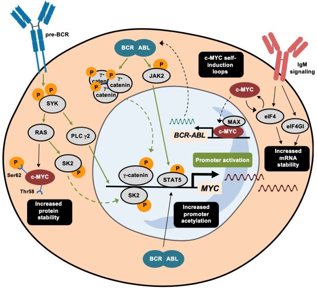
Figure 2.2. Activating
Activatingmechanisms
mechanisms of c‐MYC
of c-MYC in leukemia
in leukemia with
with the the BCR‐ABL1
BCR-ABL1 rearrangement.
rearrangement. A summary A
summary of thetransduction
of the different different transduction signaling that
signaling pathways pathways
trigger that trigger theofactivation
the activation of MYC
MYC promoter in
promoter in BCR‐ABL1‐rearranged
BCR-ABL1-rearranged leukemia. Apartleukemia.
from Apart
direct from direct transcriptional
transcriptional activation pathways,
activation pathways, marked in
marked in green, alternative
green, alternative mechanismsmechanisms
that induce thatc-MYC
inducearec‐MYC
depicted in blackinand
are depicted black and highlighted
highlighted in
in black
black squares.
squares. Dashed Dashed
arrowsarrows
indicateindicate the translocation
the translocation of proteinsof between
proteins the
between
nucleusthe nucleus
and and the
the cytoplasm.
cytoplasm.
2.2. B lymphoblastic Leukemia with the t(v;11) MLL Rearrangement
2.2.B Translocations
lymphoblastic Leukemia with themethyltransferase
in the histone t(v;11) MLL Rearrangement
MLL gene are the most common chromosomal
alteration in infant leukemia
Translocations and,methyltransferase
in the histone in general, exhibitMLL
a very poor
gene are prognosis, such that
the most common the disease
chromosomal
is an extremely lethal malignancy in infants [76–78]. As in other types of B-cell leukemia, the MYC
alteration in infant leukemia and, in general, exhibit a very poor prognosis, such that the disease isCells 2020, 9, 523 6 of 24
gene is not commonly involved in chromosomal translocations, although rare individual cases with
t(8;22) have been reported [77]. Recently, a revised characterization of the RS4;11 leukemic cell line has
demonstrated the presence of i(8q), resulting in MYC duplication, which confers a selective growth
advantage in vitro [79]. In B-ALL, this alteration is considered a secondary hit that contributes to
disease progression. Patients with the MLL-AF4 fusion protein have a strongly enriched MYC gene
signature compared with AML patients [80,81].
Consequently, MLL-fusion proteins activate the expression of the MYC oncogene in pre-B and
pro-B-cell leukemia [82,83]. For instance, MLL protein can prompt additional activity by fusing to
USP2 deubiquitinating protein, leading to the enrichment of USP2 activity on MDM2, ending with the
enhanced degradation of p53, which, in turn, activates MYC expression [84,85].
The binding of MLL-rearranged proteins to the regulatory regions of target genes depends on the
presence of chromatin adaptors that comprise the super-elongation complex (SEC). The bromodomain
and extra-terminal domain (BET) family of proteins (BRD2/3/4) are part of this complex and contribute
to the induction of MYC [86–88]. Initial evidence showed that suppression of BRD4 induces potent cell
growth arrest and cell senescence, combined with MYC downregulation, meaning that the BET family
is a promising therapeutic target [86–89].
For instance, a small molecule inhibitor of BET (iBET-151) prevents the recruitment of BET proteins
to chromatin by inhibiting the transcription of key targets, such as BCL2 and MYC [87]. In parallel,
JQ1 (a potent inhibitor of BRD4) greatly reduces MYC expression and activity, jointly with a large set
of its target genes [88]. Moreover, JQ1 has also been tested in patient-derived xenograft from ALL
patients, confirming its ability to inhibit MYC expression [90]. BET proteins are involved in maintaining
aberrantly altered chromatin states in ALL. At this point, BRD4 cooperates with MYC in recruiting
TEFb complexes to initiate transcriptional elongation at active promoters, while the transcriptional
regulator HEXIM1 counteracts BRD4 and MYC role by inactivating the complex. Therefore, tight
regulation of BRD4, MYC, and HEXIM1 is required for proper elongation [40,91,92]. A novel oral
BRD2/3/4 inhibitor (OTX015) has been shown to reduce MYC expression and to increase HEXIM1
levels in MLL-rearranged leukemia [89]. Apart from OTX015, the specific BRD4 inhibitor CPI-0610 has
been selected for phase I clinical trials in ALL patients [93].
Histone deacetylases (HDACs) have a differential expression pattern in ALL patients with MLL
rearrangement and are commonly overexpressed. For instance, HDAC9 is associated with an adverse
prognosis, whereas SIRT1 is involved in drug resistance through its regulation of the acetylation of the
TP53, MYC, and NF-κβ genes. Consequently, HDAC inhibitors (HDACis) have emerged as potential
therapeutic options in treating hematological malignancies [94,95]. However, conceptualizing the
role of HDACs in leukemia as inducers of malignant transformation would be a too simplistic view,
given that some histone deacetylases, such as HDAC7, carry out an opposite function. As reported by
Barneda-Zahonero et al., HDAC7 is involved in the repressive transcriptional machinery of MYC and,
therefore, it is often reduced in different types of leukemia and lymphoma, including MLL-rearranged
malignancies [96].
In this sense, newly developed compounds that selectively inhibit specific HDAC subtypes
are gaining relevance in the treatment of hematological malignancies [97]. For instance, class
I/IIb-selective HDACi purinostat has demonstrated a direct effect on MYC downregulation [56], while
other selective drugs (mocetinostat, entinostat) are already undergoing clinical trials for diverse
hematological malignancies [97]. The use of combinatorial therapies merging selective HDACi and
classical treatments emerges as a promising therapeutic option, and it is tempting to speculate that its
ability to induce apoptosis resides, at least partially, in MYC negative regulation [97].
The relevance of the MYC oncogene in hematopoiesis is restricted to its functions in aberrantly
proliferating B-cell precursors and in the normal hematopoietic stem cell hierarchy. The expression of
MYC throughout this process is controlled by a super-enhancer region located 1.7 Mb downstream
of the gene [98]. This super-enhancer, known as the “blood enhancer cluster” (BENC), is comprised
of several selectively active modules that recruit a wide range of transcription factors due to theCells 2020, 9, 523 7 of 24
increased chromatin accessibility. This differential access to regulatory regions has also been reported
in murine models of MLL-AF9-driven leukemia, indicating that MYC hyperactivation during leukemia
can be driven by BENC-unbalanced modulation [99,100]. BENC deletion entails a drastic depletion
of B lymphocytes during normal development, as well as an improved prognosis in MLL-AF9+
leukemia [100]. Regarding the regulation of MYC at the promoter level, we strongly consider that
recently developed techniques for 3D chromatin architecture analysis will improve our knowledge
about the coordination of transcriptional machinery, chromatin accessibility, and 3D structure. Not in
vain, this novel methodology has already conferred a new dimension to the study of B cell development
at different stages [101].
2.3. B Lymphoblastic Leukemia with the t(12;21) ETV6/RUNX1 Rearrangement
The t(12;21) translocation, which involves the ETV6 and RUNX1 (also known as AML1) genes, is the
most frequent lesion in childhood B-ALL (20–30% of cases), at early diagnosis and remission [102,103].
The N-terminal region of ETV6 displays weak homology with the bHLH region of MYC protein [104].
This homology enables the induction of the targets of these factors through protein-protein interaction,
enhancing MYC oncogenic function [103]. Apart from its characteristic fusion to RUNX1 protein, ETV6
also forms fusion proteins with PAX5, which is a key inducer of B-cell-specific genes (such as CD19
and CD79A) [104,105]. The combination of PAX5 activity with ETV6-mediated MYC targets induction
establishes the ETV6/PAX5 fusion protein as a powerful mediator of ALL progression.
Alterations affecting the MYC gene itself should be highlighted as examples of chromosomal
aberrations. For instance, a double MYC gene translocation t(8;14)t(8;9) was reported in a B-ALL
patient with ETV6 amplification [106]. Copy number variation (CNV) was reported in a substantial
65% of relapsing ETV6/RUNX1-positive ALL patients, including MYC expression gain at chromosome
8 (q23.1-24.1) in 10% of cases [107].
An indirect pathway of MYC activation in ETV6/RUNX1-rearranged leukemia is mediated
by the GTP-binding protein RAC1, a pivotal modulator of hematopoiesis [108], that increases the
phosphorylation levels of STAT3 [109]. ETV6/RUNX1 protein enhances the activity of RAC1, increasing
MYC expression, induced by the phosphorylation of STAT3 [110]. Specific STAT3 inhibitors revert
MYC induction by blocking cell proliferation and promoting apoptosis in pro-B-ALL cells [110].
Additionally, the ETV6/RUNX1 fusion gene can be stabilized at the mRNA level by the
RNA-binding protein IGF2BP1, which is overexpressed in this type of leukemia [111]. IGF2BP1
leads to an eventual increase of MYC, linked to aberrant leukemogenesis in ETV6/RUNX1-mediated
ALL [111,112]. Finally, and similarly to the mechanism reported for BCR-ABL1-rearranged leukemia,
MYC is also stabilized at the protein level through aberrantly altered phosphorylation at the Thr58 and
Ser62 residues [69].
2.4. B Lymphoblastic Leukemia with other Chromosomal Rearrangements
Expression of B220 and CD43 determines the transition of pro-B into pre-B lymphocytes [113].
Leukemia derived from this developmental stage usually displays TCF3/PBX1 chromosomal
rearrangement, which is commonly found in leukemia derived from pre-B lymphocytes (in more than
90% of cases) [114]. Survival of TCF3/PBX1+ cells critically depends on the activity of the pre-BCR [52,53].
Immunoglobulin µ (Igµ) heavy-chain knockdown impairs the proper assembly of pre-BCR and blocks
signal transduction through the Igα-Igβ heterodimer [115]. Igµ downregulation in TCF3/PBX1-rearranged
cell lines significantly suppresses MYC expression at the mRNA and protein levels. MYC is regulated by
the pre-BCR in a FOXO-dependent manner since the forced expression of a constitutive form of FOXO1
reverts the blockade of pre-BCR signaling, and partially restores MYC expression [53].
Under physiological conditions, MYC mRNA is modulated by miR-24, which is able to bind at
0
its 3 -UTR region to reduce MYC levels, thereby controlling cell-cycle progression [116]. miR-24 is
frequently downregulated in TCF3/PBX1+ pre-B-ALL, concomitantly with other miRNAs involved
in proliferation and apoptosis modulation in various cancers (e.g., miR-126 and miR-365) [117].Cells 2020, 9, 523 8 of 24
Surprisingly, the restoration of miR-24 expression in TCF3-rearranged leukemic cell lines neither affects
the expression of some of its targets nor alters the frequency of apoptotic cells, suggesting that MYC is
regulated by a combination of mechanisms in this type of leukemia [117].
Despite not being the most frequent alteration, the IGH gene (located at chromosome 5) can also
be translocated to chromosomes 14 or 12, as is the case for the NALM-6 cell line, which harbors the
t(5;12) translocation. This cell line was recently used to identify the transcriptional cofactor apoptosis
antagonizing transcription factor (AATF) as being a direct target of MYC since it features canonical
binding motifs at the promoter region [118]. AATF promotes cell-cycle progression by inhibiting
TP53 expression and mediating the response to DNA damage [119]. It is of particular note that,
when inhibiting the expression of MYC, there is a drastic downregulation of MLL gene expression, a
previously described key mediator of pediatric leukemia. This downregulation can be counteracted by
the exogenous induction of AATF. Therefore, AATF mediates a positive feedback loop between MYC
and MLL gene in pro-B-ALL [118].
3. MYC Role in Lymphomagenesis
In hematopoietic malignancies, genomic abnormalities involving the MYC gene are almost always
found in B cell lymphomas, but rarely in T cell lymphomas. 30% of all lymphoid neoplasms are B cell
non-Hodgkin lymphomas. These can be classified further as Burkitt lymphoma (BL), diffuse large B
cell lymphoma (DLBCL), follicular lymphoma (FL), mantle cell lymphoma (MCL), and plasmablastic
lymphoma (PBL), among others.
3.1. MYC in Burkitt Lymphomas
BL arises mostly in children and young adults and has an extremely high proliferation rate.
Endemic, sporadic, and immunodeficiency-associated BLs are distinguished as clinical variants in the
World Health Organization (WHO) classification. BL has a mature B cell phenotype with expression of
GC/post-GC markers such as immunoglobulin M (IgM), CD10, and is typically negative for BCL2 [120].
The genetic hallmark that characterizes BL is the rearrangement of MYC with one of the IG gene
loci. Translocation triggers constitutive MYC hypermutation of the translocated gene in germinal
centers [121], subjected to AID-dependent SHM [41], which is susceptible to generating MYC variants
and increasing its oncogenic potential [122]. Specifically, an MYC translocation to the IG heavy chain
gene locus 14q32 (80% of the cases), or to the IGκ or IGλ light chain genes at 2p12 or 22q11 (10%) are
the main rearrangement sites [123,124]. Most mutations of the rearranged MYC gene are point SNPs or
deletions in the 3’ border of the first exon and the first intron [125,126], altering the coding sequence
but permitting its transcription from the translocated chromosome. In consequence, there is an MYC
expression that terminates inhibiting cell differentiation and inducing proliferation, probably keeping
the cells in a hyperproliferative state.
In a gene expression profile study of human samples of BL (and DLBCL), three main cytogenetic
groups within the mature aggressive B cell lymphomas were distinguished: MYC-simple, with IG-MYC
fusions and a low chromosomal complexity score, no IGH-BCL2 fusions, and no BCL6 breakpoints, and
with a favorable prognosis; MYC-complex, including IG/MYC-rearranged BLs with highly complex
karyotypes, non-IG/MYC-rearranged cases, and all IGH/BCL2 fusions and/or BCL6 breakpoints, or any
combination of these; and MYC-negative, comprising lymphomas with unaltered MYC [127].
Inhibitor of DNA binding (ID) proteins, such as ID3, bind E-proteins such as TCF3 via HLH
common motifs, preventing the binding of the latter to DNA. Schmitz et al. shed light on some oncogenic
pathways, suggesting that MYC translocation is insufficient to induce BL [128]. The next-generation
sequencing (NGS) study performed by Love et al. identified MYC and ID3 as the genes most frequently
mutated in BL [128,129].
In the setting of deregulated MYC, samples with ID3 mutations show a higher level of expression
of known MYC target genes, and give rise to increased G1-to-S-phase cell-cycle progression in BL,
suggesting a role for ID3 as a tumor suppressor in this type of lymphoma [129]. The high level ofCells 2020, 9, 523 9 of 24
ID3 expression in BL might be because the ID3 locus itself is a direct target of MYC [130] and due
to BCR triggering, as described using an Id3-/- mouse model [131]. Functional analyses suggested
that ID3-inactivating mutations and TCF3-activating mutations (by blocking ID3 binding sites) lead
to the activation of a TCF3-dependent transcriptional program that consequently promotes tonic
BCR signaling [132]. TCF3 would repress PTPN6, which encodes SHP-1, a BCR-attenuating factor
that acts by dephosphorylating the ITAM motifs of the CD79A and CD79B signaling subunits of the
BCR [39]. In addition, BCR can activate both MYC and ID3 by a sequential process in which MYC
rapidly upregulates its expression. Later, upon MYC downregulation, levels of ID3 increase [131]. This
effect may be produced by direct BCR activation, or through an indirect effect of MYC, highlighting the
existence of a positive feedback loop between BCR, ID3, and MYC regulation.
Tonic BCR activation requires PI3K signaling in mature B cells to maintain its continuity [132],
and the pro-survival pathway cooperates with MYC in BL [133]. MYC deregulation induces the
expression of the MIR17HG, a microRNA host gene amplified in ~10% of BL cases [134]. Particularly,
miR-19 is the key oncogenic component of the cluster, which antagonizes PTEN and, consequently,
activates the AKT-mTOR pathway, the consequence of which is exacerbated cell survival in MYC-driven
lymphomagenesis [128,135,136]. See [137] for an extensive review of the involvement of MYC and
miRNAs in lymphomagenesis.
BCR-induced PI3K pathway activation in BL contrasts with the absence of NF-κβ survival pathway
signaling in these tumors [138]. Reinforcing this, the study by Klapproth et al. in Myc transgenic mice
showed that constitutive NF-κβ activity is incompatible with the development of the MYC-induced
lymphomas [139]. The resting state of the NF-κβ apoptotic pathway confers a selective advantage on
MYC-driven oncogenic cells.
3.2. MYC in Diffuse Large B Cell Lymphoma
DLBCL accounts for approximately 40% of all non-Hodgkin lymphomas [120]. Two major
subtypes can be identified: germinal center B-cell-like (GCB), which has a gene expression profile
similar to that of the GC B cell; and activated B cell-like (ABC), which has a worse outcome because it
expresses genes present in activated peripheral B cells [120]. The principal translocated sites in DLBCL
are a rearrangement of BCL6 (30% of cases) and t(14;18)(q32;q21) with BCL2 rearrangement to the IGH
gene locus (20–30% of cases) [140,141]. Globally BCL2 is rearranged in 30% of cases in the GCB-DLBCL
subgroup and inCells 2020, 9, 523 10 of 24
MYC and BCL6 alterations, which account for 35% and 6% of cases, respectively [151,152]. Therefore,
in the first group, upregulated MYC expression promotes proliferation and disables the capacity to
induce apoptosis, while BCL2 expression fosters cell survival. Together their co-expression confers an
aggressive proliferating phenotype on these DHLs.
MYC overexpression is a reliable biomarker for predicting therapeutic response, since its expression
is a poor prognostic factor in DLBCL [153] but, beyond the aforementioned rearrangements, the
mechanisms underlying its overexpression are still unknown. The stability of MYC is regulated by
GSK-3β, which phosphorylates MYC at Thr58 and induces its degradation via the ubiquitin-proteasome
pathway [154]. Wang et al. demonstrated that BCR stimulation could activate downstream PI3K
signaling, phosphorylating GSK-3β at Ser9, and abolishing its ability to induce MYC degradation in
DLBCL [155]. Moreover, the PI3K pathway inhibitory elements such as PTEN are frequently lost in
GCB-DLBCL [156], while BCR mutations also result in its constitutive activation [157], leading to MYC
dysregulation in DLBCL.
Finally, the upregulation of MYC expression in DLBCL promotes BCR signaling by inducing
the MIR17HG cluster, employing a mechanism similar to that described above in the section on
BL [158,159]. Taken together, these data suggest that a positive feedback loop operates in the
BCR-PI3K-MYC signaling axis in DLBCL.
3.3. MYC In Plasmablastic Lymphoma
PBL is an aggressive, high-grade lymphoma that is most commonly diagnosed in patients with
HIV infection or an immunocompromised phenotype [120]. The cell of origin in PBL is thought to
be the plasmablast, an activated B cell that has undergone SHM and CSR, and that expresses cell
surface markers such as CD138, CD38, MUM1, and Ig, similar to a plasma cell [120]. Signaling
pathways leading to plasma cell differentiation involve gene silencing of PAX5 and BCL6 through
BLIMP1 [160,161], which also represses MYC expression through promoter binding [42]. Recurrent
somatic mutations in PRDM1 (the gene encoding BLIMP1) occur in 50% of cases, where they affect the
regulation of diverse targets, such as MYC [162]. Moreover, MYC and BLIMP1 proteins were found to
be co-expressed in 80% of diagnoses [162]. These findings are firm evidence that PRDM1 contributes
to the oncogenicity of dysregulated MYC.
Although MYC rearrangement is the genetic hallmark of BL and is characteristic of an aggressive
subset of DLBCL, it is also a common finding in PBL, along with MYC gains [163,164]. In PBL, MYC
rearrangements have been found in ~50% of cases, and the IG genes are the most frequent partners
(~85%), with t(8;14) MYC/IGH being the commonest fusion product [163]. Gene expression analysis of
PBL revealed MYC overexpression at mRNA and protein levels [165]. MYC overexpression facilitates
PBL cell apoptosis escape through cell-cycle dysregulation, and jointly with loss of TP53 [166]. Together,
these two processes enhance the aggressiveness of PBL.
In the absence of translocations, the mechanisms of MYC dysregulation are poorly understood,
suggesting that MYC may be activated by other mutated genes. Rearrangements of BCL2, BCL6,
MALT1, and PAX5, which are common in BL and DLBCL, are not detected in PBL. Conversely, gains of
these loci are frequent in PBL, where 30% of cases display amplification of three or more of them [163].
Paradoxically, Ouansafi et al. demonstrated in a single case report the concomitant presence of BCL2
and MYC translocation in a rare case of FL-to-PBL transformation [167].
3.4. MYC in Other Non-Hodgkin B Cell Lymphomas
FL is an indolent non-Hodgkin lymphoma that transforms into a high-grade lymphoma, mostly
DLBCL, in about one-third of patients. The genetic hallmark of FL is t(14;18)(q32;q21), which brings
about BCL2/IGH fusion protein [168]. Low-grade lymphomas containing a BCL2 rearrangement need
subsequent secondary genetic hits for the disease to evolve. The genetic alteration of MYC may suffice
as this secondary alteration, leading to the transformation into a high-grade B cell lymphoma [169].
Actually, the majority of FLs express MYC, but only in a small fraction of the cells (Cells 2020, 9, 523 11 of 24
Pasqualucci et al. investigated the genetic drivers of transformed follicular lymphoma (t-FL) and
determined that there is a common mutated precursor that experiences distinct genetic events that
are specifically associated with alterations deregulating cell-cycle progression and DNA damage [46],
evidence that matches perfectly with MYC oncogene among others. t-FL to DLBCL progression
occurs in 30% of the cases, mainly among GCB-DLBCL patients [170,171]. t-FL oncogenic mechanisms
are characterized by the presence of a proliferation signature, together with recurrent oncogenic
transformations such as TP53 mutation, CDKN2A loss, and c-REL amplification [171], giving rise
to a proliferative phenotype in which MYC could be involved. In fact, genetic lesions deregulating
MYC are the second most common tFL-specific lesion (including translocations, point mutations, and
CNVs) [46]. Alternative pathways involving MYC and its targets could help distinguish between two
types of morphologically similar lymphomas, such as tFL-derived DLBCL and de novo DLBCL, this
signature being more enriched in de novo cases than in transformed ones [172].
As reported by Martinez-Climent et al., when examining gene expression changes in t-FL, a
considerable number of MYC target genes are differentially expressed, although the MYC gene locus
remains unaltered in terms of copy number [173]. Consequently, MYC genetic abnormalities are
not the driving mutations of FL transformation and may only serve as a surrogate for the entire
proliferation signature.
MCL is generally an aggressive malignancy, but it is thought in some cases to remain
latently quarrelsome in an indolent phase. It is characterized by t(11;14)(q13;q32), juxtaposing
IGH, and CCND1, resulting in CCND1 overexpression, which drives the cells through the G1/S
transition [174]. Interestingly, a partnership between CCND1 and MYC has been reported in
the oncogenic transformation of B cells to MCL [175]. The coexistence of MYC and CCND1/IGH
rearrangements [176] is commonly found in double-hit (DH)-MCL [177], which is associated with
a high-risk prognostic index. As reported for FL, most of the MCL cases display an intense MYC
expression, but the percentage of positive cells is frequently low (Cells 2020, 9, 523 12 of 24
Table 1. MYC alterations in leukemia and lymphoma. Summary of the gene alterations, chromosomal translocations, regulatory pathways, and post-transcriptional
modifications involved in MYC activation that are included in this review, assigned to their corresponding subtype of leukemia and lymphoma.
Gene Alterations Myc Stabilization at
Involvement of Translocations Activating Pathways
(Mutations, CNV) Post-Transcriptional Level
Translation rate controlled by EIFs
MYC/IGH translocation is Induced by aberrant AID [47], [62–65], prevention of proteasomal
t(9;22) BCR/ABL1 Almost negligible mutation
uncommon [50]. Higher frequency in pre-BCR/SYK signaling [53], Wnt [58] degradation [67–69], induction
rearrangement rate [46]
CDKN2 WT patients [51] and JAK/STAT [60,61] pathways mediated by MIR17HG cluster
[74,75]
Indirect activation through TP53
LEUKEMIA
MYC is not commonly involved in inhibition [84,85], direct binding of
t(v;11) KMT2A i(8q) MYC duplication in
translocations. Only rare cases with MLL-fusion proteins and BET
rearrangement RS4;11 cell line [79]
t(8;22) reported [77] adaptors [86–88], regulation by
BENC super-enhancer region [99,100]
Stabilized at mRNA level by
MYC gene expression gain ETV6 also fuses to PAX5 and induces
t(12;21) ETV6/RUNX1 Double MYC gene translocation IGF2BP1 [111] and at the protein
8(q23.1-24.1). CNVs reported MYC expression [104]. Also
rearrangement reported [106] level by prevention of proteasomal
in 65% of cases [107] triggered by RAC1-STAT3 [110]
degradation [69]
MYC/IGH t(8;14q32) in 80% of the
BCR-induced PI3K pathway in
Point SNPs and deletions in cases and MYC/IGκ or Igλ t(8;2p12)
Burkitt lymphoma cooperation with TCF3, ID3 and
the 3’border [125,126] or t(8;22q11) in 10% of the diagnoses
SHP-1 [129–134]
[123,124]
Found in 10% of the cases, being
MYC/IGL the most common [144],
LYMPHOMA
GSK-3β phosphorylation
but also to BCL6, BCL2, PAX5,
Point SNPs, amplifications, Mutations in the BCR or PI3K abolishing MYC degradation [155]
Diffuse large B cell IKAROS [145–147].
gains and increased copy pathway inhibitory elements MYC upregulation promotes BCR
Lymphoma Often participate in complex
numbers of MYC [148,149] [156,157] signaling by induction of
karyotypes and associatedo a second
MIR17HG cluster [158,159]
hit such as MYC/BCL2 and
MYC/BCL6 [151,152]
Gains [163,164] and somatic
Observed in 50% of the cases, being
Plasmablastic lymphoma mutations in MYC inhibitor
MYC/IGH the most common [163]
PRDM1 [42,162]Cells 2020, 9, 523 13 of 24
Table 1. Cont.
Gene Alterations Myc Stabilization at
Involvement of Translocations Activating Pathways
(Mutations, CNV) Post-Transcriptional Level
Proliferation signature with
Remains unaltered in terms of Second most common tFL-specific oncogenic transformations such as
Follicular lymphoma
copy number [173] lesion [46] TP53 mutations, CDKN2A loss and
c-REL amplification [171]
BCR-driven
Disruption of MYC locus in Coexistence of CCND1/IGH and MYC CARD11-BCL10-MALT1 complex
CCND1/CDK4 and p16INK4a
Mantle cell lymphoma t(2;8) and MYC gains at rearrangements [176,177], and also together with Nf-κβ pathway
imbalance [183–185]
(8)(q24) [179,180] MYC/IGH t(8;14) [179,180] orchestrates MYC stability
[186,187]Cells 2020, 9, 523 14 of 24
4. Concluding Remarks
The appropriate path that lymphoid progenitors should ideally follow on their way towards
fully differentiated B cells is constantly under threat of being wrecked by the alteration of regulatory
mechanisms. Beyond its widely known function as an oncogene, MYC also plays an essential role at
different steps of B-cell differentiation, and its deregulation is one of the main hazards that can disrupt
the process. As described in this review and in a physiological context, MYC is strongly expressed on
the way to producing mature B lymphocytes, whereas its transient downregulation is required at some
specific points. However, MYC basal levels are maintained and are not completely switched off at any
point before the late memory and plasmatic B cell stages, demonstrating that only tight regulation of
MYC levels ensures that the B lymphocytes achieve their correct fate.
When the expression of MYC protein is aberrantly altered, the risk of developing a hematological
malignancy, such as leukemia or lymphoma, increases substantially through the acquisition of an
uncontrolled proliferative rate and a blockade of differentiation. Remarkably, the alterations that
trigger MYC overexpression differ between leukemia and lymphoma cells. In fact, leukemic cells have
low rates of MYC mutations and a low frequency of chromosomal translocations involving the MYC
gene, whereas the aforementioned genetic alterations are a hallmark in some types of non-Hodgkin B
cell lymphoma. Nevertheless, and even when not altered at the genetic level, the expression of MYC
is usually disrupted in the commonest types of leukemia, where it is activated by several pathways,
as well as at the post-transcriptional level. Most types of lymphoma present high levels of MYC
expression that are not always correlated with a mutated MYC gene, opening the door to speculation
that, as in leukemia, multiple pathways may act to facilitate its dysregulation.
In terms of therapeutic perspectives, the possibilities for interfering with MYC activity are still to
be adequately explored. However, the identification of regulatory cascades and other mechanisms
that trigger its induction opens a wide range of possibilities for indirectly impairing MYC function.
As reported here, we believe that the disruption of altered epigenetic regulation with HDAC inhibitors,
the blockade of microRNAs function, or the use of BET inhibitors that obstruct scaffold transcriptional
activating machinery, are but a few examples of the promising therapeutic strategies that will lead to
an improved prognosis of hematological disorders, mostly mediated by the maintenance of MYC at
physiological levels.
Author Contributions: O.d.B. and A.M. did the bibliographic research and wrote the manuscript. M.P. conceived
the manuscript, supervised the work, and provided critical comments on the review. All authors have read and
agreed to the published version of the manuscript.
Funding: This manuscript was funded by grants to M.P. by the Spanish Ministry of Science, Innovation, and
Universities (SAF2017-87990-R and EUR2019-103835) and elaborated at the Josep Carreras Leukaemia Research
Institute (IJC, Badalona, Barcelona). O.d.B. is funded by a Juan de la Cierva - Formación fellowship from the Spanish
Ministry of Science, Innovation, and Universities (FJCI-2017-32430). A.M. is funded by the Spanish Ministry of
Science, Innovation and Universities, which is part of the Agencia Estatal de Investigación (AEI), through grant
PRE2018-083183 (co-funded by the European Social Fund.). We thank the CERCA Programme/Generalitat de
Catalunya and the Josep Carreras Foundation for institutional support.
Conflicts of Interest: The authors declare no conflict of interest.
References
1. Adolfsson, J.; Månsson, R.; Buza-Vidas, N.; Hultquist, A.; Liuba, K.; Jensen, C.T.; Bryder, D.;
Yang, L.; Borge, O.-J.; Thoren, L.A.M.; et al. Identification of Flt3+ lympho-myeloid stem cells lacking
erythro-megakaryocytic potential. Cell 2005, 121, 295–306. [CrossRef]
2. Kondo, M.; Weissman, I.L.; Akashi, K. Identification of clonogenic common lymphoid progenitors in mouse
bone marrow. Cell 1997, 91, 661–672. [CrossRef]
3. Victora, G.D.; Nussenzweig, M.C. Germinal centers. Annu. Rev. Immunol. 2012, 30, 429–457. [CrossRef]
[PubMed]
4. Huang, C.; Melnick, A. Mechanisms of action of BCL6 during germinal center B cell development. Sci. China
Life Sci. 2015, 58, 1226–1232. [CrossRef] [PubMed]Cells 2020, 9, 523 15 of 24
5. Coppola, F.; Cole, M. Constitutive c-myc oncogene expression blocks mouse erythroleukaemia cell
differentiation but not commitment. Nature 1986, 320, 760–763. [CrossRef] [PubMed]
6. Leon, J.; Ferrandiz, N.; Acosta, J.C.; Delgado, M.D. Inhibition of cell differentiation: A critical mechanism for
MYC-mediated carcinogenesis? Cell Cycle 2009, 8, 1148–1157. [CrossRef] [PubMed]
7. Sheiness, D.; Bishop, J.M. DNA and RNA from uninfected vertebrate cells contain nucleotide sequences
related to the putative transforming gene of avian myelocytomatosis virus. J. Virol. 1979, 31, 514–521.
[CrossRef]
8. Varmus, H. The molecular genetics of cellular oncogenes. Annu. Rev. Genet. 1984, 18, 553–612. [CrossRef]
9. Langdon, W.Y.; Harris, A.W.; Cory, S.; Adams, J.M. The c-myc oncogene perturbs B lymphocyte development
in Eµ-myc transgenic mice. Cell 1986, 47, 11–18. [CrossRef]
10. Delgado, M.D.; León, J. Myc roles in hematopoiesis and leukemia. Genes Cancer 2010, 1, 605–616. [CrossRef]
11. Laurenti, E.; Varnum-Finney, B.; Wilson, A.; Ferrero, I.; Blanco-Bose, W.E.; Ehninger, A.; Knoepfler, P.S.;
Cheng, P.F.; MacDonald, H.R.; Eisenman, R.N.; et al. Hematopoietic stem cell function and survival depend
on c-Myc and N-Myc activity. Cell Stem Cell 2008, 3, 611–624. [CrossRef] [PubMed]
12. Reavie, L.; Della Gatta, G.; Crusio, K.; Aranda-Orgilles, B.; Buckley, S.M.; Thompson, B.; Lee, E.; Gao, J.;
Bredemeyer, A.L.; Helmink, B.A.; et al. Regulation of hematopoietic stem cell differentiation by a single
ubiquitin ligase-substrate complex. Nat. Immunol. 2010, 11, 207–215. [CrossRef] [PubMed]
13. He, C.; Hu, H.; Braren, R.; Fong, S.Y.; Trumpp, A.; Carlson, T.R.; Wang, R.A. C-Myc in the hematopoietic
lineage is crucial for its angiogenic function in the mouse embryo. Development 2008, 135, 2467–2477.
[CrossRef] [PubMed]
14. Nie, Z.; Hu, G.; Wei, G.; Cui, K.; Yamane, A.; Resch, W.; Wang, R.; Green, D.R.; Tessarollo, L.; Casellas, R.; et al.
c-Myc Is a universal amplifier of expressed genes in lymphocytes and embryonic stem cells. Cell 2012, 151,
68–79. [CrossRef]
15. Smith, R.K.; Zimmerman, K.; Yancopoulos, G.D. Transcriptional down-regulation of N-myc expression
during B-cell development. Mol. Cell Biol. 1992, 12, 1578–1584. [CrossRef]
16. Klemsz, M.J.; Justement, L.B.; Palmer, E.; Cambier, J. Induction of c-fos and c-myc expression during B cell
activation by IL-4 and immunoglobulin binding ligands. J. Immunol. 1989, 143, 1032–1039.
17. Larsson, L.G.; Schena, M.; Carlsson, M.; Sallstrom, J.; Nilsson, K. Expression of the c-myc protein is
down-regulated at the terminal stages during in vitro differentiation of B-type chronic lymphocytic leukemia
cells. Blood 1991, 77, 1025–1032. [CrossRef]
18. Huang, C.Y.; Bredemeyer, A.L.; Walker, L.M.; Bassing, C.H.; Sleckman, B.P. Dynamic regulation of c-Myc
proto-oncogene expression during lymphocyte development revealed by a GFP-c-Myc knock-in mouse.
Eur. J. Immunol. 2008, 38, 342–349. [CrossRef]
19. Habib, T.; Park, H.; Tsang, M.; De Alborán, I.M.; Nicks, A.; Wilson, L.; Knoepfler, P.S.; Andrews, S.;
Rawlings, D.J.; Eisenman, R.N.; et al. Myc stimulates B lymphocyte differentiation and amplifies calcium
signaling. J. Cell Biol. 2007, 179, 717–731. [CrossRef]
20. Vaux, D.L.; Cory, S.; Adams, J.M. Bcl-2 gene promotes haemotopoietic cell survival and cooperates with
c-myc to immortalize pre-B cells. Nature 1988, 335, 440–442. [CrossRef]
21. Morrow, M.A.; Lee, G.; Gillis, S.; Yancopoulos, G.D.; Alt, F.W. Interleukin-7 induces N-myc and c-myc
expression in normal precursor B lymphocytes. Genes Dev. 1992, 6, 61–70. [CrossRef] [PubMed]
22. De Alboran, I.M.; O’Hagan, R.C.; Gärtner, F.; Malynn, B.; Davidson, L.; Rickert, R.; Rajewsky, K.; DePinho, R.A.;
Alt, F.W. Analysis of c-Myc function in normal cells via conditional gene-targeted mutation. Immunity 2001,
14, 45–55. [CrossRef]
23. Dominguez-Sola, D.; Victora, G.D.; Ying, C.Y.; Phan, R.T.; Saito, M.; Dalla-Favera, R.; Nussenzweig, M.C.
c-MYC is required for germinal center selection and cyclic re-entry. Nat. Immunol. 2012, 13, 1083–1091.
[CrossRef]
24. De Silva, N.S.; Klein, U. Dynamics of B cells in germinal centres. Nat. Rev. Immunol. 2015, 15, 137–148.
[CrossRef]
25. Ci, W.; Polo, J.M.; Cerchietti, L.; Shaknovich, R.; Wang, L.; Shao, N.Y.; Ye, K.; Farinha, P.; Horsman, D.E.;
Gascoyne, R.D.; et al. The BCL6 transcriptional program features repression of multiple oncogenes in
primary B cells and is deregulated in DLBCL. Blood 2009, 113, 5536–5548. [CrossRef]You can also read