Ultrastructural and molecular analysis of the origin and differentiation of cells mediating brittle star skeletal regeneration - BMC Biology
←
→
Page content transcription
If your browser does not render page correctly, please read the page content below
Piovani et al. BMC Biology     (2021) 19:9
https://doi.org/10.1186/s12915-020-00937-7

 RESEARCH ARTICLE                                                                                                                                   Open Access

Ultrastructural and molecular analysis of
the origin and differentiation of cells
mediating brittle star skeletal regeneration
Laura Piovani1,2,3†, Anna Czarkwiani2,4†, Cinzia Ferrario1,5, Michela Sugni1,5,6* and Paola Oliveri2,3*

  Abstract
  Background: Regeneration is the ability to re-grow body parts or tissues after trauma, and it is widespread across
  metazoans. Cells involved in regeneration can arise from a pool of undifferentiated proliferative cells or be recruited
  from pre-existing differentiated tissues. Both mechanisms have been described in different phyla; however, the
  cellular and molecular mechanisms employed by different animals to restore lost tissues as well as the source of
  cells involved in regeneration remain largely unknown. Echinoderms are a clade of deuterostome invertebrates that
  show striking larval and adult regenerative abilities in all extant classes. Here, we use the brittle star Amphiura
  filiformis to investigate the origin and differentiation of cells involved in skeletal regeneration using a combination
  of microscopy techniques and molecular markers.
  Results: Our ultrastructural analyses at different regenerative stages identify a population of morphologically
  undifferentiated cells which appear in close contact with the proliferating epithelium of the regenerating aboral
  coelomic cavity. These cells express skeletogenic marker genes, such as the transcription factor alx1 and the
  differentiation genes c-lectin and msp130L, and display a gradient of morphological differentiation from the aboral
  coelomic cavity towards the epidermis. Cells closer to the epidermis, which are in contact with developing spicules,
  have the morphology of mature skeletal cells (sclerocytes), and express several skeletogenic transcription factors
  and differentiation genes. Moreover, as regeneration progresses, sclerocytes show a different combinatorial
  expression of genes in various skeletal elements.
  Conclusions: We hypothesize that sclerocyte precursors originate from the epithelium of the proliferating aboral
  coelomic cavity. As these cells migrate towards the epidermis, they differentiate and start secreting spicules.
  Moreover, our study shows that molecular and cellular processes involved in skeletal regeneration resemble those
  used during skeletal development, hinting at a possible conservation of developmental programmes during adult
  regeneration. Finally, we highlight that many genes involved in echinoderm skeletogenesis also play a role in
  vertebrate skeleton formation, suggesting a possible common origin of the deuterostome endoskeleton pathway.
  Keywords: Regeneration, Echinodermata, Amphiura filiformis, Osteogenesis, Cell differentiation, Biomineralization,
  Gene expression

* Correspondence: michela.sugni@unimi.it; p.oliveri@ucl.ac.uk
†
 Laura Piovani and Anna Czarkwiani have contributed equally to this work
and are co-first authors.
1
 Department of Environmental Science and Policy, University of Milan, Via
Celoria, 2, 20133 Milan, Italy
2
 Department of Genetics, Evolution and Environment, University College
London, London, UK
Full list of author information is available at the end of the article

                                        © The Author(s). 2021 Open Access This article is licensed under a Creative Commons Attribution 4.0 International License,
                                        which permits use, sharing, adaptation, distribution and reproduction in any medium or format, as long as you give
                                        appropriate credit to the original author(s) and the source, provide a link to the Creative Commons licence, and indicate if
                                        changes were made. The images or other third party material in this article are included in the article's Creative Commons
                                        licence, unless indicated otherwise in a credit line to the material. If material is not included in the article's Creative Commons
                                        licence and your intended use is not permitted by statutory regulation or exceeds the permitted use, you will need to obtain
                                        permission directly from the copyright holder. To view a copy of this licence, visit http://creativecommons.org/licenses/by/4.0/.
                                        The Creative Commons Public Domain Dedication waiver (http://creativecommons.org/publicdomain/zero/1.0/) applies to the
                                        data made available in this article, unless otherwise stated in a credit line to the data.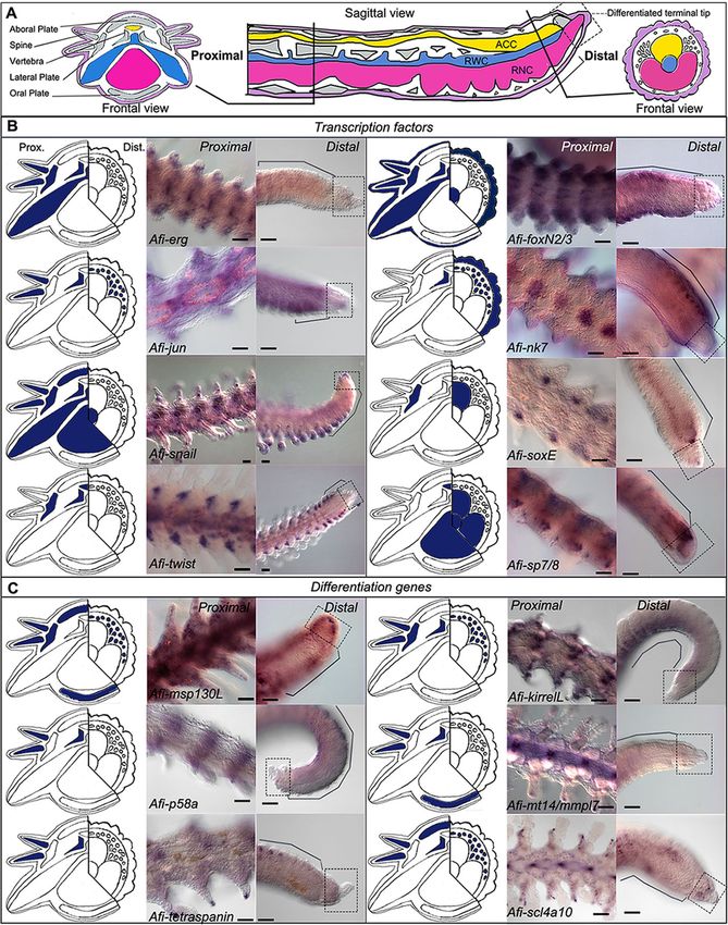
Piovani et al. BMC Biology   (2021) 19:9                                                                           Page 2 of 19

Background                                                       lectin, msp130, collagens, and many more [23–28]. Many
Regeneration is the ability to re-grow lost body parts           of these genes show conserved expression in the sclero-
after trauma. After an initial phase of wound healing,           cytes of both sea urchin and brittle star embryos [24].
new cells are produced, specified, and finally reorganized          As for adult skeletal regeneration, most of the work car-
into functional tissues and complex structures.                  ried out so far has focused on spine or test regeneration in
   Among metazoans, the ability to regenerate is highly          sea urchins, and arm regeneration in starfish [29–31] and
variable. Some animals can regenerate their entire body          the brittle star A. filiformis [32–34]. This burrowing brittle
from small fragments, such as cnidarians or flatworms,           star is ideal for studying cellular and molecular aspects of
whereas others can restore only a few tissues or organs,         development and regeneration due to its small size, fast
like some vertebrates [1–4]. Not only does the extent of         regeneration, almost transparent regenerating arms, and
regenerative ability vary among metazoans, different ani-        accessible annotated transcriptome [24, 25, 34–37].
mals employ diverse cellular mechanisms to regenerate               Like other brittle stars, adults of A. filiformis possess a
lost body parts. New cells can originate from prolifera-         central disc and five segmented arms with three main
tion of pre-existing undifferentiated cells (such as stem        continuous axial structures running along them (see
cells) or from remodelling of stump differentiated tis-          Fig. 1A). These axial structures are from the aboral to
sues. The origin of cells involved in regeneration and the       the oral side: the aboral coelomic cavity (ACC; yellow in
molecular processes underlying their differentiation re-         Fig. 1A), the radial water canal (RWC; blue in Fig. 1A),
main hotly debated topics [2, 5, 6].                             and the radial nerve cord (RNC; pink in Fig. 1A). In each
   Investigating the cellular and molecular mechanisms           segment, there is a set of five different skeletal elements
involved in regeneration of different phyla can help             (see Fig. 1B), namely a pair of lateral arm plates, an ab-
understand the evolutionary origin of this phenomenon            oral and an oral arm plate, a central vertebra and a num-
[4, 7, 8]. Among deuterostomes, echinoderms show the             ber of spines, four muscle bundles (red in Fig. 1A),
most striking regenerative capacities. In fact, both adults      ligaments (green in Fig. 1A), and a pair of podia (or
and planktonic larvae of all five extant classes can exten-      tube feet), which are finger-like lateral extensions of the
sively regenerate [5, 9–12].                                     RWC surrounded by muscles, nervous system, and epi-
   Comparative morphological studies have shown that             dermis (from the inner to the outer layer).
echinoderms use different types of regenerative strategies          The early stages of arm regeneration in A. filiformis
that rely either on cells of existing tissues, which dediffer-   have been divided by Czarkwiani and co-workers [34]
entiate and then re-differentiate, or on proliferating stem      into a five-stage process which begins with the closure
cells [8, 11, 13, 14]. Besides regeneration, another striking    of the wound (stage 1) and is followed by the formation
feature of echinoderms is their mesoderm-derived endo-           of a regenerative bud (stage 2). It has been shown that
skeleton, which is composed of a three-dimensional mesh-         this bud consists of five different regenerating tissues:
work, called stereom, made of magnesium-rich calcite             the epidermis, the developing dermal layer, the ACC, the
deposited on an organic matrix [15, 16]. Ultrastructural         RWC, and the RNC. The emerging axial structures,
studies of non-regenerating skeleton show that sclerocytes       however, become visible in whole mount only at around
(cells responsible for building the skeletal structures) are     stage 3. At stage 4, the first newly formed segment be-
characterized by mononucleated and roundish cell bodies          comes visible, and at stage 5, several segments are de-
anchored to the calcite trabeculae by stalks and are sur-        tectable. The most proximal segment, closest to the
rounded by a secondary boundary layer [17–19].                   amputation plane, is the first segment formed and the
   Most of what is known about cellular and molecular            most developed, while the most distal, closest to the pro-
mechanisms of skeleton formation in echinoderms comes            liferative zone, is the last segment formed and the least
from embryonic data from sea urchins and, more recently,         differentiated. After stage 5, the terminal tip of each re-
from the brittle star Amphiura filiformis [20–25]. In both       generating arm becomes fully differentiated as shown by
sea urchins and brittle stars, the skeleton is secreted by a     the presence of the terminal podium and the terminal
population of mesodermal cells that detaches from the            ossicle. Proximally to this differentiated structure, a
vegetal pole of the embryo and undergoes an epithelial-to-       highly proliferative zone produces new segments, which
mesenchymal transition (EMT) [20]. The developmental             intercalate between the stump and this terminal element
programme from early cell specification to final differenti-     in a proximal-distal orientation (i.e. the proximal seg-
ation has been revealed by systems-level studies and             ments are more developed than the distal ones) [34].
genome-wide analysis of gene regulatory networks                 This is consistent with the process of regeneration by
(GRNs). These studies identified several transcription fac-      distalization-intercalation where the distal part of the
tors (TFs) responsible for the specification of cell identity    body, or limb/arm, is regenerated first and gives pos-
and the activation of a large set of terminal differentiation    itional information to start restoring original structures
genes, such as skeletogenic matrix (SM) genes, p19, c-           [38]. Previous studies have shown that around stage 3
Piovani et al. BMC Biology     (2021) 19:9   Page 3 of 19

 Fig. 1 (See legend on next page.)
Piovani et al. BMC Biology (2021) 19:9 Page 4 of 19
(See figure on previous page.)
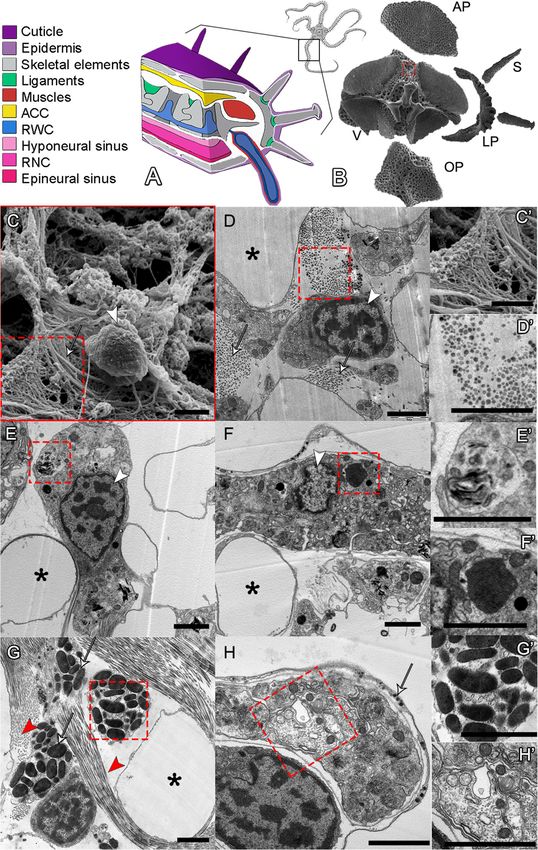
Fig. 1 A. filiformis non-regenerating arm skeletal elements and ultrastructure of cell types. A 3D schematic representation of a non-regenerating
arm showing the main anatomical structures colour coded as in legend on the left. B SEM of the skeletal elements present in a single metameric
unit and displayed in their corresponding anatomical position. AP, aboral arm plate; LP, lateral arm plate; OP, oral arm plate; S, spine; V, vertebra.
C SEM micrograph of a cell in the stroma of the skeleton of the vertebra: arrowhead indicates the nucleus, white arrow points at fibrils with
morphology typical of collagen, magnified in C’. D TEM micrograph of a sclerocyte in the vertebra: white arrows indicate fibrils in cross section
likely of collagen, magnified in D’. E TEM micrograph of a presumptive pigment cell in the oral arm plate with an evident nucleolus and spindle-
shaped electron-dense structures typical of pigment granules, magnified in E’. F TEM micrograph of a phagocyte in the aboral arm plate
characterized by the presence of phagosome, magnified in F’. G TEM micrograph of a granulocyte present in the vertebra: red arrowheads
highlight fibrils, present in both cross and longitudinal sections; arrows point at large electron-dense granules of different shapes and sizes,
magnified in G’. H TEM micrograph of a nerve cell in the aboral arm plate characterized by a nerve process magnified in H’; white arrow
indicates the small electron-dense roundish vesicles generally containing neurotransmitters or other neuro-signalling molecules. Scale bars =
2 μm. In all images: asterisks indicate the presence of biomineralized skeletal tissue (stereom calcareous elements or trabeculae) now empty, i.e.
electron-transparent due to fixation and decalcification processes; white arrowhead shows the nucleus with hetero- and euchromatin; and red
dotted box indicates magnified part of the figure
the primordia of new ossicles, called spicules, appear point of the regenerative process and the morphology of
in the developing dermal layer and that the cells of differentiated cells can be clearly distinguished. Histo-
this area express skeletogenic markers, such as alx1, logical and ultrastructural analyses were performed on
ets1/2, gataC, c-lectin, p19, and p58b [33, 34]. Cell the five skeletal elements at different positions in the
proliferation assays using 5-ethynyl-2′-deoxyuridine arm. As shown in Fig. 1, we observed a variety of cell
(EdU) showed that the cells of this layer are not proliferat- morphologies typical of sclerocytes (C, D); presumptive
ing and that even at later stages of regeneration sclero- pigment cells (E), mainly found in the aboral and oral
cytes marked by differentiation markers do not proliferate arm plates; phagocytes (F); granule cells (G); and neur-
[34]. Since the number of sclerocytes increases consider- onal cells (H). Among these cell types, sclerocytes repre-
ably during regeneration, but they do not proliferate, the sent the minority of the populations: they are
origin of these cells remains unknown. characterized by a roundish cell body with little cyto-
This work aims to understand the source of sclerocytes plasm and a patchy nucleus, and are immersed in a
during arm regeneration using microscopy at different re- matrix rich in fibrils and microfibrils, likely collagen
generative stages combined with molecular markers. In (Fig. 1C, D). In fact, both the fibril diameters (visible in
order to do this, we first identified various cell types cross section) and D-period (visible in longitudinal sec-
present in the stroma of the fully differentiated skeleton of tion) are compatible with those of collagen fibrils.
non-regenerating arms to assess their appearance and dif- We then studied the cell types present in the regenerat-
ferentiation during regeneration. We then characterized ing arms at different stages using similar structural and ul-
regenerating cells at both ultrastructural and molecular trastructural approaches. Consistent with previous studies,
level using a combination of new and known skeletogenic our analysis shows that the regenerative bud (stages 2 and
genes. Finally, we analysed the spatial expression of 19 3) is composed of the covering epidermis (shown in pur-
genes at early and advanced regenerative stages to unravel ple in most schematics) and an inner bulk made by the re-
the mechanisms used to re-establish the complexity of the generating axial structures, i.e. the ACC (yellow), the
skeletal elements in the brittle star arms. RWC (blue), and the RNC (pink) [34]. Between the epi-
Our data support the hypothesis that sclerocytes dif- dermis and the axial structures, the dermal layer, where
ferentiate from a population of progenitor cells emerging skeletal elements appear, is also present [34].
from the epithelium of the aboral coelomic cavity. Fur- In the regenerating dermal layer, we observed only
thermore, we show that different molecular signatures three distinct cell morphologies. Two out of these three
characterize the development of the different skeletal el- were observed more rarely, and only from stage 3 on-
ements (e.g. vertebrae and arm plates). wards, and present the morphology of morula (or spher-
ule) cells (Additional file 1 Fig. S1 A) and phagocytes
Results (Additional file 1 Fig. S1 B). The more abundant cell
Histological and ultrastructural characterization of population is characterized by a population of mesen-
sclerocyte precursors chymal cells with highly patchy and heterochromatin-
To better characterize cells involved in the regeneration rich nuclei, abundant rough endoplasmic reticulum
of the skeleton in A. filiformis, we first performed histo- (RER) and mitochondria, electron-dense vesicles, and a
logical and ultrastructural analyses of non-regenerating secondary boundary layer (Fig. 2). Among cells of this
arms to identify mature cell types. This is because fully population, we can observe some gradual differences be-
developed non-regenerating arms represent the end tween cells closer to the axial structures (ACC, RWC,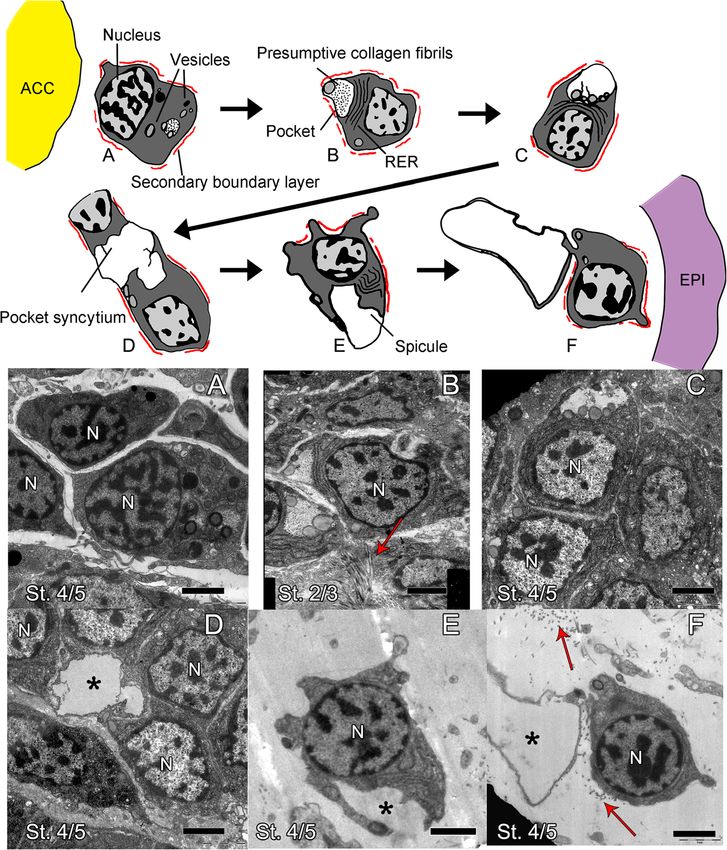
Piovani et al. BMC Biology       (2021) 19:9                                                                                                 Page 5 of 19

 Fig. 2 The main cell population of the dermis at early regenerative stages shows a gradient of cellular differentiation. Schematics (top) and TEM
 micrographs (a–f). a Mesenchymal cells at stage 4/5 located at the distal tip near the axial structures look rather undifferentiated. Main features
 are a secondary boundary layer (always highlighted in red in the schematics), a few vesicles, and a patchy nucleus. b Mesenchymal cells at stage
 2/3 in the area next to where the ACC and RNC are adjacent on either side of the RWC. Cells show large RER, many vesicles, and a cytoplasmic
 pocket containing fibrils likely of collagen. c Mesenchymal cells at stage 4/5 at the very distal tip of the regenerate. Cells show large RER, vesicles,
 and a more electron-transparent cytoplasmic pocket. d Mesenchymal cells at stage 4/5 at the distal-most tip of the regenerate show a pocket
 syncytium. e, f Mesenchymal cells at stage 4/5 right under the epidermis show growing spicules. The main cellular features are indicated in the
 schematic the first time they appear. Red arrows indicate collagen fibrils, capital N indicates nucleus, and asterisks indicate growing spicules. ACC,
 aboral coelomic cavity; EPI, epidermis; St., stage. Scale bars = 2 μm

and RNC) and those closer to the epidermis (Fig. 2 and                         onwards), we can also find them in the space between
Additional file 1 Fig. S2). Cells closer to the axial struc-                   the RWC and the ACC. Here, we can observe numer-
tures (Fig. 2a, b and Additional file 1 Fig. S2 A-C)                           ous cells containing phagosomes (Additional file 1
appear very tightly packed and rather undifferentiated;                        Fig. S2 C, red triangle) and apoptotic nuclei, charac-
they are overall roundish and have little cytoplasm                            terized by condensation of chromatin on the nuclear
and few cell inclusions. At all stages, these cells are                        membrane (Additional File 1 Fig. S2 C, red circle),
localized where the ACC and the RNC meet at either                             signs of tissue remodelling that are scarcely detectable
side or in front of the RWC in the most distal part                            elsewhere. This is the area where later in the regener-
of the regenerative bud (see Additional file 1 Fig. S2).                       ation process (from stage 5) vertebral primordia will
At later regenerative stages (from around stage 4                              start to form [34] (see Additional file 1 Fig. S3).
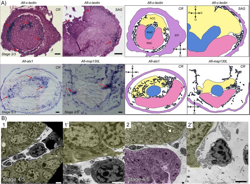
Piovani et al. BMC Biology        (2021) 19:9                                                                                                  Page 6 of 19

  In all areas described above, cells in the developing                         and E). Here, serial sections from different samples at dif-
dermal (or mesenchymal) layer are in very close contact                         ferent stages seem to show a few cells detaching from the
with the epithelium of the ACC, the RWC, and the                                ACC epithelium towards the mesenchymal space (Fig. 3b).
RNC. However, they have a very different morphology                             Cells closer to the epidermis display gradually more cyto-
from nearby cells of the axial structures which have a big-                     plasmic projections, often have a secondary boundary
ger, more electron-dense cytoplasm, large nucleolus, and                        layer resembling those described for sclerocytes by Märkel
many cell inclusions (compare mesenchymal cells in Fig. 2                       and Röser [17], and display a pocket-like structure con-
and Additional file 1 Fig. A2 with Additional file 1 Fig. S4                    taining presumptive collagen fibrils. They also have a
A”, C, E and G). Cell boundaries within the axial struc-                        growing number of electron-opaque vesicles gathering
tures are difficult to detect (see Additional file 1 Fig. S4                    next to this plasma membrane invagination (Fig. 2a–d).
A”, C, E, G); however, the RWC (Additional file 1 Fig. S4                       The content of the pocket of cells near the axial structures
D, see arrow), the RNC (Additional file 1 Fig. S4 F, see                        appears more electron-opaque than that of cells closer to
arrow), and the epidermis (Additional file 1 Fig. S4 G’)                        the epidermis (compare Fig. 2b with Fig. 2c, d) possibly in-
show a clear basal lamina while the ACC apparently does                         dicating that the composition of the pocket content
not (Fig. S4 B). The epithelium lining of the ACC loses its                     changes. The short cell projections delimiting the pocket
continuity and “disaggregates”, especially on its oral side,                    of one single cell never fuse together; therefore, this space
facing the RWC and RNC (see Additional file 1 Fig. S4 A,                        remains “extracellular”. However, in some cases, different
A’ and A” and compare with Additional file 1 Fig. S4 C                          neighbouring cells “fuse” their projections to form a

 Fig. 3 Regenerating mesenchymal cells express skeletogenic gene markers and appear to detach from the ACC epithelium. a Semi-thin sections
 of whole mount ISH for skeletal genes: Afi-c-lectin (embedded in resin), Afi-alx1 and Afi-msp130L (embedded in wax). Red arrows point at areas
 where gene expression (dark blue/purple staining) is present, as schematized on the right. Yellow, aboral coelomic cavity; blue, radial water canal;
 pink, radial nerve cord; CR, cross section; SAG, sagittal section. Crossed arrows further indicate the orientation of the section: A, aboral; O, oral; R,
 right; L, left; P, proximal; D, distal. Scale bars = 10 μm. b TEM micrographs of cells likely detaching from the ACC epithelium. (1’) and (2’) are details
 of (1) and (2), respectively. Yellow, ACC; pink, RNC. Scale bars = 2 μm
Piovani et al. BMC Biology (2021) 19:9 Page 7 of 19
syncytium delimitating a shared extracellular space, i.e. To further characterize the molecular signature of these
pocket space (Fig. 2d). Cells immediately under the epider- cells, we then selected 16 genes with known skeletogenic
mis are scattered in a relatively abundant matrix, composed roles. Among these genes, we chose ten TFs of which six
of banded collagen fibrils and microfibrils. Several sections have a role in the sea urchin and/or brittle star embryonic
show that these cells are often in very close proximity to de- skeletogenic gene regulatory networks (GRNs) (erg, foxN2/3,
veloping spicules (Fig. 2e, f and Additional file 1 Fig. S2 D), jun, nk7, snail, and twist) [23, 24, 26, 39–42]. We additionally
and ultrastructural analysis shows a morphology similar to cloned four TFs known to be involved in bone formation in
that of mature sclerocytes (compare Fig. 1D with Fig. 2e, f). vertebrates (pax1/9, soxE, sp5, and sp7/8) [43–48].
We observed that, morphologically, this population of Afi-erg and Afi-jun show staining in the developing
mesenchymal cells adjacent to the epidermis appears dermal layer (Fig. 4) with Afi-jun presenting additional
more differentiated (i.e. they have a lower nucleus/cyto- staining in the epidermis. Afi-nk7 stains the epidermis,
plasm ratio, several cytoplasmic projections, and various the developing dermal layer, and the ACC, starting from
inclusions) closer to the amputation plane compared to stage 4/5. Afi-snail signal is also in the developing der-
the tip as well as in later stages of development (stage 4/ mal layer, but it is only localized at the distal end of the
5) compared to early ones (stage 2/3) (see the top of arm (Fig. 4 and Additional file 2 Fig. S5).
Additional file 1 Fig. S2). All cell morphologies described The transcription factors Afi-soxE and Afi-twist are
above have been consistently observed across different expressed in the distal ACC epithelium with Afi-soxE
samples and stages unless otherwise stated in the text. showing additional staining in the developing lateral arm
To summarize, by analysing serial sections of samples at plates in the proximal region of the regenerates starting
different regenerative stages, we observed a population of from stage 4/5 (Fig. 4 and Additional file 2 Fig. S5).
mesenchymal cells, which appears to bud off from the oral Both Afi-foxN2/3 and Afi-sp7/8 show expression in the
side of the ACC that faces the RWC/RNC. These cells ACC epithelium as well as in the tip of the RNC (Fig. 4)
show a gradient of morphologies with cells closer to the with Afi-foxN2/3 showing additional staining in the epi-
axial structures having a more undifferentiated morphology dermis as well (see Additional file 1 Figs. S3 and S5).
than those closer to the epidermis. Cells right under the Lastly, Afi-sp5 show expression only in the epidermis
epidermis are often in close contact with developing spic- and Afi-pax1/9 show no staining at all stages analysed
ules and resemble mature sclerocytes. Gene expression of (Additional file 2 Fig. S6). To control for correct func-
molecular markers might help elucidate their true identity. tioning of the Afi-pax1/9 probe, we conducted whole
mount in situ hybridization in developing larvae of A.
filiformis. Here, the probe shows clear expression in a re-
Localization of skeletogenic gene expression at early gion of the ectoderm of the blastula and in ectodermal
stages of regeneration cells of the lower arms, adjacent to the mesenchymal
To assess the identity of the cells described above, we car- cells at the gastrula stage (Additional file 2 Fig. S6).
ried out a large-scale gene expression study using in situ The other six genes selected are skeletogenic differentiation
hybridization (ISH) of both transcription factors (TFs) and genes that are either expressed in the skeletogenic cells of devel-
differentiation genes known to be involved in skeletogen- oping brittle star and sea urchin embryos [23, 26, 49] or are
esis. All ISH experiments were conducted and analysed in proteins found in adult skeletal components of sea urchin and
at least three replicates for each stage, and only consistent brittle star [50–52]. During the early stages of regeneration, Afi-
results are reported here. Whole mount samples were ori- msp130L, Afi-slc4a10, Afi-mt14/mmpl7, and Afi-p58a were
ented for imaging, and both oral and aboral sides were found expressed specifically in the dermal layer (Fig. 4). Afi-kir-
analysed to determine the expression in ACC and RNC. relL and Afi-tetraspanin only show expression in the dermal
Firstly, we analysed the expression of the three skeleto- layer at stage 4/5, while no signal is detectable before (Fig. 4).
genic markers msp130-L, c-lectin, and alx1; the latter two Quantitative analysis of six TFs (Afi-erg, Afi-foxN2/3, Afi-jun,
were previously shown to be broadly expressed in the der- Afi-nk7, Afi-snail, and Afi-twist) and of all six differentiation
mal layer of A. filiformis regenerating arms [33, 34]. To genes agrees with what was observed in ISH (see Additional file
gain better cellular resolution, we performed whole mount 2 Fig. S7 and Table S1). Moreover, most differentiation genes
ISH and then sectioned the arms after embedding in wax show upregulation of their expression as regeneration pro-
and/or resin. Our sections show that not only cells under gresses (see Additional file 2 Fig. S7 and Table S1).
the epidermis but also those in close contact with axial
structures and those present between the ACC and RWC Localization of skeletogenic gene expression at late
at later stages show expression of these molecular markers stages of regeneration
(Fig. 3, red arrows). Moreover, our sections clearly show We then looked at gene expression of the same molecu-
that Afi-alx1 is also expressed in the oral side of the ACC lar markers at later stages of regeneration when 50% or
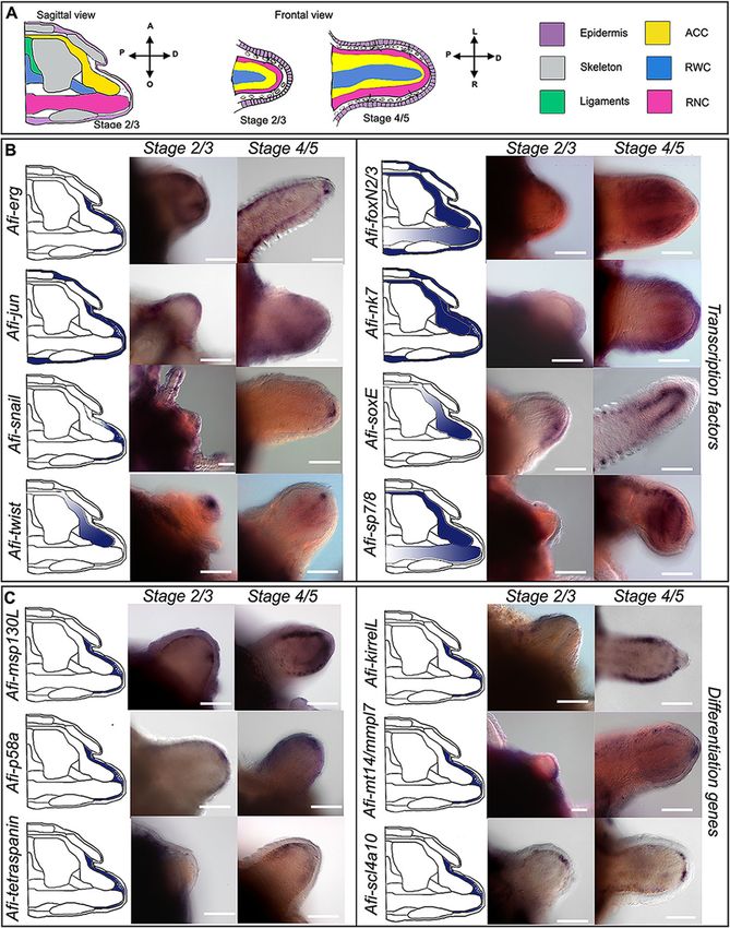
epithelium (Fig. 3a). more of the arm had already differentiated (DI:Piovani et al. BMC Biology (2021) 19:9 Page 8 of 19 Fig. 4 Gene expression at early stages of regeneration (stage 2/3 and stage 4/5). a Colour coded sagittal scheme of stage 2/3 and frontal schemes of stages 2/3 and 4/5. Crossed arrows near schematic indicate the axis of the section. Schematics of gene expression in b and c follow the sagittal scheme as this view allows clear distinction of all tissues. Pictures of gene expression in whole mount are taken in frontal view from the aboral side after orienting the samples, unless otherwise specified. b Whole mount ISH at two regenerative stages (stage 2/3 and stage 4/5) as indicated at the top of the columns using antisense probes for transcription factors. Probe name is indicated on the left of the summary schematics. c Whole mount ISH at two regenerative stages (stage 2/3 and stage 4/5) as indicated at the top of the columns using antisense probes for known differentiation genes. Probe name is indicated on the left of the summary schematics. In b and c, summary schematics of expression are based on several images of different focal plane observations of multiple samples; however, only one focal plane is shown here. A, aboral; O, oral; R, right; L, left; P, proximal; D, distal. Dark blue/purple indicates probe-specific signal. Scale bars = 100 μm differentiation index). At this point, the complex archi- dermal domain, whereas Afi-soxE is again localized to tecture of the skeletal elements starts forming. In gen- the ACC epithelium. Afi-nk7 is expressed in both the eral, although many of these genes were expressed epidermis and the dermal layer in a repetitive pattern ubiquitously in the dermis at early stages, at later stages, that follows the segments. Afi-snail, Afi-foxN2/3, and the situation is quite different (Fig. 5). Afi-sp7/8 show expression in the tip of the RWC, in In the distal undifferentiated end of the regenerate what is called the terminal podium (see Fig. 5 and Add- proximal to the tip (where the terminal differentiated itional file 2 Fig. S6). The latter two also show expres- structures are present), Afi-jun is expressed in a broad sion respectively in the epidermis (Afi-foxN2/3) and the
Piovani et al. BMC Biology      (2021) 19:9                                                                                               Page 9 of 19

 Fig. 5 Gene expression at late stages of regeneration. a Sagittal schematic of regenerating arm at late stages of regeneration with proximal
 (Prox.) and distal (Dist.) schematics of cross sections. Purple, epidermis; grey, skeleton; yellow, ACC; blue, RWC; pink, RNC. b Whole mount ISH
 (proximal and distal position as indicated at the top of the columns), using antisense probes for transcription factors. c Whole mount ISH
 (proximal and distal position as indicated at the top of the columns), using probes for known differentiation genes. For b and c, probe name is
 indicated at the bottom of the proximal image. On the left of each pair of ISH pictures, there is a summary of expression (blue) in cross
 schematics for the proximal (Prox.) and distal (Dist.) arm. Summary schematics of expression are based on focal plane observations of multiple
 samples; however, only one focal plane is shown here. In the distal figures, dotted squares indicate the differentiated terminal structure (terminal
 ossicle and podium) and black lines indicate the proliferating area. The summary expression data in the schematic is taken from the proliferating
 area. Scale bars = 50 μm

ACC epithelium and the RNC (Afi-sp7/8) (Fig. 5). Afi-                        foxN2/3 which additionally show expression in the lateral
erg, Afi-sp5, and Afi-twist are not expressed in this re-                    arm plates (Fig. 5). Afi-snail shows a wider expression in
gion (Fig. 5 and Additional file 2 Fig. S6).                                 all skeletal elements except the oral arm plates (Fig. 5).
   In proximal segments, the differentiating skeletal ele-                   Afi-soxE and Afi-twist show staining, respectively, only in
ments express various combinations of transcription fac-                     the lateral arm plates, vertebrae, and ACC (Fig. 5). Afi-nk7
tors (Fig. 5). For example, Afi-jun and Afi-rreb1 show                       expression is confined to the vertebrae only (Fig. 5). Afi-
expression in both vertebrae and spines (Fig. 5 and Add-                     sp5, Afi-sp7/8, and Afi-pax1/9 are not expressed at all in
itional file 2 Fig. S6). This is also true for Afi-erg and Afi-              the skeleton (Fig. 5 and Additional file 2 Fig. S6).
Piovani et al. BMC Biology (2021) 19:9 Page 10 of 19
Three of the downstream genes analysed (Afi- S8) suggesting that sclerocytes in the regenerating arm
msp130L, Afi-kirrelL, and Afi-scl4a10) show staining in are secreting at least these types of collagens.
the dermal layer of the proximal regenerating arm at
later stages as well as during early stages. Consistent Discussion
with their role as terminal differentiation genes, the This work aims to shed light on the origin of sclerocyte
other downstream genes analysed (Afi-p58a, Afi-tetra- precursors and analyse their differentiation process in the
spanin, and Afi-mt14/mmpl7) do not show any staining regenerating arm of the brittle star Amphiura filiformis.
in the distal-most part of the regenerating arm, but only Using a combination of ultrastructural and molecular ana-
in more developed proximal metameric units. lysis, we show that sclerocyte precursors likely originate
As for the proximal part, Afi-msp130L is the most from the epithelium of the aboral coelomic cavity. More-
widely expressed and is present in all external skeletal over, we characterize the morphology and molecular sig-
domains (all arm plates and spines) but not in the verte- nature of these cells as they differentiate. Finally, we show
brae (Fig. 5). Afi-p58a and Afi-tetraspanin are restricted that at advanced regenerative stages unique combinatorial
to vertebrae and lateral arm plates, whereas Afi-kirrelL gene expression underlies the patterning of the different
also shows expression in the spines (see Fig. 5). Afi- skeletal elements. Altogether, our results highlight many
slc4a10 is expressed in vertebrae, spines, and aboral arm similarities between adult and embryonic skeletogenesis in
plates, and Afi-mt14/mmpl7 is expressed in spines, ver- echinoderms and may help to unravel the origin and evo-
tebrae, and oral arm plates (see Fig. 5). lution of the deuterostome skeleton.
Altogether, a pattern arises with vertebrae, lateral arm
plates, and spines expressing more TFs and downstream The origin of sclerocytes in the brittle star regenerating
genes than oral and aboral arm plates. skeleton
Similarly to what shown for early stages of regeneration, The aim of this work was to understand the origin of
quantitative data of six TFs (Afi-erg, Afi-foxN2/3, Afi-jun, sclerocytes as well as their differentiation during arm re-
Afi-nk7, Afi-snail, and Afi-twist) and of all six differenti- generation in A. filiformis, following traumatic amputa-
ation genes at later stages are consistent with the ISH pat- tion. For this reason, we analysed the ultrastructure of
terns observed (see Additional file 2 Table S1). and gene expression in cells of the developing dermal
layer where the skeleton forms [34]. As cells in the de-
Collagen deposition veloping dermis had been previously shown to be non-
The skeletal stroma of the non-amputated arm and the proliferative, part of this work was aimed at finding a
developing dermis of the regenerating arm show large potential alternative source. Apart from the developing
quantities of collagen fibrils and microfibrils (Fig. 1C, D, dermis, there are only four other tissues in the regener-
G; Fig. 2f; Additional file 1 Fig. S2 A, D). Additionally, ating arm of A. filiformis, all of which are proliferative
the above described pocket-like structure of the sclero- from stage 2/3: the epidermis, the ACC, the RWC, and
cyte precursors contains presumptive collagen fibrils, the RNC [34]. Our gene expression results at early stages
which are comparable in terms of ultrastructural appear- of regeneration (stages 2/3 and 4/5; Figs. 3 and 4 and
ance and size with those widespread in the dermal tissue Additional file 2 Fig. S5) combined with previously pub-
(Fig. 2b). Fibroblasts, cells involved in collagen depos- lished data [33] show that the ACC epithelium is charac-
ition, have been described in ophiuroids as rather undif- terized by the expression of several TFs, which includes
ferentiated cells that resemble sclerocytes but lack alx1, ets1/2, foxN2/3, gataC, nk7, soxE, and twist (Fig. 6a).
branches and distal processes [17]. As they do not The expression of several TFs implicated in embryonic
present a clear morphology, we could not distinguish skeletogenic lineage specification in the ACC epithelium
them from sclerocytes or sclerocyte precursors. There- supports the hypothesis that this tissue could be a po-
fore, we tried to investigate whether collagen secretion tential source of skeletogenic cells among other cell
and biomineralization were performed by the same cells, types. Those TFs could be responsible for the specifica-
at least during regeneration, as this would allow us to tion of cells of the ACC before detachment similarly to
better characterize the role of sclerocytes during regen- the specification of the skeletogenic mesodermal cells of
eration. For this purpose, we selected two collagen genes sea urchin and brittle star embryos before ingression.
that previous studies identified as expressed in the der- However, the exact regulatory state of specified sclero-
mal layer: Afi-alpha-collagen and Afi-col-L C [33, 53] cyte precursors in the ACC will be revealed by further
and one well-established skeletogenic marker (Afi-c-lec- studies using double in situ and/or single cell sequen-
tin) and performed fluorescent double whole mount in cing. Once specified, sclerocyte precursors could per-
situ hybridizations. Our results on late stages of regener- form EMT, detaching from the epithelium to enter the
ation show that cells in the skeleton co-express c-lectin developing dermal layer. This hypothesis is supported by
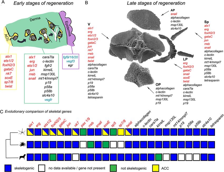
and these two types of collagen (Additional file 2 Fig. our TEM data (Fig. 3b) on serial sections, which showPiovani et al. BMC Biology (2021) 19:9 Page 11 of 19 Fig. 6 Summary of the origin of skeletogenic cells and the evolution of the molecular signature in deuterostomes. a Model of main hypothesis of sclerocyte origin and differentiation at early stages of regeneration (stage 2/3 and 4/5): new sclerocyte precursors detach from the aboral coelomic cavity (ACC) epithelium and differentiate while moving towards the epidermis together with summary of regulatory (red), differentiation (black), and signalling (blue) gene expression of the ACC, the dermis, and the epidermis. Schematics are based on data shown in Fig. 2 and Additional file 1 Fig. S2; gene expression data is based on Figs. 3 and 4 and Additional file 2 Figs. S5 and S6 combined with already published data (References in Table S2). b Summary of molecular signature of different skeletal elements at the proximal side of late stages of regenerating arms. Gene expression data is based on proximal differentiating skeletal elements shown in Fig. 5 and Additional file 2 Figs. S5 and S6 together with published data. AP, aboral arm plate; LP, lateral arm plate; OP, oral arm plate; Sp, spine; V, vertebra. c Phylogenetic tree with conserved role of skeletogenic genes between the brittle star Amphiura filiformis (present work), Strongylocentrotus purpuratus, and vertebrates. Genes encoding for transcription factors are in red, and the differentiation genes are in black. For details and relative references, see Additional file 2 Table S2. For this summary figure, we considered the gene present if we could find data on a member of the gene family; therefore, it is not a strict 1:1 orthology, considering also the two rounds of genome duplication occurred at the base of vertebrate evolution cells likely detaching from the ACC, whereas no cells and S4 A and A’). The ACC has already been proposed were captured detaching from the RWC or the RNC in as a possible source of cells during regeneration of both all TEM images analysed. From a mechanical point of A. filiformis and Ophioderma longicaudum [36] and of view, it would be easier for cells to detach from the ACC other echinoderms as well [11, 55, 56]. In fact, during epithelium due to the lack of a thick basal lamina in the crinoid and starfish arm regeneration, as well as in holo- regenerating tip, which instead is present in the regener- thuroid visceral regeneration, cells detach from the epi- ating RNC and RWC. The RWC, particularly, is nor- thelia of the regenerating coelomic (somatocoel) mally provided with a very thick basal lamina [54] (in compartment and ingress the underlying mesenchymal Additional file 1 compare Fig. S4 D with B, F and G’). tissue. Nevertheless, the RWC has also been previously Moreover, cells in the oral epithelium of the ACC ap- proposed as a source of cells during brittle star regener- pear more loosely connected (Additional file 1 Fig. S2 ation [57].
Piovani et al. BMC Biology   (2021) 19:9                                                                        Page 12 of 19

  To corroborate our hypothesis, which is based on              and thin filopodia, but only very short extensions. This
static TEM and ISH images, it will be fundamental to            is possibly linked to the physical constrictions experi-
perform lineage tracing experiments and develop tools           enced by the population of skeletogenic cells, which in
for knockouts of TFs expressed in these cells to confirm        the embryo are freely moving in a cell-deprived fluid of
their role in sclerocyte specification.                         the blastocoel, but in the regenerating arm are highly
                                                                crowded in an extracellular matrix-rich dermal layer.
Progression of sclerocyte differentiation during                   Finally, cells reach the region just beneath the epidermis
regeneration                                                    where they appear scattered in a dermal layer filled with
Assuming that the cells described detach from the epithe-       collagen fibrils and growing spicules. At this stage, cells
lium of the ACC, our work suggests that, as they migrate        acquire a more differentiated morphology, resembling that
towards the epidermis, they start differentiating as in the     of mature sclerocytes: the cytoplasm surrounding the nu-
model presented in Fig. 6a and in Fig. 2. In fact, mesenchy-    cleus is reduced, with cell projections extending towards
mal cells closer to the axial structures (and the ACC) look     the growing spicules (Fig. 2e, f). The expression of several
less differentiated than those adjacent to the epidermis, al-   biomineralization genes specifically in the dermal layer is
though they display some characteristics of mature sclero-      consistent with the fact that in this location cells are in-
cytes of other ophiuroids and of cidaroid echinoids: patchy     deed producing the skeleton (Figs. 6a, 3, and 4) [33].
nucleus, abundant RER, several mitochondria, and a sec-            Recent studies have shown that fgf9/16/20 and vegf3
ondary boundary layer [17, 18, 32, 58]. We hypothesize that     signals from the epidermis of A. filiformis are necessary
most of these cells are sclerocyte precursors.                  for the formation of biomineralized spicules during re-
   Consistent with this hypothesis, we show that cells          generation [25]. This signal could drive the migration
close to the axial structures already express skeletogenic      and final differentiation of sclerocyte precursors and ini-
genes, such as c-lectin, alx1, and msp130L (Fig. 3).            tiation of deposition of the biomineralized spicule that
   As these cells move towards the epidermis, they in-          we show ultrastructurally (see Fig. 2e, f and Additional
crease the amount of cytoplasm, project cytoplasmic             file 1 Fig. S2 D).
branches, and develop a pocket-like structure that de-             It is worth noting that the process of apparent migration
limits an extracellular space, which is initially filled with   towards the epidermis and subsequent differentiation just
presumptive collagen fibrils and then becomes more              described is valid for the external skeletal elements of the
electron-transparent (in Fig. 2 compare b with c). As           arm (arm plates and spines). The sclerocyte precursors of
TEM images show, the cytoplasmic projections of the             the vertebrae, which have been briefly described here as
pockets later fuse with those of other nearby cells and         the cells present in between the ACC and the RWC at
this is where spicules begin to form, similarly to what         stage 4/5 (Additional file 1 Fig. S2 C), might undergo
happens in both sea urchin and brittle star embryos             slightly different cellular and molecular processes, as they
(Fig. 6a and Fig. 2) [59, 60]. Consistent with the acquisi-     never come in contact with the epidermis. Here, other tis-
tion of cell differentiation features, cells in the dermal      sues, such as the RWC, might function as “inductive ele-
layer express a large set of skeletogenic differentiation       ments” promoting sclerocyte differentiation.
genes (for summary see Fig. 6a) and have a specific regu-          Besides sclerocytes, very few cell types were observed
latory state (combination of TFs) that partially overlaps       in the derma. Presumptive pigment cells, granulated
with that of the ACC. The two transcription factors alx1        cells, and nerve cells identified in the mature stroma of
and ets1/2 in common between the ACC and the differ-            the skeleton were not found at early stages of regener-
entiated sclerocyte are known in the sea urchin embryo          ation, suggesting that they might populate this tissue
to constitute the key regulators of the skeletogenic gene       only later. Fibroblasts, which are normally involved in
battery and cellular features [61]. Therefore, our data         collagen secretion, could not be unambiguously identi-
suggest that the programme underlying the cellular              fied either in the mature or in the regenerating arm al-
mechanism of spicule formation within a cellular syncyt-        though large quantities of collagen and microfibrils fill
ium takes place not only in embryonic but also in post-         the stroma and the developing dermis. Since in the lit-
metamorphic skeleton development, and it is a shared            erature fibroblasts are described as morphologically
feature with other echinoderm classes (e.g. Echinoidea).        similar to sclerocytes, we wanted to further investigate if
Consistent with this, we also show that Afi-kirrelL,            in A. filiformis they are indeed two distinct cell types
which in sea urchins is responsible for the fusion of filo-     [17]. Our results at advanced stages of regeneration
podia necessary for larval skeleton deposition [62], is         show that cells in the skeleton co-express two collagen
expressed from stage 4/5 in the dermal cells, when skel-        genes with the skeletogenic marker c-lectin (Additional
etal primordia are actively deposited [34]. Differently         file 2 Fig. S8). This suggests that sclerocytes also play a
from sea urchin embryos, in adult regenerating arms of          role in collagen secretion and gives a molecular signifi-
A. filiformis, sclerocyte precursors do not produce long        cance to the ultrastructural observations made by
Piovani et al. BMC Biology (2021) 19:9 Page 13 of 19
Märkel and Röser [17] and Heatfield and Travis [32]. observations). In conclusion, we suggest that the differ-
Moreover, two other collagen genes (Afi-col-L B and C) ences in molecular signatures may reflect the complexity
have recently been shown to be expressed in the devel- of morphologies of the different skeletal elements as well
oping dermal layer of A. filiformis at early regeneration as anatomical position.
stages (3/4) and in the skeleton at later stages [53]. Moreover, our optimized technique for WMISH sec-
Altogether, our cellular and molecular findings suggest tioning in resin/wax could be a powerful tool to verify
a conserved mechanism of sclerocyte differentiation dur- whether there is a correlation between different stereom
ing development and regeneration in echinoderms. This densities and molecular signatures. This might be of par-
comprehensive, molecular, and ultrastructural study ticular interest in the field of palaeontology since the pore
forms the basis for future functional work. size and thickness of trabeculae vary depending on the
type of soft tissues attached to the skeleton, and hence on
Unique combinatorial gene expression underlies the their specific functions [64]. It would be also interesting to
patterning of the different skeletal elements during explore the role of signalling coming from different tissues
regeneration in skeleton development. It has been recently confirmed
Once sclerocytes are fully developed, they start secret- that signalling from the epidermis is crucial for the onset
ing calcium carbonate in the form of single spicules. of biomineralization in arm plates. However, we know
These spicules then grow and fuse to form the 3D very little about signalling from other tissues [25]. The
meshwork typical of the stereom and the final ossicles vertebrae, for example, which have no contact with the
of the arm. If we look at gene expression at this ad- epidermis, could receive signalling from other tissues,
vanced stage of regeneration, we notice two very in- such as the ACC, the RWC, and the RNC, and this could
teresting patterns arising. orchestrate the formation of the different grooves.
First, we observe that in the distal-most part of the re-
generating arm, right underneath (proximal to) the dif- Evolutionary origin of skeletogenesis in deuterostomes
ferentiated terminal ossicle and podium, gene expression We have so far discussed in detail skeletal regeneration in
of seven genes highly resembles that of early stages (Afi- the mature arms of A. filiformis and highlighted some simi-
jun, Afi-nk7, Afi-soxE, Afi-sp7/8, Afi-msp130L, Afi-kir- larities with sea urchin development. Other deuterostome
relL, and Afi-scl4a10). This further supports the idea groups, such as vertebrates, are in some cases able to regen-
that echinoderm arm regeneration follows the erate the endoskeleton of their appendages [65–67].
distalization-intercalation mode of regeneration as pro- We therefore analysed a number of genes that are
posed previously [34, 63]. known to have a role in both sea urchin skeletal devel-
Secondly, although many of the genes analysed showed opment and in vertebrate chondrogenesis and/or bone
a broad dermal domain at early stages of regeneration, at formation. We compare skeleton formation in echino-
later stages in the proximal region (older and most devel- derms with both cartilage and bone formation in verte-
oped segments), we observed that different sets of genes brates as the earliest vertebrate skeleton was likely made
are expressed in different skeletal elements. In particular, of unmineralized cartilage [68].
vertebrae, lateral arm plates, and spines show expression There are clear differences in the way that vertebrates
of many more genes (both TFs and differentiation genes) and echinoderms produce their biomineralized tissues;
than oral and aboral arm plates (Figs. 5 and 6b). This is notably, they use different minerals: calcium phosphate
consistent with the degrees of stereom organization com- for vertebrates and calcium carbonate for echinoderms
plexity and densities in these different skeletal elements [68]; the vertebrate crystallization pathway involves
(Additional file 2 Fig. S9). Vertebrae, for example, show mineralization of extracellular matrix, while echinoderm
the highest degree of complexity, with different pore sizes mineralization is enclosed in a cell syncytium [69]. These
and rod thickness in the area where muscles or joints are differences are reflected in the evolution of clade-specific
present as well as grooves for the passage of the RNC, the mineralization genes, such as the echinoderm skeleto-
RWC, and the ACC. Lateral arm plates, which are shaped genic matrix (SM) genes, or the specific co-option of the
like a half-moon, also possess highly dense protrusions vascularization programme regulated by VEGF in sea ur-
where spines attach. Spines themselves have a conical cal- chins [70]. Mineralization in both vertebrates and echi-
cite structure and display an array of different shapes (long noderms, however, takes place in an extracellular space,
and thin, slightly thicker, and with hammer-shape tip) (see and it is secreted by mesenchymal cells. Our aim is not
Fig. 6b). By contrast, the oral and aboral arm plates have a to compare individual cell types across phyla (i.e. echino-
simpler, almost flat structure with even pore size and rod derm sclerocytes with vertebrate osteoblasts and chon-
thickness across the plate. Additionally, besides sclero- drocytes), which might be ineffective given the long
cytes, the set of cell types present in different mature skel- independent evolutionary history of these two deutero-
etal elements can be different ([54] and personal stome clades (at least 540 millions of years), but ratherPiovani et al. BMC Biology (2021) 19:9 Page 14 of 19 to explore the extent of conservation of the basic deu- share several genes [70]. Our cellular and molecular terostome biomineralization toolkit present in the last characterization of adult echinoderm sclerocytes will common ancestor before the divergence of echinoderms help further investigating the origin and evolution of the and chordates. deuterostome skeleton. Figure 6c summarizes the occurrence of our set of genes together with other published data in vertebrates, Conclusions sea urchin, and A. filiformis and their roles in skeleton This study provides cellular and molecular insights into formation (mineralized or not). the possible origin and differentiation of sclerocytes in Twenty-three out of 25 of these genes are expressed in brittle star arm regeneration following traumatic ampu- the skeletogenic tissues of A. filiformis during regener- tation. Our findings strengthen the hypothesis that adult ation. Of those 23 genes, 20 also show expression in the regeneration in echinoderms re-uses a developmental skeletogenic mesoderm of S. purpuratus embryos, indicat- programme. Moreover, our molecular data identify sev- ing a conserved role in both development and regener- eral commonalities between echinoderm skeletogenesis ation of the skeleton (for references see Additional file 2 and skeletal development in other deuterostomes. Table S2). Consistent with the studies carried out by Czarkwiani and colleagues [25], this observation supports Materials and methods the idea that skeletogenesis during regeneration highly re- Animal maintenance and handling sembles embryonic skeleton formation not only from a Adult (disc diameter ~ 0.5 cm) specimens of Amphiura cellular but also from a molecular point of view. filiformis O. F. Müller, 1776 were collected at the Sven Fifteen of the 25 genes chosen (alx1, erg, ets1/2, gataC, Lovén Centre for Marine Sciences in Kristineberg jun, rreb, snail, soxE, twist, alpha-collagen, cara7la, c- (Sweden). Experimental animals were kept in aerated lectin, mt1/4/mmpl7, slc4a10, and tetraspanin) also play aquaria of artificial seawater (ASW) (Instant Ocean®) at a role in skeleton (bone or cartilage) formation in verte- 14 °C and 34‰ salinity, and chemical-physical ASW pa- brates (for references see Additional file 2 Table S2). rameters were constantly checked. Animals were fed twice Additionally, we specifically considered three vertebrate a week with Microvore Microdiet (Brightwell Aquatics). sclerotome/chondrogenic markers, i.e. pax1, sox8/9/10, During all manipulations, animals were anesthetized in so- and sp7 or osterix [43–48, 69, 71]. Pax 1/9, the echino- lution of 3.5% MgCl2 in distilled water (dH2O) and derm homologue of vertebrate pax1, showed no staining ASW (1:1). To minimize stress, a maximum of two arms [43, 47]. Sp5 and sp7/8 are the sea urchin homologues of out of five per brittle star were artificially amputated with vertebrate sp7 (or osterix) [44, 46, 71]. Afi-sp5 was a scalpel at the level of natural autotomy planes (i.e. be- expressed only in the epidermis (at early stages of regen- tween plates/intervertebral articulations). Despite our ef- eration); however, Afi-sp7/8 was expressed at all stages forts to mimic pseudo-autotomic conditions, in the of regeneration in the ACC and in the RNC. Finally, present work, we specifically investigated post-traumatic soxE, the sea urchin orthologue of vertebrate sox8, sox9, regeneration, which might partially differ from natural and sox10 genes, was localized in the ACC at early stages post-autotomic regeneration [72, 73]. After amputation, and in the lateral arm plates at later stages of regener- the animals were returned to the aquarium and left to re- ation. The expression of Afi-sp7/8 and Afi-soxE in the generate for a given amount of time to reach the desired ACC agrees with our hypothesis on the coelomic origin stages: 2, 3, 4, 5, 50% differentiation index (DI) and 95% of sclerocytes; however, Afi-soxE skeletogenic role is DI [14, 34]. Following this step, regenerating arms (includ- additionally confirmed by its expression in skeletal ele- ing few segments of stumps) were collected for whole ments at later stages of regeneration. mount in situ hybridization (WMISH) experiments and These observations, together with the expression of microscopy analyses. alx1 and involvement of fgf signalling [25], identify an overlap in the molecular pathways used by sclerocytes in Transmission electron microscopy A. filiformis and skeleton-forming cells in vertebrates. As Samples for transmission electron microscopy (TEM) vertebrate osteoblasts and chondrocytes and echinoderm were processed as described by Ferrario et al. [54]. For sclerocytes all derive from mesenchymal precursors, we each stage (non-regenerating, stage 2, stage 3, stage 4, cannot rule out that this overlap could be due to a com- stage 5, 50% DI, 95% DI), at least four samples were used mon mesenchymal regulatory programme rather than a for semi-thin sectioning; the most informative were then conserved role in skeletogenesis. The use of regulatory used for ultrathin sectioning (at least two per stage). For modules encoding for specific cell/tissue processes, such each sample, we cut approximately 40 sections, a sub- as EMT or tubulogenesis, is in line with what recently sample of which were observed at the TEM. Semi-thin presented for the regulatory programme of vertebrate and ultrathin sectioning was performed with a Reichert endothelial cells and echinoderm sclerocytes, which Jung Ultracut E. Serial semi-thin sections (~ 1 μm thick)
You can also read