Regulation of Iron Homeostasis and Use in Chloroplasts - MDPI
←
→
Page content transcription
If your browser does not render page correctly, please read the page content below
International Journal of
            Molecular Sciences

Review
Regulation of Iron Homeostasis and Use
in Chloroplasts
Gretchen E. Kroh and Marinus Pilon *
 Department of Biology, Colorado State University Department of Biology, Fort Collins, CO 80523, USA;
 gkroh@colostate.edu
 * Correspondence: marinus.pilon@colostate.edu; Tel.: +1-970-491-0803

 Received: 30 March 2020; Accepted: 9 May 2020; Published: 11 May 2020

 Abstract: Iron (Fe) is essential for life because of its role in protein cofactors. Photosynthesis, in
 particular photosynthetic electron transport, has a very high demand for Fe cofactors. Fe is commonly
 limiting in the environment, and therefore photosynthetic organisms must acclimate to Fe availability
 and avoid stress associated with Fe deficiency. In plants, adjustment of metabolism, of Fe utilization,
 and gene expression, is especially important in the chloroplasts during Fe limitation. In this review,
 we discuss Fe use, Fe transport, and mechanisms of acclimation to Fe limitation in photosynthetic
 lineages with a focus on the photosynthetic electron transport chain. We compare Fe homeostasis in
 Cyanobacteria, the evolutionary ancestors of chloroplasts, with Fe homeostasis in green algae and in
 land plants in order to provide a deeper understanding of how chloroplasts and photosynthesis may
 cope with Fe limitation.

 Keywords: photosynthesis; iron homeostasis; green lineage; Fe–S; plants; Cyanobacteria;
 Chlamydomonas; chloroplast

1. Introduction
     In the early, anoxic, reducing environment of Earth, life was built on iron (Fe). In the low
oxygen environment, the bioavailable form of Fe, Fe2+ , was abundant and readily reacted with
sulfur (S) to form pyrite (Fe2 S). Early life began to incorporate pyrite into biochemistry for catalytic
functions such as electron transport [1]. Fe–S clusters were especially important in the evolution of
the oxygenic photosynthetic electron transport chain [2]. After oxygenic photosynthesis evolved,
atmospheric oxygen levels began to increase from less than 0.001% to 21% [3]. The oldest fossils of
Cyanobacteria—the earliest contributors to oxygenic photosynthesis—are dated to 2.7 billion years
ago (BYA) [3]. The rise in oxygen in the Earth’s atmosphere is estimated to have begun about 2.4 BYA
(Figure 1). However, modern oxygen levels are thought to be the result of land plant evolution
420–400 million years ago (Figure 1) [4]. The rise of oxygenic photosynthesis resulted in geological
shifts where the poorly bioavailable, oxidized Fe3+ now accumulates [2].
     While Cyanobacteria are considered to be the earliest oxygenic photosynthetic organisms,
endosymbiosis of a Cyanobacterial cell allowed expansion of photosynthesis to a eukaryotic lineage,
giving rise to Archaeplastida, which includes land plants [5,6]. The predominance of oxidized Fe
presents a major problem for most organisms, but even more so for modern photosynthetic organisms,
as the photosynthetic electron transport chain has an exceptionally high demand for Fe, requiring an
estimated 28 Fe atoms per chain [7]. In general, the chloroplast is a strong sink for Fe and contains
60%–80% of total Fe in leaves [8].

Int. J. Mol. Sci. 2020, 21, 3395; doi:10.3390/ijms21093395                         www.mdpi.com/journal/ijms
Int. J. Mol. Sci. 2020, 21, 3395 2 of 30
Int. J. Mol. Sci. 2020, 21, x 2 of 30
Figure 1. Conceptual model of relationship between atmospheric oxygen and Fe availability during
Figure 1. Conceptual model of relationship between atmospheric oxygen and Fe availability during
Earth’s history. Percent atmospheric oxygen is presented on the y-axis with time on the x axis.
Earth’s history. Percent atmospheric oxygen is presented on the y‐axis with time on the x axis. The
The early earth’s atmosphere contained about 0.001% oxygen until oxygenic photosynthesis began
early earth’s atmosphere contained about 0.001% oxygen until oxygenic photosynthesis began in
in Cyanobacteria about 2.7 billion years ago (BYA). The increase in oxygen formation led to lowered
Cyanobacteria about 2.7 billion years ago (BYA). The increase in oxygen formation led to lowered
availability of Fe. Banding iron formations in strata are thought to represent times of intermittent Fe
availability of Fe. Banding iron formations in strata are thought to represent times of intermittent Fe
oxide formation from free oceanic Fe as atmospheric oxygen began to increase. Banding iron formations
oxide formation from free oceanic Fe as atmospheric oxygen began to increase. Banding iron
are not found prior to 3 BYA, suggesting the low oxygen content allowed for free Fe. Banding iron
formations are not found prior to 3 BYA, suggesting the low oxygen content allowed for free Fe.
formation frequency peaks around 2.5 BYA and these are again not found following 1.8 BYA suggesting
Banding iron formation frequency peaks around 2.5 BYA and these are again not found following 1.8
that Fe oxidation became constant. The start of soil Fe oxidation is dated to around 2.3 BYA. Timeline
BYA suggesting that Fe oxidation became constant. The start of soil Fe oxidation is dated to around
depicts billions of years ago. Blue line represents the general trend of increasing oxygen to current
2.3 BYA. Timeline depicts billions of years ago. Blue line represents the general trend of increasing
levels. Red gradient represents estimated amount of free Fe2+ based on frequency of banding Fe
oxygen to current levels. Red gradient represents estimated amount of free Fe2+ based on frequency
patterns in strata with darker red representing more free Fe2+ . Dotted line represents onset of soil
of banding Fe patterns in strata with darker red representing more free Fe2+. Dotted line represents
Fe oxidation in the geological record. First occurrence of Cyanobacteria (C), the Great Oxygen Event
onset of soil Fe oxidation in the geological record. First occurrence of Cyanobacteria (C), the Great
(GOE) and land plants (LP) are noted along the x axis. Trends are based on data reviewed in [3,9].
Oxygen Event (GOE) and land plants (LP) are noted along the x axis. Trends are based on data
reviewed in [3,9].
Fe deficiency results in a lack of activity in Fe-requiring pathways, such as photosynthetic electron
transport, cofactor assembly, and sulfur and nitrogen metabolism, which compromises an organism’s
growthFe deficiency
[10]. During results in a lackexpression
Fe deficiency, of activityofinspecific
Fe‐requiring
reactivepathways,
oxygen speciessuch (ROS)-scavenging
as photosynthetic
electron transport, cofactor assembly, and sulfur and nitrogen metabolism,
molecules is up- or down- regulated, suggesting a need to prevent potential ROS-induced which compromises
damage [11].an
organism’s
The potential growth [10]. During
accumulation of ROS Fe may
deficiency,
arise fromexpression
impairmentof specific reactive oxygen
of photosynthetic species
electron (ROS)‐
transport as
scavenging molecules is up‐ or down‐ regulated, suggesting a need to prevent
a result of Fe deficiency [12,13]. In stress conditions when electron transport is impaired, the organismpotential ROS‐induced
damage
must adapt [11].toThe potential
scavenge accumulation
harmful ROS that of may ROS mayfrom
result ariseexcess
fromexcitation
impairment of photosynthetic
of photosystems [14].
electron transport as a result of Fe deficiency [12,13]. In stress conditions
During Fe deficiency, mechanisms to increase Fe uptake and economize Fe for most important when electron transport
cellularis
impaired, the organism must adapt to scavenge harmful ROS that may
functions must be employed. Thus, Fe deficiency, while more common in nature than Fe excess, may result from excess excitation
of photosystems
also have a greater [14]. During
impact on Fe
an deficiency,
organisms’mechanisms
health. to increase Fe uptake and economize Fe for
most The
important cellular functions must be employed.
strong requirement for Fe in photosynthesis has led Thus, Fe todeficiency, while of
the evolution more common in
mechanisms in
nature than Fe excess, may also have a greater impact on an organisms’ health.
photosynthetic organisms to minimize stress from Fe deficiency. When Fe is limiting, photosynthetic
The strong
organisms requirement
can respond for Fe in Fe
by (1) increasing photosynthesis has led to
uptake; (2) remodeling the evolution
metabolism; of mechanisms
(3) increasing in
efficiency
photosynthetic organisms to minimize stress from Fe deficiency. When Fe is limiting,
of Fe utilization; (4) remobilization of stored Fe. For example, several Cyanobacteria and the unicellular photosynthetic
organisms can respond
eukaryotic algae, by (1) increasing
such as Chlamydomonas Fe uptake;
reinhardtii, (2) remodeling
can increase Fe uptake on metabolism; (3) increasing
the cell membrane [15–17].
efficiency of Fe utilization; (4) remobilization of stored Fe. For example,
Land plants must regulate increased Fe uptake at the root epidermis, and also mediate Fe transport several Cyanobacteria and
the unicellular eukaryotic algae, such as Chlamydomonas
into the xylem from the pericycle for Fe translocation to shoots [18–21]. reinhardtii, can increase Fe uptake on the cell
membrane [15–17]. Land
It is essential plants must regulate
that photosynthetic organismsincreased
respondFe uptake at the in
to changes root
Feepidermis, and also
status to maintain
mediate Fe transport into the xylem from the pericycle for Fe translocation
photosynthesis and chloroplast integrity without aberrant production of ROS resulting from the to shoots [18–21].
It is of
presence essential that photosynthetic
free Fe atoms and decreased organisms
photosynthetic respond to changes
performance. As in Fe status toare
Cyanobacteria maintain
closely
photosynthesis and chloroplast
related to the earliest ancestors ofintegrity without
chloroplasts, theiraberrant
responses production of ROSmay
to Fe deficiency resulting
providefrom the
insight
presence of free Fe atoms and decreased photosynthetic performance.
into chloroplast Fe homeostasis and regulation. Chlamydomonas and land plants share a common, As Cyanobacteria are closely
related to the earliest ancestors
chloroplast-containing ancestor.ofTherefore,
chloroplasts, their responses
comparative analyses tobetween
Fe deficiency may provideand
Chlamydomonas insight
land
into
plantschloroplast
can uncover Fe homeostasis and regulation.
conserved, ancestral strategiesChlamydomonas
for responding andto land plants share
Fe deficiency. a common,
Similarities in
chloroplast‐containing
Fe deficiency responsesancestor. across greenTherefore,
lineages comparative
(Chlorophyte analyses between Chlamydomonas
and Streptophyte lineages [22]) haveand
land plants can uncover conserved, ancestral strategies for responding to Fe deficiency. SimilaritiesInt. J. Mol. Sci. 2020, 21, 3395 3 of 30
given insight into the evolution of the regulation of Fe homeostasis required for photosynthesis.
The comparison between Chlamydomonas and land plants is especially of interest as both are
eukaryotes with nuclear control of regulation. However, in Chlamydomonas, acclimation to Fe
deficiency will differ drastically from plants because the unicellular algal cell needs to only allocate Fe
between different cellular compartments, and not across different tissues.
Today, Fe is the fourth most abundant element in the earth’s crust [23] but is not readily
bioavailable. Fe availability in soil depends on many factors, including pH and availability of other
elements, but in general, soil Fe is predominantly found in Fe oxide, which is poorly bioavailable
for plants [24]. In aqueous environments, dissolved Fe is more abundant in freshwater systems
than marine systems [25]. In marine environments, dissolved Fe is one of the main limiting factors
for photosynthetic output in areas of high nitrogen and phosphorous [26]. Land plants are found
in varying soil habitats. Chlamydomonas reinhardtii is found in temperate soil environments [27].
Different Cyanobacterial strains are found in almost all environments including marine, freshwater,
and terrestrial habitats [28]. While the Fe availability of these organisms’ natural environments may
influence their responses to Fe limitation, most studies on regulation of Fe homeostasis are done in
artificial environments. Chlamydomonas and Cyanobacteria are typically grown in agar or liquid
culture, and plants are grown on agar or hydroponic conditions where few factors, other than Fe, are
limiting. For plants on soil in laboratory settings, Fe availability can be decreased by addition of lime,
which raises pH, while Fe chelates can be added to increase Fe absorption [29].
Here, we will review mechanisms of acclimation to Fe deficiency across green lineages,
by comparing Fe metabolism of chloroplasts in land plants and in Chlamydomonas with Cyanobacteria.
2. Chloroplast Fe Use
The majority of chloroplast proteins are encoded in the nucleus, translated on cytoplasmic 80S
ribosomes and imported into the organelle before maturation and assembly [30]. The chloroplast
genome encodes a set of proteins that function in photosynthesis or chloroplast gene expression [31].
Both plant development and the environment affect chloroplast function, and therefore the expression
and maturation of plastid-encoded and nucleus-encoded chloroplast proteins must be coordinated to
respond to developmental and environmental cues [30]. Micronutrient availability (including Fe) is
one important environmental variable. Due to its very low bioavailability, and the high photosynthetic
requirement [7], Fe is one of the main nutrients limiting plant productivity.
Fe is required for biological processes because of its role as a protein cofactor. Fe cofactors exist in
three main forms (heme, nonheme, and Fe–S clusters) to allow proteins to carry out functions such as
catalysis, electron transport, and ROS-scavenging [10]. Fe is the most common metal cofactor and Fe
cofactors provide a range of redox potentials for different protein functions [10]. The photosynthetic
electron transport chain requires all three forms of Fe cofactors. The highest demand is for Fe–S
clusters, with Photosystem I (PSI) subunits requiring three 4Fe-4S clusters, each Rieske subunit of the
Cytochrome-b6 f (Cyt-b6 f ) complex requiring a 2Fe-2S cluster and, Ferredoxin (Fd) requiring a 2Fe-2S
cluster [32–34]. The Cyt-b6 f complex also contains multiple heme cofactors for electron transport and
exists as a dimer, for a total of 12 Fe atoms spanning the subunits [7]. Photosystem II (PSII) requires
one nonheme Fe cofactor, but, unlike Fe in the rest of the photosynthetic electron transport chain, it is
unlikely that this cofactor is involved in electron transport [35]. PSII also contains a cytochrome heme
cofactor that has a photoprotective role [7].
Fe Cofactor Assembly in Plastids
Relatively little is known about the maturation of nonheme Fe proteins in plants. In contrast,
the synthesis and assembly of heme and Fe–S clusters is understood in greater detail. In plants,
the synthesis pathway of heme and siroheme is localized in plastids. Siroheme, heme, and chlorophyll
synthesis all branch off from the plastid tetrapyrrole pathway (Figure 2a) [36–38]. The tetrapyrrole
pathway begins with three enzymatic steps whereby glutamate is used to form aminolevulinic acidInt.Int. J. Mol.
J. Mol. Sci.Sci. 21, 21,
2020,
2020, x 3395 4 of4 30
of 30
(ALA), the tetrapyrrole precursor [38]. ALA is proposed to be maintained in two separate pools for
(ALA), the tetrapyrrole precursor [38]. ALA is proposed to be maintained in two separate pools for
heme and chlorophyll biosynthesis [39] and heme synthesis is directly linked to the amount of ALA
heme and chlorophyll biosynthesis [39] and heme synthesis is directly linked to the amount of ALA
present [40]. Eight molecules of ALA are used to form uroporphyrinogen III, which has the basic
present [40]. Eight molecules of ALA are used to form uroporphyrinogen III, which has the basic
tetrapyrrole-conjugated ring structure. The pathway branches at uroporphyrinogen III to form on
tetrapyrrole-conjugated ring structure. The pathway branches at uroporphyrinogen III to form on
one hand siroheme, which requires the 2Fe-2S enzyme, Sirohydrochlorin Ferrochelatase B (SirB) [41],
one hand siroheme, which requires the 2Fe-2S enzyme, Sirohydrochlorin Ferrochelatase B (SirB) [41],
or on the other hand protoporphyrin IX (PPIX), the common precursor for chlorophyll and heme
or on the other hand protoporphyrin IX (PPIX), the common precursor for chlorophyll and heme
production [38]. Fe insertion into PPIX by Ferrochelatase leads to heme formation while Mg-ion
production [38]. Fe insertion into PPIX by Ferrochelatase leads to heme formation while Mg-ion
insertion leads to functional chlorophyll [36]. High Chlorophyll Fluorescence 164 (HCF164/CCS5), a
insertion leads to functional chlorophyll [36]. High Chlorophyll Fluorescence 164 (HCF164/CCS5),
thioredoxin, and Cytochrome-c Deficient A (CCDA), a thylakoid thiol disulfide transporter, are
a thioredoxin, and Cytochrome-c Deficient A (CCDA), a thylakoid thiol disulfide transporter, are
proteins that are required for the correct insertion of heme into plastid cytochromes [42,43]. It is
proteins that are required for the correct insertion of heme into plastid cytochromes [42,43]. It is
notable that several enzymes of heme and chlorophyll metabolism are Fe–S-cluster-dependent
notable that several enzymes of heme and chlorophyll metabolism are Fe–S-cluster-dependent enzymes
enzymes (Figure 2a).
(Figure 2a).
Figure 2. Biosynthesis of Fe cofactors in the chloroplast. (a) Tetrapyrrole biosynthesis. Tetrapyrrole
biosynthesis
Figure producesofheme,
2. Biosynthesis siroheme,
Fe cofactors andchloroplast.
in the chlorophyll.(a) Enzymes requiring
Tetrapyrrole Fe–S clusters
biosynthesis. are denoted.
Tetrapyrrole
Tetrapyrroleproduces
biosynthesis biosynthesis
heme, begins with Glutamate
siroheme, (Glu) which
and chlorophyll. Enzymesis used to form Fe–S
requiring aminolevulinic
clusters are acid
(ALA) which
denoted. is converted
Tetrapyrrole into protoporphyrin
biosynthesis IX (PPIX).
begins with Glutamate Thewhich
(Glu) pathway then
is used tosplits to either produce
form aminolevulinic
heme
acid (ALA)by insertion
which is of Fe by Ferrochelatase
converted (FeCH)IX
into protoporphyrin or (PPIX).
chlorophyll a and b by
The pathway theninsertion
splits toofeither
Mg by
produce heme by insertion of Fe by Ferrochelatase (FeCH) or chlorophyll a and b by insertion of MgMg
Magnesium Chlelatase (MgCH). Chlorophyll biosynthesis is catalyzed, in part, by the enzymes,
byProto IX Monomethyl
Magnesium ChlelataseEster Cyclase
(MgCH). (MgCy) and
Chlorophyll Chlorophyllide
biosynthesis A Oxygenase
is catalyzed, in part,(CAO)
by thewhich require
enzymes,
Mg Fe–S clusters.
Proto Siroheme cofactor
IX Monomethyl production
Ester Cyclase (MgCy) branches before protoporphyrin
and Chlorophyllide A Oxygenase IX is produced
(CAO) which and is
catalyzed
require Fe–Sby Sirohydrochlorin
clusters. Ferrochelatase
Siroheme cofactor (SirB).branches
production Each arrow signifies
before one enzymatic
protoporphyrin IX step. (b) Sulfur
is produced
andUtilization Factor
is catalyzed (SUF) Fe–S assembly.
by Sirohydrochlorin Fe–S assembly
Ferrochelatase begins
(SirB). Eachwith
arrowcysteine desulferase
signifies via SUFS
one enzymatic and
step.
SUFE. The Fe–S cluster is produced on the SUFBCD scaffold and then may be transferred
(b) Sulfur Utilization Factor (SUF) Fe–S assembly. Fe–S assembly begins with cysteine desulferase via to candidate
carrier
SUFS andmolecules,
SUFE. Theincluding
Fe–S clusterSUFA, High Chlorophyll
is produced Fluorescence
on the SUFBCD scaffold101and(HCF101),
then mayNitrogen Fixation
be transferred
to U-Like (NFU),
candidate and monothiol
carrier molecules,glutaredoxins
including SUFA, (GRX),High
for delivery to target
Chlorophyll proteins. Enzymes
Fluorescence necessary
101 (HCF101),
for cysteine
Nitrogen desulfurase
Fixation are orange,
U-Like (NFU), enzymes ofglutaredoxins
and monothiol the major scaffold
(GRX),arefor
green, and to
delivery transfer
targetproteins
proteins.are
black. Dashed lines for the carrier proteins indicate biochemical evidence
Enzymes necessary for cysteine desulfurase are orange, enzymes of the major scaffold are green, of their role. Solidand
lines
indicate genetic evidence of their role.
transfer proteins are black. Dashed lines for the carrier proteins indicate biochemical evidence of their
role. Solid lines indicate genetic evidence of their role.
Fundamental mechanisms of Fe–S cluster synthesis were first uncovered in nitrogen-fixing
bacteria that utilize the Nitrogen Fixation (nif) operon [44,45]. It became apparent that housekeepingInt. J. Mol. Sci. 2020, 21, 3395 5 of 30
Fundamental mechanisms of Fe–S cluster synthesis were first uncovered in nitrogen-fixing bacteria
that utilize the Nitrogen Fixation (nif ) operon [44,45]. It became apparent that housekeeping Fe–S
proteins in bacteria requires another operon called Iron Sulfur Cluster (isc) [45], while a third bacterial
Fe–S system—functioning under oxidative or low sulfur stress conditions—is encoded in the Sulfur
Utilization Factor (suf ) operon [46,47]. Fe–S cluster assembly by either the nif/isc or suf systems can be
divided into three steps: mobilization of S from cysteine (Cys) by a Cys desulfurase, assembly of S
with Fe on scaffolds to form an Fe–S cluster, and transfer to and insertion into apoproteins, for reviews
see [48–50]. The first discovered Cys desulfurase was NifS (in the nif cluster), and later IscS and SufS
were found to have the same function in the isc and suf operons, for a review see [51].
Homologues of the bacterial nif, isc and suf genes have been discovered in other organisms
including yeast and plants for reviews see [50,52]. In plants there are two major Fe–S cluster
biosynthesis machineries, one in mitochondria and one in plastids [52]. However, in analogy to other
eukaryotes [50], the mitochondrial machinery is linked with a cytosolic Fe–S assembly system [53–55].
The ATP-binding cassette (ABC) transporter called ABC Transporter of the Mitochondrion 3, (ATM3)
is required for cytosolic Fe–S assembly and likely exports glutathione disulfide that is thought to be
used to provide S for cytosolic Fe–S assembly [55]. The mitochondrial machinery is homologous to
that encoded by the bacterial isc operons, while several of the genes encoding the plastid machinery
resemble bacterial suf operons (Figure 2b) [52,56].
Evidence for a plastid Fe–S cluster assembly system was obtained when Fd maturation was
observed after in vitro import into isolated chloroplasts, which have their own Fe–S biosynthetic
machinery [57,58]. Fe–S cluster assembly for Fd in isolated chloroplasts used cysteine as the sulfur
donor and further required light or ATP and NADPH [59–61]. However, the Fe source for plastid Fe–S
cofactor formation is unclear [62].
CpNifS/SUFS (called SUFS from hereon) is the only plastid stroma protein with Cys-desulfurase
activity [63,64]. Arabidopsis SUFS is constitutively expressed in all major plant tissues at about equal
levels [63]. Stromal fractions can mediate Fe–S cluster insertion into apoferredoxin in vitro, but this
activity was lost when SUFS was depleted [65]. Inducible RNAi knockdown mutants of SUFS led to a
gradual loss of all plastidic Fe–S proteins, causing chlorosis, thylakoid degradation, severe impairment
of photosynthesis, and eventually death; thus, SUFS is required for all Fe–S formation in plastids [66].
SUFS is a type-II Cys-desulfurase, which in bacteria is activated by SufE. Plants have three nuclear
encoded, plastid-targeted SufE-like proteins. SUFE1 forms a complex with SUFS in vitro and stimulates
Cys-desulfurase activity 40–60-fold [67]. Homozygous sufe1 knockout mutants are not viable [68].
Next to a SUFE-domain found in both prokaryotes and eukaryotes, the mature SUFE1 protein in plants
has a BolA-like domain that is unique to higher plants and that may play a role in regulation via
interaction with monothiol glutaredoxins, perhaps in response to redox status [69]. There are two
additional SUFE proteins in plants: SUFE2 functions in Fe–S assembly in pollen and SUFE3 plays a
role in plastid NAD synthesis and carries a 4Fe-4S cluster required for this activity [70].
In the bacterial suf system a complex of SufB, C and D forms an Fe–S assembly scaffold with
ATPase activity [56]. Homologues of SufB, C and D are also present in plastids where they form a
complex [71–74] (Figure 2b). Arabidopsis SUFB, SUFC and SUFD are known to be essential proteins,
and inducible RNAi knockdown mutants of the sufbcd scaffold proteins had lower accumulation of
Fe–S-requiring photosynthetic proteins, suggesting that the SUFBCD complex is required for the
synthesis of all photosynthetic Fe–S clusters [74]. Other components of the plastid Fe–S machinery
are the potential transfer proteins that serve to insert Fe–S clusters into Fe–S-requiring proteins [75].
Potential transfer proteins include Nitrogen Fixation U-Like 1-3 (NFU1-3) [64,76], CpIscA/SUFA, hereon
called SUFA [77], and High Chlorophyll Fluorescence 101 (HCF101) [78,79]. Mutational loss of NFU2
or HCF101 results in defects in the maturation of specific subsets of Fe–S cluster proteins [76,79–81].
However, for a sufa loss-of-function mutant, no phenotype was described in mutant plants grown on
regular soil [77,82]. Furthermore, two monothiol glutaredoxin (GRX) proteins (GRXS14/GRXS16) could
serve as Fe–S scaffolds or transfer proteins, based on in vitro studies, but no strong phenotypes forInt. J. Mol. Sci. 2020, 21, 3395 6 of 30
loss of function mutants have been reported [83]. Finally, a plastid and mitochondrial dual localized
protein called AtNEET, named for the NEET amino acid motif, has the capacity to bind an Fe–S
center and it was proposed that AtNEET may participate in Fe–S export from the chloroplast [84].
A dominant negative mutation in AtNEET-affected chloroplast biogenesis, ROS homeostasis and plant
Fe metabolism, but did not seem to affect expression of the Cyt-b6 f Rieske protein [84,85].
3. Chloroplast Fe Transport and Storage
In photosynthetic plant cells, chloroplast Fe uptake systems are induced in the light, suggesting a
link between Fe requirement and photosynthetic capacity [86–88]. Fe uptake in isolated pea chloroplasts
was reported to be dependent on the pH gradient across the inner membrane which is maintained
in the light [89]. Further, in mature chloroplasts, Fe homeostasis has been closely tied to circadian
rhythm [90] and light–dark cycles through the Time for Coffee clock regulator (TIC) [91]. Circadian
rhythms in etiolated seedlings (which have undeveloped, non-photosynthetic, chloroplasts) were
unresponsive to low Fe, suggesting that as the chloroplast develops as a sink for Fe, a signal is produced
to modulate the circadian clock based on Fe status [90]. Here we will briefly discuss Fe transport
systems. For comprehensive reviews on Fe transport, see [92,93].
3.1. Chloroplast Fe Uptake
Because Cyanobacteria are related to the evolutionary ancestors of chloroplasts, Fe uptake
mechanisms in Cyanobacteria may be related to chloroplast Fe uptake. Both the chloroplast
and Cyanobacteria contain an outer and inner membrane that Fe must cross for import [92].
In photosynthetic organisms several mechanisms can be used and combined for extracellular Fe
uptake. In one mechanism, the organism produces siderophores, Fe-chelating molecules, which are
secreted extracellularly to chelate Fe, and then the siderophore–Fe complex is taken up by the organism.
In a second mechanism, Fe is reduced from Fe3+ to Fe2+ by a membrane-bound reductase enzyme,
and then is taken up by a transmembrane Fe uptake protein. Cyanobacteria employ both strategies
to import Fe across the outer and inner membranes (Figure 3a) [94–96]. On the outer membrane,
Cyanobacterial species are thought to take up Fe into the periplasmic space through TonB Dependent
Transporters (TBDT) [97]. Anabaena PCC 7120 secretes a siderophore and takes up the Fe siderophore
complex by using a TBDT [98,99]. A Synechocystis PCC 6803 TBDT quadruple mutant had decreased Fe
uptake rates compared to wild type [100]. Chloroplast outer membrane Fe specific transport proteins
have not been characterized [92] but the relatively large pore size of outer membrane porins [101]
may allow diffusion of Fe-chelator complexes to reach the inner membrane surface. In the periplasm,
Synechocystis PCC 6803 binds Fe3+ by FutA of the Fe Uptake Transport system, FutABC. There is
convincing evidence that reduction of FutA bound Fe3+ results in Fe2+ release from FutA, which is
then transported through the inner membrane by the Fe uptake protein Ferrous Iron Transporter B
(FeoB) [96]. However, there is also evidence for non-reduction-based uptake of Fe3+ by the FutABC
uptake system in Synechocystis PCC 6803, in which Fe3+ is released from one molecule of FutA in the
periplasm, and is transported through the FutB transmembrane protein where it is bound to a second
molecule of FutA in the Cyanobacterial cytosol [96].Int. J. Mol. Sci. 2020, 21, 3395 7 of 30
Int. J. Mol. Sci. 2020, 21, x 7 of 30
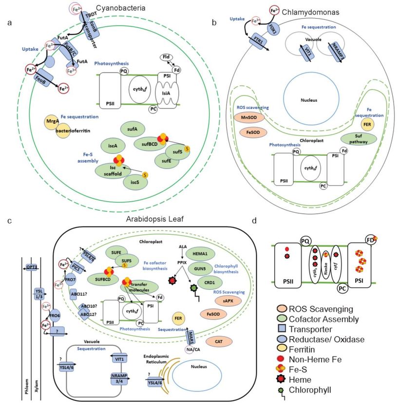
Figure 3. Fe-responsive proteins with relevance for photosynthesis in Cyanobacteria, Chlamydomonas,
and land3.plants.
Figure Proteins thatproteins
Fe-responsive are discussed
within this review for
relevance eachphotosynthesis
for species are presented (a) Fe-responsive
in Cyanobacteria,
proteins in Cyanobacteria, relatives to the ancestors of chloroplasts. On the outer membrane, Fe3+
Chlamydomonas, and land plants. Proteins that are discussed in this review for each species are
is chelated by a siderophore and taken up through a TonB Dependent Transporter (TBDT). TonB
presented (a) Fe-responsive proteins in Cyanobacteria, relatives to the ancestors of chloroplasts. On
spans the periplasmic space and the inner membrane to facilitate uptake through the TBDT. On the
the outer membrane, Fe3+ is chelated by a siderophore and taken up through a TonB Dependent
inner membrane, Fe is taken up as Fe3+ via the Fe Uptake Transporter, FutABC, system. Fe can
Transporter (TBDT). TonB spans the periplasmic space and the inner membrane to facilitate uptake
also be taken up as Fe2+ after reduction, through Ferrous Iron Transporter B (FeoB). Fe is required
through the TBDT. On the inner membrane, Fe is taken up as Fe3+ via the Fe Uptake Transporter,
for the photosynthetic electron transport chain. During Fe deficiency, Iron Stress Induced protein A
FutABC, system. Fe can also be taken up as Fe2+ after reduction, through Ferrous Iron Transporter B
(IsiA) can protect Photosystem I (PSI) and Ferredoxin (Fd) can be replaced by the non-Fe-requiring
(FeoB). Fe is required for the photosynthetic electron transport chain. During Fe deficiency, Iron Stress
Flavodoxin (Fld) in several species. Two operons exist for Fe–S cluster assembly: Iron Sulfur Cluster
Induced protein A (IsiA) can protect Photosystem I (PSI) and Ferredoxin (Fd) can be replaced by the
(isc) and Sulfur Utilization Factor (suf ). Fe is sequestered by ferritin molecules: bacterioferritin and
non-Fe-requiring Flavodoxin (Fld) in several species. Two operons exist for Fe–S cluster assembly:
MrgA. (b) Fe-responsive proteins in Chlamydomonas reinhardtii. Chlamydomonas takes up Fe3+ by a
Iron Sulfur Cluster (isc) and Sulfur Utilization Factor 2+ (suf). Fe is sequestered by ferritin molecules:
ferric iron permease yeast homologue (FTR1) after Fe is oxidized by Ferric Oxidase 1 (FOX1). Fe is
bacterioferritin and MrgA. (b) Fe-responsive proteins in Chlamydomonas reinhardtii. Chlamydomonas
sequestered in the vacuole via Vacuolar Iron Transporter 1 (VIT1) and can be exported from vacuoles via
takes up Fe3+ by a ferric iron permease yeast homologue (FTR1) after Fe2+ is oxidized by Ferric Oxidase
Natural Resistance Associated Macrophage-like Protein 3 (NRAMP3). Chloroplast Fe is sequestered by
1 (FOX1). Fe is sequestered in the vacuole via Vacuolar Iron Transporter 1 (VIT1) and can be exported
Ferritin (FER). The ROS-scavenging SuperOxide Dismutases (SOD), FeSOD and MnSOD, are regulated
from vacuoles via Natural Resistance Associated Macrophage-like Protein 3 (NRAMP3). Chloroplast
in response to Fe deficiency. (c) Fe-responsive proteins in the leaf mesophyll cell with a focus on
Fe is sequestered by Ferritin (FER). The ROS-scavenging SuperOxide Dismutases (SOD), FeSOD and
chloroplast proteins. From the xylem, Fe can be loaded into the phloem by Oligo Peptide Transporter 3
MnSOD, are regulated in response to Fe deficiency. (c) Fe-responsive proteins in the leaf mesophyll
cell with a focus on chloroplast proteins. From the xylem, Fe can be loaded into the phloem by Oligo
Peptide Transporter 3 (OPT3) or to the mesophyll cell. For import into the mesophyll cell, Fe is
exported from the xylem by Yellow-Stripe Like 1/3 (YSL1/3) presumably in a Fe3+-nicocianamine (NA)Int. J. Mol. Sci. 2020, 21, 3395 8 of 30
(OPT3) or to the mesophyll cell. For import into the mesophyll cell, Fe is exported from the xylem by
Yellow-Stripe Like 1/3 (YSL1/3) presumably in a Fe3+ -nicocianamine (NA) complex. Fe3+ is reduced
at the leaf plasma membrane by Ferric Reductase Oxidase 6 (FRO6) and Fe2+ is taken up into the
cell. Fe is reduced at the chloroplast envelope by FRO7 and taken up into the stroma by Permease
in Chloroplasts 1 (PIC1). ATP Binding Cassette (ABC) proteins, ABCI11, ABCI10, and ABCI12 may
also take up Fe into the chloroplast. YSL4/6 is proposed to be a chloroplast Fe exporter. Multiple
Antibiotic Resistance1 (MAR1) may transport NA or citrate (CA) into the chloroplast to sequester free
Fe. FER is also required to sequester Fe. Fe–S clusters are formed by the SUF pathway in the chloroplast
and transfer molecules insert these Fe–S clusters into photosynthetic proteins. Heme and chlorophyll
are produced in the tetrapyrrole pathway. Many enzymes for chlorophyll and heme production are
Fe-responsive, including Genome Uncoupled 5 (GUN5), Glutamyl-tRNA reductase 1 (HEMA1), and
Copper Response Defect 1 (CRD1). Within the chloroplast, during Fe deficiency, ROS-scavenging
molecules, Stromal Ascorbate Peroxidase (sAPX) and FeSOD are downregulated in Fe deficiency.
Catalase (CAT) is maintained or slightly downregulated during Fe deficiency. Fe is sequestered in the
vacuole, where it is imported by VIT1 and exported by NRAMP3/4. YSL4/6 may also be a vacuolar Fe
exporter. (d) Fe requirement of photosynthetic electron transport chain proteins. Symbols are explained
in the legend.
In higher plants, at both the plasma membrane and chloroplast membranes, Fe is reduced
before uptake (Figure 3c). Arabidopsis Ferric Reduction Oxidase 6 (FRO6) functions to reduce Fe for
import into the leaf cell and FRO7 reduces Fe for import from the cytoplasm into the chloroplast [29].
Expression of both FRO6 and FRO7 is induced during differentiation of leaf cells as chloroplasts
mature [87]. Additionally, FRO6 expression is driven by light-responsive elements in its promoter [87].
FRO7 is especially linked with photosynthetic need, as Arabidopsis knockout mutants exhibited
stunted growth, low electron transport, and decreases in the accumulation of Cyt-b6 f, when grown
on agar without sucrose [29]. However, increasing Fe supply to fro7 could recover this phenotype,
suggesting that there is either an alternative chloroplast reductase enzyme, or an alternative, currently
unknown, Fe uptake pathway that does not require reduction.
While the chloroplast seems to employ FRO7 to acquire Fe, it is unclear if the reductase activity for
Fe is tied to a specific Fe transporter or if Fe reduced via FRO7 can be taken up by multiple Fe importers.
Likely, there are multiple high- and low-affinity transport systems to modulate Fe movement into or
out of the chloroplast based on chloroplast Fe need [102]. The most viable candidate chloroplast Fe
importer is the inner-membrane-localized Permease In Chloroplast 1 (PIC1) (Figure 3c). PIC1 and its
homologue, sll1656 from Synechocystis PCC 6803, complemented a yeast Fe uptake deficient mutant,
suggesting a role in Fe import for both transporters [103]. Arabidopsis pic1 mutants had severely
underdeveloped chloroplasts along with an increased expression of the stromal Fe storage molecule,
ferritin (FER), suggesting an increased capacity to store Fe. FER expression is presumably induced
by ROS accumulation as a consequence of decreased accumulation of photosynthetic proteins [103].
PIC1 was originally characterized as a subunit of the chloroplast protein importer (TIC21) [104], but
all expressed chloroplast proteins in pic1 were processed to their mature form, indicating that they
were imported into the chloroplast [103]. Therefore, Duy et al. [105] propose that PIC1 may closely
associate with a TIC protein on the inner chloroplast membrane to coordinate Fe import and Fe cofactor
assembly with protein maturation.
A second candidate transfer system that may mediate chloroplast Fe uptake is comprised of
the ATP-Binding Cassette proteins, ABCI10, ABCI11/NAP14, and ABCI12. Arabidopsis ABCI10 and
ABCI11/NAP14 GFP fusion proteins were localized to the chloroplast, and most likely to the inner
envelope [106,107]. Both ABCI10 and ABCI11/NAP14 knockout mutants share the severely undeveloped
chloroplast phenotype of PIC1 and also exhibit decreases in FRO7, and PIC1 transcripts [107]. ABCI10
is predicted to interact with ABCI12, a transmembrane protein localized to the chloroplast inner
membrane and to provide ATPase activity for ABCI12. However, the interaction of ABCI10 and
ABCI12 is only based on colocalization in fluorescence microscopy for ABCI10:GFP and ABCI12:YFP,Int. J. Mol. Sci. 2020, 21, 3395 9 of 30
and ATPase activity of ABCI10 has not been directly measured [107]. While ABCI11/NAP14 may be
important for chloroplast Fe, many questions remain about its involvement in transport, for instance
the direction of ABCI11/NAP14-mediated transport is unknown [106]—i.e., it could be used with an Fe
import system, an Fe export system or not involved in Fe uptake.
3.2. Chloroplast Fe Export
Fe export from the chloroplast is equally important as Fe uptake both to ensure that the chloroplast
is not overloaded with Fe, which can lead to production of free radicals by Fenton reactions, and
to export Fe from the chloroplast during senescence. Export from chloroplasts in Arabidopsis may
be mediated by two Yellow Stripe Like proteins (YSL4 and YSL6). The double mutant, ysl4/6 over
accumulated Fe in the chloroplast and induced FER expression [108]. However, localization of
YSL4/6 has been a matter of debate based on the method used. In immunolocalization experiments,
fluorescently tagged antibodies for YSL4/6 were localized to the chloroplast [108], while YSL4/6-GFP
fusion proteins localized to the tonoplast and the endoplasmic reticulum (Figure 3c) [109]. Both a
proposed function as a chloroplast Fe exporter or as a tonoplast Fe importer could explain the
accumulation of Fe in the chloroplast. Clearly, the loss of a chloroplast Fe exporter would result in
higher levels of Fe remaining in the chloroplast. However, the loss of a tonoplast Fe importer could also
lead to increases in chloroplast Fe, as less Fe can be distributed into the tonoplast it may be sequestered
instead by FER in the chloroplast. Regardless of the location of YSL4/6, it does appear to have a role
in modulating intercellular Fe. However, the ability of YSL4/6 to transport Fe has not been directly
measured and the sensitivity to Fe of ysl4/6 seems to be dependent on the growth conditions; thus it
was suggested that YSL4/6 may transport manganese or nickel instead of Fe [109].
3.3. Fe Sequestration
Fe is thought to be transported while bound to Fe chelators, such as nicotianamine (NA) and citrate.
Brassica napus chloroplasts, had a higher rate of Fe uptake when Fe was supplied as Fe(III)-citrate
compared to when it was supplied as Fe(III)- NA or Fe(II)-NA [110]. Once inside the chloroplast,
the majority of the Fe is found as heme or Fe–S clusters [111]. A possible chelate transporter, Arabidopsis
Multiple Antibiotic Resistance 1 (MAR1) has been localized to the chloroplast inner envelope. MAR1
was discovered as a chloroplast importer of antibiotics but more likely evolved to function in Fe uptake,
as its expression has been linked to Fe status [112]. Most likely, MAR1 functions to couple import of Fe
chelators with that of Fe so that once inside the chloroplast (Figure 3c), Fe is chelated to avoid Fenton
reactions. However, the specific chelator that may be imported by MAR1 is unknown.
Ferritin (FER) is the major Fe storage molecule located in the chloroplast and is vital to modulating
the amount of free Fe in the chloroplast for use (Figure 3c) [113]. Arabidopsis has four ferritin genes:
FER1, FER3, and FER4 are major leaf Ferritins, while FER2 is expressed in the seed [114]. Ferritins
are found in bacteria and animals as well as across green lineages [115]. Ferritin proteins complexes
make a shell-like structure that holds Fe3+ bound to organic phosphate [116]. One Ferritin shell is
composed of 24 FER molecules [116] and ferritin from legumes was found to hold at least 1000 Fe
atoms [117]. The major function of FER in plastids is to scavenge free Fe to eliminate the threat of
ROS production (Figure 4) [113]. Free Fe in the presence of oxygen can lead to the accumulation
of harmful hydroxyl radicals (•OH) and hydrogen peroxide (H2O2) via Fenton and Haber–Weiss
reactions, which disrupt membrane integrity and protein structure (Figure 4) [118]. FER is especially
important for chloroplast protection during Fe toxicity, where its expression is stabilized to strengthen
Fe storage. FER triple mutants (fer1/fer3/fer4) had limited growth and enhanced catalase and ascorbate
peroxidase activity at high levels of Fe, suggesting an increase in ROS protection mechanisms [113].
Cyanobacterial bacterioferritin is required for buffering Fe stores and a second class of FER (MrgA) is
suggested to provide redundancy for bacterioferrtins in Fe deficiency to avoid oxidative damage [119],
which may be a consequence of Fe deficiency depending on the severity of Fe limitation. The function
of FER in Fe homeostasis will be further discussed in the following sections.Int. J. Mol. Sci. 2020, 21, x 10 of 30
Int. J. Mol. Sci. 2020, 21, 3395 10 of 30
Figure 4. Role of Ferritin (FER) in preventing Haber–Weiss and Fenton reactions that can perpetuate
the production
Figure of Ferritin
4. Role of reactive(FER)
oxygenin species (ROS).
preventing FER is expressed
Haber–Weiss under reactions
and Fenton normal conditions to prevent
that can perpetuate
accumulation of free Fe and avoid excessive build up of ROS. If FER cannot capture free Fe,
the production of reactive oxygen species (ROS). FER is expressed under normal conditions to preventHaber–Weiss
reactions,
accumulationwhich are catalyzed
of free by free
Fe and avoid Fe, canbuild
excessive lead to
upthe build If
of ROS. upFER
of hydroxyl radicals
cannot capture (•OH).
free These
Fe, Haber–
reactions occur in two steps. First, Fe 2+ and oxygen are produced from Fe3+ and superoxide. Second,
Weiss reactions, which are catalyzed by free Fe, can lead to the build up of hydroxyl radicals (•OH).
the Fe2+
These can reactoccur
reactions with in
hydrogen peroxide
two steps. First, (H2O2)
Fe2+ andtooxygen
produceare 3+ and harmful hydroxyl radicals in a
Feproduced from Fe3+ and superoxide.
Fenton reaction.
Second, the Fe can react with hydrogen peroxide (H2O2) to produce Fe3+ and harmful hydroxyl
2+
radicals in ato
4. Acclimation Fenton
Low reaction.
Fe
During Fe deficiency
4. Acclimation to Low Fe a photosynthetic organism must acclimate both locally and systemically to
alter growth and metabolism. The systemic response to low Fe becomes more complex as the number
During Fe deficiency a photosynthetic organism must acclimate both locally and systemically to
of cells and structures in the organism increases. For example, unicellular Cyanobacteria will have one
alter growth and metabolism. The systemic response to low Fe becomes more complex as the number
overall response as the organism will sense Fe and respond in the single cell. Filamentous Cyanobacteria,
of cells and structures in the organism increases. For example, unicellular Cyanobacteria will have
such as Anabaena variabilis, that have specialized cells for different metabolic functions, such as nitrogen
one overall response as the organism will sense Fe and respond in the single cell. Filamentous
fixation, may sense Fe locally in photosynthetic cells but will have to respond systemically to acclimate
Cyanobacteria, such as Anabaena variabilis, that have specialized cells for different metabolic
other cells to the deficiency in photosynthetic output [120]. In contrast, the unicellular eukaryotic alga,
functions, such as nitrogen fixation, may sense Fe locally in photosynthetic cells but will have to
Chlamydomonas will have local responses in different cellular compartments, and these responses
respond systemically to acclimate other cells to the deficiency in photosynthetic output [120]. In
will need to be coordinated by signaling between organelles and nucleus [121]. In land plants, not
contrast, the unicellular eukaryotic alga, Chlamydomonas will have local responses in different
only do responses need to be coordinated in each cell, but also between different organs. For instance,
cellular compartments, and these responses will need to be coordinated by signaling between
plants induce root Fe uptake based on shoot Fe needs. Therefore, in land plants, there are distinct
organelles and nucleus [121]. In land plants, not only do responses need to be coordinated in each
but coordinated local responses to Fe deficiency in leaves, vasculature, roots, and reproductive
cell, but also between different organs. For instance, plants induce root Fe uptake based on shoot Fe
structures [122,123].
needs. Therefore, in land plants, there are distinct but coordinated local responses to Fe deficiency in
In green organisms, photosynthesis is a major target for regulation under low Fe availability. During
leaves, vasculature, roots, and reproductive structures [122,123].
Fe deficiency, photosynthetic organisms become chlorotic as they decrease activity of the chlorophyll
In green organisms, photosynthesis is a major target for regulation under low Fe availability.
biosynthesis pathway [124]. Chlorophyll biosynthesis requires Fe for Chlorophyllide A Oxygenase
During Fe deficiency, photosynthetic organisms become chlorotic as they decrease activity of the
(CAO) and the Copper Response Defect 1 (CRD1/CHL27) subunit of Mg Proto IX Monomethyl Ester
chlorophyll biosynthesis pathway [124]. Chlorophyll biosynthesis requires Fe for Chlorophyllide A
Cyclase (MgCY) [38]. In plants, chlorosis begins in the young leaves with developing chloroplasts,
Oxygenase (CAO) and the Copper Response Defect 1 (CRD1/CHL27) subunit of Mg Proto IX
reflecting an inability to remobilize Fe from mature tissue to developing leaves. Photosynthetic electron
Monomethyl Ester Cyclase (MgCY) [38]. In plants, chlorosis begins in the young leaves with
transport activity when measured by chlorophyll fluorescence is specifically inhibited downstream of
developing chloroplasts, reflecting an inability to remobilize Fe from mature tissue to developing
PSII as indicated by a lower ϕPSII parameter (indicating electron transport downstream of PSII) that
leaves. Photosynthetic electron transport activity when measured by chlorophyll fluorescence is
is decreased to a stronger extent than maximum efficiency of PSII (Fv /Fm ) [125,126]. Comparatively,
specifically inhibited downstream of PSII as indicated by a lower φPSII parameter (indicating
cellular respiration rates are much less affected in a mild Fe deficiency in Arabidopsis [126], suggesting
electron transport downstream of PSII) that is decreased to a stronger extent than maximum
that chloroplast metabolism, as opposed to that of the mitochondria, is a major target of the Fe
efficiency of PSII (Fv/Fm) [125,126]. Comparatively, cellular respiration rates are much less affected in
deficiency response.
a mild Fe deficiency in Arabidopsis [126], suggesting that chloroplast metabolism, as opposed to that
The regulated response to Fe deficiency depends greatly on the severity of the iron deficiency and
of the mitochondria, is a major target of the Fe deficiency response.
developmental stage of the organism. In Chlamydomonas and Arabidopsis, severity of Fe deficiency
The regulated response to Fe deficiency depends greatly on the severity of the iron deficiency
can be determined by both the length of Fe deficiency and the fold change in available Fe [121,125,127].
and developmental stage of the organism. In Chlamydomonas and Arabidopsis, severity of Fe
A more severe Fe deficiency can lead to irreversible damage to photosynthetic electron transport chain
deficiency can be determined by both the length of Fe deficiency and the fold change in available Fe
and secondary stress responses [121]. Symptoms of less severe Fe deficiency in Arabidopsis can be
[121,125,127]. A more severe Fe deficiency can lead to irreversible damage to photosynthetic electron
recovered by resupplying plants with sufficient levels of Fe [126]. A list of Arabidopsis chloroplast
transport chain and secondary stress responses [121]. Symptoms of less severe Fe deficiency in
homeostasis proteins and their orthologues in Cyanobacteria and Chlamydomonas are listed in Table 1.
Arabidopsis can be recovered by resupplying plants with sufficient levels of Fe [126]. A list ofInt. J. Mol. Sci. 2020, 21, 3395 11 of 30
Table 1. Arabidopsis thaliana chloroplast Fe homeostasis genes and their Chlamydomonas reinhardtii and
Synechocystis PCC 6803 (Cyanobacterial) orthologues. Arabidopsis protein name and ID are listed for
plastid Fe homeostasis proteins discussed in this review. Studies that identified Chlamydomonas and
Cyanobacterial orthologues for these proteins are referenced next to the gene name. In cases where an
orthologue has not been previously identified in the literature, the Arabidopsis protein sequence was
used as a query sequence in NCBI BLAST. Orthologues were determined if the Chlamydomonas and
Cyanobacterial sequences had at least 20% sequence similarity for at least 50% of the query sequence.
When possible, Cyanobacterial gene names are listed for Synechocystis PCC 6803. Otherwise, the NCBI
gene ID is listed.
Arabidopsis Gene Synechocyst Is
Function A. thaliana C. reinhardtii
Name PCC 6803
Transport FRO7 AT5G49740 Cre04.g227400.t1.2
PIC1 AT2G15290 Cre10.g454734.t2.1 sll1656 [103]
ATP-Binding Cassette
AT5G14100 PNW71978/Cre16.g687550.t1.1 [107] slr0354 [107]
I11 (ABCI11)/NAP14
ABCI10 AT4G33460 XP_001703542/Cre03.g164150.t1.1 [107] sll1623 [107]
ABCI12 AT3G21580 PNW75614/Cre12.g533950.t1.1 [107] slr1978 [107]
Multiple Antibiotic
AT5G26820 Cre03.g175200.t1.2 lap75
Resistance 1 (MAR1)
Yellow Stripe Like 4/6 (4) AT5G41000,
(YSL4/6) (6) AT3G27020
AT4G25100
Fe SuperOxide
ROS homeostasis AT5G51100 Cre10.g436050.t1.2 WP_010872652
Dismutase (FSD)
AT5G23310
Stromal Ascorbate
AT4G08390 Cre02.g087700.t1.2
Peroxidase (sAPX)
Conserved in Green
Linage and Diatoms 27 AT5G67370 Cre05.g237050.t1.1 WP_010873853
(CGLD27)
Suf Fe–S assembly SUFB AT4G04770 Cre15.g643600.t1.2 [128] slr0074 [128]
SUFC AT3G10670 Cre07.g339700.t1.2 [128] slr0075 [128]
SUFD AT5G44316 Cre12.g513950.t1.2 * [128] slr0076 [128]
SUFS AT1G08490 Cre12.g525650.t1.2 [128] slr0077 [128]
SUFE AT4G26500 Cre06.g309717.t1.1 [128] slr1419 [128]
SUFA AT1G10500 Cre07.g349600.t1.2 [128] slr1417 [128]
(1) AT4G01940 (1) Cre18.g748447.t1.1
Nitrogen Fixation U-Like
(2) AT5G49940 (2) Cre12.g504150.t2.1 ssl2667 [128]
(NFU)
(3) AT4G25910 (3) Cre17.g710800.t2.1 [128]
High Chlorophyll
Fluorescence 101 At3g24430 Cre01.g045902.t1.1 [128] slr0067 [128]
(HCF101)
Monothiol (14) AT3G54900 (14) Cre07.g325743.t1.1
WP_010871706
Glutaredoxins (GRXS) (16) AT2G38270 (16) Cre01.g047800.t1.1 [128]
NEET AT5G51720 Cre01.g050550.t1.2
AT5G26030
Heme biosynthesis Ferrochelatase (FC) Cre07.g339750.t2.1 WP_010873751
AT2G30390
Sirohydrochlorin
Cre04.g214100.t1.2 WP_010874018
Ferrochelatase B (SirB)
(1) AT5G01600 sll1341 ** [129]
pSequestration Ferritin (FER) (3) AT3G56090 Cre09.g387800.t1.2, Cre13.g574500.t1.1 slr1890 **
(4) AT2G40300 slr1894 ** [130]
* C. reinhardtii SUFD was more similar to A. thaliana SufB. The C. reinhardtii gene ID listed is for the annotated SUFD
gene; ** No sequence similarity was found for A. thaliana ferritin in Synechocystis PCC 6803. Genes listed are for
Bacterioferritin A and B, and MrgA.Int. J. Mol. Sci. 2020, 21, 3395 12 of 30
4.1. Increase Fe Uptake
The increase in Fe uptake in response to Fe deficiency in photosynthetic organisms has been well
documented and upregulation of uptake systems are common markers of Fe deficiency [16,17,131–133].
Cyanobacteria are known to increase Fe uptake by upregulating FutC and FeoB (Figure 3a) [16,17].
Chlamydomonas employs an oxidation strategy for Fe uptake, similar to yeast, where Fe2+ is oxidized
by a multicopper ferroxidase (FOX1) and then taken up by a ferric Fe permease yeast homologue
(FTR1) [15]. Chlamydomonas increases Fe3+ uptake by upregulation of FOX1 and FTR1 expression in
response to deficiency (Figure 3b) [15,133].
In plants, Fe uptake at the root has been thoroughly studied [21,123,134]. In short, the dicot plant,
Arabidopsis, acidifies the rhizosphere by the proton pump, H+ATPase 2 (AHA2); reduces the poorly
bioavailable Fe3+ to Fe2+ via the Fe Reductase Oxidase 2 (FRO2) enzyme; and finally, takes up Fe2+
from the soil through Iron Regulated Transporter 1 (IRT1) [21,134].
4.2. Metabolic Remodeling
Plants remodel metabolism locally, in both the shoots and roots to respond to Fe deficiency.
In the roots, C metabolism, ROS metabolism and N metabolism have all been found to be targets of
metabolic remodeling in response to Fe deficiency [122,135]. Leaf metabolic remodeling coordinates
downregulation of photosynthetic output with photoprotective mechanisms [124]. Early in the Fe
deficiency response, gene expression of mRNAs that encode enzymes in the tetrapyrrole pathway are
downregulated. The targeted mRNAs include HEMA1 which encodes for an enzyme needed for ALA
synthesis in the tetrapyrrole pathway, and a geranylgeranyl reductase (CHLP), a phytochlorophyllide
oxidoreductase (PORB), and Genome Uncoupled 5 (GUN5) which are all required for chlorophyll
production (Figure 3c) [124]. In addition, N metabolism has been found to be altered in leaves, with
decreased expression of nitrate reductase, which requires Fe, and altered amino acid accumulation [136].
Chlorophyll and chloroplast protein biosynthesis, both require N assimilation [137], and therefore,
the alterations to N metabolism could represent adjustments these pathways during Fe deficiency.
Additionally, mRNA encoding the Fe-responsive protein, Conserved in Green Lineage and Diatoms
27 (CGLD27) is upregulated and this is predicted to function in quenching of ROS that results from
photooxidative damage [124]. Other ROS-scavenging molecules in the plastid, such as the Fe-requiring
stromal ascorbate peroxidase (sAPX), are downregulated, at least at the transcriptional level, during Fe
deficiency [126], suggesting that specific ROS reduction systems are employed for photoprotection.
Overall, metabolic remodeling in response to Fe deficiency suggests the plant decreases chlorophyll
synthesis and increases mechanisms to protect from ROS.
The metabolic remodeling on low Fe in Chlamydomonas depends on the growth requirements of
the organism, as Chlamydomonas can grow both photoautotrophically and photoheterotrophically [121].
When grown photoautotrophically, carbon assimilation by means of photosynthesis is maintained
on low Fe. When grown photoheterotrophically, in which cells are supplied with light for energy
production and an organic carbon source, Fe deficiency results in a similar response as seen in plants
with a maintenance of respiration and strong decreases to photosynthetic electron transport [121].
In photoheterotrophic conditions, the ascorbate concentration was reported to increase tenfold in
the cell during Fe deficiency [138]. Chlamydomonas grown photoheterotrophically had increased
xanthophyll cycle pigments but a decreased induction of Non-Photochemical Quenching (NPQ) during
Fe deficiency, suggesting that a decreased rate of electron transport did not allow for acidification
of the lumen needed for induction of NPQ [139]. In photoautotrophic conditions, Chlamydomonas
maintained NPQ in response to Fe limitation [139]. These changes to metabolism in chloroplasts of
Fe-deficient Chlamydomonas suggest a need to increase photoprotective mechanisms and defenses
against ROS.You can also read