Plague Prevention and Therapy: Perspectives on Current and Future Strategies
←
→
Page content transcription
If your browser does not render page correctly, please read the page content below
biomedicines
Review
Plague Prevention and Therapy: Perspectives on Current and
Future Strategies
Raysa Rosario-Acevedo, Sergei S. Biryukov, Joel A. Bozue                          and Christopher K. Cote *

                                          Bacteriology Division, United States Army Medical Research Institute of Infectious Diseases (USAMRIID),
                                          Fort Detrick, MD 21702, USA; raysa.rosarioacevedo.mil@mail.mil (R.R.-A.);
                                          sergei.s.biryukov.civ@mail.mil (S.S.B.); joel.a.bozue.civ@mail.mil (J.A.B.)
                                          * Correspondence: Christopher.k.cote.civ@mail.mil; Tel.: +1-(301)-619-4936

                                          Abstract: Plague, caused by the bacterial pathogen Yersinia pestis, is a vector-borne disease that has
                                          caused millions of human deaths over several centuries. Presently, human plague infections continue
                                          throughout the world. Transmission from one host to another relies mainly on infected flea bites,
                                          which can cause enlarged lymph nodes called buboes, followed by septicemic dissemination of the
                                          pathogen. Additionally, droplet inhalation after close contact with infected mammals can result
                                          in primary pneumonic plague. Here, we review research advances in the areas of vaccines and
                                          therapeutics for plague in context of Y. pestis virulence factors and disease pathogenesis. Plague
                                          continues to be both a public health threat and a biodefense concern and we highlight research that is
                                          important for infection mitigation and disease treatment.

                                          Keywords: Yersinia pestis; plague; antibiotic; vaccine; antibodies


Citation: Rosario-Acevedo, R.;
Biryukov, S.S.; Bozue, J.A.; Cote, C.K.   1. Plague History and Disease Presentation
Plague Prevention and Therapy:                  Yersinia pestis is a Gram-negative coccobacillus that is the etiologic agent of plague,
Perspectives on Current and Future        an acute infectious disease that has played an important role in human history [1–5].
Strategies. Biomedicines 2021, 9, 1421.
                                          This bacterium has been responsible for three devastating pandemics that led to millions of
https://doi.org/10.3390/biomedicines
                                          deaths [6–8]. The first pandemic, referred to as the “Justinian Plague”, occurred in 541 AD,
9101421
                                          originated in Central Africa, and spread to the Mediterranean region [7]. The “Black Death”
                                          of 1347 was the second major pandemic and occurred during the 14th century, originating
Academic Editor: Maria Grau
                                          in Central Asia and eventually reaching the Middle East, North Africa, and Europe. The
                                          third pandemic took place in 1855 in Yunnan, China and spread globally via infected rats
Received: 1 September 2021
Accepted: 4 October 2021
                                          on ships likely originating from Hong Kong [7,9,10]. This bacterium has impacted human
Published: 9 October 2021
                                          civilization throughout history and continues to be of concern [5].
                                                Dissemination of Y. pestis among wild rodents and humans takes place in two different
Publisher’s Note: MDPI stays neutral
                                          cycles, enzootic and epizootic [11]. Enzootic refers to when the infection, at low levels,
with regard to jurisdictional claims in
                                          cycles naturally among wild rodents (e.g., rats, squirrels, marmots, and prairie dogs).
published maps and institutional affil-   Sporadically, other susceptible species can be infected, causing an outbreak—this is referred
iations.                                  to as epizootic plague. In this disease cycle, animals become infected and ultimately die
                                          as fleas from infected natural reservoir species begin to seek other blood sources and the
                                          probability of human transmission is increased [11].
                                                Y. pestis can cause three forms of plague disease: bubonic, septicemic, and pneumonic
Copyright: © 2021 by the authors.
                                          (Figure 1). Historically, the bubonic form of plague has been the most common and can be
Licensee MDPI, Basel, Switzerland.
                                          transmitted to humans by a bite from an infected flea or by handling an animal infected with
This article is an open access article
                                          Y. pestis [12–15]. After transmission, the Y. pestis bacteria disseminate to the nearest lymph
distributed under the terms and           node where they colonize and subsequently proliferate. The incubation time ranges from
conditions of the Creative Commons        approximately 2–8 days and during that period patients will experience flu-like symptoms,
Attribution (CC BY) license (https://     such as chills, fever and headaches [16–19]. As the bacteria continue to multiply, the lymph
creativecommons.org/licenses/by/          nodes become swollen and extremely painful. Infected lymph nodes (referred to as buboes
4.0/).                                    when attributable to plague) are generally found in specific areas of the body, such as

Biomedicines 2021, 9, 1421. https://doi.org/10.3390/biomedicines9101421                                https://www.mdpi.com/journal/biomedicines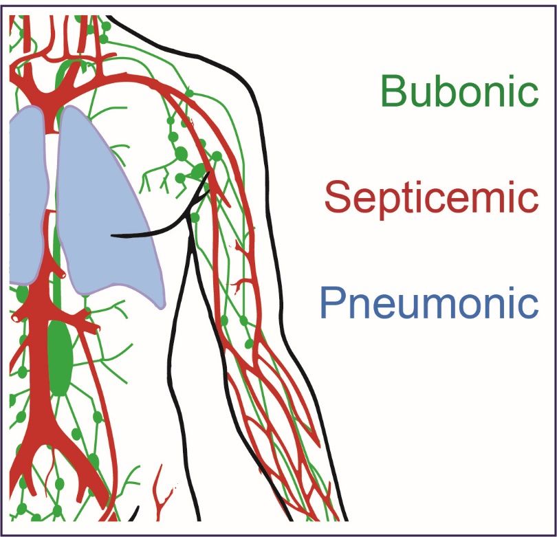
Biomedicines 2021, 9, x FOR PEER REVIEW 2 of 17
Biomedicines 2021, 9, 1421 2 of 18
(referred to as buboes when attributable to plague) are generally found in specific areas
of the body, such as the neck, under the chin, armpits and groin. If left untreated, these
buboes
the neck,can
underbecome necrotic
the chin, and cause
armpits hemorrhaging
and groin. ultimately
If left untreated, leading
these to lethal
buboes disease
can become
in approximately 50–60% of cases [20]. A culture isolated from a blood
necrotic and cause hemorrhaging ultimately leading to lethal disease in approximately sample or fluid
from the
50–60% ofswollen lymph
cases [20]. node may
A culture be necessary
isolated for thesample
from a blood diagnosis and identification
or fluid from the swollen of Y.
lymph nodeSeveral
pestis [17]. may beclasses
necessary for the diagnosis
of antibiotics and in
are effective identification of Y. pestis
treating bubonic plague [17].
andSeveral
if treat‐
classes
ment isofinitiated
antibiotics are effective
promptly, in treating
antibiotics bubonic plague
can significantly and survival
increase if treatment
ratesis [19,21,22].
initiated
promptly, antibioticssuch
Aminoglycosides, can significantly
as streptomycin increase
andsurvival rates tetracyclines,
gentamicin, [19,21,22]. Aminoglycosides,
and the fluoro‐
such as streptomycin
quinolone and gentamicin,
ciprofloxacin tetracyclines,
are some effective and thefor
antibiotics fluoroquinolone
bubonic plague ciprofloxacin
treatment
are some effective
[20,23,24]. antibiotics for bubonic plague treatment [20,23,24].
Figure1.1.Y.Y.pestis
Figure pestisinfection
infectioncan
canlead
leadtotothree
threeforms
formsofofplague.
plague.Bubonic
Bubonicplague
plaguecan
canresult
resultfrom
fromflea
flea
bites or handling infected animals and can result in a severe localized infected lymph node
bites or handling infected animals and can result in a severe localized infected lymph node referred referred
to as a buboe (green). Septicemic plague can result as a secondary issue arising from bubonic disease
to as a buboe (green). Septicemic plague can result as a secondary issue arising from bubonic disease
or if the bacteria are introduced directly to the blood stream (red). Pneumonic plague is the most
or if the bacteria are introduced directly to the blood stream (red). Pneumonic plague is the most
severe form of the disease and can result in person‐to‐person spread if untreated and initiated by
severe form of the disease and can result in person-to-person spread if untreated and initiated by the
the deposition of Y. pestis bacteria into the respiratory tract of individuals (blue).
deposition of Y. pestis bacteria into the respiratory tract of individuals (blue).
In other more severe cases, the bacteria can enter the blood stream directly and mul‐
In other more severe cases, the bacteria can enter the blood stream directly and
tiply, causing septicemic plague. The increased number of bacteria in the blood causes the
multiply, causing septicemic plague. The increased number of bacteria in the blood causes
release of endotoxins, leading to ischemic necrosis. In those cases, Y. pestis directly infects
the release of endotoxins, leading to ischemic necrosis. In those cases, Y. pestis directly
the blood without developing bubonic plague symptoms (resulting in primary septicemic
infects the blood without developing bubonic plague symptoms (resulting in primary
plague) [25]. Furthermore, septicemia may induce intravascular coagulation that may lead
septicemic plague) [25]. Furthermore, septicemia may induce intravascular coagulation
thatgangrene
to may leadoftothe extremities.
gangrene of theThe prognosis The
extremities. for these patients
prognosis is poor,
for these with mortality
patients is poor,
rates reaching 100% in untreated individuals. Even when the appropriate
with mortality rates reaching 100% in untreated individuals. Even when the appropriate treatment is ad‐
ministered, mortality rates remain high [26]
treatment is administered, mortality rates remain high [26]
AAthird
thirdmanifestation
manifestation ofof plague,
plague, pneumonic
pneumonic plague,
plague, cancan be transmissible
be transmissible either
either per‐
person-
son‐to‐person
to-person or byor by secondary
secondary pneumonia,
pneumonia, resulting
resulting from thefrom the colonization
colonization of the of the with
lungs lungs
with Y. pestis via hematogenous spreading [1,27]. If droplets containing
Y. pestis via hematogenous spreading [1,27]. If droplets containing plague bacteria are plague bacteria
are inhaled,
inhaled, the result
the result can becan be primary
primary pneumonic
pneumonic plague.plague. Secondary
Secondary pneumonic
pneumonic plagueplague
may
may be associated with
be associated with primary septicemic plague or as a complication of bubonic plague.plague.
primary septicemic plague or as a complication of bubonic In the
In the States,
United Unitedapproximately
States, approximately 5–10% of
5–10% of patients patients
develop developpneumonic
secondary secondaryplague
pneumonic
[28].
plague [28].
Symptoms of Symptoms of plague
plague include chills, include chills,
fever, body fever,
pains, body pains,
headache, headache,
weakness, weakness,
dizziness, and
dizziness,
chest and chest
discomfort [29]. discomfort
These rather[29]. These rather
non-descript non‐descript
symptoms mirrorsymptoms
many othermirror many
infectious
diseases, which can lead to frequent misdiagnosis and subsequent poor disease outcomes
for the patients. Blood cultures or a sputum sample can be used to diagnose pneumonicBiomedicines 2021, 9, 1421 3 of 18
plague [30]. Pneumonic plague is considered highly contagious and is nearly 100% lethal if
appropriate treatment is not administered within the first 20 h after onset of symptoms [30].
Due to rapid spread, high fatality rates, and the ability to transmit via aerosols, Y. pestis
has been classified as a Tier 1 select agent by the United States Department of Health and
Human Services and is considered an agent of biothreat concern [31,32]. Numerous studies
have been performed in mice and other species in order to elucidate Y. pestis pathogenesis
and pneumonic plague disease progression in animal models of human disease [25,33–35].
It has been reported that during the first 36 h post infection in mice, there is an increase in
bacterial replication in the lungs with no appreciable changes in cytokines or chemokines
levels [36,37]. In this pre-inflammatory phase, it has been shown that Y. pestis suppresses
the host immune response in order to facilitate pulmonary infection [38,39]. After 36 h, the
pro-inflammatory phase begins, where there is an upregulation of cytokine or chemokine
levels, resulting in a critical point that can lead to death [40].
2. Yersinia pestis Virulence Plasmids
Y. pestis must be able to survive in and infect very diverse hosts, including both fleas
and mammals. There are numerous essential virulence factors and effector proteins that
are either encoded on the chromosome of Y. pestis or carried on its plasmids. It is believed
that Y. pestis evolved from the genetically related Yersinia species Yersinia pseudotuberculosis
(usually an intestinal pathogen) through the process of gaining plasmids, most likely
several thousands of years ago [4,41,42].
Y. pestis carries three plasmids, two of which are unique to this species: pMT1 (or
pFra), which encodes the F1 capsular antigen and pPCP (or pPst or pPla), which carries
the gene for the virulence factor plasminogen activator. The third plasmid is common
to the human pathogenic Yersiniae and is known as pCD1 (calcium dependence), pYV
(Yersinia virulence), or pLcr (low calcium response). This plasmid is responsible for the
synthesis of a number of anti-host factors and is an absolute requirement for virulence.
The pMT1/pFra plasmid (100–110 kb) carries two important Y. pestis virulence factors
involved with survival/infection of these two different hosts: the murine toxin Ymt and
the F1 capsular protein. Ymt is a phospholipase D which has toxic properties for mice
and rats but not for other animals [43–45]. More importantly, Ymt is required for survival
within the midgut of the flea by protecting it against some components of plasma [46].
In addition, the caf operon encoding for the F1 capsular antigen protein (referred to here
as F1) is also located on pMT1. The F1 antigen has been described both as capsular and
fimbrial-like as it is composed of fibers [47–50]. The F1 protein is produced abundantly
in vivo during infection of a mammal and in vitro when grown at 37 ◦ C. F1 is thought to
protect Y. pestis from uptake by host phagocytic cells [51]. However, naturally derived
and genetically engineered F1-negative Y. pestis strains have been described and remain
virulent in animal models of plague [52,53].
The other Y. pestis specific plasmid (pPCP/pPst/pPla) is 9.5 kb and carries genes that
encode for pesticin, pesticin immunity protein, and plasminogen activator (Pla) [54]. Pla
appears to be a multifunctional protein and belongs to the omptin family of enterobacterial
surface proteins. One of the functions of Pla during infection is to cleave numerous host
substrates, such as those involved in coagulation and fibrinolysis [55]. During bubonic
plague, Pla activates host fibrolysis to allow bacterial dissemination from the site of infec-
tion [56,57]. Furthermore, this protein is believed to alter the host immune response during
pneumonic plague to allow outgrowth of the bacteria within the lungs [58,59]. In addition
to its ability to cleave proteins, it has a role in mediating bacterial adhesion to host cells
and extracellular matrices [60–64].
The final plasmid is pCD1/pYV/pLcr (68–75 kb), which is common to all human
pathogenic Yersiniae, is essential for virulence, and carries genes encoding for the Type III
secretion system (T3SS) and effector proteins known as Yops (Yersinia outer protein). The
T3SS forms a syringe-like structure (injectisome) made up of 27 proteins, including the LcrV
protein, which is both a structural and effector protein and is critical for virulence [65–76].Biomedicines 2021, 9, 1421 4 of 18
The injectisome is able to “inject” host cells when in close contact with Yops. Two of the
Yops (YopB and YopD) function to transport the other six effector proteins into the host cell
(YopO, YopH, YopM, YopT, YopJ, and YopE). Toxic activities of the Yops include disruption
of the cytoskeleton, interference with phagocytic activity, prevention of proinflammatory
cytokine synthesis, inhibition of the oxidative burst, and induction of programmed cell
death (apoptosis). The overall effect of the Yops is to block phagocytic uptake of the bacteria
by host macrophages and polymorphonuclear leukocytes [77–82].
3. Outbreak Prevention
Despite the efforts of public health agencies to monitor and prevent this disease, re-
ports of plague outbreaks continue to exist in various parts of the word [83–85]. The United
States, China, India, Vietnam, Democratic Republic of the Congo, Peru, and Madagascar
are among the countries that have confirmed plague cases annually and their respective cli-
mates, public health infrastructure, surveillance programs, and socio-economic differences
make outbreak prevention strategies and severity of outbreaks unique in each case [86–88].
Madagascar is currently the most affected country with the highest numbers of recently
reported cases of plague, representing approximately 75% of the reported cases in the
world [87,89,90]. Differences in weather and temperature patterns affect the abundance of
fleas [89,91] which in turn influences the start of annual transmission that generally arises
between the months of September to April in Madagascar [92]. Given the right conditions,
it may be possible for person-to-person transmission mediated by human ectoparasites—
especially in underdeveloped but densely populated areas [93]. A total of 2417 suspect
plague cases, including 209 deaths, were reported by the Word Health Organization (WHO)
from August to November in 2017 in Madagascar [94]. Generally, bubonic plague cases in
Madagascar are commonly concentrated in rural areas, while pneumonic cases are more
prevalent in urban locations [95]. In 2017, the majority (77%; 1854 total) reported were clini-
cally classified as pneumonic plague whereas 15% (355) were classified as bubonic plague.
Only one of the reported cases was diagnosed as septicemic plague while the remaining
207 cases remain unclassified [94]. Control and prevention of plague cases in Madagascar
has proven to be challenging. Most of the prevention strategies are focused on the rodent
host and the flea vectors [91]. Inappropriate use of chemical insecticides inadvertently
selects for flea resistance, which leads to the survival of Y. pestis-infected fleas [91]. Food
handling/storage procedures, community sanitation standards, and environmental factors
of the household and its surroundings are parameters that can increase human interactions
with rodent reservoirs [96]. Importantly, distribution of supplies and equipment including
hand washing facilities, sanitation standards, disinfectants (e.g., laundry soap, chlorine
powder, disinfectant sprays), and personal protective equipment contribute to an efficient
plague prevention strategy [94].
4. Current Antibiotic Therapies
Due to the rapid onset and rather non-descript disease characteristics of plague, the key
to positive patient outcomes, particularly after infection with aerosolized bacteria, is early
diagnosis and rapid implementation of appropriate medical countermeasures. The gold-
standard for plague diagnostics continues to be culture-positive samples (normally blood
or sputum samples) but other molecular diagnostic assays have been used or continue to
be developed [2].
If diagnosed accurately and early after infection, human plague cases can be controlled
by the appropriate administration of antimicrobial drugs, including aminoglycoside, tetra-
cyclines, fluroquinolones, and sulfonamides [91,97–101]. Generally, successful treatment
of Y. pestis infections requires early recognition and the administration of an effective
antibiotic during the first 24 h after the onset of symptoms that often present within 24 to
48 h post-infection [18]. According to the United States Centers for Disease Control and
Prevention (CDC), the majority of human plague cases can be treated successfully with
antibiotics [18]. The most effective antibiotics against Y. pestis are aminoglycosides, suchBiomedicines 2021, 9, 1421 5 of 18
as streptomycin and gentamicin, and accordingly, streptomycin is the first-line antibiotic
for treatment of plague in most cases; both can be administered and are recommended
for all adults, including pregnant women, immunocompromised patients, and children
(although reduced dosages may be warranted in some cases) [102]. Chloramphenicol
is another suitable agent for bubonic or septicemic plague and can be administered in
conjunction with aminoglycosides [18,102]. Doxycycline and tetracycline are acceptable
alternate antimicrobial agents as primary treatment for patients with uncomplicated plague
and can also be used in conjunction with other antibiotics or in patients intolerant of amino-
glycosides [18]. Fluoroquinolones, such as ciprofloxacin, have been demonstrated to have
pharmacokinetic properties that make them suitable for plague therapy. Ciprofloxacin
possesses bactericidal activity and its efficacy has been tested in vitro, in vivo, and in pa-
tients with bubonic plague [103,104]. Individuals in close contact with people afflicted with
pneumonic plague, exposed to Y. pestis infected fleas, or who have been handling body
fluids or tissues infected with Y. pestis should receive prophylactic antibiotic therapy.
The emergence of multidrug-resistant (MDR) strains of bacterial pathogens, includ-
ing Y. pestis, is one of the most critical issues facing public health due to the difficulty in
treatment, the high cost associated with medical care (particularly in developing countries)
and increased mortality rates associated with the drug-resistant phenotypes [105]. This
increased incidence of antibiotic resistance is likely due to the close proximity between
humans and rodents, the extensive use of antibiotics in animal husbandry, and/or the
presence of antibiotics in contaminated hospital waste. In addition to being the most recent
epicenter of plague infections, Madagascar is also a focus for MDR Y. pestis strains. In 1995,
two different strains of naturally occurring antibiotic-resistant Y. pestis were isolated in
Madagascar. The first one, Y. pestis 17/95 was isolated from a 16-year-old boy, and the
second strain 16/95 was isolated from a 14-year-old boy; both cases were diagnosed with
bubonic plague [97,106,107]. Galimand et al. determined that strain 17/95 isolated from the
city of Ambalavao was resistant to eight antimicrobial agents, including those for therapy
and prophylaxis—while strain 16/95, isolated from the city of Ambohimahasoa, demon-
strated high levels of streptomycin-resistance [97,106]. The multiple antibiotic resistance
genes in both MDR strains of Y. pestis were all located on plasmids belonging to different
incompatibility (Inc) groups [108]. Y. pestis strain 17/95 encoded MDR genes on pIP1202 in
the Inc6-C group of plasmids, while Y. pestis strain 16/95 harbored MDR genes on pIP1203,
which belongs to the IncP group of plasmids [97]. In 1998, a novel doxycycline-resistant
strain was isolated from the spleen of a rat (Rattus norvegicus) in Antananavo, Madagas-
car [109]. To date, these characterized MDR Y. pestis strains were isolated from distinct
hosts, at different times, and from distant locations [109]. In addition, Cabanel, et al., 2018
cited that independent horizontal transfer may be the reason for unrelated plasmids among
these MDR strains. Other drug-resistant strains and antimicrobial resistance mechanisms
have been reported and all aspects of drug-resistance in Y. pestis must remain an important
research priority [110,111].
5. Vaccines
Due to the relative ease of transmissibility, rapid course of disease, non-descript
clinical signs and symptoms, high mortality, and antibiotic resistance potential, effective
vaccine strategies are needed. An effective and safe plague vaccine is important from a
public health perspective but also in context of national biodefense strategies [2,33,112,113].
The first plague vaccines, developed late in the 19th century, consisted of killed whole cells
of Y. pestis [114].
An immunogenic and somewhat less-reactogenic licensed vaccine (USP) containing
a formalin-killed highly virulent 195/P strain of Y. pestis, was effective in preventing or
ameliorating bubonic disease, as seen by the low incidence of plague cases in military
personnel serving in Vietnam. However, the extent of efficacy remains in question, and
it is thought that the protection was almost entirely based upon titers to the F1 capsu-
lar antigen [115–118]. In vivo data suggested that this vaccine might not offer optimalBiomedicines 2021, 9, 1421 6 of 18
protection against pneumonic plague [119–125]. Such vaccines may not protect against
genetically engineered or naturally occurring F1-negative strains, which often maintain
significant virulence despite the loss of capsule. The killed whole-cell preparation vac-
cines, in the absence of frequent boosting, failed to provide long-term protection against
bubonic plague.
Live bacterial vaccines are protective, and in many cases, contain a nearly complete
array of native antigens—thereby reducing the chances of breakthrough infections. How-
ever, live attenuated vaccines are also considered more reactogenic than other vaccination
approaches, and may cause safety concerns in certain subsections of the community (e.g., el-
derly or immune-compromised), and depending upon the vaccination strategy may elicit
only short-term immunity [114]. Candidate live vaccines have included recombinant
Y. pestis, Y. pseudotuberculosis, and Salmonella strains [126–128].
Live attenuated Y. pestis vaccines, such as the EV strain (derived and attenuated
in the 1920s by serial passaging a virulent Y. pestis parent strain isolated from a patient
identified as EV), have been in use in various parts of the world for decades [129]. Evidence
demonstrated that these live vaccine strategies were able to protect against pneumonic and
bubonic plague and induced high antibody titers [119,130]. Unfortunately, these vaccines
may have severe side effects and systemic reactions in non-human primates [119] and
humans [131], and only induce short-lived protection that require annual boosters [132,133].
Recently, additional live attenuated vaccine strategies using attenuated Y. pestis strains
are also being developed and their corresponding immune responses evaluated [134,135].
Bubeck and Dube achieved significant protection when using a ∆yopH live vaccine strain [136]
and Bozue et al., demonstrated that a Y. pestis ∆yscN mutant strain also offered significant
protection against challenge with a fully virulent strain of Y. pestis [134,137]. Follow-on
studies further characterized the vaccine potential of ∆yscN strains of Y. pestis as well as a
strain lacking both the pigmentation (pgm) locus and the pPst virulence plasmid [138–141].
In addition, promising results were obtained with Y. pseudotuberculosis based live vaccines
that conferred protection and immunological memory [142–145].
Besides whole-cell based vaccines, considerable work on subunit vaccine strategies
has been completed. With recombinant DNA technology and improved protein chemistry,
antigens can be purified for subunit vaccine development. Protein-based subunit vaccines
have the potential of increased stability and consistency between vaccine preparations along
with reduction in adverse effects that are often correlated with live vaccines [146]. Both
the United States and the United Kingdom have focused attention on the development of
subunit vaccines based on the fraction 1 (F1) capsular antigen and the low calcium response
protein V (LcrV) antigens [123,147,148] (Figure 2). As discussed earlier, Y. pestis expresses a
capsule antigen F1, encoded by the caf1 gene, which has been shown to have significant an-
tiphagocytic activity [51,149–153]. LcrV is a secreted virulence protein which is essential for
survival in the host, acting as an immunosuppressive factor [65]. Early work demonstrated
a protective effect associated with active vaccination with V fractions [154–157]. Several
groups reported that anti-V antibodies may be protective by promoting phagocytosis of
bacteria and reducing bacteria-induced cytotoxicity [158–160]. In addition to being a pro-
tective antigen, LcrV was later shown to be a multi-factorial protein that is an important
structural component of the T3SS (injectisome) and a secreted protein that is trafficked into
eukaryotic cells; furthermore, the LcrV antigen can regulate aspects of the host immune
response extracellularly by induction of anti-inflammatory IL-10 [65–76,156]. Unfortunately,
important differences have been documented between LcrV proteins isolated from various
Y. pestis strains which may hamper protective efficacy of subunit vaccines that rely on a
specific polymorph for induction of immunological protection but may have a limited
cross-reactive response [161]. Nevertheless, both F1 and V have been studied as components
of subunit vaccines and have been shown to confer significant protection against bubonic
and pneumonic plague induced by encapsulated strains of Y. pestis, with few documented
concerns about tolerability or safety [155,157,162–165].of subunit vaccines that rely on a specific polymorph for induction of immunological pro‐
tection but may have a limited cross‐reactive response [161]. Nevertheless, both F1 and V
have been studied as components of subunit vaccines and have been shown to confer sig‐
nificant protection against bubonic and pneumonic plague induced by encapsulated
Biomedicines 2021, 9, 1421 strains of Y. pestis, with few documented concerns about tolerability or safety 7 of 18
[155,157,162–165].
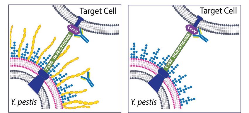
Figure 2.
Figure 2. Schematic
Schematicofof Antibodies
Antibodies that target
that Y. pestis
target and and
Y. pestis are effective at ameliorating
are effective disease.
at ameliorating Y. pestisY.is pestis
disease. a Gram‐negative
is a Gram-
bacterium that expresses multiple potential targets that have been exploited for the development of novel medical coun‐
negative bacterium that expresses multiple potential targets that have been exploited for the development of novel medical
termeasures. Antibodies generated by active vaccination or administered via passive immunization have been successful
countermeasures. Antibodies generated by active vaccination or administered via passive immunization have been
at protecting cell culture and animals from Y. pestis infection. These targets include The F1 capsular antigen (yellow struc‐
successful
tures depictedat protecting
in the left cell culture
panel) andLcrV
and the animals from Y.component
(V antigen) pestis infection.
of theThese targets include
T3SS injectisome. TheThe F1 capsular
capsule, while not antigen
abso‐
(yellow structures depicted in the left panel) and the LcrV (V antigen) component of the T3SS
lutely necessary for virulence, has been the target of both vaccine and therapeutic strategies. The LcrV protein has beeninjectisome. The capsule,
while
shownnotto absolutely
be essential necessary for virulence,
for virulence and has has
beenbeen the targettarget
a successful of both
invaccine and therapeutic
both active and passivestrategies.
immunization The LcrV protein
studies and
has beencombined
is often shown towith be essential for virulence
the F1 capsular antigen and hasthe
(i.e., been
F1‐V a successful
vaccine or target in both experiments).
mAb cocktail active and passive immunization
(Left Panel) In most
cases, the
studies and Y.ispestis
oftenbacterium
combinedwill withinteract directly with
the F1 capsular host
antigen target
(i.e., cells via
the F1-V the T3SS
vaccine or mAbinjectisome and a robust anti‐phago‐
cocktail experiments). (Left Panel)
cytic capsule will also be present; thus, both anti‐F1 and anti‐V antibodies are potentially
In most cases, the Y. pestis bacterium will interact directly with host target cells via the T3SS injectisome effective. (RightandPanel) Non‐
a robust
encapsulated strains of Y. pestis can be found in nature or engineered in the laboratory. In cases of
anti-phagocytic capsule will also be present; thus, both anti-F1 and anti-V antibodies are potentially effective. (Right Panel) infection caused by
non‐encapsulated Y. pestis, the antibodies to the F1 capsular antigen are no longer effective and the protective immune
Non-encapsulated strains of Y. pestis can be found in nature or engineered in the laboratory. In cases of infection caused
response generated by a vaccine or the effective therapeutic mAb relies solely on the anti‐V antibodies. A lipopolysaccha‐
by non-encapsulated Y. pestis, the antibodies to the F1 capsular antigen are no longer effective and the protective immune
ride structure (LPS) often observed in Y. pestis grown at 37 °C is depicted in blue in both panels. Other protective antigens
response
have beengenerated
identifiedbyanda vaccine or theantigen
other novel effectivetargets
therapeutic mAb relies
will almost solely
certainly beon the anti-V
identified inantibodies. A lipopolysaccharide
ongoing research efforts.
structure (LPS) often observed in Y. pestis grown at 37 ◦ C is depicted in blue in both panels. Other protective antigens have
been identified and other novel antigen targets will
The primary almost
subunit certainly
vaccine be identified
candidate in the in ongoing research
United States is efforts.
the recombinant F1‐V,
a fusion protein of the F1 capsular antigen and the lcrV gene product [166–170]. The
The primary subunit vaccine candidate in the United States is the recombinant F1-V,
United Kingdom pursued a similar strategy for vaccination but retained the F1 and V
a fusion protein of the F1 capsular antigen and the lcrV gene product [166–170]. The
immunogens as separate proteins [171–173]. PharmAthene further advanced this concept
United Kingdom pursued a similar strategy for vaccination but retained the F1 and V
and developed a recombinant dual antigen vaccine for plague, composed of rF1 + rV,
immunogens as separate proteins [171–173]. PharmAthene further advanced this concept
named RypVax™ [174,175]. A truncated LcrV antigen, rV10, developed by Schneewind
and developed a recombinant dual antigen vaccine for plague, composed of rF1 + rV,
and colleagues in 2011 was also assessed [176]. Both vaccines, rF1 + rV and rV10, were
named RypVax™ [174,175]. A truncated LcrV antigen, rV10, developed by Schneewind
tested and demonstrated
and colleagues efficacy
in 2011 was against pneumonic
also assessed [176]. Bothplague infection
vaccines, rF1 +in
rVmice,
and guinea pigs
rV10, were
and Cynomolgus macaques [176,177] but not in African green monkeys [125]. Further
tested and demonstrated efficacy against pneumonic plague infection in mice, guinea pigs in‐
vestigations of enhancing
and Cynomolgus immunogenicity
macaques [176,177] but or delivery
not of these
in African subunit
green vaccines
monkeys have
[125]. been
Further
attempted or are ongoing by several groups [178–185].
investigations of enhancing immunogenicity or delivery of these subunit vaccines have
been attempted or are ongoing by several groups [178–185].
The two-component vaccines have been variably effective and are vulnerable to break-
through infection. Researchers found that the combination of these subunits significantly
enhances protection against bubonic and pneumonic plague in different animal mod-
els [176,186,187]. The protection afforded by this vaccine strategy against F1-negative
strains relies entirely on the LcrV antigen component of the F1-V fusion protein. Since
there is evidence for V heterogeneity within Yersinia species, the potential exists that nat-
urally occurring or engineered strains harboring altered LcrV antigens could overcome
F1-V-induced immunity [161].Biomedicines 2021, 9, 1421 8 of 18
Other vaccine strategies have attempted to identify vaccine antigens that would
protect animals against non-encapsulated strains. Andrews demonstrated that active
vaccination against YopD could protect mice against an F1-negative strain of Y. pestis [188].
Later studies by Ivanov et al. further characterized the protective efficacy of YopD and
created fusion proteins of YopBD and YopBDE [189]. Ivanov et al. achieved protection
with both active and passive vaccination strategies using these proteins, but these data
suggested that anti-Yop immune responses were most protective against non-encapsulated
Y. pestis [189]. While there has been progress in vaccine development, the need remains for
other reliably protective and readily deployable countermeasures against plague.
6. Antibodies
There has been significant interest in and effort towards identifying antibody ther-
apies in animal models of plague. In 1963, Lawton and coworkers identified protection
associated with anti-LcrV polyclonal serum passively administered to mice. Importantly,
it was determined that the protection associated with this polyclonal serum could not be
completely attributable to the LcrV antigen due to the likelihood of other Yersinia proteins
present in the material used to generate the sera [190]. Later, Motin et al. demonstrated
with more modern techniques that a highly purified preparation of the V protein could be
produced and used for polyclonal sera generation [190]. These anti-sera directed against
native LcrV antigen or recombinant LcrV antigens were able to provide substantial passive
protection against Y. pestis infection.
Tables 1 and 2 and the following paragraphs provide summary information extracted
from noteworthy publications describing particularly effective monoclonal antibodies in
mouse models of plague. Hill et al. demonstrated that a monoclonal antibody directed
against the LcrV could protect against a fully virulent strain of Y. pestis and went on to
demonstrate which regions of the LcrV antigen appeared to be protective epitopes [156].
Quenee et al. and Amemiya et al. further elaborated on the importance of the specific
binding sites on protective anti-LcrV monoclonal antibodies [191,192]. The mAb 7.3 was
shown to protect macrophages from Y. pestis-induced cell death and also to promote
phagocytosis of the bacteria [158,193]. Importantly, much of the protection afforded by
mAb 7.3 can be attributed to the blocking of the T3SS machinery. When anti-V antibodies
block this injectisome, the immunomodulation and anti-phagocytic Yop effectors are not
efficiently secreted, thus blocking cytotoxic phenotypes and promoting phagocytosis of
the bacterial cells [158]. This concept was further supported by Eisele and Anderson
when they demonstrated that both blocking the T3SS as well as inducing phagocytosis are
required from an anti-LcrV mAb in order to optimally protect against pneumonic plague
in a mouse model of disease [194]. Novel Adenovirus-mediated delivery has also been
used to demonstrate the protection associated with anti-LcrV antigen mAbs [195].
Partly due to the known heterogeneity amongst LcrV protein sequences from different
bacterial isolates [161,196–198], a multi-antigen strategy was postulated to be required to
optimize protection. Anderson et al. also demonstrated that it was possible to passively
protect mice against either bubonic or pneumonic plague [199] with anti-F1 antibodies.
These anti-F1 monoclonal antibodies, however, would not protect against F1-negative
(non-encapsulated) strains of Y. pestis (Figure 2). Since immunity to F1 is not sufficient to
protect in all cases, there was renewed focus on the incorporation of the LcrV antigen or
other proteins for multifactorial strategies.Biomedicines 2021, 9, 1421 9 of 18
Table 1. Monoclonal antibodies shown to protect mice in bubonic plague models (subcutaneous challenge with bacteria).
mAb Antibody/Therapeutic Administration Y. pestis Challenge Challenge
Antigen a Description Ref.
Route Concentration Schedule Strain d Dose (LD50 )
F1; LcrV; F1V 24 h pre–120 h
human IgG1 IP 500 µg CO92 25–40 202
+ LcrV post
LcrV mouse IP 350 µg 24 h pre GB 12 156
mouse IgG1,
LcrV b IP 1–500 µg 24 h pre CO92 or C12 21–39 192
IgG2a
LcrV; F1 +
mouse IgG1 IP 0.7–100 µg 4 h pre–96 post GB 9.6–91,000 200
LcrV
LcrV mouse IgG1 IP 200 µg 1 h pre CO92 20 191
F1 c mouse IgG1 IP 125–500 µg 6 h or 24 h pre CO92 48–54 199
F1 not reported IV 100 µg 24 h pre 141 600 203
a, unless otherwise noted mAbs tested in BALB/c female mice; b, tested in BALB/c and Swiss Webster mice; c, tested in Swiss Webster
female mice; d, bacteria delivered via subcutaneous injection.
Table 2. Monoclonal antibodies shown to protect mice in pneumonic plague models (intranasal instillation or exposure to
aerosolized bacteria).
mAb Antibody/Therapeutic Administration Y. pestis Challenge Challenge
Antigen a Ref.
Description Route Concentration Schedule Strain f Dose (LD50 )
LcrV mouse IgG1 IP 35 µg 4 h pre–96 h post GB 88 156
b mouse IgG1 IP 200 or 400 µg 1 h pre CO92 g 15–20 194
LcrV
100–500 µg, 94 h pre–24 h
LcrV c mouse IgG2b IP, IV CO92 g 363–9080 195
1011 pu e post
F1 d mouse IgG1 IP 125–500 µg 6 h or 24 h pre CO92 29–74 199
F1 + LcrV mouse IgG1 IT 77.5 µg (each) 2 h post GB 27 200
a, unless otherwise noted mAbs tested in BALB/c female mice; b, tested in C57BL/6 female mice; c, tested in BALB/c female and C57BL/6
male mice; d, tested in Swiss Webster female mice; e, antibodies delivered as hybridoma supernatant or via adenovirus vector; f, unless
otherwise noted aerosolized bacteria were delivered; g, intranasal instillation was used for challenge
Combinations of F1 and LcrV monoclonal antibodies were shown to protect mice
against both bubonic and pneumonic plague either prophylactically or as post-exposure
therapy [200,201]. More recent efforts have continued to examine strategies combining
anti-F1 and anti-LcrV antibodies [202]. Xiao et al. identified three human mAbs: m252 for
anti-F1, and m253 and m254 for anti-LcrV. They found that anti-F1 human mAb (m252)
provided better protection in mice than anti-LcrV human mAbs. However, an apparent
synergistic effect was found when they combined all three antibodies. Another three
anti-F1 (F5C10, F6E5, and F2H5) mAbs were tested against subcutaneous challenge with
Y. pestis 141 and showed different protection levels. Liu et al. demonstrated that mAb F2H5
from a mouse hybridoma provides complete protection from bubonic plague in BALB/c
mice [203]. Altogether, it would be feasible that these mAbs against F1 or LcrV can be
utilized as a potential treatment or prophylaxis for humans against plague. Additional anti-
Y. pestis human antibodies have recently been produced and are currently being evaluated
for therapeutic application [204].
7. Advantages of Monoclonal Antibodies (mAbs) and Final Perspectives
Currently, there are only a few mAbs licensed for use against bacterial infectious
agents. To date, mAb against Clostridium difficile and Bacillus anthracis toxins are licensed in
Europe and the USA [205–207]. Other work has focused on mAbs against Staphylococcus
aureus or Pseudomonas aeruginosa [208]. The renewal of interest in mAb therapies is evenBiomedicines 2021, 9, 1421 10 of 18
more relevant given the current era of worrisome multidrug resistance in bacterial isolates
of clinical importance. Several clinically relevant bacteria such as Burkholderia pseudomallei
and Burkholderia mallei are naturally resistant to many antibiotics [209]. Examples of
emerging MDR bacteria include but are not limited to S. aureus, C. difficile, P. aeruginosa,
Acinetobacter baumeii, Enterococci, etc [210–213]. This MDR trait has been observed with
Y. pestis, and the lack of effective therapies for such an infection could result in catastrophic
loss of life, whether from a naturally emerging outbreak or an intentional attack utilizing
purposefully engineered bacteria.
Recent strategies have focused on nimble and rapid platforms that can be leveraged
when combatting outbreaks and emerging threats. Advances in rapid mAb discovery,
optimization, and production make this an attractive strategy that could significantly
decrease the time required to get a product to civilian populations in the midst of an
outbreak or to military personnel deployed to areas where endemic disease could be an
issue and an effective vaccine has yet to be identified. Other more novel strategies involve
administering nucleic acids that encode the protective mAb to patients. In that case, a
nucleic acid encoding the immunoglobulin may be manufactured or obtained from a
cDNA library or nucleic acid isolated from any tissue or cells expressing the antibody.
These nucleic acids can be cloned into a suitable vector and administered into an infected
patient [202].
These concepts have been useful recently during West African Ebola virus out-
breaks [214]. In this epidemic, mAbs were demonstrated to be important novel treatment
strategies. Products such as ZMapp®and others were critical in the response to the Ebola
virus epidemic [214]. Viruses of recent or continual concern including HIV, Zika, and
COVID-19 have also been targets of these novel strategies [215–220]. Monoclonal anti-
bodies will likely be of the utmost importance moving forward when developing novel
medical countermeasures against existing and rapidly emerging infectious diseases.
Funding: This review article was written with funding provided by the U.S. Defense Threat reduction
Agency (DTRA) CB10392.
Acknowledgments: We thank William Discher for assistance with illustrations. Opinions, interpreta-
tions, conclusions, and recommendations are those of the authors and are not necessarily endorsed
by the U.S. Army.
Conflicts of Interest: The authors declare no conflict of interest.
References
1. Pechous, R.D.; Sivaraman, V.; Stasulli, N.M.; Goldman, W.E. Pneumonic plague: The darker side of Yersinia pestis. Trends Microbiol.
2016, 24, 190–197. [CrossRef] [PubMed]
2. Demeure, C.E.; Dussurget, O.; Fiol, G.M.; Le Guern, A.S.; Savin, C.; Pizzarro-Cerdá, J. Yersinia pestis and plague: An updated
view on evolution, virulence determinants, immune subversion, vaccination, and diagnostics. Genes Immun. 2019, 20, 357–370.
[CrossRef] [PubMed]
3. Stenseth, N.C.; Atshabar, B.B.; Begon, M.; Belmain, S.R.; Bertherat, E.; Carniel, E.; Gage, K.L.; Leirs, H.; Rahalison, L. Plague: Past,
present, and future. PLoS Med. 2008, 5, e3. [CrossRef] [PubMed]
4. Achtman, M.; Zurth, K.; Morelli, G.; Torrea, G.; Guiyoule, A.; Carniel, E. Yersinia pestis, the cause of plague, is a recently emerged
clone of Yersinia pseudotuberculosis. Proc. Natl. Acad. Sci. USA 1999, 96, 14043–14048. [CrossRef]
5. Glatter, K.A.; Finkelman, P. History of the plague: An ancient pandemic for the age of COVID-19. Am. J. Med. 2021, 134, 176–181.
[CrossRef]
6. Brubaker, R.R. Yersinia pestis. Mol. Med. Microbiol. 2015, 3, 1845–1865.
7. Frith, J. The history of plague—Part 1. The three great pandemics. J. Mil. Vet. Health 2012, 20, 11–16.
8. Barbieri, R.; Signoli, M.; Chevé, D.; Costedoat, C.; Tzortzis, S.; Aboudharam, G.; Raoult, D.; Drancourt, M. Yersinia pestis: The
natural history of plague. Clin. Microbiol. Rev. 2020, 34, e00044-19. [CrossRef]
9. Drancourt, M.; Raoult, D. Molecular history of plague. Clin. Microbiol. Infect. 2016, 22, 911–915. [CrossRef]
10. Bramanti, B.; Dean, K.R.; Walløe, L.; Stenseth, N.C. The third plague pandemic in Europe. Proc. Biol. Sci. 2019, 286, 20182429.
[CrossRef]
11. Eisen, R.J.; Gage, K.L. Adaptive strategies of Yersinia pestis to persist during inter-epizootic and epizootic periods. Vet. Res. 2009,
40, 1. [CrossRef] [PubMed]Biomedicines 2021, 9, 1421 11 of 18
12. Raoult, D.; Mouffok, N.; Bitam, I.; Piarroux, R.; Drancourt, M. Plague: History and contemporary analysis. J. Infect. 2013, 66,
18–26. [CrossRef] [PubMed]
13. Melman, S.D.; Ettestad, P.E.; VinHatton, E.S.; Ragsdale, J.M.; Takacs, N.; Onischuk, L.M.; Leonard, P.M.; Master, S.S.; Lucero, V.S.;
Kingry, L.C. Human case of bubonic plague resulting from the bite of a wild Gunnison’s prairie dog during translocation from a
plague-endemic area. Zoonoses Public Health 2018, 65, e254–e258. [CrossRef]
14. Dai, R.; Wei, B.; Xiong, H.; Yang, X.; Peng, Y.; He, J.; Jin, J.; Wang, Y.; Zha, X.; Zhang, Z.; et al. Human plague associated with
Tibetan sheep originates in marmots. PLoS Negl. Trop. Dis. 2018, 12, e0006635. [CrossRef]
15. Richgels, K.L.; Russell, R.E.; Bron, G.M.; Rocke, T.E. Evaluation of Yersinia pestis transmission pathways for sylvatic plague in
prairie dog populations in the Western U.S. Ecohealth 2016, 13, 415–427. [CrossRef] [PubMed]
16. CDC. Fatal laboratory-acquired infection with an attenuated Yersinia pestis Strain—Chicago, Illinois, 2009. MMWR Morb. Mortal.
Wkly. Rep. 2011, 60, 201–205.
17. Worsham, P.L.; McGovern, T.W.; Vietri, N.J.; Friedlander, A.M.; Bozue, J. Plague. In Textbook of Military Medicine: Medical Aspects of
Biological Warfare; Bozue, J.A., Cote, C.K., Glass, P.J., Eds.; Borden Institute US Army Medical Department Center and School
Health Readiness Center of Excellence: Houston, TX, USA, 2018; pp. 247–284.
18. Yang, R. Plague: Recognition, treatment, and prevention. J. Clin. Microbiol. 2017, 56, e01519-17. [CrossRef] [PubMed]
19. Nikiforov, V.V.; Gao, H.; Zhou, L.; Anisimov, A. Plague: Clinics, diagnosis and treatment. Adv. Exp. Med. Biol. 2016, 918, 293–312.
20. Dennis, D.T.; Gage, K.L.; Gratz, N.G.; Poland, J.D.; Tikhomirov, E. Plague Manual: Epidemiology, Distribution, Surveillance and
Control; No. WHO/CDS/CSR/EDC/99.2; World Health Organization: Geneva, Switzeralnd, 1999.
21. Sebbane, F.; Lemaitre, N. Antibiotic therapy of plague: A review. Biomolecules 2021, 11, 724. [CrossRef]
22. Bossi, P.; Tagnell, A.; Baka, A.; Loock, F.V.; Hendriks, J.; Werner, A.; Maidhof, H.; Gouvras, G. Bichat guidelines for the clinical
management of plague and bioterrorism-related plague. Euro Surveill 2004, 9, E5–E6. [CrossRef]
23. Lemaître, N.; Ricard, I.; Pradel, E.; Foligné, B.; Courcol, R.; Simonet, M.; Sebbane, F. Efficacy of ciprofloxacin-gentamicin
combination therapy in murine bubonic plague. PLoS ONE 2012, 7, e52503. [CrossRef]
24. Boulanger, L.L.; Ettestad, P.; Fogarty, J.D.; Dennis, D.T.; Romig, D.; Mertz, G. Gentamicin and tetracyclines for the treatment of
human plague: Review of 75 cases in New Mexico, 1985–1999. Clin. Infect. Dis. 2004, 38, 663–669. [CrossRef]
25. Lawrenz, M.B. Model systems to study plague pathogenesis and develop new therapeutics. Front. Microbiol. 2010, 1, 119.
[CrossRef]
26. Dillard, R.L.; Juergens, A.L. Plague; StatPearls: Treasure Island, FL, USA, 2020.
27. Cleri, D.J.; Marton, R.; Rabbat, M.; Vernaleo, J. Pneumonia Caused by Yersinia pestis: Plague Pneumonia, in Community-Acquired
Pneumonia; Marrie, T.J., Ed.; Springer: Boston, MA, USA, 2001; pp. 777–799.
28. Kool, J. Risk of person-to-person transmission of pneumonic plague. Clin. Infect. Dis. 2005, 40, 1166–1172. [CrossRef] [PubMed]
29. Dennis, D.T.; Mead, P.S. Plague. Trop. Infect. Dis. 2006, 1, 471–481.
30. Gage, K.L.; Beard, C.B. 126—Plague. In Infectious Diseases, 4th ed.; Cohen, J., Powderly, W.G., Opal, S.M., Eds.; Elsevier:
Amsterdam, The Netherlands, 2017; pp. 1078–1084.e1.
31. Wagar, E. Bioterrorism and the role of the clinical microbiology laboratory. Clin. Microbiol. Rev. 2016, 29, 175–189. [CrossRef]
[PubMed]
32. Ansari, I.; Grier, G.; Byers, M. Deliberate release: Plague—A review. J. Biosaf. Biosecur. 2020, 2, 10–22. [CrossRef]
33. Du, Z.; Wang, X. Pathology and pathogenesis of Yersinia pestis. Adv. Exp. Med. Biol. 2016, 918, 193–222.
34. Hewitt, J.A.; Lanning, L.L.; Campbell, J.L. The African green monkey model of pneumonic plague and US Food and Drug
Administration approval of antimicrobials under the animal rule. Clin. Infect. Dis. 2020, 70 (Suppl. S1), S51–S59. [CrossRef]
35. Warren, R.; Lockman, H.; Barnewall, R.; Krile, R.; Bermeo Blanco, O.; Vasconcelos, D.; Price, J.; House, R.V.; Bolanowksi, M.A.;
Fellows, P. Cynomolgus macaque model for pneumonic plague. Microb. Pathog. 2011, 50, 12–22. [CrossRef]
36. Bubeck, S.S.; Cantwell, A.M.; Dube, P.H. Delayed inflammatory response to primary pneumonic plague occurs in both outbred
and inbred mice. Infect. Immun. 2007, 75, 697–705. [CrossRef]
37. Lathem, W.W.; Crosby, S.D.; Miller, V.L.; Goldman, W.E. Progression of primary pneumonic plague: A mouse model of infection,
pathology, and bacterial transcriptional activity. Proc. Natl. Acad. Sci. USA 2005, 102, 17786–17791. [CrossRef]
38. Pechous, R.D.; Sivaraman, V.; Price, P.A.; Stasulli, N.M.; Goldman, W.E. Early host cell targets of Yersinia pestis during primary
pneumonic plague. PLoS Pathog. 2013, 9, e1003679. [CrossRef]
39. Banerjee, S.K.; Crane, S.D.; Pechous, R.D. A dual role for the plasminogen activator protease during the preinflammatory phase
of primary pneumonic plague. J. Infect. Dis. 2020, 222, 407–416. [CrossRef] [PubMed]
40. Pechous, R.D.; Broberg, C.A.; Stasulli, N.M.; Miller, V.L.; Goldman, W.E. In vivo transcriptional profiling of Yersinia pestis reveals
a novel bacterial mediator of pulmonary inflammation. mBio 2015, 6, e02302-14. [CrossRef] [PubMed]
41. Zhou, D.; Yang, R. Molecular Darwinian evolution of virulence in Yersina pestis. Infect. Immun. 2020, 77, 2242–2250. [CrossRef]
42. Skurnik, M.; Peippo, A.; Ervelä, E. Characterization of the O-antigen gene clusters of Yersinia pseudotuberculosis and the cryptic
O-antigen gene cluster of Yersinia pestis shows that the plague bacillus is most closely related to and has evolved from Y.
pseudotuberculosis serotype O:1b. Mol. Microbiol. 2000, 37, 316–330. [CrossRef] [PubMed]
43. Rudolph, A.E.; Stuckey, J.A.; Zhao, Y.; Matthews, H.R.; Patton, W.A.; Moss, I.; Dixon, J.E. Expression, characterization, and
mutagenesis of the Yersinia pestis murine toxin, a phospholipase D superfamily member. J. Biol. Chem. 1999, 274, 11824–11831.
[CrossRef]Biomedicines 2021, 9, 1421 12 of 18
44. Schar, M.; Meyer, K.F. Studies on immunization against plague. XV. The pathophysiologic action of the toxin of Pasteurella pestis
in experimental animals. Schweiz Z Pathol. Bakteriol. 1956, 19, 51–70.
45. Bergdoll, M.S.; Enterotoxins, T.C.; Montie, S.J.A. Microbial Toxins; Academic Press: New York, NY, USA, 1970; Volume 3,
pp. 265–326.
46. Hinnebusch, B.J.; Rudolph, A.E.; Cherepanov, P.; Dixon, J.E.; Schwan, T.G. Role of Yersinia murine toxin in survival of Yersinia
pestis in the midgut of the flea vector. Science 2002, 296, 733–735. [CrossRef]
47. Chen, T.H.; Crocker, T.T.; Meyer, K.F. Electron microscopic study of the extracellular materials of Pasteurella pestis. J. Bacteriol.
1956, 72, 851–857.
48. Davis, K.J.; Fritz, D.L.; Pitt, M.L.; Welkos, S.L.; Worsham, P.L.; Friedlander, A.M. Pathology of experimental pneumonic plague
produced by fraction 1-positive and fraction 1-negative Yersinia pestis in African green monkeys (Cercopithecus aethiops). Arch.
Pathol. Lab. Med. 1996, 120, 156–163.
49. Engelsberg, E.; Levy, J.B. Studies on immunization against plague. VI. Growth of Pasteurella pestis and the production of the
envelope and other soluble antigens in a casein hydrolyzate mineral glucose medium. J. Bacteriol. 1954, 67, 438–449. [CrossRef]
50. Runco, L.M.; Myrczek, S.; Bliska, J.B.; Thanassi, D.G. Biogenesis of the fraction 1 capsule and analysis of the ultrastructure of
Yersinia pestis. J. Bacteriol. 2008, 190, 3381–3385. [CrossRef]
51. Du, Y.; Rosqvist, R.; Forsberg, Å. Role of fraction 1 antigen of Yersinia pestis in inhibition of phagocytosis. Infect. Immun. 2002, 70,
1453. [CrossRef] [PubMed]
52. Welkos, S.L.; Davis, K.M.; Pitt, L.M.; Worsham, P.L.; Friedlander, A.M. Studies on the contribution of the F1 capsule-associated
plasmid pFra to the virulence of Yersinia pestis. Contrib. Microbiol. Immunol. 1995, 13, 299–305. [PubMed]
53. Winter, C.C.; Cherry, W.B.; Moody, M.D. An unusual strain of Pasteurella pestis isolated from a fatal human case of plague. Bull.
World Health Organ. 1960, 23, 408–409.
54. Sebbane, F.; Uversky, V.N.; Anisimov, A.P. Yersinia pestis plasminogen activator. Biomolecules 2020, 10, 1554. [CrossRef] [PubMed]
55. Caulfield, A.J.; Lathem, W.W. Substrates of the plasminogen activator protease of Yersinia pestis. Adv. Exp. Med. Biol. 2012, 954,
253–260.
56. Degen, J.L.; Bugge, T.H.; Goguen, J.D. Fibrin and fibrinolysis in infection and host defense. J. Thromb. Haemost. 2007, 5 (Suppl. S1),
24–31. [CrossRef]
57. Sodeinde, O.A.; Subrahmanyam, Y.V.; Stark, K.; Quan, T.; Bao, Y.; Goguen, J.D. A surface protease and the invasive character of
plague. Science 1992, 258, 1004–1007. [CrossRef]
58. Caulfield, A.J.; Walker, M.E.; Gielda, L.M.; Lathem, W.W. The Pla protease of Yersinia pestis degrades fas ligand to manipulate
host cell death and inflammation. Cell Host Microbe 2014, 15, 424–434. [CrossRef]
59. Lathem, W.W.; Price, P.A.; Miller, V.L.; Goldman, W.E. A plasminogen-activating protease specifically controls the development
of primary pneumonic plague. Science 2007, 315, 509–513. [CrossRef]
60. Kienle, Z.; Emödy, L.; Svanborg, C.; O’Toole, P.W. Adhesive properties conferred by the plasminogen activator of Yersinia pestis. J.
Gen. Microbiol. 1992, 138, 1679–1687. [CrossRef]
61. Lähteenmäki, K.; Virkola, R.; Sarén, A.; Emödy, L.; Korhonen, T.K. Expression of plasminogen activator Pla of Yersinia pestis
enhances bacterial attachment to the mammalian extracellular matrix. Infect. Immun. 1998, 66, 5755–5762. [CrossRef]
62. Lobo, L.A. Adhesive properties of the purified plasminogen activator Pla of Yersinia pestis. FEMS Microbiol. Lett. 2006, 262,
158–162. [CrossRef]
63. Cowan, C.; Jones, H.A.; Kaya, Y.H.; Perry, R.D.; Straley, S.C. Invasion of epithelial cells by Yersinia pestis: Evidence for a
Y. pestis-specific invasin. Infect. Immun. 2000, 68, 4523–4530. [CrossRef]
64. Lähteenmäki, K.; Kukkonen, M.; Korhonen, T.K. The Pla surface protease/adhesin of Yersinia pestis mediates bacterial invasion
into human endothelial cells. FEBS Lett. 2001, 504, 69–72. [CrossRef]
65. Fields, K.A.; Straley, S.C. LcrV of Yersinia pestis enters infected eukaryotic cells by a virulence plasmid-independent mechanism.
Infect. Immun. 1999, 67, 4801–4813. [CrossRef] [PubMed]
66. Derewenda, U.; Mateja, A.; Devedjiev, Y.; Routzahn, K.M.; Evdokimov, A.G.; Derewenda, Z.S.; Waugh, D.S. The structure of
Yersinia pestis V-antigen, an essential virulence factor and mediator of immunity against plague. Structure 2004, 12, 301–306.
[CrossRef] [PubMed]
67. Price, S.B.; Cowan, C.; Perry, R.D.; Straley, S.C. The Yersinia pestis V antigen is a regulatory protein necessary for Ca2(+)-dependent
growth and maximal expression of low-Ca2+ response virulence genes. J. Bacteriol. 1991, 173, 2649–2657. [CrossRef]
68. Hamad, M.A.; Nilles, M.L. Structure-function analysis of the C-terminal domain of LcrV from Yersinia pestis. J. Bacteriol. 2007, 189,
6734–6739. [CrossRef]
69. DiMezzo, T.L.; Ruthel, G.; Brueggemann, E.E.; Hines, H.B.; Ribot, W.J.; Chapman, C.E.; Powell, B.S.; Welkos, S.L. In vitro
intracellular trafficking of virulence antigen during infection by Yersinia pestis. PLoS ONE 2009, 4, e6281. [CrossRef] [PubMed]
70. Sing, A.; Roggenkamp, A.; Geiger, A.M.; Heesemann, J. Yersinia enterocolitica evasion of the host innate immune response by
V antigen-induced IL-10 production of macrophages is abrogated in IL-10-deficient mice. J. Immunol. 2002, 168, 1315–1321.
[CrossRef] [PubMed]
71. Perry, R.D.; Haddix, P.; Atkins, E.B.; Soughers, T.K.; Straley, S.C. Regulation of expression of V antigen and outer membrane
proteins in Yersinia pestis. Contrib. Microbiol. Immunol. 1987, 9, 173–178. [PubMed]You can also read