Adrenocortical Carcinoma: Updates of Clinical and Pathological Features after Renewed World Health Organisation Classification and Pathology ...
←
→
Page content transcription
If your browser does not render page correctly, please read the page content below
biomedicines
Review
Adrenocortical Carcinoma: Updates of Clinical and Pathological
Features after Renewed World Health Organisation
Classification and Pathology Staging
Alfred King-yin Lam 1,2,3

                                          1   School of Medicine, Griffith University, Gold Coast, QLD 4222, Australia; a.lam@griffith.edu.au
                                          2   Pathology Queensland, Gold Coast University Hospital, Southport, Gold Coast, QLD 4215, Australia
                                          3   Faculty of Medicine, The University of Queensland, Herston, Brisbane, QLD 4006, Australia

                                          Abstract: Adrenocortical carcinoma (ACC) is a heterogenous group of diseases with different clinical
                                          behaviour between adult and paediatric patients. In addition, three histological variants, oncocytic,
                                          myxoid and sarcomatoid are noted on the recent World Health Organisation (WHO) classification of
                                          ACC. A review of recent literature showed that the different types of ACC have distinctive demo-
                                          graphic data, clinical presentation, pathology, biological behaviour, genomic and patients’ prognosis.
                                          In addition, recent updates of pathology staging for ACC allow refinement of prognostic grouping for
                                          planning treatment of the patients with ACC. These advances in genomic, pathology and staging have
                                          driven the development of standardisation of pathology reporting. International standardisation of
                                          pathological reporting of adrenocortical carcinoma and adaption to local pathology communities

                                   provide universal platforms for clinicians and researchers involved in the management of patients
                                          with ACC. To conclude, all these advances in the field of pathology will improve development of
Citation: Lam, A.K.-y.
Adrenocortical Carcinoma: Updates
                                          management strategies including improvement of clinical care, development of prognostic markers
of Clinical and Pathological Features     and testing of novel therapeutic approaches for patients with adrenocortical carcinoma.
after Renewed World Health
Organisation Classification and           Keywords: adrenocortical carcinoma; adrenal; staging; pathology; oncocytic; myxoid; sarcomatoid
Pathology Staging. Biomedicines 2021,
9, 175. https://doi.org/10.3390/
biomedicines9020175
                                          1. Introduction
Academic Editors:
                                               Adrenal cortical carcinoma (ACC) is a rare cancer but is the most common primary
Chitra Subramanian and
                                          cancer in the adrenal gland [1]. It is the second most common malignant tumour of the
Yasuhiro Nakamura
                                          endocrine organ after anaplastic thyroid carcinoma [2]. In large pathology series, ACC
Received: 31 December 2020
                                          accounts for 6.8% of all primary adrenal tumours [3]. In the most recent population
Accepted: 8 February 2021
                                          database (based on approximately 2000 cases) from the Surveillance, Epidemiology, and
Published: 10 February 2021
                                          End Results (SEER) published in 2018 from the United States of America (USA) of ACC-
                                          diagnosed patients between 1973 to 2012, the annual incidence of the cancer was 1.02 per
Publisher’s Note: MDPI stays neutral
with regard to jurisdictional claims in
                                          one million [4]. This figure has reflected the increase incidence of the cancer due to increased
published maps and institutional affil-
                                          use of sensitivity imaging studies in current years. In addition, in a recent large series of
iations.
                                          adrenal incidentalomas (n = 3672), ACC accounts for 1.4% of cases [5].
                                               There is an increase of new knowledge about ACC in terms of genomic characterisa-
                                          tion based on the findings of The Cancer Genome Atlas (TCGA) [6–9]. Further genomic
                                          studies showed that mutational and expression profiles of advanced and metastatic ACC
                                          are very similar to those from primary ACC as well as low mutation rates, few major
Copyright: © 2021 by the author.
                                          oncogenic drivers and loss of function mutations in several epigenetic regulators suggest
Licensee MDPI, Basel, Switzerland.
                                          an epigenetic basis of ACC such as DNA methylation [10,11]. In a similar period, the eighth
This article is an open access article
distributed under the terms and
                                          American Joint Committee on Cancer (AJCC) Cancer Staging Manual and UICC (Union for
conditions of the Creative Commons
                                          International Cancer Control) Tumour, Lymph node, Metastasis (TNM) staging Manual
Attribution (CC BY) license (https://     published in 2016 (adopted for use in 2017) have updated the pathological staging of ACC
creativecommons.org/licenses/by/          based on the European Network for the study of Adrenal Tumours (ENSAT) [12]. In addi-
4.0/).

Biomedicines 2021, 9, 175. https://doi.org/10.3390/biomedicines9020175                                https://www.mdpi.com/journal/biomedicines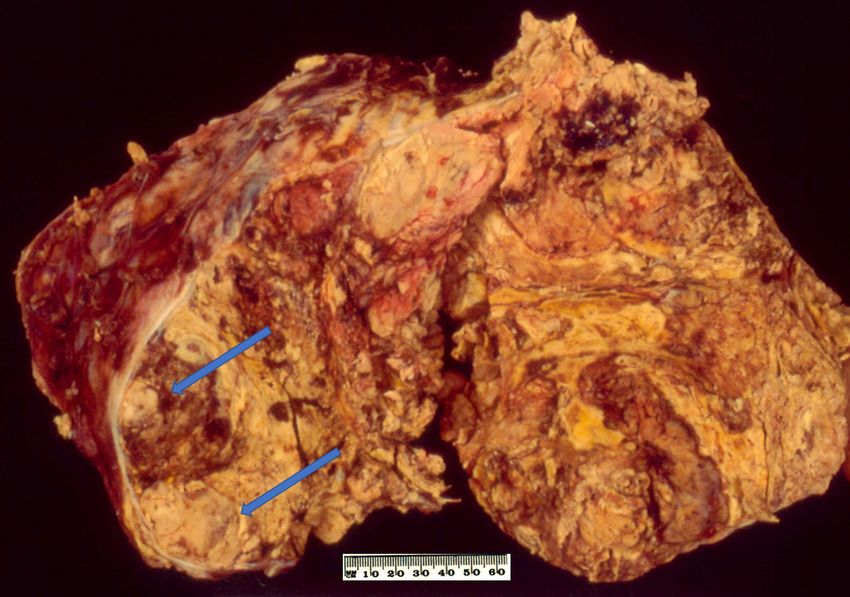
Biomedicines 2021, 9, 175                                                                                             2 of 25

                            tion, the fourth edition of the World Health Organization (WHO) book series on tumour
                            classification published in 2017 has updated the histological classification of ACC [13].
                                  The present review focuses on knowledge about these advances in the pathological
                            field as well as updates of current advances of the ACC. In the 4th (current) WHO clas-
                            sification of tumours of the endocrine system, apart from the conventional ACC, three
                            histological variants of ACC are proposed for the first time in WHO classification [13].
                            Emphases on the updates are on the unique clinical and pathological features of conven-
                            tional as well as new histological variants accepted by the WHO classification of tumours
                            of endocrine system.

                            2. Conventional Adrenocortical Carcinoma
                            2.1. Demographic Characteristics
                                 Based on the small number of cases of other histological variants of ACC, conventional
                            ACC comprises more than 90% of cases. Thus, data noted in epidemiological studies likely
                            represent the features of conventional ACC. Like other endocrine tumours, ACC is more
                            common in females. From the pooled series of different countries, ACC is more common
                            in females with male to female ratio of approximately 1 to 1.4 [3,14,15]. The average age
                            at presentation of patients with ACC in larger series range from 47- to 55-year-olds [15].
                            Although some series demonstrate a bimodal age distribution with peaks in the paediatric
                            group and middle age group, SEER data [4] show that there is a steady increase of ACC
                            with increased age peaking at the sixth and seventh decades.

                            2.2. Clinical Features
                                  Approximately half of ACC is functioning with signs and symptoms of hormone(s)
                            secretion [16]. Patients with functioning ACC were younger, more likely to be females
                            and present with metastatic disease. Of functioning tumours, almost half present with
                            signs or symptoms of cortisol excess (Cushing syndrome). The second most common
                            functional presentation was sex hormone secretion accounting for 20% of cases. These sex
                            hormone-producing ACCs are mainly androgens. Rarely, oestrogen secreting/feminising
                            ACC were reported [17–21]. Aldosterone secreting ACC is uncommon and accounted for
                            approximately 8% of cases. In addition, mixed hormone production was seen in 15% to
                            25% of functioning ACCs [15,22].
                                  Non-functioning ACCs commonly present with abdominal mass, abdominal pain
                            as well as general symptoms of malignancy. Paraneoplastic manifestations of having
                            ACC such as hypoglycaemia (due to insulin growth factor 2 [IGF2] production) [23] and
                            clinical manifestations related to adrenocorticotropic hormone ACTH production have been
                            reported [24]. Rare manifestations of patients with ACC include cancer-related thrombotic
                            microangiopathy [25] and tumour rupture with retroperitoneal haemorrhage [26–28].
                                  ACC could be a cancer in the setting of several hereditary syndromes. These syn-
                            dromes could occur in high as 5–10% of patients with ACC [29]. In adult patients with
                            ACC, these syndromes include multiple endocrine neoplasia type 1 (MEN1, approxi-
                            mately 20 cases reported) [30], Lynch syndrome (mismatch repair genes) (approximately
                            10 cases) [31], Li-Fraumeni syndrome (TP53, more than 10 cases) [32] and neurofibromato-
                            sis type 1 (NF1, approximately 10 cases) [33]. Rarely, ACC can occur in patients with
                            Carney complex (protein kinase A regulatory subunit 1A [PRKARIA]) [34,35], Gardner’s syn-
                            drome [36] and familial adenomatous polyposis (adenomatous polyposis coli [APC]) [37,38].
                            Although extremely rare, ACCs have been reported in patients with congenital adrenal
                            hyperplasia and myelolipoma [39–41]. It is likely these are collision tumours rather than
                            having been aetiologically related.

                            2.3. Pathology
                                ACC is often noted on left adrenal (left to right ratio = 1.2 to 1). Bilateral ACC is uncom-
                            mon and accounted for approximately 1% of the cases [4]. Ectopic ACCs, presumably arise
                            from ectopic adrenal rests, have been reported in the retroperitoneum [42], ovary [43,44],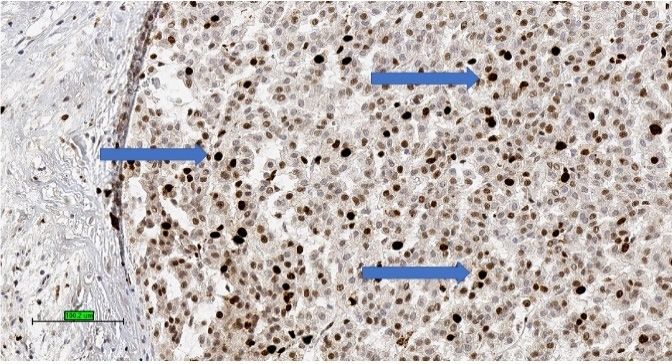
2.3. Pathology
Biomedicines 2021, 9, 175 ACC is often noted on left adrenal (left to right ratio = 1.2 to 1). Bilateral ACC 3 of 25is un-
common and accounted for approximately 1% of the cases [4]. Ectopic ACCs, presumably
arise from ectopic adrenal rests, have been reported in the retroperitoneum [42], ovary
[43,44],
spinal spinal
region [45],region [45],and
liver [46] liver [46] andwall
abdominal abdominal wallof[47].
[47]. A case A case of retroperitoneal
retroperitoneal ACC was
ACC was noted in 68-year-old man
noted in 68-year-old man with Lynch syndrome [48].with Lynch syndrome [48].
OnOnmacroscopic
macroscopic examination,
examination, ACC
ACC is often
is often yellow
yellow to tan
to tan colour
colour (Figure
(Figure 1). 1). Areas of
Areas
ofnecrosis
necrosisand
andhaemorrhage
haemorrhage are are common
common whichwhich give
give aa heterogenous
heterogenousappearance
appearanceon oncutcut sec-
tions. The
sections. tumour
The tumour is often
is oftenlarge with
large withmedian
median maximum
maximum dimension
dimension of of
100100mm mmto 120
to mm
[4,49].
120 mm From
[4,49]. the
From findings in recent
the findings literature,
in recent the largest
literature, non-functioning
the largest non-functioning ACC ACC was re-
was reported
ported in ain39-year-old
a 39-year-old Greekwoman
Greek woman withwith aamaximum
maximum dimension
dimension of 237
of mm
237 [50]
mm [50]
whereas
whereasthe thelargest
largestfunctioning
functioning(androgen-secreting)
(androgen-secreting) ACC ACC was
was noted
notedinina a48-year-old
48-year-old Ca-
Canadian woman with a maximum dimension of 230 mm [51]. Also,
nadian woman with a maximum dimension of 230 mm [51]. Also, Weiss and colleagues Weiss and colleagues
have
havementioned
mentioned anan ACCACC with
with280280
mmmm in dimension
in dimension in their series
in their published
series in 1989
published in [52]
1989 [52]
but no clinicopathological details about the case. The tumour could
but no clinicopathological details about the case. The tumour could be seen to grossly be seen to grossly
extend to adjacent organs as well as to regional veins and the right atrium which could
extend to adjacent organs as well as to regional veins and the right atrium which could
lead to pulmonary embolism [53].
lead to pulmonary embolism [53].
Figure
Figure 1. 1. Conventional
Conventional adrenocortical
adrenocortical carcinoma
carcinoma (ACC)(ACC) showing
showing variegated
variegated cut surface
cut surface of yellow
of yellow
tumour with foci of haemorrhages and necrosis (arrows).
tumour with foci of haemorrhages and necrosis (arrows).
Uponmicroscopic
Upon microscopic examination,
examination, ACC
ACC often
often has
has eosinophilic
eosinophilic cytoplasm.
cytoplasm. There
There areare of-
ten thick
often fibrousbands
thick fibrous bandsand
andcapsules
capsules (Figure
(Figure 2A).2A). Necrosis
Necrosis is common
is common (Figure
(Figure 2B). Mitotic
2B). Mitotic
figures
figuresare
areoften prominent.
often prominent.ForForprognostic
prognostic purposes, ACCACC
purposes, was was
subdivided into low
subdivided into low
grade or high grade depending on mitotic frequency (dividing line is between
grade or high grade depending on mitotic frequency (dividing line is between ≤20 and ≤ 20 and
>20 2 ) [52].
>20mitosis
mitosis per 5050
per high power
high powerfields/10 mm
fields/10 mm 2) [52].
Biomedicines 2021, 9, 175 4 of 25
Biomedicines 2021, 9, x FOR PEER REVIEW 4 of 25
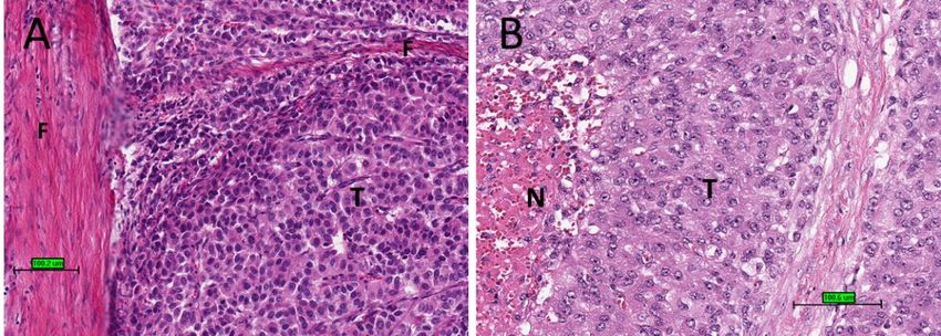
Figure 2.
Figure 2. Histological
Histologicalfeatures
featuresofofACC.
ACC.(A)
(A)Tumour
Tumourcells (T)(T)
cells with eosinophilic
with cytoplasm
eosinophilic with
cytoplasm thick
with fibrous
thick bands
fibrous (F). (F).
bands (B)
Tumour cells (T) with necrosis (N) (haematoxylin and eosin stain) (scale bar–100 µm).
(B) Tumour cells (T) with necrosis (N) (haematoxylin and eosin stain) (scale bar–100 µm).
It
It is often not
is often not difficult
difficultto todifferentiate
differentiateACC ACCfromfrom benign
benign cortical
cortical adenoma
adenoma or border-
or borderline
line cortical tumour. Nevertheless, in some instances, the diagnosis
cortical tumour. Nevertheless, in some instances, the diagnosis of ACC needs to rely of ACC needs to rely
on
on assessment of multiple pathological parameters. The most
assessment of multiple pathological parameters. The most widely accepted system adopted widely accepted system
adopted
by WHOby WHO classification
classification for assessment
for assessment is Weissiscriteria
Weiss criteria
published published
in 1984in 1984 [52,54].
[52,54]. Using
Using this system,
this system, ACC ACC couldcould be diagnosed
be diagnosed on on at least
at least three
three ofofthe
the99histological
histological features
features— —
high nuclear grade (Fuhrman
high nuclear grade (Fuhrman III III or IV), high mitotic rate (>5 mitoses per 50 high power
field, atypical
atypicalmitotic
mitoticfigures,
figures,≤25%
≤25% clear
clearcells, diffuse
cells, diffusearchitecture,
architecture, tumourtumour confluent
confluent ne-
crosis,
necrosis,venous
venous invasion,
invasion, sinusoidal
sinusoidalinvasion,
invasion,and andcapsular
capsularinvasion.
invasion.Modified
Modified Weiss Weiss has
been proposed which which is is based
based on on 55 of
of the
the 99 histological
histological features
features of of Weiss
Weiss (mitotic
(mitotic counts,
counts,
clear
clear cells, atypical
atypical mitotic figures, tumour necrosis and capsular invasion) [55]. In this
system, mitotic
mitotic rate and clear cells have twice the points (‘weight”) compared with other
features. TheThe outcome
outcomeofofuse useofof modified
modified Weiss
Weiss is highly
is highly correlated
correlated with the withoriginal
the original
Weiss
Weiss system [55].
system [55].
Diffuse architecture
Diffuse architecture must be present in >33% >33% and could be identified by disruption
of the reticulin [56] and interruption of basal lamina (antibodies to laminin or collagen
type IV). A reticulin algorithm has been used for diagnosis of ACC which involves an
type
abnormal/absentreticulum
abnormal/absent reticulumframework
frameworkand andatatleast
least one
one ofof the
the 3 of histological features
(tumour necrosis, presence of venous invasion and mitotic raterate
(tumour necrosis, presence of venous invasion and mitotic of >5/50
of >5/50 high power
high power field)
Biomedicines 2021, 9, x FOR PEER REVIEW
field)
[56,57].[56,57]. 5 of 25
These systems for predicting malignant potential of adrenocortical
These systems for predicting malignant potential of adrenocortical tumours are ap- tumours are ap-
plicable mainly
plicable mainly in in conventional
conventional ACC. ACC. It It is
is important
important to to note
note that
that no
no single
single microscopic
microscopic
criterion
criterion on its own is indicative of malignancy and there
often noted [15]. Common epithelial markers such as cytokeratin, EMA, CEA are in
on its own is indicative of malignancy and there is
is subjective
subjective variability
variability in the
the
gener-
interpretation
interpretation [58].
ally negative.[58]. Studies
Studies
Although have proposed
havecould
ACC proposed the use
the usefor
be positive of proliferative
of synaptophysin, index
proliferative index (Ki-67
it is(Ki-67
negative index
index > 5%)
for>chro-
5%)
(Figure
(Figure
mogranin.3) [59]
3) [59] and
and IGF2
IGF2 over-expression
over-expression to to confirm
confirm thethe diagnosis
diagnosis of of ACC
ACC [60].
[60].
A relatively new system published by a European group in 2015 is the Helsinki score
which relay on mitotic rate, necrosis and Ki-67 index (3× mitotic count [>5/50 high power
fields] + 5× presence of necrosis + Ki-67 proliferative index in the most proliferative index
of the tumour) of ACC and focus on the predicting diagnosis as well as prognosis of ACC
[61,62]. A Helsinki score >8.5 is associated with metastatic potential and warrant the diag-
nosis of ACC.
The most common malignant tumour in adrenal gland is metastatic carcinoma [63].
In a biopsy specimen, it is important to differentiate ACC from metastatic carcinoma as
well as phaeochromocytoma by clinical history, biochemical studies, radiology, and a
panel of immunohistochemical stains [64]. ACC expresses markers specific for steroid-
producing cells which often include steroidogenic factor-1 (SF-1) and inhibin alpha (Fig-
Figure
ure 4). 3.
Figure 3.Adrenocortical
TheAdrenocortical
tumour is also carcinoma
carcinoma ofofhigh
positive high
for Ki-67
markers
Ki-67 index with
expressed
index with high
high by percentage
other tumour
percentage of nuclear
of nucleartypes, stain
stain such
(brownas
(brownA
melan
colour) colour)
forand
Ki-67 for
(3,3Ki-67
calretinin. (3,3′-diaminobenzidine/haematoxylin
0 -diaminobenzidine/haematoxylin
In addition, abnormal beta-catenin stains)
stains) (blue (blue arrows)
intracellular
arrows) (scale (scale bar—
accumulation
bar—100 µm).is
100 µm).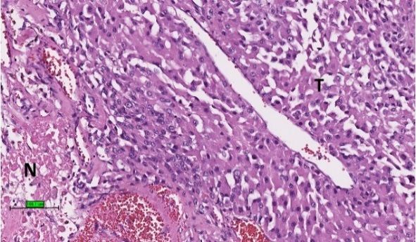
Biomedicines 2021, 9, x FOR PEER REVIEW 5 of 25
Biomedicines 2021, 9, 175 5 of 25
often noted [15]. Common epithelial markers such as cytokeratin, EMA, CEA are gener-
allyAnegative. Although
relatively ACCpublished
new system could be by
positive for synaptophysin,
a European group in 2015itisisthe
negative forscore
Helsinki chro-
mogranin.
which relay on mitotic rate, necrosis and Ki-67 index (3× mitotic count [>5/50 high power
fields] + 5× presence of necrosis + Ki-67 proliferative index in the most proliferative index
of the tumour) of ACC and focus on the predicting diagnosis as well as prognosis of
ACC [61,62]. A Helsinki score >8.5 is associated with metastatic potential and warrant the
diagnosis of ACC.
The most common malignant tumour in adrenal gland is metastatic carcinoma [63].
In a biopsy specimen, it is important to differentiate ACC from metastatic carcinoma
as well as phaeochromocytoma by clinical history, biochemical studies, radiology, and
a panel of immunohistochemical stains [64]. ACC expresses markers specific for steroid-
producing cells which often include steroidogenic factor-1 (SF-1) and inhibin alpha (Figure 4).
The tumour is also positive for markers expressed by other tumour types, such as melan
A and calretinin. In addition, abnormal beta-catenin intracellular accumulation is often
Figure 3. Adrenocortical carcinoma of high Ki-67 index with high percentage of nuclear stain
noted [15]. Common epithelial markers such as cytokeratin, EMA, CEA are generally nega-
(brown colour) for Ki-67 (3,3′-diaminobenzidine/haematoxylin stains) (blue arrows) (scale bar—
tive. Although ACC could be positive for synaptophysin, it is negative for chromogranin.
100 µm).
Figure4.4.Tumour
Figure Tumourstain
stainpositive
positive(brown
(browncolour)
colour)for
forinhibin
inhibin(3,3
(3,3′-diaminobenzidine/haematoxylin
0 -diaminobenzidine/haematoxylin
stains) in cytoplasm of ACC (green arrows). The necrotic area is not stained up (blue arrow). (scale
stains) in cytoplasm of ACC (green arrows). The necrotic area is not stained up (blue arrow). (scale
bar—100 µm).
bar—100 µm).
Distantmetastases
Distant metastases at at presentation
presentation occurred
occurred in approximately
in approximately one third
one third of theofpatients
the pa-
tients
with with
ACC ACC
[15]. The[15]. The common
common sites ofmetastases
sites of distant distant metastases
of ACC are ofliver,
ACClung,
are liver, lung,[65].
and bone and
bone [65]. Unusual descripted metastatic locations of ACC have been
Unusual descripted metastatic locations of ACC have been reported in the stomach [66], reported in the
stomach [66], pancreas [67], skin [68], spleen [69], tongue [70] and brain
pancreas [67], skin [68], spleen [69], tongue [70] and brain including meninges [71,72].including menin-
ges [71,72].and
Recurrence Recurrence and distant
distant metastases of ACCmetastases of ACC
often occur often
quickly occur2 years).
(within quicklyHowever,
(within 2
years). However, late locoregional recurrence could occur 18 years after
late locoregional recurrence could occur 18 years after surgery [73] and liver metastasissurgery [73] and
liverbeen
have metastasis have
reported in abeen reported
patient 23 yearsin after
a patient 23 years
surgical afterofsurgical
resection resection of ACC
ACC [74].
[74].
2.4. Prognosis
2.4. Prognosis
Prognosis of the conventional ACC depends on the cancer stage [12]. In addition,
Prognosisvalue
the prognostic of theofconventional
Helsinki scoreACChas depends on the cancer stage
been demonstrated to be [12]. In addition, the
an outperforming
prognosticindex
prognostic value[56].
of Helsinki scorelarge
In addition, has been
tumour demonstrated to be an outperforming
size and cortisol-secreting tumour wereprog-
nostic index
additional [56].for
factors In ACC-specific
addition, large tumour
death [49]. size and cortisol-secreting
Moreover, tumour were
there are many predictive addi-
features
proposed for the
tional factors for prognosis
ACC-specific of patients with
death [49]. ACC such
Moreover, as are
there mitotic
many grading [52],features
predictive Ki-67
index [75], for
proposed mi-RNAs expression
the prognosis [76], expression
of patients with ACC such of PDL-1 [77], grading
as mitotic SF-1 [78][52],
andKi-67
sterol-O-
index
acyl
[75],transferase 1 (SOAT1)[76],
mi-RNAs expression [79].expression
Preliminary data [77],
of PDL-1 also SF-1
shows[78]that
andfor localised ACC,
sterol-O-acyl trans-
molecular makers [79].
ferase 1 (SOAT1) (expression,
Preliminarymethylation, and chromosome
data also shows alterations)
that for localised could predict
ACC, molecular mak-
cancer recurrence methylation,
ers (expression, [9]. Nevertheless,
and many of these alterations)
chromosome markers need validation
could andcancer
predict some (such
recur-
asrence
molecular markers) aremany
[9]. Nevertheless, difficult to apply
of these in clinical
markers settings. and some (such as molec-
need validation
ular markers) are difficult to apply in clinical settings. A recent Danish study on 160 pa-
Overall, the prognosis of patients with ACC is poor.
tients Overall,
with ACC theshowed that of
prognosis a median
patientssurvival
with ACC of patients
is poor.with ACC Danish
A recent was 35 months
study on[14]160
whereas
patientsin the ACC
with studyshowed
based on thea USA
that medianSEER database,
survival the median
of patients with survival
ACC wasof35 patients
months
with ACC was 17 months [4]. The 5-year cancer specific survival rate of patients with ACC
as noted from USA SEER database was 38% [1]. It is worth noting that surgery on the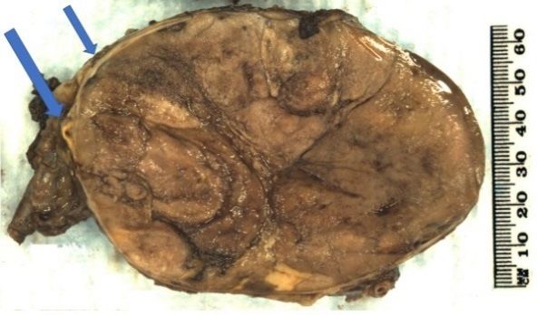
Biomedicines 2021, 9, 175                                                                                            6 of 25

                            primary site even in metastatic ACC significantly improved overall and cancer-specific
                            survival of patients with ACC [80]. The 5 year-survival of adult patients from multiple
                            datasets with ACC after surgery range from 40% to 70% [49,81]. The estimated five-year
                            overall survival rate for patients with ACC in recent cohorts is slightly less than 50% [11]

                            3. Paediatric Adrenocortical Carcinoma
                            3.1. Demographic Characteristics
                                  ACC in paediatric patients deserves separate grouping as they are different from ACC
                            in adults in terms of biological behaviour. The incidence of paediatric ACC is 0.2 cases per
                            million children per year in SEER data from USA [82]. A recent national cohort study in
                            France suggested the incidence of paediatric ACC to be approximately 0.1 cases per million
                            children per year [83]. On the other hand, the incidence of paediatric ACC is reported to be
                            high in southern regions of Brazil in which 4 case per million was reported in children of
                            less than 10 years which may be related to high incidence of germline mutation of TP53
                            (R337H) detected in the region [84,85].
                                  ACC in paediatric group is more commonly seen in girls (male to female ratio of 1
                            to 2). Paediatric patients with ACC accounted for 6.5% of patients with ACC [4]. The
                            disease is more common in the first decade with peak in 1 to 4 years [82] and with the
                            mean age at presentation at 5 [86]. Infants of less than 6-months-old with ACC have been
                            reported [87–89].

                            3.2. Clinical Features
                                 Paediatric ACCs are often functioning tumours. In the largest single-institution
                            study [90] involving 41 paediatric ACC, only 15% of the patients were non-functioning.
                            In the functioning tumours, many secrete more than 1 hormones (54%) and the most
                            common hormonal manifestation is related to sex hormones. The sex hormones produced
                            by paediatric ACC are mostly androgen (virilising effects) [91] with a couple of paediatric
                            ACCs oestrogen secreting [92].
                                 A few paediatric ACCs occur in the setting of hereditary syndromes. They were found
                            in Li-Fraumeni syndrome [87,93,94], multiple endocrine neoplasia type 1 [95], neurofibro-
                            matosis type 1 [96] and familial adenomatous polyposis [97]. In addition, paediatric ACCs
                            were found in Beckwith–Wiedemann syndrome [98,99].

                            3.3. Pathology
                                  ACC is often noted in the left adrenal gland in paediatric patients (left to right ratio =
                            1.4 to 1). The median size of paediatric ACC was 95 mm (range, 2 to 200 mm) [90,100].
                                  Like adult ACC, paediatric ACC may sometimes need criteria to differentiate from
                            cortical adenoma and borderline cortical tumour. Weiss criteria used for adult ACC is not
                            applicable to paediatric tumours as it will label many tumours of benign behaviour as ACC.
                            Thus, Wieneke and colleagues from the Armed Forces Institute of Pathology (AFIP) of the
                            USA developed a set of criteria in 2003 and have tested on 83 paediatric adrenal cortical
                            neoplasms in USA, for the purpose of predicting the behaviour of these tumours [101].
                            Wienke criteria comprises 9 parameters which incorporate macroscopic features (tumour
                            weight > 400 g; tumour size > 105 mm) as well as microscopic features (extension into
                            periadrenal soft tissues and/or adjacent organs, invasion into the vena cava, venous
                            invasion (emboli, independent of main tumour), capsular invasion (beyond the capsule),
                            tumour necrosis (confluent), atypical mitotic figures and high mitotic count (defined by
                            more than 15 mitoses/20 high-power fields). Tumours with two or fewer criteria were
                            cortical adenoma, those with 3 as “indeterminate” for malignancy and tumours with
                            4 or more criteria as ACC. The criteria were reported to be superior to Weiss by a few
                            studies in the USA, Australia, and India [90,102–104]. Recently, in 2019, the criteria have
                            been validated by Picard and colleagues in 95 paediatric adrenal cortical neoplasms from
                            France [83]. In addition, the use of the Ki-67 index (>15%) was proposed to use to predict
                            the outcome of paediatric patients with ACC.
Biomedicines 2021, 9, 175 7 of 25
3.4. Prognosis
Different from adult patients with ACC, paediatric patients with ACC often have
better survival rates than adult patients with the disease [105]. Due to the small number of
cases, studies in literature have shown different 5-year survival rates reported for paediatric
patients with ACC, ranging from 34% to 100% [106]. The 5-year survival reported by Gupta
and colleagues in a largest single-institution study on 41paediatric patients with ACC was
61% [90]. Recurrence was not detected in over 75% of the patients after treatment.
Approximately one third (31%) of paediatric patients with ACC presented with
metastatic disease at the time of diagnosis [100]. Common sites of metastases are liver and
lung [90]. Gulack and collages, in analysis 111 paediatric patients on USA national cancer
database reported that age, size, extension of tumour, metastatic disease and margins status
were associated with the survival of the patients with paediatric ACC [100]. Also, McAtter
and colleagues based on 85 cases in SEER showed that older age and distant metastases
were significant predictors of cancer-specific death [82]. Furthermore, Picard and colleagues
showed that histological features in Wienke score (tumour necrosis, capsular invasion,
venous invasion, high mitotic count) as well as high Ki-67 index are associated with worse
outcomes [83]. In addition, a recent meta-analysis (published in 2021) on 42 studies with
1006 patients showed age, non-secreting tumours, complete surgical resection, small tu-
mour (weight, volume and dimension) and low tumour stage are associated with better
outcome [107]. Patients affected by Cushing syndrome showed a worse outcome.
4. Oncocytic Adrenocortical Carcinoma
4.1. Demographic Characteristics
Oncocytic adrenocortical carcinoma is defined as ACC with abundant oncocytic
cytoplasm which is due to the accumulation of mitochondria. This variant was first
mentioned in the third edition of WHO classification but without description of histological
details [108]. Amongst the histological variants of ACC, oncocytic ACC is the most common
variant. Different from other variants, oncocytic ACC often being studied together with
other members of oncocytic adrenocortical neoplasms, namely adrenal oncocytoma and
borderline oncocytic tumour.
In 2018, Kanitra undertook a systemic review on 140 adrenocortical oncocytic tumours
with documented details reported in the literature and noted that 35% were benign, 41%
were borderline and 24% were malignant (oncocytic ACC) [109]. Since the review by Kani-
tra, there are couples with oncocytic ACC and two large series of oncocytic adrenocortical
tumours presented on the importance of Ki-67 expression and genomic profiles [110,111].
These 2 series do not provide data on the clinical pathological data of the individuals.
The unique features of oncocytic ACC have never been studied in separation from the
oncocytic adenoma or borderline oncocytic neoplasm in the literature. In this review, all
the features of documented oncocytic ACC were reviewed. The first case of oncocytic ACC
was confirmed in 1991 by electron microscopy showing the closely packed mitochondria in
an ACC from a 56-year-old man [112]. Up to the year 2020, there were 56 cases of oncocytic
ACC in the adrenal gland with documented features in the English literature [112–137].
Apart from these cases, a giant ectopic oncocytic ACC (280 mm in greatest dimension;
2250 g in weight) have been reported in a 26-year-old Hispanic woman in tissue around
the left adrenal gland and kidney {Wadhwani 33,281,927} [138].
In the WHO classification, the Lin–Weiss–Bisceglia (LWB) criteria proposed in 2004 is
adopted to differentiate the different members of oncocytic neoplasms [117]. The Weiss
score is not used as three of the criteria in the Weiss score—high nuclear grade, 5 mitoses
per 50 high-power field), atypical mitotic figures and venous invasion—indicates the
oncocytic adrenal tumour is the oncocytic variant of ACC. These criteria are amongst the
nine features noted in the Weiss score [13]. Any of the minor criteria in the system in
adrenal oncocytic cortical neoplasm (necrosis, sinusoidal invasion, capsular invasion, andBiomedicines 2021, 9, 175 8 of 25
large size/weight (size >100 mm and/or weight >200 g) indicates that it is of uncertain
malignant potential.
According to the review by Kanitra, male patients with oncocytic adrenocortical
tumour were more likely to be malignant [109]. It follows that oncocytic ACC do not show
female gender predilection as in conventional ACC. From the pooled data in literature,
the male to female ratio for oncocytic ACC is 1 to 1.1. The mean age at presentation of
the patients with oncocytic ACC was 48 (range, 1 to 83), which is like that of conventional
ACC [15]. On the other hand, the carcinoma is most often seen in younger patients in the
fourth decade; with 30% of the cases reported in this age range. There are only 2 patients
with oncocytic ACC in the paediatric group. They are both sex hormone producing
oncocytic ACC; one is an 18-month body with androgen production tumour and co-existing
rhabdomyosarcoma [137] and one is a 19-year-old female with testosterone secreting [124].
The oldest case of oncocytic ACC was noted in an 83-year-old female with non-functioning
oncocytic ACC [130].
4.2. Clinical Features
In the literature, half of the documented cases of oncocytic ACC were functioning.
The non-functioning cases most often presented with abdominal mass or pain as in con-
ventional ACC. The most common presenting syndrome was related to the secretion of sex
hormones, accounting for approximately half of the functioning cases. The other common
functioning status was due to cortisol. Oncocytic ACC with Conn syndrome (aldosterone
secreting) without other hormonal changes was reported in detail only in a 25-year-old
man from Canada [121]. A patient with oncocytic ACC with multi-hormonal syndromes
was associated with Lynch syndrome [133].
4.3. Pathology
From the pooled data of the 56 cases in the literature, oncocytic ACC occurs predomi-
nately on the left side with left to right ratio of 1.6 to 1. The mean and median maximum
dimension of the reported case were both 130 mm. The size is slightly larger than the
size range reported for conventional ACC (100 to 120 mm) as mentioned. The mean and
median weight of the adrenal gland with the tumour was 829 g and 552 g respectively
(range, 50 g to 5720 g). The largest oncocytic ACC was reported by Wong and colleagues in
Australia who reported in their series an oncocytic ACC of maximum tumour dimension
of 285 mm and with weight 5720 g in a 41-year-old woman [125].
Macroscopically, in contrast to conventional ACC with yellow cut sections, the tumour
is tan or brown on cut sections (Figure 5). Necrosis is common [134]. Microscopically, the
tumour cells have abundant granular eosinophilic cytoplasm (oncocytes) and high-grade
nuclear features (Figure 6). Intranuclear inclusions and frequent atypical mitotic figures
were noted. The diagnosis of oncocytic ACC is often obvious after exclusion of adrenal
oncocytoma and borderline neoplasm by LWB system. In addition, oncocytic tumours
that are positive for synaptophysin and negative for vimentin are more often benign [109].
Myelolipomatous component have been demonstrated in a oncocytic ACC in left adrenal
gland of a 69-year-old woman [125].should be granular
and finely used in case of doubt.
staining Wong
pattern and colleagues recommended
of anti-mitochondrial a strong,
antibody, mES-13, diffuse,
to document
and finely granular
the oncocytic staining pattern
differentiation of anti-mitochondrial
of the tumour [125]. antibody, mES-13, to document
the oncocytic differentiation of the tumour [125].
The differential diagnoses could include malignant oncocytic neoplasms in the adja-
The differential
cent region diagnoses
such as from could
the kidney orinclude malignant
liver. Rarely, oncocytic
oncocytic neoplasms in thecould
phaeochromocytoma adja-
cent region such as from the kidney or liver. Rarely, oncocytic phaeochromocytoma
occur in the adrenal gland [139]. Chromogranin is important to differentiate them 9asofun- could
Biomedicines 2021, 9, 175 25
occur in the cortical
like adrenal adrenaloncocytoma,
gland [139]. Chromogranin
chromogranin have is important
not beento differentiate
reported them asACC
in oncocytic un-
like adrenal
but is alwayscortical
positiveoncocytoma,
in oncocyticchromogranin have not[109,139].
phaeochromocytoma been reported in oncocytic ACC
but is always positive in oncocytic phaeochromocytoma [109,139].
Figure5.5.Oncocytic
Figure Oncocyticadrenocortical
adrenocorticalcarcinoma
carcinomashowing
showingtantanlobular
lobulartumour
tumour onon macroscopic
macroscopic exami-
examina-
Figure
nation. 5.
TheOncocytic
tumour adrenocortical
is rimmed by carcinoma
normal showing
adrenal cortextan lobular
(yellow tumour
colour, on macroscopic exami-
arrows).
tion. TheThe
nation. tumour is rimmed
tumour by normal
is rimmed by normaladrenal cortex
adrenal (yellow
cortex colour,
(yellow arrows).
colour, arrows).
Figure 6. Oncocytic adrenocortical carcinoma: microscopic examination showing tumour cells
Figure
Figure 6.6.Oncocytic
Oncocytic
with oncocytic (pink)adrenocortical
cytoplasm (T)
adrenocortical carcinoma:
carcinoma: microscopic
with necrosis examination
(N) (haematoxylin
microscopic and
examination showing tumour
eosin stain)
showing tumour cells
(scale bar—
cells with
with
100 oncocytic
µm). (pink) cytoplasm (T) with necrosis (N) (haematoxylin and eosin stain) (scale bar—
oncocytic (pink) cytoplasm (T) with necrosis (N) (haematoxylin and eosin stain) (scale bar—100 µm).
100 µm).
The immunohistochemical profiles of oncocytic ACC is like that of conventional ACC.
4.4. Prognosis
4.4. Prognosis only half of the oncocytic ACC express alpha-inhibin and slightly more than
Nevertheless,
Distant metastases were relatively uncommon for oncocytic ACC and documented
one third
Distantof oncocytic
metastases ACC express melanin A [109].forThus, a panel of and
markers should
in 7 cases: accounting forwere
13% relatively uncommon
of cases. The tumour could oncocytic ACC
metastasise to liver,documented
lung, bone,
bein used
7 cases: in case of
accountingdoubt.
for Wong
13% of and
cases. colleagues
The tumour recommended
could a
metastasise strong,
to diffuse,
liver, lung,forand
bone,
and ovary. Kanitra and colleagues reported that the 5-year overall survival rates pa-
finely
and granular staining pattern of anti-mitochondrial antibody, mES-13, to document the
tientsovary. Kanitra and
with oncocytic ACC colleagues
was found reported
to be 47%that [109].
the 5-year overall the
In addition, survival
pooledrates
dataforfrom
pa-
oncocytic
tients withdifferentiation
oncocytic ACC of the
was tumour
found [125].
to be 47% [109]. In addition, the pooled data from
the literature revealed that median survival of 60 months for patients with oncocytic ACC.
the The differential diagnoses could includeofmalignant oncocytic neoplasms in the adja-
Theliterature
prognosisrevealed
appearsthat median
to be bettersurvival 60 months
than conventional ACCfor and
patients with oncocytic
Helsinki score appearACC.to
cent
The region
prognosissuchappears
as from to thebekidney
better or liver.
than Rarely, oncocytic
conventional ACC phaeochromocytoma
and Helsinki score couldto
appear
predict the prognosis of the patients with oncocytic ACC [110].
occur
predictin the
the adrenal
prognosisgland [139].
of the Chromogranin
patients is important
with oncocytic to differentiate them as unlike
ACC [110].
adrenal cortical oncocytoma, chromogranin have not been reported in oncocytic ACC but
5. Myxoid Adrenocortical Carcinoma
is5.always
Myxoid positive in oncocytic
Adrenocortical phaeochromocytoma [109,139].
Carcinoma
5.1. Demographic Characteristics
5.1.Prognosis
4.4. Demographic Characteristics
The myxoid variant of ACC is defined as ACC with abundant extracellular connec-
The myxoid
tive Distant
tissue mucin. variant
metastases
In thewereof ACC
3rd is defined
relatively
edition uncommon
of WHO as classification,
ACC forwith abundant
oncocytic extracellular
ACCmentioned
it was and documented connec-
that in
ACC
tive
7could tissue
cases:have mucin.
accounting
“myxoid In the 3rd
forchanges” edition
13% of cases. The
[108]. of WHO
tumour
Only classification,
in thecould it
metastasise
current 2017 WHOwas mentioned
to liver, that
lung, bone,
classification ACC
and
(fourth
could Kanitra
ovary. have “myxoid changes”reported
and colleagues [108]. Only
thatinthethe5-year
current 2017 WHO
overall survivalclassification (fourth
rates for patients
with oncocytic ACC was found to be 47% [109]. In addition, the pooled data from the
literature revealed that median survival of 60 months for patients with oncocytic ACC. The
prognosis appears to be better than conventional ACC and Helsinki score appear to predict
the prognosis of the patients with oncocytic ACC [110].
5. Myxoid Adrenocortical Carcinoma
5.1. Demographic Characteristics
The myxoid variant of ACC is defined as ACC with abundant extracellular connective
tissue mucin. In the 3rd edition of WHO classification, it was mentioned that ACC could
have “myxoid changes” [108]. Only in the current 2017 WHO classification (fourth edition),
it was formally labelled as a variant of ACC [13]. Myxoid ACC is an uncommon variant of
ACC and first reported by Tang and colleagues in 1979 in a 41-year-old woman [140]. Up
to the year 2020, 47 cases of myxoid ACC have been reported in the literature [140–154].Biomedicines 2021, 9, 175 10 of 25
Unlike conventional ACC, myxoid ACC do not show gender predilection (24 males
and 23 females). The mean age at presentation of patients with the tumour was 48 (range,
19 to 82) which is like that of conventional ACC [15]. The carcinoma is most often seen in
the fifth decade. All the patients of myxoid ACC noted in the literature are adult patients.
The youngest patient was noted in a 19-year-old girl [149].
5.2. Clinical Features
In the literature, 57% (n = 27) of the documented cases of myxoid ACC were function-
ing. The most common presenting syndromes were Cushing syndrome (n = 23), accounting
for 49% of the documented cases. There were also 4 patients with Conn syndrome as
well as 4 of the 23 patients with Cushing also had increased aldosterone secretions. No
cases presented with sex hormone secretion. Recently, Harada and colleagues reported a
functioning myxoid ACC in a 68-year-old Japanese woman with increased aldosterone and
cortisol in the setting of MEN 1 [153]. Tang and colleague, who reported the first case of
myxoid ACC, noted that the patient had parathyroid hyperplasia and pituitary tumour
which may be a case with MEN 1 [140].
5.3. Pathology
The laterality of the myxoid ACC was not documented in many cases. Nevertheless,
myxoid ACC appears occur in the left adrenal more commonly (Left to right ratio = 1.5 to 1).
The mean and median maximum dimension of the reported case was 125 cm and 100 mm,
respectively. The mean weight of the adrenal gland with the myxoid ACC was 707 g (range,
38.5 g to 3200 g).
Macroscopically, the tumour had a yellow-brown appearance with variegated appearance
like that of conventional ACC. In addition, gelatinous myxoid areas were noted [148,151].
Microscopically, the tumour is characterised by the cords or trabeculae of tumour cells
floating in stroma with diffuse pools or lack extracellular mucin which is positive for alcian
blue. The staining pattern favour acid mucopolysaccharides of connective tissue type
mucin [146]. A minority of cases also positive for mucicarmine or periodic acid Schiff
stains. This contrasts with conventional ACC which may have focal myxoid changes in the
stroma [147].
In the literature, 81% (38/47) of myxoid ACC documented the proportion of myxoid
component in the ACC. The median proportion of the myxoid component was 30%. Some
features of myxoid ACC, in contrast to conventional ACC, are trabecular/micro acinar
growth pattern, small, uniform cell size, mild nuclear atypia and scant, eosinophilic cell
cytoplasm [147]. Thus, the lack of diffuse growth pattern and nuclear atypia (two Weiss
criteria) make the use of Weiss parameters problematic in the application to assess the
malignant potential. In addition, the myxoid stroma make the identification of invasive
areas (another Weiss parameter) difficult. Myxoid changes could occur in cortical adenoma
though uncommon [148]. By applying Weiss parameters, 3 cases of borderline myxoid
adrenal tumours have been reported [147,155]. One case developed local and peritoneal
metastases and should be reclassified as myxoid ACC [147].
Lipometaplasia were reported in 21% (n = 5) of myxoid ACC [143,149,150,152]. The
morphology is a reactive degenerative or metaplastic process in tumour cells which appears
to be detected in myxoid ACC.
The immunohistochemical expression pattern of the ACC component is like that of
conventional ACC in general. Neurofilament and CD56 have also been noted in the myxoid
areas [147].
Due to the myxoid appearance, the differential diagnoses of the tumour in biopsy
includes many tumours with myxoid stroma such as chordoma, myxoma, extra skeletal
myxoid chondrosarcoma, lipoma, liposarcoma, benign or malignant nerve sheath tumours,
myxoid leiomyoma, myxoid leiomyosarcoma, gastrointestinal stromal tumour and myxoid
myxofibrosarcoma [152]. Besides, the differential diagnoses must include metastatic car-
cinoma, as in the settings of conventional ACC. Throughout examination of the resectedBiomedicines 2021, 9, 175 11 of 25
tumour with immunohistochemical and clinicopathological correlations would be able to
come to the correct diagnosis.
5.4. Prognosis
Distant metastases occur often in patients with myxoid ACC. In the literature, 68%
(32/47) of the patients had documented the sites of metastases in myxoid ACC. The two
commonest sites of metastatic myxoid ACC were in liver (n = 17) and lung/pleura (n = 16).
Metastatic carcinoma was also reported in the bones (n = 4). An uncommon metastatic
myxoid ACC to the brain was noted in a 63-year-old woman [142].
Myxoid ACC appears to be slightly more aggressive than conventional ACC [152].
Tumour recurrence was documented in 20 patients with myxoid ACC. The median time
for recurrence after surgery was 4.8 months. On pooling the data from the reported cases
of myxoid ACC in the literature, the median survival of the patients was 29 months and
the longest survival reported for patients with myxoid ACC was 69 months [149]. Helsinki
score (based on mitotic count, necrosis, and Ki-67 index) was proposed to be of value
in predicting the prognosis of patients with myxoid ACC, but the cases were too few to
perform specific survival analysis [61].
6. Sarcomatoid Adrenocortical Carcinoma
6.1. Demographic Characteristics
Sarcomatoid adrenocortical carcinoma is characterised by the presence of mesenchy-
mal differentiation in additional to the carcinomatous component. The carcinoma is the
least common variant of adrenocortical carcinoma. All the reported cases were reported
as case reports. In the 2nd edition of the WHO classification and the 3rd edition of the
WHO classification of endocrine tumours, the carcinoma is labelled as “carcinosarcoma”
{WHOs} [108,156]. Sarcomatoid ACC is now recognised as a distinct histological variant of
ACC in the current WHO classification [13]. The carcinoma appeared first in Japanese litera-
ture in 1987 [157] and in English literature in 1989 [158]. Up to the year 2020, 28 patients with
sarcomatoid adrenocortical carcinoma were documented in the literature [15,152,159–179].
From pooled data in the literature, sarcomatoid ACC is slightly more common in
females with female to male of 1.3 to 1. The mean age at presentation was 56 (range, 23
to 79) which is within the upper range of those reported in a large series of conventional
ACC [15]. The carcinoma was most often seen in the sixth and seventh decades. No
paediatric patient with sarcomatoid ACC was reported in the literature.
6.2. Clinical Features
Most of the patients presented with non-functioning sarcomatoid adrenocortical
carcinoma. This contrasts with conventional ACC, in which approximately half of patients
had functional tumour [57]. In the literature, symptoms of sex hormone production
(virilization) was noted in a 29-year-old American women [160] and aldosteronism was
present in a 79-year-old American woman (the oldest patient with the disease reported in
the literature) [161]. In addition, a 69-year-old Japanese woman with bilateral sarcomatoid
ACCs presented with symptoms and signs of hypoadrenalism likely because of destruction
of the adrenal glands [175]. Other than these, patients with sarcomatoid ACC often
presented with localized pain (abdominal pain, loin pain, flank pain, back pain, etc.)
related to the mass effect of the big adrenal tumour (18/28; 64%). One fourth (7/28; 25%)
of patients with sarcomatoid adrenocortical carcinoma had non-specific symptoms or
incidental findings.
6.3. Pathology
Two of the patients with sarcomatoid ACC had bilateral tumours [174,175]. The
carcinomas were slightly more commonly seen on the right side (right to left = 1.4 to 1).
They are often big with the mean and median maximum dimension of 129 mm and 127 mm
respectively (range, 55 to 240 mm). This is slightly larger than the size range reported forBiomedicines 2021, 9, 175 12 of 25
conventional ACC (100 to 120 mm) [14,49]. The mean weight of the adrenal gland with
sarcomatoid ACC was 620 g (range, 20 to 6500 g).
Macroscopically, like other ACC, many of the sarcomatoid ACCs were yellow and
necrotic [152,171,172]. The necrotic changes could be extensive and giving cystic appearance.
In the literature, 6 of the 28 sarcomatoid ACCs had cystic appearance [159,160,163,164,176].
In addition, the adrenal gland in sarcomatoid ACC was often extensively replaced by the
tumour [164,175]. The sarcomatoid area may stand out as white fleshy foci [173].
Microscopically, sarcomatoid ACC comprises a mixture of conventional ACC compo-
nent mixed with sarcomatoid component. The sarcomatoid component of ACC is mainly
composed of spindle tumour cells. Prominent nuclear pleomorphism, atypical mitotic
figures and tumour giant cells could be identified. There is no definition of proportion of
spindle tumour cells required for the diagnosis of sarcomatoid ACC. However, the spindle
cancer cells may comprise 70% of the tumour [160]. On histological examination, especially
in small biopsy examination, it may be difficult to differentiate sarcomatoid ACC with
predominant spindle cell differentiation from retroperitoneal sarcoma, leiomyosarcoma
or gastrointestinal stroma tumour. It is important to sample the tumour to identify the
carcinomatous component of the tumour. The carcinomatous component of sarcomatoid
ACC staining profiles is the same as the conventional ACC. However, the sarcomatous
component may have lost or partial lost in immunoreactivity to melan A, inhibin and SF-1.
Cytokeratins may be present in the spindle cell components.
There are cases reported to have heterologous elements. In the literature, 4 cases had a rhab-
domyosarcoma component [159,160,166,169] and 3 had osteosarcoma component [161,167,176].
One of the 3 cases with an osteosarcoma component had a chondrosarcoma component and
was noted in a patient presenting with aldosterone excess [161]. There were two ACC with
an oncocytic component, but the tumours should be classified as sarcomatoid ACC because
the presence of sarcomatoid component carries a more biological aggressive potential.
These include the case reported by Thway and colleagues of a 45-year-old Afro-Caribbean
man with oncocytic features in ACC and with sarcomatous metastases [169] as well as
the case reported by Kao and colleagues of a 48-year-old American woman having ACC
with an oncocytic carcinomatous component and primitive neuroectodermal-like features
(perivascular clustering of tumour cells and pseudorosettes) [170].
Due to its rarity, no detailed molecular works on sarcomatoid ACC was noted. Papath-
omas and colleagues have demonstrated in 6 cases of sarcomatoid ACC, that dysregulation
of Wnt/beta-catenin signalling pathway and p53 mutation were common [176]. In addition,
the stem cell makers and epithelial mesenchymal transition (EMT) markers were noted.
6.4. Prognosis
Distant metastases occur often in patients with sarcomatoid ACC. In the literature, 75%
(21/28) of the patients had documented the sites of metastases in sarcomatoid ACC. These
metastases either occur at presentation or within a few months after surgery. It is worth
noting that for conventional ACC, only approximately one third of cases had distant metas-
tases at presentation. The most common site of metastatic sarcomatoid ACC was in the
liver (n = 16). The second most common site was in the thorax (lung/pleura/mediastinum)
(n = 8). The other sites could include spleen, brain, and heart [152,157,169].
Sarcomatoid ACC appears to be more clinically aggressive than conventional ACC.
Recurrence of the cancer was often noted shortly after surgical resection. Most (approxi-
mately 80%; 13/16) of the documented recurrence occurs within 4 months after surgery.
The median survival of reported cases from the pooled data in the literature was 7 months
and the longest survival reported for patients with sarcomatoid ACC was 30 months.
7. Comparison of Different Types of Adrenocortical Carcinoma
Table 1 shows the comparison of features of different variants of ACC summarised
from the literature. This highlights the clinical and biological differences between theBiomedicines 2021, 9, 175 13 of 25
different histological variants of ACC justifying dividing them into distinctive groups
in research.
Table 1. Characteristics of different types of Adrenocortical carcinoma.
Characteristics Conventional Oncocytic Myxoid Sarcomatoid
Adult Paediatric
Number of cases ~8000 ~200 56 47 28
Mean Age 47 to 55 5 (median = 4) 48 48 56
Most common age
sixth/seventh first (Biomedicines 2021, 9, 175 14 of 25
Figure 7. Survival analysis showing the difference in survival for patients with different types of adrenocortical carcinoma.
Renaudin and colleagues in France presented the largest series of oncocytic adrenocor-
tical tumours in literature, also noted that patients with oncocytic ACC showed significant
survival than those with conventional ACC [110]. It is worth noting that the findings
concur that oncocytic ACC had lower mutation burden compared with the conventional
or myxoid variants of ACC [111]. Renaudin and colleagues proposed that Helsinki score,
which incorporates the Ki-67 proliferation index, was the most specific prognostic score for
this group of adrenocortical tumours [110].
Genomic characterisation of ACC by next-generation sequencing published in 2014 to
2016 showed that ACC had multiple driver gene mutations [8,9,182]. Common mutated
driver genes in ACC are TP53, β-catenin (CTNNB1), IGF 2, zinc and ring finger 3 (ZNRF3) and
telomerase reverse transcriptase (TERT). In addition, Pinto and colleagues had also identified
genomic changes in paediatric ACC in 37 patients with ACC [7]. The driver genes muta-
tions in paediatric ACC include IGF-2, TP53 as well as mutations in alpha-thalassemia/mental
retardation, X-linked (ATRX), CTNNB1 and integration of human herpesvirus-6 in chromo-
some 11p. The authors noted that paediatric ACC could be divided into 3 groups based on
TP53 and ATRX mutations (group 1—TP53 mutated and ATRX mutated; group 2—TP53
mutated and ATRX wide type; group 3—TP53 wide type and ATRX wide type) which may
be associated with disease progression.
Molecular genomic characterisation of ACC on different types of ACC were studied
by Vatrano and colleagues in a study in Italy [111]. The study noted difference patterns in
gain and loss of copy number variations of common driver genes—RB1, CDKN2A, ZNRF3,
TERT, CDK4 between conventional, myxoid and oncocytic variants of ACC. In addition,
p53/Rb1 pathway was the adverse molecular signatures associated with high tumour
stage, high Ki-67 index, aggressive disease status and shorter disease-free survival.
8. Tumour Staging
The new 8th edition of the cancer staging manual of the AJCC adopted the information
from European Network for the Study of Adrenal Tumours (ENSAT) [183]. Table 2 shows
the details and the difference between the 7th edition and the current 8th edition of AJCC
for ACC [7,184].Biomedicines 2021, 9, 175 15 of 25
Table 2. Comparison between 7th edition and 8th edition of American Joint Committee on Cancer
(AJCC) in TNM prognostic stage grouping of ACC.
Prognostic Grouping 7th Edition 8th Edition
Stage I T1 N0 M0 T1 N0 M0
Stage II T2 N0 M0 T2 N0 M0
Stage III T3 N0 M0 T3 N0 M0
T1/2 N1 M0 T1/2 N1 M0
T4 N0 M0
T3/4 N1 M0
Stage IV T4 N0 M0
T3/4 N1 M0
Any T N M1 Any T N M1
In the T-stage grouping for ACC, T1 is ACC ≤ 50 mm in greatest dimension with
no extra-adrenal invasion; T2 is ACC more than 50 mm with no extra-adrenal invasion;
T3 is ACC of any size with local invasion but not invading adjacent organs; T4 is ACC
of any size that invades adjacent organs (kidney, diaphragm, pancreas, spleen, or liver)
or large blood vessels (renal vain or vena cava). Invasion of large blood vessels (renal
vein or vena cava) as thrombus, previously considered as M1 in AJCC 7th edition is now
classified as T4. Overall, the only major change in the current prognostic group is to refine
the division between Stage III and Stage IV. In the current prognostic staging, only ACC
with distant metastases (M1) were classified as stage IV. In the 7th Edition of AJCC cancer
staging manual, carcinoma of either T4 N0 M0 or T3/T4 N1 which was labelled as Stage
IV in 7th Edition now re-classified into stage III. As noted for the discussion mentioned
above, prognostic information like age, tumour grade based on mitotic counts, tumour size,
functional status of the patients, tumour weight, vascular invasion, Ki-67 index and Weiss
score were recommended information to be collected by the staging Manual. Furthermore,
Poorman and colleagues in the USA (US adrenocortical carcinoma study group), based on
265 patients with ACC, have suggested the use of lymphovascular invasion as a criterion
to better differentiate T2 and T3 but this option has not been validated [185].
Paediatric patients with ACC are different in biology from adult patients with ACC.
The International Pediatric Adrenocortical Tumor Registry’s (IPACTR) proposed a modified
tumour staging system and validated by the Children’s Oncology Group (COG) [186]. In
the system, Stage I is ACC completely resected with negative margins, weight ≤100 g
or ≤200 cm3 , no metastasis; Stage II is ACC completely resected with negative margins,
weight >100 g or >200 cm3 , no metastasis; Stage III is residual or inoperable ACC and Stage
IV is metastasis at presentation.
9. Pathological Reporting of Adrenocortical Carcinoma
With the refinement of WHO classification and pathological staging of ACC, stan-
dardization of pathological reporting of the disease is necessary for incorporation of the
new information for the management and development of new therapeutics for this group
of patients. Thus, shortly after the publication of new editions of the WHO classification
and cancer staging protocol, the International Collaboration of Cancer Reporting (ICCR)
developed a universal dataset for standardization of reporting for carcinomas of the adrenal
cortex which was published on the website in 2019 [187]. Protocols for pathological re-
porting of adrenal gland tumours is not new and have been used in different countries
such as Australia which developed a protocol of adrenal tumour in the year 2013 [188].
The ICCR is an international collaboration sponsored by major pathological bodies in the
world. The expert group collected the protocols of different pathology groups in the world
and modified into a universal dataset with updates of information from the new WHO
classification and staging manual for adrenocortical carcinoma.
The ICCR dataset for adrenal cortex cancer comprises 23 core or required items to
be available to clinicians after pathological assessment [187]. These core features includeYou can also read