Collagen Mimetic Peptides - Review - MDPI
←
→
Page content transcription
If your browser does not render page correctly, please read the page content below
bioengineering

Review
Collagen Mimetic Peptides
Yujia Xu * and Michele Kirchner

                                           Department of Chemistry, Hunter College of the City University of New York, 695 Park Ave.,
                                           New York, NY 10065, USA; mkirchne@hunter.cuny.edu
                                           * Correspondence: Yujia.xu@hunter.cuny.edu; Tel.: +1-(212)-772-4310

                                           Abstract: Since their first synthesis in the late 1960s, collagen mimetic peptides (CMPs) have been
                                           used as a molecular tool to study collagen, and as an approach to develop novel collagen mimetic
                                           biomaterials. Collagen, a major extracellular matrix (ECM) protein, plays vital roles in many physio-
                                           logical and pathogenic processes. Applications of CMPs have advanced our understanding of the
                                           structure and molecular properties of a collagen triple helix—the building block of collagen—and
                                           the interactions of collagen with important molecular ligands. The accumulating knowledge is
                                           also paving the way for developing novel CMPs for biomedical applications. Indeed, for the past
                                           50 years, CMP research has been a fast-growing, far-reaching interdisciplinary field. The major
                                           development and achievement of CMPs were documented in a few detailed reviews around 2010.
                                           Here, we provided a brief overview of what we have learned about CMPs—their potential and their
                                           limitations. We focused on more recent developments in producing heterotrimeric CMPs, and CMPs
                                           that can form collagen-like higher order molecular assemblies. We also expanded the traditional
                                           view of CMPs to include larger designed peptides produced using recombinant systems. Studies
                                           using recombinant peptides have provided new insights on collagens and promoted progress in the
                                           development of collagen mimetic fibrillar self-assemblies.

                                           Keywords: collagen mimetic peptides; fibril-forming collagen peptide; homotrimer triple helix;
                                           heterotrimeric triple helix; recombinant collagen peptides; design of collagen mimetic peptides;
                                 collagen receptors; collagen-based biomaterials; extracellular matrix; synthetic collagen


Citation: Xu, Y.; Kirchner, M.
Collagen Mimetic Peptides.
Bioengineering 2021, 8, 5.                 1. Introduction
https://doi.org/10.3390/
                                                The term collagen mimetic often conjures up two different ideas: Those that intend
bioengineering8010005
                                           to capture the biological functions of collagen by mimicking the structural hierarchy of
Received: 23 November 2020
                                           collagen building up from the triple helix, and those “inspired” by the properties of collagen
Accepted: 31 December 2020                 and trying to mimic its nano-scale structure and function using non-biological polymers.
Published: 5 January 2021                  Examples of the latter include the molecular scaffold made of electrospun polymers with a
                                           similar diameter and morphology as collagen fibrils, or nano-scale tubes self-assembled
Publisher’s Note: MDPI stays neu-          from non-peptide building blocks but decorated with certain amino acid residues on
tral with regard to jurisdictional clai-   the surface mimicking the functions of collagen [1–5]. Applications of collagen mimetic
ms in published maps and institutio-       peptides (CMPs) belong to the former. Peptides are developed to resemble collagens in
nal affiliations.                          their amino acid sequence, in their structure, and in their bioactivity. The principle of such
                                           an approach falls within the general premise of structural biology that at the foundation of
                                           the biological functions of a biomolecule is its molecular structure.
Copyright: © 2021 by the authors. Li-      1.1. The Macromolecular Assembly of Collagen
censee MDPI, Basel, Switzerland.
                                                 Collagen is a family of extracellular matrix proteins with considerable diversity both
This article is an open access article
distributed under the terms and con-
                                           in structure and in function. A total of 28 different types of collagen have been identified
ditions of the Creative Commons At-
                                           in this super family, among which the fibrillar collagens are the most abundant and are
tribution (CC BY) license (https://        also the best characterized [6–8]. The major fibrillar collagens include collagen types I, II,
creativecommons.org/licenses/by/           and III. Collagen type I is the major collagen in bones, skin, and tendon. Collagen type II
4.0/).                                     presents primarily in cartilage. Type III collagen often coexists with type I in skin, and in

Bioengineering 2021, 8, 5. https://doi.org/10.3390/bioengineering8010005                              https://www.mdpi.com/journal/bioengineering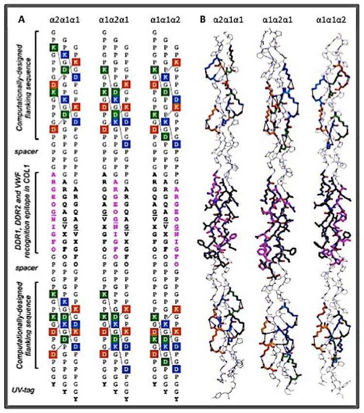
Bioengineering 2021, 8, 5                                                                                                   2 of 24

                            blood vessel walls. Other types of fibrillar collagens are present at a lower amount and are
                            often found coexisting with the three major types.
                                  The structural hierarchy of all collagens starts from the building block: The collagen
                            triple helix [8,9]. A collagen triple helix consists of three polypeptide chains (often referred
                            to as the α chains) coming together in parallel with a precise one residue staggering at the
                            ends [9,10]. The three chains tightly wrap around each other about a common axis in a
                            right-handed helical twist to form a rod-like helical conformation (Figure 1A). The tight
                            packing of the triple helix requires a Gly residue at every third position, giving rise to the
                            characteristic (Gly-X-Y)n repeating sequence. The obligatory Gly residues are buried at
                            the center of the helix, the side chains of X and Y residues are largely exposed to solvent.
                            The triple helix is often considered a “linear molecule” because of its uniform backbone
                            conformation characterized by an ~0.86 nm helical rise per Gly-X-Y tripeptide [11–14]. The
                            side chains of the X and Y residues can be described as a linear sequential array in an
                            N-to-C directionality spiraling around the surface of the molecule (Figure 1A).

                            Figure 1. The rod-shaped conformation of the triple helix. (A) The structure of the homotrimer triple
                            helix T73–785 [15] was generated using DeepView–Swiss-PdbViewer (PDB: 1bkv). The helix is shown
                            with the N-terminus on top; the Thr residues are shown in green, Arg in blue, hydrophobic residues
                            in dark gray. (B) In order to show the asymmetric structure of a heterotrimer associated with different
                            chain registers, a type I collagen like the AAB heterotrimer model was created using DeepView by
                            replacing the Thr of one of the three chains of 1bkv to Ala (side chain shown in black). The amino
                            acid residues included in this heterotrimer model are ITGARGLAG for the two identical strands, and
                            IAGARGLAG for the third, mutated strand. The three structures are shown in the identical view of
                            the backbone, with the N-terminus on top.

                                  The three polypeptide chains of a triple helix can be identical in the form of a ho-
                            motrimer, or they can be different in amino acid sequences forming a heterotrimeric triple
                            helix [8]. Collagen type II and collagen type III are homotrimers, while collagen type I
                            is a heterotrimer consisting of two identical α1 chains, and one α2 chain. There is about
                            72% sequence similarity between the two α chains in the triple helix domain of type I
                            collagen. Because of the one residue stagger between the adjacent strands in the triple
                            helix, the analogous residues in each strand are unique even in a homotrimer environment
                            (Figure 1A) [10,16–18]. The three strands are usually called leading, middle, and trailing,
                            as viewed from their N-termini. The chain-stagger-related asymmetry in structure is par-
                            ticularly pronounced in a heterotrimer (Figure 1B). Thus, for type I collagen the surface
                            features of the triple helix can be very different depending on which chain is in the leading,
                            middle, or trailing position. There are three possible chain registers for type I collagen:
                            α1α1α2, α1α2α1, and α2α1α1, which are often referred to as α2 trailing, α2 middle, and
                            α2 leading, respectively. Unfortunately, determining the chain register is not at all easy.
                            The correct chain register of type I was accepted to be α1α2α1 [19,20]. Emerging data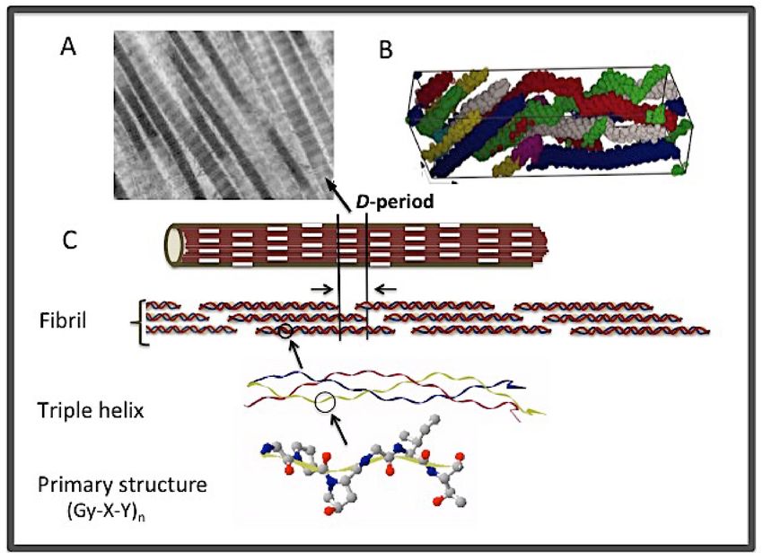
Bioengineering 2021, 8, 5                                                                                                        3 of 24

                            from studies using CMPs, however, are challenging this chain alignment in favor of an
                            α1α1α2 register with the α2 chain in the trailing position (details below) [21]. Inside the
                            cell, the C-terminal globular domain, the C-propeptide, was believed to be responsible
                            for both chain selection and chain registration [22]. Structural studies of type I collagen
                            C-propeptide have provided a mechanism for heterotrimerization of the C-propeptide.
                            How the structure of the C-propeptide determines the chain alignment of the triple helix
                            domain, however, remains a mystery [23,24].
                                  Collagens in tissues are higher order, supramolecular assemblies of triple helices.
                            Fibrillar collagens self-associate laterally with a specific 67 nm staggering at the ends to
                            form fibrils (Figure 2A,C) [25–30]. Fibrillar collagens are large molecules consisting of
                            more than 1000 residues per single polypeptide chain in uninterrupted (Gly-X-Y) repeating
                            sequences, forming a long triple helix about 300 nm long and ~1.5 nm in diameter. Each
                            triple helix compromises about 4.4 × 67 nm in its total length. The staggered arrangement
                            would thus generate long, smooth fibrils with alternating gap-and-overlap regions every
                            67 nm. This 67-nm structure is termed a D-period, which consists of a 0.4D overlap zone
                            and a 0.6D gap region. The overlap and the gap zones appear as light and dark bands,
                            respectively, when examined using an electron microscope, giving rise to the characteristic
                            striation appearance of collagen fibrils (Figure 2A). In the fibrils, the triple helix further
                            adopts a right-handed super-twist around the microfibrils [31–35]. Because of this super-
                            twist, there is an uneven exposure of different parts of the triple helix on the fibril surface
                            (Figure 2B); as to which specific sections of the triple helix might be exposed on the surface
                            of the fibrils is still under debate [32,36,37].

                            Figure 2. The structural hierarchy of fibrillar collagen. (A) Electron micrograph of collagen fibrils
                            showing the characteristic striation pattern of the D-period and the tipped ends. (B) The unit
                            cell of collagen fibril showing the staggered and intertwined arrangement of five triple helices
                            (in different colors) due to the super-twist of the triple helices in fibrils [33]. (C) The different stages
                            of fibrillogenesis from the primary structure to fibrils.

                                  The D-period of collagen fibrils is an important feature that has been linked to the
                            tensile strength of bones, the stiffness of the extracellular matrix, and other biomechanical
                            properties of tissues [38–43]. It is the holy grail of all development of collagen mimetic
                            biomaterials to capture the structure and function of this unique, yet ubiquitous molecular
                            scaffold of all connective tissues. Some of the recent progress in achieving collagen mimetic
                            fibrils is mentioned later in this review. Fibrillogenesis in tissues is a complex process and
                            remains poorly understood. The fibril assembly process per se, however, is a self-assembly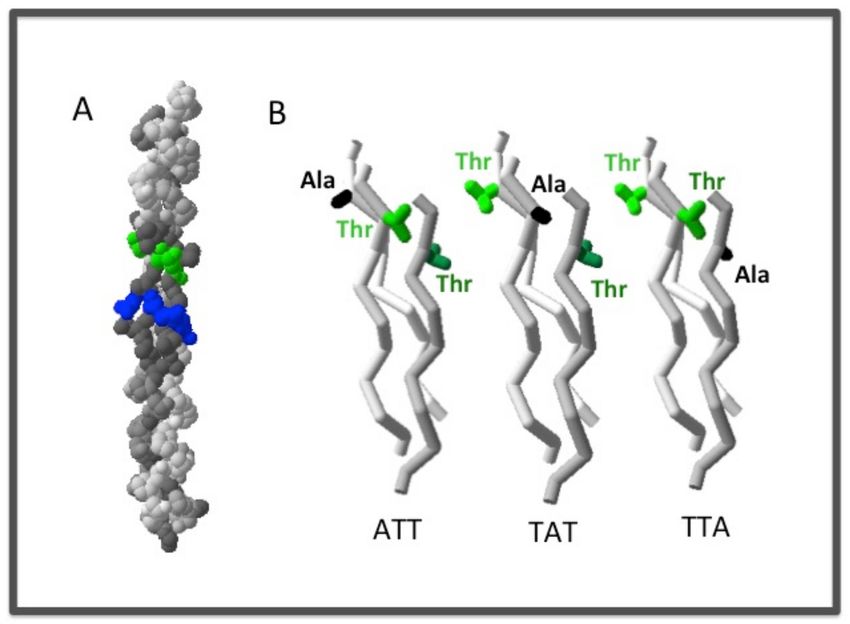
Bioengineering 2021, 8, 5                                                                                              4 of 24

                            process that can be reproduced in vitro from acid-dissolved fibrils [44,45]. It is generally
                            considered that the axial repeating D-period of fibril assembly is determined by the molec-
                            ular interactions contained in each triple helix, although the exact molecular mechanism is
                            not resolved [11–13]. Alternatively, some studies have attributed the deterministic factor of
                            fibrillogenesis to the involvement of the telopeptides: Two short stretches of peptides at
                            the N- and C-termini of the triple helix domain that do not confer to the Gly-X-Y sequence
                            pattern and do not adopt to a triple helix conformation [46]. Later studies have shown that
                            triple helices without the telopeptides can form fibrils, albeit with a slower kinetics [47–49].
                                  Collagen plays much more than a structural role in tissues. It is a dynamic molecular
                            scaffold that supports cell adhesion, cell migration, and cell differentiation [36,50–54]. Cell
                            receptors recognize specific regions of the triple helix. Studies using CMPs to identify
                            these recognition sites are described in Section 2 below, and a comprehensive review of the
                            distribution and organization of these epitopes is given by San Antonio et al. in this special
                            issue. One major family of collagen-binding proteins is integrin. Integrins are the major
                            cell adhesion proteins that bind to the extracellular matrix (ECM) and function as signal
                            transducers of various signaling pathways that can induce global cell responses and affect
                            gene expression [55]. Epithelial and endothelial cells will undergo programmed cell death,
                            or apoptosis, when they lose contact with the ECM. Cells can also control their affinity to
                            collagen through inside-out signaling. The intricate interplay between cells and the ECM is
                            integral to the development of all tissues and organs. Other important collagen receptors
                            include discoidin domain receptor (DDR1 and DDR2), platelet glycoprotein GPVI, immune
                            receptors, the plasma protein von Willebrand factor (vWF), and other macromolecules and
                            proteins [21,36,51,56].
                                  Collagen catabolism is another critical interaction of collagen for maintaining the
                            ECM homeostasis [57]. The hydrolysis of fibrillar collagens is due to the action of matrix
                            metalloproteinases (MMPs) MMP-1, MMP-8, and MMP13. All three enzymes cut collagens
                            into 34 and 14 length fragments, but with different kinetics to different collagens. MMP-8
                            preferentially cleaves type I collagen, while MMP-1 has greater catalytic activity on the
                            type III collagen. MMP-13 cleaves type II collagen 5 and 6 times faster than collagens
                            type I and type III, respectively. MMP-2 and MMP-9, which are broadly categorized as
                            gelatinases, also participate in the homeostasis of the ECM by hydrolyzing a partially
                            unfolded, or gelatin form of collagens [58,59]. The well-regulated digestion activity of
                            the MMPs controls the turnover of collagen in normal growth and in tissue remodeling.
                            The unregulated proteolysis by MMP is also the cause of pathological conditions such as
                            arthritis, periodontal diseases, and cancer metastasis [60–62].
                                  Another type of collagen which has often been studied using CMPs is the base-
                            ment membrane collagen–collagen type IV. Unlike fibrillar collagen, type IV collagen
                            self-assembles into a chicken wire-like molecular network joined at the ends through the
                            N- and C-globular domains. The molecular composition of type IV collagen is also quite
                            complicated [8]. There are six different α chains of type IV collagen that form at least three
                            distinct triple helices with the stoichiometries of 2α1 α2 , α3 α4 α5 , and 2α5 α6 , respectively.
                            Different isoforms are found in different tissues, and in different developmental stages.
                            While the molecular assembly of the type IV collagen may not have the biomechanical prop-
                            erties of fibrillar collagen, it is the critical molecular scaffold of the basement membrane
                            supporting angiogenesis, and has many cell recognition sites [63]. Molecular interactions
                            with type IV collagens are involved in cancer metastasis; these interactions are potential
                            targets of cancer drugs [64–66].
                                  We are just starting to understand the structural and the molecular details of collagen,
                            and the interactions involving collagen. As is described in more detail, CMPs have been
                            indispensable in these studies. Many reactions take place in an overcrowded ECM environ-
                            ment where collagen triple helices organize into different molecular scaffolds or networks.
                            How the supramolecular structures of collagen modulate the cell-ECM interactions is yet
                            to be fully elucidated.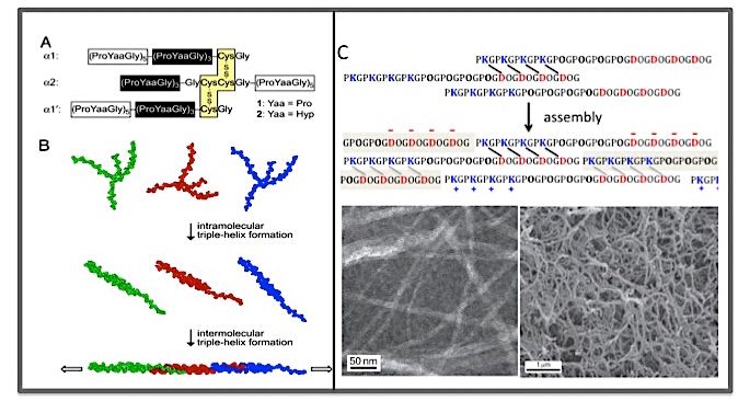
Bioengineering 2021, 8, 5                                                                                            5 of 24

                            1.2. Collagen-Based Biomaterials
                                  Collagen-based biomaterials in the form of sutures and films for wound healing
                            have been used for more than a century [67]. The field has grown tremendously since
                            then to include a broad range of potential medical products based on collagen. The clear
                            advantage of collagen devices is their ability to interact with the host. These products
                            can act as a scaffold for new tissue formation prior to resorption, or be used for soft
                            tissue augmentation. Collagens from animals are often the source for manufacturing these
                            materials. However, the purification process of collagen from animal tissues is a difficult
                            and expensive process. Because of the constant tissue remodeling and modification of
                            collagen, collagen preparations isolated from tissues are often heterogeneous with a high
                            degree of variations in terms of covalent modifications and composition. While collagens
                            per se are considered poor immunogens, the impurities in collagen preparations are known
                            to elicit immunogenic responses. In recent years, there have been increasing concerns of
                            the pathogenicity of collagen devices made from animal collagens or from collagen of
                            human tissues.
                                  Collagen mimetic peptides, thus, represent a desirable alternative for being safer and
                            potentially less expensive. The one other great advantage of mimetic materials, whether
                            produced by chemical synthesis or by genetic engineering, is the ability to modify the
                            amino acid sequences for a better control of the properties and activities of the material.
                            The goal of the collagen mimetic materials should mimic both the physical and biochemical
                            properties of native collagen. Additionally, control of the turnover rate is another important
                            factor for the proper tissue remodeling process, and also for the life span of the material.
                            CMP-based biomaterials bearing epitopes for platelet activation have already demonstrated
                            their potential for application [68,69]. The effectiveness and value of the collagen mimetic
                            biomaterials will largely depend on the ability to modulate and to fine-tune the bioactivities
                            of the material for specific applications.

                            2. Collagen Mimetic Peptides by Chemical Synthesis
                            2.1. The Homotrimeric CMPs
                                  Synthetic peptides have been an integral part of protein science. The collagen field is
                            no exception. What is unique to collagen is the requirement to have three peptide chains
                            come together and fold into a triple helix conformation with a specific one-residue stagger
                            at the ends. An early approach was to use chemical cross-links at the C-terminus to bring
                            about the correct association of the three polypeptide chains [70,71]. Later, it was found that
                            peptides with repeating sequences of (Gly-Pro-Hyp)n with n > 6 can self-assemble into a
                            stable triple helix without the need of cross-links, and the thermal stability increases with an
                            increase in the number of tripeptide units [72]. Thus, while the triple helix (Gly-Pro-Hyp)6
                            was only marginally stable with a melting temperature (Tm ) of 10 ◦ C, that of a (Gly-Pro-
                            Hyp)10 can reach 68 ◦ C. Peptides with (Gly-Pro-Pro)n repeating sequences also form a
                            triple helix, but have a much lower thermal stability; in comparison with (Gly-Pro-Hyp)10 ,
                            the thermal stability of (Gly-Pro-Pro)10 is only about 27 ◦ C [73,74]. Since these pioneering
                            studies, it has become quite feasible to synthesize CMP 21–50 residues in sizes with more
                            sequencing variety. The only requirement is to follow the (Gly-Xxx-Yyy)n sequence pattern
                            with Gly at every third position, although repeating (Gly-Pro-Hyp)n or (Gly-Pro-Pro)n ,
                            with n = 2–4, are frequently included at the N- and/or C-termini for added stability [10,75].
                            Later studies showed that the multiple (Gly-Pro-Hyp)n or (Gly-Pro-Pro)n at the end of the
                            peptide can also function as the nucleation domain to facilitate the association of the three
                            chains in the desired mutual one residue staggering [76–79]. The spontaneous folding of
                            such CMPs inevitably leads to homotrimeric triple helices consisting of three identical
                            polypeptide chains.

                            2.1.1. The Sequence–Structure Relationship
                                 Studies using the CMP have clearly demonstrated that the rod-shaped triple helix is
                            not uniform in structure or stability [10,80,81]. The three polypeptide chains of a triple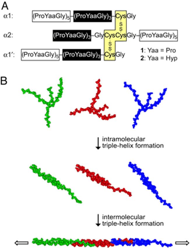
Bioengineering 2021, 8, 5                                                                                            6 of 24

                            helix are connected by a set of hydrogen bonds between the backbone NH of Gly and the
                            backbone CO (X-position) of the neighboring chain [15,82–84]. These main-chain H-bonds
                            present rather uniformly throughout the helix. Additional water-mediated H-bonds can be
                            found between the CO of the Gly with the NH of an X-residue if the X-residue is not a Pro.
                            Crystal structures of CMPs also revealed a sequence-dependent variation in the helical
                            twist. Regions with a high content of Gly-Pro-Hyp or Gly-Pro-Pro form a tighter 7/2 helix
                            (3.5 residues/turn), while the “imino acid-poor” regions adapt to a more relaxed 10/3
                            symmetry (3.3 residues/turn) [9,10]. This sequence-dependent variation can potentially
                            play a role in the molecular recognition of collagen.
                                  The stability of the CPMs is sensitive to the residues in the X and/or Y positions.
                            Using a set of host–guest peptides, Brodsky and colleagues examined the effects of all
                            20 amino acid residues plus hydroxyproline in X and/or Y positions(s), and in different
                            combinations [80,81,85–88]. The Gly-Pro-Hyp was the most stable tripeptide; the host
                            peptide (Gly-Pro-Hyp)8 had a Tm of 47.3 ◦ C in neutral buffer. Replacing the Pro in the guest
                            site positioned in the middle of the peptides often resulted in a decrease of Tm ranging
                            from 2–15 ◦ C depending on the identity of the substituted residue, while that caused by
                            replacing Hyp in the Y position ranged from 0 to 21 ◦ C. Having a charged residue like
                            Glu, Asp, Arg, or Lys generally had an unfavorable effect with the exception of Arg in
                            the Y position, which appeared to have a similar stabilizing effect as a Hyp. However,
                            when the charged residues were present in pairs in the sequences of Lys-Gly-Glu (KGE) or
                            Lys-Gly-Asp (KGD), significant stabilizing effects were reported: The Tm of a host–guest
                            peptide with a sequence G_KGE_ or G_KGD_ can increase by 15.4 to 17.5 ◦ C, respectively.
                            This significant stabilizing effect was attributed to a set of inter-chain salt bridges between
                            a pair of oppositely charged residues. In the extended backbone conformation of the
                            triple helix, little interactions could take place between the charged groups in the same
                            chain. Furthermore, the effects of different residues on the overall stability of the triple
                            helix appeared to follow a simple additive rule. A stability calculator was developed
                            based on these studies that could provide reasonably accurate estimations of the thermal
                            stability of CMP 18–50 residues in size, and has been used broadly in the sequence design
                            of homotrimeric CMPs [80].
                                  As the peptide becomes longer, the triple helix is more stable until it reaches a plateau
                            at about 50 residues or so. An empirical curve was used to predict the length dependence
                            of the CMPs [80]. For longer chains, the decrease in entropy of the polypeptide chains in
                            the more constrained folded structure could off-balance the enthalpy contribution in the
                            form of H-bond formation and other interactions. This entropy penalty is more significant
                            for the folding of a longer triple helix. A more quantitative interpretation of the length
                            dependence on the thermal stability would require thermodynamic studies under an
                            equilibrium condition. The thermal unfolding process of CMPs generally does not satisfy
                            this condition [9,81]. How to extrapolate the sequence–stability relationship of CMPs to
                            natural collagen also remains an intriguing question, since natural collagens are not only
                            much longer, but also have much more diverse amino acid sequences. This subject will be
                            revisited in Section 3 on the discussion of recombinant peptides. The findings of the CMPs
                            are generally applicable to larger triple helices, at least on the qualitative level.

                            2.1.2. The Binding Sites of Collagen Receptors
                                 Defining the binding sites of cell receptors and other macromolecules on collagen
                            is considered one of the crown accomplishments of CMPs [21,50]. Given the complex
                            supramolecular structure of collagen in the ECM, defining the site where a collagen-
                            binding ligand may interact is difficult. The collagen-binding proteins themselves are also
                            frequently membrane-bound molecular complexes, or in the case of vWF, a complex multi-
                            domain protein. CMPs carrying 6–27 residues modeling a selected section of the α1 chain
                            of collagen type I were used to study the interaction of collagen with integrin [89,90]. The
                            I-domain (or A-domain) of the α-subunit of integrin α2 β1 and α1 β1 was identified as the
                            domain to interact with both collagen types I and IV in a metal-dependent fashion [91–93].
Bioengineering 2021, 8, 5 7 of 24
Peptides containing the sequence GFOGER were first selected as potential binding sites
because of their high affinity to the isolated I-domain and ability to support α2 β1 -mediated
cell adhesion. Subsequent studies using mutations indicated that the recognition of the
I-domain is entirely contained in the six-residue sequence, with the residues Arg (R) and
Glu (E) being the most critical for the binding [90].
The molecular recognition mechanism of collagen binding by integrin was revealed
by the crystal structure of the complex of the I-domain with a 27 mer CMP containing
GFOGER at the guest site [94]. The Glu from the middle strand of the triple helix formed
a critical interaction with the required divalent ion, and the Arg residue from the middle
strand formed a salt bridge with Asp219 of the I-domain to stabilize the complex. The
fact that both the Glu and Arg involved in specific interactions with the I-domain all came
from the middle strand of the triple helix appeared to offer an explanation as to why the
homotrimeric peptide binds the I-domain with similar or even higher affinity as that of
heterotrimeric collagen type I. The sequence of the alpha 2 chain of type I collagen in the
equivalent location is GPOGES. In the molecular complex, the binding site of the I-domain
had close contacts with only one out of the three chains; the interactions with the residues
of the other two chains may have facilitated the binding with less specific interactions.
The Phe in the middle, and the trailing strands made hydrophobic contacts with the I-
domain, while the one in the leading strand was exposed on the surface of the complex.
The hydrophobic residues Ala and Leu in the other high-affinity sequence GAOGER and
GLOGER, respectively, were expected to provide similar interactions during binding [95].
A specific involvement of Hyp in the six-residue binding site was suspected because of
its common presence in the identified high affinity sites. The Hyp of the trailing strand
was buried in the interface of the complex, a replacement of residues with larger side
chains are likely to create steric clashes and decrease the affinity. No other, more specific
interactions involving this Hyp were identified. Later studies found replacing the Hyp
with Pro reduced binding affinity but did not abolish the integrin activation [96]. Studies
using cross-linked heterotrimeric peptides have indicated the binding affinity of the three
different chain registers showed similar affinity to the I-domain. Interestingly, the binding
affinity of the heterotrimers was significantly lower than that of the homotrimer.
The peptide approach was later developed into a system known as the ToolKit
III [50,95,97]. The ToolKit III consists of 57 peptides with overlapping sequences at the
27-residue guest site that cover the entire sequence of type III collagen. Repeating GPO
or GPP sequences flanking the guest site were included to facilitate the triple helix for-
mation. Since sequences of (GPO)n can interact with the GPVI of platelet, the (GPP)n
sequence is preferred especially for studies with platelets [98]. The ToolKit approach is
particularly good when studying the interactions of collagen receptors with collagen type
III and collagen type II, due to their homotrimeric nature. Applications using the ToolKit
peptides led to the identification of the epitopes of integrin α1β1, α2β1, the vWF on type III
collagen, and the DDR2, DDR1, and the immune receptors on type III collagen and type II
collagen [97,99–103]. Ideally, these peptides should be covalently crosslinked as well, since
the trimerization process of triple helix folding is sensitive to peptide concentration. For
this reason, the peptides of the ToolKit III have a Cys at the N- and the C-termini. Given the
triple helix conformation, two adjacent Cys residues are often needed in order to cross-link
all three chains in a set of disulfide bonds; a single Cys in a peptide can only cross-link one
other neighboring chain and leaves the –SH on the third chain unpaired. Peptides with
free –SH groups are often prone to non-specific aggregations.
In tissues, these molecular interactions with collagen receptors take place with colla-
gens in fibril form or in other supramolecular structures. Some epitopes showing a high
affinity in a CPM study may not be fully exposed on the surface of the packed fibrils
(Figure 1B) [32,36,37,104]. Even if the specific molecular interactions are available, the
binding kinetics and binding affinity are likely to be affected by the structural context
of the epitopes. Molecular modeling indicates that an I-domain should be able to bind
to a triple helix without steric clashes with neighboring helices on the fibrils based onBioengineering 2021, 8, 5 8 of 24
the center-to-center distance of the closely packed triple helices being 15 Å in fibrils [94].
However, the I-domain is also just a part of the integrin complex and does not work in
isolation. It remains to be tested in studies of the I-domain and other collagen-binding
proteins and/or domains with the triple helix in a higher molecular complex.
2.2. The Heterotrimeric CMPs
In the past decade or so, progress has been made in designing and creating heterotrimeric
collagen peptides. Two of the major collagens, collagen type I and collagen type IV, are
heterotrimers, and are known to interact extensively with cell receptors. Heterotrimeric
peptides are used as model systems to understand these interactions [19,21,96]. Making
heterotrimeric triple helices with the correct register faces many challenges; not the least of
them is the lack of clear knowledge of the actual chain registration of the native collagens.
Because of the one residue staggered arrangement, a triple helix formed from two or three
different peptide chains will create a different structure environment for the interaction
with cell receptors (Figure 1B).
2.2.1. The Chain Register Affects both the Stability and the Binding Affinity of the
Triple Helix
As it was in the case for the homotrimer CMPs, terminal cross-links were first used to
circumvent the problems of the chain selection and chain registration. To gain more detailed
characterization of the mechanism of proteolysis of collagen type I by the metalloproteinase,
a hetero CMP was synthesized to mimic the MMP-1 digestion site between residues 772–783
of the α1 and α2 chains of type I collagen [19,105]. Altogether, 4 Cys residues were included
at the C-terminus of the peptide chains which were designed to cross-link the three chains
through two disulfide bonds. The chain register was fixed to α1α2α1 (α2 as the middle
chain). An additional (GPO)5 at the N-terminus was also included to give the peptide a
thermal stability of ~33 ◦ C. The enzyme digest assays performed at room temperature
indicated the triple-helical substrates were cleaved with a single cut through the three
chains in a manner similar to those observed in studies using collagens isolated from tissues.
This similar enzyme activity was also used to argue that the chain register of type I collagen
must be α1α2α1. However, an NMR study of the peptide showed the C-terminal half of
the peptide appeared to be disordered. It is not clear if the disordered conformation was
due to the low helix propensity of the sequences or the structural constraint imposed by
the C-terminal cross-link. Later studies by crystallography also indicated that the regions
surrounding the interchain disulfide bonds are often flexible and the chain register may
not be fixed by their use. Additionally, there is a chance the disulfide bond may reshuffle,
and lead to unexpected cross-links [106,107].
Another set of similar cross-linked hetero-CMPs were used to model the α1β1 binding
sites of collagen type IV. The study revealed the chain register affected both the stability
and the folding kinetics of the triple helix [108,109]. The trimer of the α1α2α1 register was
more stable (Tm of 42 ◦ C) but had lower affinity to integrin, while the trimer with a α2α1α1
was less stable (Tm of 30 ◦ C), but showed higher affinity [108,109]. The high stability was
postulated to affect the binding because collagen needs to undergo conformational changes
in the integrin adhesion region upon binding, and a less rigid conformation may favor
such a conformational adjustment.
2.2.2. The Self-Assembled Heterotrimeric Triple Helix
A heterotrimeric triple helix formed by self-association of three polypeptide chains
without cross-linking is not just an interesting, fundamental problem to solve for protein
design, but one that would offer more flexibility in chemical synthesis and more versatility
for applications. The challenge is the control of both the chain composition and the chain
register. The control of composition is a tractable problem if different compositions lead
to different stability. By simply mixing two peptides A and B, there will be 8 possible
combinations of trimers: AAA, BBB, ABB, BAB, BBA, AAB, ABA, and BAA; where the
AAB and ABA have the same chain composition but represent two different triple helicalBioengineering 2021, 8, 5 9 of 24
structures due to the different chain register. The population of each of the 8 possible
configurations is partitioned into a Boltzmann distribution based on their stability; the
desired configuration can be the dominant species if its stability is significantly higher
than any of the other competing species. The control of the chain register turns out to be a
closely related problem (see below), since the chain registers also affect the stability of the
triple helix.
One stabilizing factor that can be exploited for the design of a stable heterotrimeric
triple helix is the inter-chain salt bridges [80,86,88]. By strategically placing a pair of
oppositely charged residues on neighboring chains, the interactions between the charged
pair lead to the formation of a salt bridge (an ionic H-bond) that can significantly stabilize
the triple helix. In contrast, unpaired charged residues in the X or Y position are known
to cause destabilizing effects on the triple helix due to charge repulsion, and can be used
to discourage unwanted conformations. Nanda and coworkers implemented this idea
and computationally generated three peptide sequences A, B and C, which by mixing
in a 1:1:1 ratio formed an ABC heterotrimer (here ABC stands for chain composition not
chain registration) [110,111]. By several rounds of optimization to increase the stability
of the ABC trimer while destabilizing other competing configurations, the ABC trimer
emerged as the only triple helix in the solution. However, the ABC heterotrimer could
have 6 possible chain alignments. The most likely alignment was the one designated as
abc with A, B, C chains in the leading, middle, and trailing positions, respectively. Based
on the computer simulation, abc had the most favorable charge-paired interactions and
fewest charge repulsions and thus, should have been the one that had the highest stability.
Later, a crystal structure study of the designed ABC heterotrimer confirmed the abc chain
alignment [112]. The amino acid sequences of the A, B, and C peptides, however, were
nothing close to that of natural collagen. It will be interesting to find out if some of the
sequence features can be utilized to generate heterotrimers with more sequence diversity.
Using an intuitive experimental approach, Hartgerink and coworkers exploited the
geometric and sequence specificity of electrostatic interactions of charged residues nearly
exhaustively in a series of 30-residue peptides [113–121]. Among the major findings are
that peptides carrying 5 or more like charges such as (DOG)10 or (PKG)10 will not form
homotrimers because of a strong charge repulsion. However, when such decapositive
and decanegative peptides are mixed with (POG)10 in a 1:1:1 molar ratio, they form stable
ABC heterotrimers. Most of all, the NMR study shows that such heterotrimers are sta-
bilized by a set of axially oriented charge–pair interactions between a Lys residue in the
leading and middle chains with a Glu or Asp residue in the middle and trailing chains,
respectively; with the interactions between the Lys-Asp being particularly strong. Fur-
thermore, the orientation bias of the Lys-Asp interaction means only in the alignment of
(PKG)10 (EOG)10 (POG)10 (i.e., with (PKG)10 , (EOG)10 , and (POG)10 in the leading, middle,
and trailing positions, respectively) can all 10 sets of salt bridges be satisfied, and this maxi-
mized stability predetermined that only heterotrimer in this chain register will dominate in
the solution. The same type of the axial Lys-Asp salt bridge was also found in the crystal
structure of the heterotrimer mentioned above by Zheng et al. [112]. This directional inter-
chain charge–pair interaction has since been used to develop self-assembled heterotrimers
or a long super-helix [21,122,123].
Another equally important consideration in designing a stable heterotrimer is to desta-
bilize the competing compositions. While the original ABC heterotrimer based on the
inter-chain charge–charge interaction was remarkably strong, with a Tm above 56 ◦ C, they
cannot outcompete the homotrimer (POG)10 , which has a Tm of 58 ◦ C [115]. Thus, mixing
of the three peptides generated a mixture of heterotrimer and the (POG)10 homotrimer. In
another effort, Hartgerink and coworkers developed a stable ABC heterotrimer using a de-
capositive (PKG)10 and a decanegative (EPG)10 mixed with a zwitterionic peptide (DKG)10 .
Since the zwitterionic peptide does not form a stable homotrimer, the ternary mixture
produced a single component ABC heterotrimer with a single chain registration (117). The
peptide has a Tm of 38 ◦ C which is quite remarkable considering this trimer has no Hyp.Bioengineering 2021, 8, 5 10 of 24
By maximizing the axial Lys-Asp interactions while reducing the possibility of like-
charge repulsions at the same time, a stable heterotrimer in the AAB arrangement was
achieved in the composition of 2(GPKGEO)5 (POGDOG)5 [119,120]. The heterotrimer has
a unique AAB chain register because only in this chain alignment are the interactions of
the directional, axial Lys-Asp salt bridge maximized, while unfavorable, unpaired charge
residues are minimized. This study is a clear demonstration that, by strategically placing
the charged residues, both the chain composition and the chain register can be controlled.
At the same time, however, the need to have several of the charged residues in fixed
locations can restrict the sequence diversity of the peptides and limit their applications.
In a new development, it was found that short stretches of peptides of five (Gly-X-Y)
tripeptide units consisting of the charge-pairing residues in the optimal positions can
be used as a hetero-nucleation site to develop peptides carrying sequences from natural
collagens [21]. A set of heterotrimeric peptides in the AAB composition modeling the cell
adhesion epitopes or the vWF binding site of type I collagen in all three possible chain
alignments were developed. These heterotrimeric peptides have the hetero-nucleation
sites at both the N- and the C-termini, flanking 12 residue-specific CBP (collagen-binding
protein)-binding epitopes in the center (Figure 3). The specific amino acid sequences of the
nucleation sites were computationally generated using a genetic algorithm; a group of Lys
and Asp residues were strategically positioned to form a unique set of optimally oriented
Lys-Asp salt bridges which stabilize the triple helix having the desired chain alignment.
The folded structure was further “covalently captured” by converting the salt bridges into
an amide bond between the ammonium group of the Lys side chain and the carboxylate
group of the Asp. Based on the structural, stability, and binding studies, the triple helix
in the α1α1α2 alignment was found to be less thermally stable than those in the other
two possible chain registers, but had the highest binding affinity to DDR1 and vWF. The
α1α1α2 heterotrimer was also found to induce higher levels of cellular DDRI and DDR2
kinase activation. These findings appeared to indicate that the α1α1α2, rather than the
α1α2α1 as proposed by previous studies, was most likely to be the correct chain alignment
of type I collagen. In this conclusion, it is implied that the correct chain register of collagen
was not necessarily the one with the highest stability, but the one with highest bioactivity.
Figure 3. The heterotrimeric peptides modeling the collagen-binding epitopes on type I collagen (21).
(A) The design and the amino acid sequences of the heterotrimers mimicking type I collagen in three
different registers: α2α1α1, α1α2α1, and α1α1α2; here X is norleucine, a methionine bioisostere.
The N- and C-termini flanking sequences are generated by a computer program utilizing genetic
algorithm to optimize the location of the Lys-Asp charge-pairings in the helix. (B) The corresponding
crystal structure of the heterotrimers in three registers.Bioengineering 2021, 8, 5 11 of 24
The covalently captured heterotrimeric triple helix can extend the ToolKit approach to
study the binding and MMP specificity of heterotrimeric collagens. The covalent-capture
can potentially be a more effective alternative than Cys-based cross-linking strategies to
generate cross-linked heterotrimeric peptides that are more homogeneous, and more stable.
Such systems can find a wide range of applications for collagen research.
2.3. Applications of CMPs
Collagen rarely functions as an individual triple helix in tissues. To truly “mimic”
collagen, CMPs need to further associate into fibril-like supramolecular structures. Devel-
oping higher order molecular assemblies through the self-association of CMPs, however,
has proven to be challenging: Small sizes and limited sequence diversity are two likely
limiting factors.
2.3.1. Self-Assembled Fibrillar Structures
A common approach to form a long triple helix through self-assembly is to use
the so called “sticky-end” strategy. In the approach by the Raines’ group, the sticky-
end CMPs were created using a cross-linked “core” that brought together polypeptide
chains with different lengths extending from the C- and N-termini of the core as branches
(Figure 4A,B) [124]. The branches were peptides with (POG)n or (PPG)n sequences that
had a high propensity to trimerize to form a triple helix. The three strands at the N-
terminal end of the core formed an intramolecular triple helix but with an overhang that
was complementary to the segment sticking out from the C-terminal end of the core, thus
the “sticky ends”. The intermolecular assembly between the complementary sticky ends
then produced helices that could reach a length of 200 nm or longer. The functionality of
these long triple helices has yet to be fully evaluated. In another approach using disulfide
bond cross-links, Koide and colleagues developed long triple helix assemblies containing
the integrin-binding epitope GFOGER sequence [125,126]. This supramolecular CMP
formed a hydrogel and was found to support integrin-mediated cell adhesion in a fashion
“comparable to that of native collagen”.
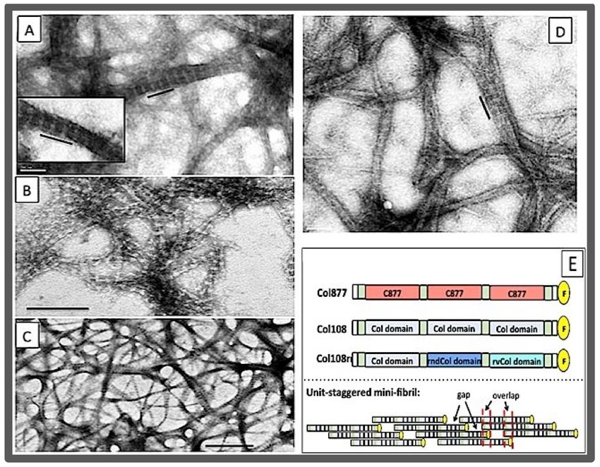
Figure 4. The "sticky end” approach for the self-assembly of collagen mimetic peptides (CMPs).
(A) The cross-linked CMP with sticky ends. (B) The self-assembly process to form a super triple
helix [124]. (C) The charge-pair directed self-assembly of the KOD peptide, and the TEM images of
the KOD fibrils (left, the scale bar is 50 nm), and the hydrogel formed by KOD (right, the scale bar is
1 µm). The directional, interchain Lys-Asp salt brides of the nucleation site are highlighted by short,
slanting bars.
The sticky end approach used by the Hartgerink’s lab was based entirely on self-
association (Figure 4C) [123]. The first successful case was the KOD super-triple helix.Bioengineering 2021, 8, 5 12 of 24
The building block was the KOD peptide which had a modular amino acid sequence
composition made of three units: (PKG)4 (the K), (POG)4 (the O), and (DOG)4 (the D). The
core of the sticky end was stabilized by a set of Lys-Asp salt bridges. In addition to growing
long, the unsatisfied Lys and Asp residues also provided additional interacting surfaces
for further lateral association of the triple helices. The result was a hydrogel sharing many
features with those created using natural collagens. Furthermore, because of the (GPO)4
units in the long helix, the KOD hydrogel was found to be able to activate platelet and
clot whole blood plasma [69]. It is one of the CMP materials with a high potential for
biomedical applications as a synthetic hemostat. By rearranging the modular units of K, O,
and D in a CMP, it was found that the size of the core (the nucleation site) was a crucial
design factor that determined if the self-assembly of the CMP would produce hydrogels or
amorphous aggregates [127].
A recent work in the Raines’ group further pointed out that the Lys-Asp charge-pair-
based sticky-end approach could be optimized by including the “elements of symmetry”
in order to afford identical interactions for every peptide in the assembly [122]. Under
this symmetry condition, all Lys and Asp residues engage in interchain charge–pairing
interactions, thus maximally stabilizing the assembly and at the same time eliminating un-
paired Lys and Asp residues that are prone to form non-specific aggregates. The symmetry
consideration was developed into a set of design rules for CMPs that could self-assemble
into a long triple helix. As it was shown in their study, peptides generated using these
design rules formed long triple helices that could match or even exceed the natural collagen
in length.
It worth pointing out that the self-assembled long triple helices or fibrillar structures
of CMPs have one fundamental difference from that of the collagen fibrils. Collagen fibrils
are assembled through lateral, staggered association of triple helices. While these self-
assembled triple helices are wonderfully long, they lack certain structural elements of the
native collagen fibrils, chief among them the axial structure of the D-period. Similarly, the
mechanical support provided by the CMP hydrogels are likely to be different from the sup-
port of the collagen molecular scaffold in the ECM, despite similar morphology as shown
by TEM images. None of the supramolecular CMPs have the D-period-like structure seen
in collagen fibrils. In one report, it was proposed that a D-periodic microfibril was formed
from blunt-end self-assembly of a 36-resdiue triple helix [128]. However, the proposed
17.9 nm D-period of the microfibrils is nearly twice the length of the constituent triple helix.
The microfibrils must have formed through a very different molecular arrangement than
the D-periodic collagen fibrils.
2.3.2. Interaction of CMPs with Damaged Collagens
There have been many interesting studies using CMPs to produce nano-structures in
different shapes, sizes, and compositions. Some of these molecular assemblies have shown
promise for biomedical and other applications [129–139]. There is a good account of some
of these works in a recent review [56]. We would, however, like to end this section on
synthetic CMPs with the application of CMPs that covers the other end of the spectrum—by
going small. This application uses CMPs in an unfolded, single chain conformation to
study damaged collagen in tissues.
As indicated in some of the studies above, multiple (Gly-Pro-Hyp) peptide repeats
have a high tendency to trimerize with two other peptides to form a triple helix. This
tendency makes it a high affinity ligand which can bind to unfolded or partially unfolded
collagens. Based on this property, Yu and colleagues have developed several effective
peptide probes to detect damaged collagen using a (POG)9 peptide with a fluorophore
conjugated to the N-terminus [140,141]. These collagen hybridizing peptides (CHPs) were
found to have a high tendency to form a hybridized triple helix with unfolded chains
in damaged collagen, both under in vitro and in vivo conditions. The CHPs are also
remarkably stable and resistant to proteolysis in serum. However, the high propensity of
(GPO)9 for triple helix formation has its downside in this application, since the affinity toBioengineering 2021, 8, 5 13 of 24
damaged collagen diminishes once the CHPs trimerize themselves to form a triple helix. To
prevent the self-trimerization, the peptides had to be heated above 80 ◦ C before injection
for in vivo applications; the Tm of a (GPO)9 -based triple helix can reach above 50 ◦ C. To
overcome this problem, new features were included in the CHPs. In one clever approach, a
nitrobenzyl (NB) group was attached to the backbone nitrogen of a Gly residue located at
the center of the peptides [142]. This “NB-caged” CHP cannot trimerize to form a triple
helix due to steric clashes of the bulky NB group. Once delivered to the site of detection,
the NB group can be removed by a brief radiation of UV light and free the CHPs so they
can hybridize with damaged collagen. In a more recent approach, 2S,4S-fluoroproline (flp,
or f) was used to replace Pro [143,144]. The homotrimer (GfO)9 has a low stability at room
temperature, but its strands can effectively bind damaged collagen [145]. The range of
applications of the CHP probe is remarkable. It has been used to visualize matrix turnover
caused by proteolytic migration of cancer cells in a 3D collagen gel, and to detect the ECM
changes associated with mechanical stress, different types of cancers, and connective tissue
diseases in mouse models [143,146,147].
Works in the Raines’ lab utilized a similar concept to develop CHP probes that can
hybridize to the cell surface collagenous protein of Streptococcus pyogenes, which is a
bacterium responsible for serious infections of wounds and connective tissues [148]. These
CHPs utilized (GPP)7 peptides, which have a low tendency for trimerization (thermal
stability of (GPP)7 triple helix < 27 ◦ C), but maintain the ability to hybridize with damaged
collagen at room temperature. Such CHPs can potentially be used to detect bacterial
infection at the wound bed. Another modified CHP with the sequence Cy5-G(SG)2 -(fOG)7
was used to assess the wound healing and tissue remodeling process in injured human
skin, the burned damages in tissues, and the abnormal ECM in bone associated with
developmental detects [149–152].
3. The Recombinant Collagen Peptides
There has been an increase in the use of recombinant peptides produced by a recombi-
nant system using designed genes for collagen research. The CMPs produced by chemical
synthesis are often limited toBioengineering 2021, 8, 5 14 of 24
size, which can be produced either by a eukaryotic expression system or, more frequently,
by a prokaryotic expression system.
3.1. The Sequence–Stability Relationship Revisited
Further expanding their work on the triple helix propensity of amino acid sequences,
Brodsky and coworkers analyzed the different factors in the overall thermal stability of
a series of recombinant peptides derived from bacterial collagen Scl2 [155,158–160]. The
size of the triple helix domain in the peptides ranged from 75 to 237 residues. Despite
lacking any hydroxyproline, these recombinant collagens were stable, with a Tm between
23.5 and 35.6 ◦ C depending on the specific sequence and the length of the peptide. The
unexpected stability was attributed to the high content of Pro in X positions, and the high
level of charge-based interactions. As a result, the Tm was found to be sensitive to pH
and the ionic strength of the buffer; a 3–14 ◦ C decrease was reported for some peptides
as the pH was reduced from 7 to 2.8 [158]. The stability was found to increase with chain
length and reach a plateau when the size reached about 150 residues. For peptides longer
than 150 residues, the value of the Tm was more or less stabilized at 36–39 ◦ C with less
dependence on the amino acid sequence. Replacing the stabilizing Pro residues with
residues with a lower propensity reduced the thermal stability of the triple helix, although
the exact extent of change in the Tm did not quantitatively follow the simple additive rules
of short CMPs [155].
3.1.1. Defining the Sequence Requirements of Fibronectin Binding, and of the Proteolysis
of MMPs
The ability to include longer stretches of a native sequence of collagen makes it possible
to study interactions of collagen with larger molecular complexes using the recombinant
peptides. Peptides derived from Scl2 are particularly good as a model for such studies.
Because of its prokaryotic origin, Scl2 has a low affinity to the human collagen receptors
and low susceptibility to MMPs. For this reason, Scl2 was often considered a collagen
“blank slate” [160].
By including an amino acid sequence of 3 to 8 Gly-X-Y triplets (9–24 residues) taken
from type II collagen in the center guest site of Scl2, an 18-residue amino acid sequence was
identified as the binding site of fibronectin on type II collagen [161]. Fibronectin is a large,
dimeric glycoprotein that interacts with collagen to maintain the integrity of the ECM.
The 18-residue binding site of fibronectin is nearly three times the size of the footprints
of the collagen-binding domain of integrin or of vWF on collagen, and would be very
difficult to characterize using short CMPs. Similar studies of the recombinant peptides with
other collagen receptors further expanded our understanding of the molecular recognition
process of collagen [154,162].
The Scl2-based peptides were also used to define the sequence selectivity of MMPs [163].
Peptides with an insertion of 12–18 amino acid residues from type III collagen at the guest
site of Scl2 were produced as the substrate of MMP-1 and MMP-13. A 5-tripeptide sequence
was identified as the minimum requirement for MMP digestion, including 4 residues before
and 11 residues after the cleavage site. The ratio of kcat /Km of the reaction was close to
that observed using human collagen as a substrate, but the values of both Km and kcat were
about 10-fold higher. This discrepancy was partially attributed to the lack of Hyp in the
recombinant peptides, which can affect the affinity of the peptide as a substrate. The same
cleavage site and the high kcat indicate the mechanism of the enzymatic reaction is similar
for the two substrates.
3.1.2. The Impact of Gly Substitution Mutations
CMP 30–45 residues in size have been used to elucidate the structural and stability
effects on collagen by mutations linked with brittle bone disease (osteogenesis imperfecta
or OI). The majority of OI mutations are missense mutations that lead to the replacement
of the obligatory Gly to a different amino acid. One unique feature of the OI mutations
is that the same type of Gly substitution often results in phenotypes of the disease withBioengineering 2021, 8, 5 15 of 24
very different severity depending on the location of the Gly. Because of their limited sizes,
studies using CMPs could not fully resolve why the location of a mutation has such a
profound impact. In one study, a recombinant peptide containing the 63-residue Hyp-
free region of the α1 chain of type I collagen was produced using a bacterial expression
system [164]. Several Gly substitution mutations in this region were linked to OI with
very different phenotypes. For the same Gly to Ser substitution, the OI was mild when
it takes place at Gly901 , but just 12 residues away at Gly913 it causes a severe type of OI
characterized by prenatal death. In this 90-residue model peptide F877, the authors were
able to demonstrate that the structural impact of the Gly replacing mutation is modulated
by the local stability and sequence context of the mutation site. Located next to a relatively
unstable region consisting of no imino acids, the conformational alteration related to the
Ser substitution at Gly913 triggered a large scale unfolding of the triple helix, while the
effects at Gly901 were better confined to the close vicinity of the mutation site. A region
of more than 20 residues in size was found to be unfolded because of the substitution of
Gly913 . Conformational change of this scale is difficult to study using short CMPs. In a
study using the Scl2 consisting of about 300 residues, Brodsky and coworkers were also
able to demonstrate that the relative location of the Gly substitution site to the folding
nucleation domain had a different overall impact on the structure and stability of the triple
helix [165,166].
3.2. The Heterotrimeric Recombinant Peptides
The recombinant peptides offer a different strategy to make heterotrimers by utilizing
a heterotrimeric nucleation site appended to the triple helical domain. The nucleation
site functions as the C-propeptide of type I or type IV collagen to bring together three
different polypeptide chains. Recent studies of the non-collagenous domains of collagen
type IX indicate that the NC2 domain of collagen type IX can function as a nucleation
site of heterotrimers [167]. Collagen type IX is a heterotrimer consisting of three different
chains, and is a member of the fibril-associated collagens with interrupted triple helices
(FACIT). The isolated NC2 domain itself consists of 3 different polypeptide chains, each
about 38 residues long, and forms a stable α-helix coiled coil. It was further demonstrated
that the NC2 domain can be used to direct the folding of a hetero-trimeric triple helix
containing the vWF binding site of type I collagen [106,168]. For each peptide chain of the
NC2 domain (IXα1, IXα2, and IXα3), two fusion proteins were expressed by connecting to
either the peptide mimicking the α1 chain of type I collagen (Iα1) or the one mimicking the
α2 chain (Iα2). For instance, by mixing Iα1-IXα1, Iα1-IXα2, and Iα2-IXα3 in a 1:1:1 ratio, a
molecular chimera forms with the triple helix domain with a specific register referred to
as 112. It should be noted that, since the chain register of the NC2 domain is not known,
the register 112 is not the same as α1α1α2, which generally denotes the chain alignment
with the two α1 chains in the leading and middle positions, and the α2 in the trailing.
However, by simply mixing the group of 6 different fusion proteins, the vWF domain in
all three different chain registers can be produced. After stability and binding studies, the
heterotrimer with the 112 arrangement showed the highest thermostability and the highest
binding affinity. It was, therefore, concluded that by connecting the peptide Iα1 to the
IXα1 and IXα2 chains, and peptide Iα2 to the IXα3 chain, the NC2 domain will lead to the
formation of a heterotrimer in the same chain register as that of native collagen type I [167].
This approach to heterotrimers is still in its early stages; further studies are needed to fully
establish its effectiveness and applicability.
3.3. The Collagen Mimetic Fibrils
The longer triple helices with more diverse amino acid side chains on its surface
also facilitated the lateral self-association of the triple helix. Using bacterial collagen,
Brodsky and coworkers showed that peptides with a 79 GXY tripeptide can form fibrous
bundles at neutral pH (Figure 5) [159]. When the size of the peptide was doubled to
include two identical triple helix units, in a construct of CL-CL where CL = 79 tripeptides,You can also read