WG2/WG4 COST Action CM1201: Biomimetic Radical Chemistry - Inter-Working Group Meeting Carton House, Co. Kildare 23rd - 25th July 2015
←
→
Page content transcription
If your browser does not render page correctly, please read the page content below
              COST Action CM1201: Biomimetic Radical
                            Chemistry
                   Inter-Working Group Meeting
                            WG2/WG4

                     Carton House, Co. Kildare
                        23rd – 25th July 2015
CM1201 WG2/WG4 Welcome Dear Colleagues, It is my great pleasure to welcome you all to CM1201: Biomimetic Radical Chemistry Inter-‐Working Group 2 & 4 Meeting at Carton House, Kildare, Ireland. On behalf of the Chair of the Action, Dr. Chryssostomos Chatgilialoglu, Dublin City University, and the other local organisers from NUI Maynooth and the Institute of Technology Tallaght, Dublin, we hope you all have an enjoyable and productive stay here in Ireland. We are delighted to have so many of you attend this meeting and the programme promises to deliver high quality scientific talks and discussion in the areas of Radical-‐ Induced DNA Damage and Bio-‐Inspired Synthetic Strategies. In conjunction with our COST CM1201 network, we are delighted to welcome Investigators from the Marie Skłodowska-‐Curie Innovative Training Network (ITN) ClickGene: Click Chemistry for Future Gene Therapies to Benefit Citizens, Researchers and Industry. This meeting is therefore an excellent opportunity to showcase how European Cooperation in Science and Technology (COST) can bring together researchers within a diversity of chemistry-‐related fields to produce new, and exciting, collaborative opportunities. I would like to thank COST Action CM1201, the ClickGene Network funded under Horizon 2020, baseclick GmbH (DE), ATDbio Ltd (UK), and LipiNutraGen srl (IT) for their kind sponsorship and participation at this event. I wish you all a successful meeting and pleasant stay in Kildare. Andrew Kellett On behalf of the Organising Committee. 2
                         CM1201
  WG2/WG4


Organising
  Committee


Dr.
  Andrew
  Kellett,
  Dublin
  City
  University

Miss
  Zara
  Molphy,
  Dublin
  City
  University

Miss
  Creina
  Slator,
  Dublin
  City
  University

Dr.
  Malachy
  McCann,
  NUI
  Maynooth

Dr.
  Bernie
  Creaven,
  Institute
  of
  Technology
  Tallaght,
  Dublin


                                                 www.clickgene.eu


                                                 www.baseclick.eu


                                                 www.atdbio.com


                                               www.lipinutragen.it


                                                     www.dcu.ie


                                                                                                        3
Scientific
Programme
Thursday
23rd/7
Friday
24th/7
Saturday
25th/7
8:00
–
8:45
Registration
8:45
–
9:00
Introductory
remarks
Chair
C.
Chatgilialoglu
C.
Ferreri
B.
Creaven
9:00
–
9:45
T.
Carell
T.
Brown
H.
Zipse
9:45
–
10:15
A.
Monari
P.
Trouillas
F.
Denes
10:15
–
10:45
J.
Rak
S.
Sasson
A.
Prisecaru
10:45
–
11:15
Coffee
Coffee
Coffee
Chair
B.
Golding
M.
McCann
A.Kellett
11:15
–
11:45
M.
Bietti
C.
Ollivier
C.
Ferreri
11:45
–
12:15
A.
Martín
A.
Masi
M.
McCann
12:15
–
12:45
E.I.
Saygili
D.
H.
Guerra
Closing
remarks
12:45
–
14:30
Lunch
Lunch
Lunch
Chair
J-‐L.
Ravanat
U.
Jahn
Working
group
14:30
–
15:15
M.
Dizdaroglu
B.
Golding
discussions
and
15:15
–
15:45
A.
Georgakilas
J.
Kaizer
STSM
planning
15:45
–
16:15
Coffee
Coffee
Chair
K.
Nolan
S.
Sasson
16:15
–
17:00
U.
Jahn
J-‐L.
Ravanat
17:00
–
17:30
J.M.
Kelly
Z.Molphy
/
C.Slator
-‐
Conference
event
18:30
–
21:00
Walking
tour
of
and
dinner
Carton
House
and
Maynooth
4
CM1201
WG2/WG4
List
of
Abstracts
First
Name
Surname
Title
Thursday
23rd/7
Thomas
Carell
DNA
Bases
(hmC
fC,
caC)
Beyond
Watson
and
Crick
Antonio
Monari
Modeling
DNA
Under
External
Stress:
Photosensitization
and
Oxidation
Janusz
Rak
Two
Shades
of
5-‐Thiocyanto-‐2’-‐Deoxyuridine
Toxicity
Induced
by
Electrons.
ESR,
Photoelectron
Spectroscopy
and
DFT
Studies
Massimo
Bietti
Hydrogen
atom
transfer
from
cyclohexanes
and
decalins
to
alkoxyl
radicals.
The
role
of
structural
effects
on
the
equatorial
vs
axial
C−H
bond
reactivity
Ángeles
Martín
Cyclodextrins
and
Radical
Chemistry:
a
Successful
Match
E.İlker
Saygili
Myeloperoxidase
In
Chronic
Lymphocytic
Leukemia
and
Multiple
Myeloma
Miral
Dizdaroglu
Free
Radical
Damage
to
DNA:
Mechanisms
and
Measurement
Alexandros
Georgakilas
Mechanisms
of
response
to
ionizing
radiation
from
bacteria
to
G.
humans:
A
holistic
approach
Ullrich
Jahn
Toward
the
Total
Synthesis
of
Diketopiperazine
Alkaloids
Using
the
Persistent
Radical
Effect
John
M.
Kelly
Transient
spectroscopic
studies
of
enantiomerically-‐resolved
intercalating
photo-‐oxidising
ruthenium
dipyridophenazine
(dppz)
complexes
bound
to
defined
sequence
DNA
th
Friday
24 /7
Tom
Brown
Click
nucleic
acid
ligation:
Chemistry
and
applications
Patrick
Trouillas
Understanding
antioxidant
properties
of
natural
compounds
(polyphenols)
at
an
atomistic-‐scale
Shlomo
Sasson
Cell-‐based
and
kinetic
analyses
of
the
modulation
of
the
intrinsic
activity
of
glucose
transporter-‐4
by
the
non-‐metabolisbale
glucose
analogue
3-‐O-‐methyl-‐D-‐glucose
Cyril
Ollivier
Recent
Advances
in
Visible-‐Light
Photoredox
Catalysis
From
Organic
Synthesis
to
Polymer
Chemistry
Annalisa
Masi
Diastereomeric
5ʹ′,8-‐cyclo-‐2ʹ′-‐deoxypurines:
brief
overview
of
synthetic
strategies,
modeling
and
in
vitro
biological
activity
Daniel
Guerra
Direct
Intermolecular
C-‐H
Amination
of
Ethers
with
Nonaflyl
Azide
Bernard
T.
Golding
Using
All
the
Isotopes
of
Hydrogen
to
Probe
Mechanisms
of
Radical
Enzymes
József
Kaizer
Functional
ribonucleotide
reductase
and
methane
monooxygenase
models
Jean-‐Luc
Ravanat
A
brief
history
of
the
oxidative
DNA
lesion
8-‐oxodGuo
Zara/
Molphy/
DNA
Oxidation
Profiles
of
Copper
Phenanthrene
Chemical
Creina
Slator
Nucleases
Saturday
25th/7
Hendrick
Zipse
Hydrocarbon
(Aut)Oxidation
-‐
Theoretical
Aspects
Fabrice
Dénès
Biologically
active
natural
products
as
a
source
of
inspiration
for
the
development
of
new
synthetic
methods
in
radical
chemistry:
The
use
of
intramolecular
hydrogen
shifts
in
vinyl
radicals
Andreea
Prisecaru
Protein
Engineering
with
Artificial
Chemical
Nucleases
Carla
Ferreri
Cell
Membranes
and
Antitumoral
Activity:
The
Bleomycin
Model
Malachy
McCann
PHENomenal
PHENanthrolines
5
                           CM1201
  WG2/WG4


             DNA Bases (hmC fC, caC) Beyond Watson and Crick
                                                    T. Carell
       Center for Integrative Protein Science at the Department of Chemistry, Ludwig Maximilians University,
               Munich, Butenandtstr. 5-13, 81377; e-mail: thomas.carell@lmu.de; www.carellgroup.de

Epigenetic information is stored in the form of modified bases in the genome. The positions
and the kind of the base modifications determines the identity of the corresponding cell.
Setting and erasing of epigenetic imprints controls the complete development process
starting from an omnipotent stem cells and ending with an adult specialized cell. I am going
to discuss the latest results related to the function and distribution of the epigenetic marker
bases 5-hydroxymethylcytosine (hmC), 5-formylcytosine (fC), 5-carboxycytosine (caC) and
5-hydroxymethyluracil.1 These nucleobases control epigenetic programming of stem cells
and some of these bases are also detected at relatively high levels in brain tissues. Synthetic
routes to these new bases will be discussed that enable today preparation of
oligonucleotides containing the new bases. The second part of the lecture will cover mass
spectroscopic approaches to decipher the biological functions of the epigenetic bases.2 In
particular, quantitative mass spectrometry, new covalent-capture proteomics mass
spectrometry and isotope tracing techniques will be discussed, which allow us to unravel the
chemistry in stem cells and the protein networks that are controlled by epigenetic
modifications.

       References

[1] Perera, D. Eisen, M. Wagner, S. K. Laube, A. F. Künzel, S. Koch, J. Steinbacher, E. Schulze, V. Splith, N.
    Mittermeier, M. Müller, M. Biel, T. Carell, S. Michalakis Cell Rep. 2015 , 11, 1-12 TET3 Is Recruited by
    REST for Context-Specific Hydroxymethylation and Induction of Gene Expression
[2] C.G. Spruijt, F. Gnerlich, A.H. Smits, T. Pfaffeneder, P.W.T.C. Jansen, C. Bauer, M. Münzel, M. Wagner,
    M. Müller, F. Khan, H.C. Eberl, A. Mensinga, A.B. Brinkman, K. Lephikov, U. Müller, J. Walter, R.
    Boelens, H. van Ingen, H. Leonhardt, T. Carell∗, M. Vermeulen∗Cell. 2013, 152, 1146-59. Dynamic readers
    for 5-(hydroxy)methylcytosine and its oxidized derivatives


                                                                                                             6
CM1201
WG2/WG4
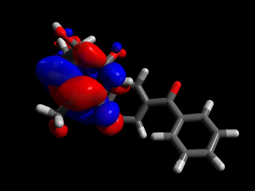
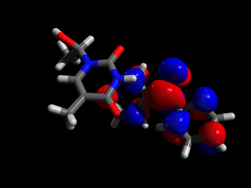
Modeling DNA Under External Stress: Photosensitization and
Oxidation
Antonio Monaria
a) Université de Lorraine and CNRS, Theory-Modeling-Simulation, SRMS, France
Cells and biological molecules are constantly exposed to the UV/vis radiations or reactive
oxygen species. This situation generates an important stress involving both complex
photochemical pathways and ground state reaction. The fine comprehension of these rather
complicated chemical mechanisms is necessary to rationalize phenomena related to aging and
to many diseases such as cancers.
The effects of the UV/vis radiation can be expanded by photosensitization, i.e. by the
interaction of biological macromolecules with organic or organometallic chromophores that
absorb light at relatively long wavelengths. Subsequently, the excited chromophore can
induce electron- or energy-transfer to the macromolecule, leading to its degradattion, or favor
the production of free radical and triplet oxygen.
In this talk we will analyze the interaction of different sensitizers with DNA also comprising
artificial nucleobases; multiscale molecular modeling will give us a better understanding of
the DNA/photosensitizers aggregates properties and structure. Hybrid QM/MM methods will
provide a detailed description of the modification induced by the environment on the
photophysical and photochemical properties of different chromophores, and will give access
to the energetic profiles related to the lesions’ induction. We will consider both the structural
and dynamical effects, in particular concerning the characterization of the sensitizer/DNA
aggregate, and the evolution of the excited states landscapes leading to sensitization. Energy-
and electron-transfer phenomena will be particularly considered together with the tuning of
the complex environment.
Moreover we will illustrate how modeling can enlighten the mechanism behind the oxidation
of guanine nucleobases in presence of singlet oxygen, and in particular explaining the
experimental observed high selectivity.
1T
A) Double inserted mode
5
1.34 Å
4 3BP
Energy (eV)
ξ=0 τ=30.1°
3
0.09 eV 0.74 eV 3T
DEDS
2
0 0.125 0.25 0.375 0.5 0.625 0.75 0.875 1
Interpolation coordinate
1BP
τ=38.5°
ξ=1
References
[1] Monari A. et al. Acc. Chem. Res. 2013 46, 596 1.49 Å
[2] Very T. et al. Chem. Eur. J. 2014 20, 12901 (2014) (1.34)
1.42 Å
[3] Dumont E., Monari A. J. Phys. Chem. Lett. 2013 4, 4119 (1.46)
[4] Dumont E. et al. J. Phys. Chem. Lett. 2015 6, 576 Natural transition orbitals for T1
[5] Bignon E. et al. Chem. Eur. J. 2015 in press
[6] Bignon E. et al. J. Am.Chem. Soc. 2015 submitted
7

                           CM1201
  WG2/WG4


 Two Shades of 5-Thiocyanto-2’-Deoxyuridine Toxicity Induced by
  Electrons. ESR, Photoelectron Spectroscopy and DFT Studies
        Janusz Rak,a Magdalena Zdrowowicz,a Lidia Chomicz,a Michał Żyndul,a Paweł Wityk,a
       Franciszek Kasprzykowski,a Tyler J. Wiegand, Cameron G. Hanson,b Amitava Adhikary,b
               Michael D. Sevilla,b Angela Buonaugurio,c Yi Wang,c and Kit H. Bowenc
       a) Faculty of Chemistry, University of Gdańsk, Wita Stwosza 63, 80-308 Gdańsk, Poland; b) Department of
        Chemistry, Oakland University, Rochester, MI 48309, USA; c) Department of Chemistry, Johns Hopkins
                         University, Baltimore, MD 21218, USA; e-mail: janusz.rak@ug.edu.pl

Incorporated into genomic DNA, 5-substituted uracils could be employed in human cancer
radiotherapy if they could be sensitized to dissociate upon reaction with electrons in water.
We demonstrated that for a uracil analogue to be an efficient electron acceptor the uracil
substituent had to possess significant electron-withdrawing power. On the other hand, in order
to assure effective dissociation of the anion, the chemical bond holding together the
substituent and uracil residue should be relatively weak. DFT modeling along with negative
ion photoelectron spectroscopy enabled 5-thiocyanatouracil, a derivative that has not been
tested so far, to be selected out of a number of uracil derivatives as a new, potential
radiosensitizer.1 ESR spectra in γ-irradiated nitrogen-saturated frozen aqueous solutions of 5-
thiocyanato-2′-deoxyuridine (SCNdU) showed that electron-induced S-CN bond cleavage
occurred to form a thiyl radical.2 Furthermore, HPLC and LC-MS/MS studies of γ-irradiated
N2-saturated aqueous solutions of SCNdU in presence of an OH-radical scavenger at ambient
temperature showed formation of the dU-5S-5S-dU dimer in preference to 2’-deoxyuridine by
about 10 to 1 ratio.2 These together with DFT calculations, suggesting the dU-5-S• and CN¯
formation is thermodynamically favored by over 15 kcal/mol (∆G) to dU• and SCN¯
production, show both possible routes of electron-induced bond cleavage to be operative.
Thus, our studies establish SCNdU as a potential radiosensitizer that could cause intra- and
interstrand crosslinking as well as DNA-protein crosslinking via S-S dimer formation.

Acknowledgements. This work was supported by the Polish National Science Centre (NCN),
Grant No. 2012/07/N/ST5/01877 (MZ), the National Institutes of Health, Grant No.
RO1CA045424 (MDS), and the National Science Foundation, Grant No. CHE-1111693
(KHB).

       References

[1] Chomicz, L.; Zdrowowicz, M.; Kasprzykowski, F.; Rak, J.; Buonaugurio, A.; Wang, Y. Bowen, K. H. J.
    Phys. Chem. Lett. 2013, 4, 2853.
[2] Zdrowowicz, M.; Chomicz, L.; Żyndul, M.; Wityk, P.; Rak, J.; Wiegand, T. J.; Hanson, C. G.; Adhikary, A.;
    Sevilla, M. D. Phys. Chem. Chem. Phys. 2015, accepted.


                                                                                                               8

                                    CM1201
  WG2/WG4


     Hydrogen atom transfer from cyclohexanes and decalins to
  alkoxyl radicals. The role of structural effects on the equatorial vs
                      axial C−H bond reactivity
                               Massimo Bietti, Michela Salamone and Vanesa B. Ortega
                        Dipartimento
  di
  Scienze
  e
  Tecnologie
  Chimiche,
  Università
  "Tor
  Vergata",

                          Via della Ricerca Scientifica, 1 00133 Roma; e-mail: bietti@uniroma2.it

Hydrogen atom transfer (HAT) reactions play a key role in a variety of important chemical
and biological processes such as lipid peroxidation, the oxidative damage to biomolecules and
polymers, the antioxidant activity of natural and synthetic radical scavenging antioxidants, the
degradation of volatile organic compounds, as well as in an increasing number of
synthetically useful C−H functionalization procedures. Among the radicals involved in these
processes, alkoxyl radicals have received considerable attention, and cumyloxyl
(PhC(CH3)2O , CumO ) has emerged as a very convenient radical for the study of these
                    •               •

reactions. CumO can be easily generated by photolysis of commercially available dicumyl
                           •

peroxide and is characterized by an absorption band in the visible region of the spectrum and
a lifetime that allow the direct measurement of rate constants for HAT from a large variety of
substrates by means of the laser flash photolysis technique.

In this framework, we have recently carried out a detailed time-resolved kinetic studies on
HAT from the C−H bonds of cycloalkanes to CumO .1 The role of structural effects on the
                                                                                     •

reactivity and selectivity patterns observed in these reactions will be discussed, emphasizing
in particular the role played by release of 1,3-diaxial strain and by torsional strain on the HAT
reactivity of tertiary axial and equatorial C−H bonds of cyclohexanes and decalins.

       References

[1] Salamone, M.; Ortega, V. B.; Bietti, M. J. Org. Chem. 2015, 80, 4710.


                                                                                                                                  9
                               CM1201
  WG2/WG4


              Cyclodextrins and Radical Chemistry: a Successful Match
                    Ángeles Martín, Dimitri Álvarez-Dorta, Elisa I. León, Inés Pérez-Martín
                                             and Ernesto Suárez
             Instituto de Productos Naturales y Agrobiología del CSIC, Avenida Astrofísico Francisco Sánchez 3,
                            38206 La Laguna, Tenerife, Spain; e-mail: angelesmartin@ipna.csic.es

       Nowadays there is a great interest to design new drug carrier systems for their applications in
       medical research for the treatment of a wide variety of diseases. In this sense, cyclodextrins
       (CDs) are considered potentially nanocarriers because of their ability to encapsulate
       biomolecules in their internal cavity.1 Thus, an important effort to modify and improve their
       chemophysical properties have been made. However, selective modifications in these
       macrostructures are not easy to carry out due to the torus shape and the large number of
       hydroxyl groups.2

       Based on our previous studies related with the intramolecular 1,8-hydrogen atom transfer
       (1,8-HAT) reactions in Hexp-(1→4)-Hexp disaccharides systems (e.g., -maltose),3 we
       wondered whether this radical protocol might be suitably deployed in more complex
       carbohydrate systems such as CDs where the glucose units are linked in similar fashion.

       The extension and scope of this radical methodology not only to monoalcohols but also to
       diols and peralcohols derived from CDs will be discussed in this lecture.4

          References

       [1] a) Todres, Z. V. in Organic Chemistry in Confining Media, Springer, Switzerland, 2013. b) Dodziuk, H.
           Cyclodextrins and Their Complexes. Chemistry, Analytical Methods, Applications; Wiley-VCH: Weinheim,
           2006.
       [2] Guieu, S.; Sollogoub, M. Advances in Cyclodextrins Chemistry; Werz, D. B., Vidal, S. Eds.; Modern
           Synthetic Methods in Carbohydrate Chemistry: From Monosaccharides to Complex Glycoconjugates; Wiley-
           VCH, Weinheim, 2014.
       [3] Francisco, C. G.; Herrera, A. J.; Kennedy, A. R.; Martín, A.; Melián, D.; Pérez-Martín, I.; Quintanal, L. M.;
           Suárez, E. Chem. Eur. J. 2008, 14, 10369−10381.
       [4] Alvarez Dorta, D.; León, E. I.; Kennedy, A. R.; Martín, A.; Pérez-Martín, I.; Suárez, E. Angew. Chem. Int.
           Ed. 2015, 54, 3674−3678.


                                                                                                                         10
                           CM1201
  WG2/WG4


          Myeloperoxidase In Chronic Lymphocytic Leukemia and
                            Multiple Myeloma
       E.İlker Saygili,a Nur Aksoyb Mustafa Pehlivanc., Tugce Severd., Mehmet Yilmazc., Iclal
                               Geyikli Cimencib and Sacide Pehlivand
        a) Vocational School of Higher Education for Health Services, b) Department of Biochemistry, c)
         Department of Hematology, d) Department of Medical Biology, Faculty of Medicine, Gaziantep
                           University, Gaziantep, Turkey e-mail: isaygili@sanko.edu.tr

                                         Assoc.Prof.Dr.E.İlker SAYGILI

                     University of SANKO, School of Medicine, Department of Biochemistry.

The aim of this study was to investigate how myeloperoxidase (MPO) G-463A gene
polymorphism and enzyme levels varied among patients with chronic lymphocytic leukemia
(CLL) and multiple myeloma (MM) and to find the relationship between the MPO gene,
enzyme levels, and clinical parameters. We studied the sera from 40 healthy volunteers,
patients with CLL (n,34) and MM (n,28). In subjects with homozygote GG genotype, MPO
levels were higher in the patients with both CLL and MM than in the control group. This
difference was statistically significant in patients with CLL. In conclusion, homozygote GG
genotype is found to be associated with an increasing amount of serum MPO. In accordance
with the results of the study, we assess that the increase in the MPO enzyme level in the
patient groups with CLL and MM generated bactericidal effects as well as the increased
formation of ROP, thus setting off a pro-cell death pathway and playing a role on the
pathogenesis of lymphoproliferative malignancies through this mechanism.1 HOCl, which is
formed by MPO in the presence of H2O2 not only causes physiological bactericidal effects in
neutrophiles but also causes formation of chlorohydrin and lysophospholipid by influencing
lipids.2 It was previously stated that formation of lysophospholipid may alter membrane
function and result in cell destruction.3 HOCl might form 5 chlorourasil by influencing DNA.
It was reported that 5 chlorourasil formation may be a marker of DNA damage.4 Harmful
effect of HOCl in target cell membrane is conducted by attacking membrane –SH or –NH2
groups and membrane denaturation occurs.

Keywords: Multiple myeloma, leukemia, myeloperoxidase, gene polymorphism
       References

[1] Saygili E.I.; Aksoy N.; Pehlivan M.; Sever T.; Yilmaz M.; Cimenci IG.; Pehlivan S. Enzyme Levels and G-
    463A polymorphism of myeloperoxidase in chronic lymphocytic leukemia and multiple myeloma. Leukemia
    & Lymphoma, 2009, 50 (12), 2030-2037.
[2]. Malle E, Marsche G, Arnhold J, Davies MJ. Modification of low-density lipoprotein by myeloperoxidase-
  derived oxidants and reagent hypochlorous acid. Biochim Biophys Acta 2006;1761:392–415.
[3]. Thukkani AK, Martinson BD, Albert CJ, Vogler GA, Ford DA. Neutrophil-mediated accumulation of 2-
  ClHDA during myocardial infarction: 2-ClHDA-mediated myocardial injury. Am J Physiol Heart Circ Physiol
  2005; 288:2955–2964.
[4]. Malle E, Furtmuller PG, Sattler W, Obinger C. Myeloperoxidase: a target for new drug development? Br J
  Pharmacol 2007;152(6):838-854.


                                                                                                         11
CM1201
WG2/WG4
a)
b)
Figure 1: Agarose gel electrophoresis of MPO DNA fragments stained with ethidium bromide (fragment lengths
are given in bp. M: DNA size standart, ND: Non-digest PCR product. a) It’s given CLL patients sample;
1,2,4,6:GG, 3,5:GA, 7:AA. b) It’s given MM patients sample; 1-3,6,7:GG, 4,5:GA, 8:AA.
Comparison of G-463A polymorphism of the MPO gene between patients with chronic lymphocytic leukemia,
multiple myeloma and control subjects.
Table
1:
MPO
-‐
463
MM
CLL
Healthy
Controls
OR*
%95
CI*
p
Genotypes
n=28
(%)
n=34
(%)
n=40
(%)
GG
18
(64)
11
(32)
26
(65)
0.258a
0.098-‐0.678
a
0.005
a
0.969
b
0.353-‐2.661
b
0.952
b
GA
9
(32)
23
(68)
12
(30)
0.221a*
0.082-‐0.595
a*
0.003
a*
0.894b*
0.304-‐2.635
b*
0.840
b*
AA
1
(4)
-‐
(0)
2
(5)
0.950a
0.885-‐1.020
a
0.186
a
0.937b*
0.071-‐12.348
b*
0.961
b*
Allele
G
45
(80)
45
(67)
64
(80)
0.489a
0.233-‐1.029
a
0.057
a
A
11
(20)
23
(33)
16
(20)
1.023
b
0.434-‐2.410
b
0.959
b
MPO
levels
150
(110-‐240)
191.5
(120-‐256)
128
(100-‐192)
0.002
a,
&,
0.030
b,
&
&,
median
test;
*, OR (95% CI) was adjusted by age and sex; a,
comparison
of
genotypes
frequencies
between
chronic
lymphocytic
leukemia
and
healthy
control
groups;
b,
comparison
of
genotypes
frequencies
between
multiple
myeloma
and
healthy
control
groups;
CLL,
chronic
lymphocytic
leukemia;
MM,
multiple
myeloma
Table
2:
Association
between
polymorphisms
of
the
MPO
gene
and
MPO
levels
MPO
-‐
463
MM
CLL
Healthy
Controls
p&
Genotip
na
MPO*
nb
MPO*
nc
MPO*
GG
18
175
(125-‐240)
11
210
(120-‐256)
26
123
(100-‐192)
0.028
bd
GA
9
132
(110-‐195)
23
180
(120-‐253)
12
129
(115-‐190)
0.081
cd
AA
1
123
(123-‐123)
-‐
-‐
2
146
(132-‐160)
0.236
ad
a,
n=28;
b,
n=34;
c,n=40;
*,
median
ng/mL;
p&,
median
test;
d,
MPO
enzyme
levels
compare
to
between
GG
genotype
and
GA
genotype;
CLL,
chronic
lymphocytic
leukemia;
MM,
multiple
myeloma;
MPO,
myeloperoxidase
12
                          CM1201
  WG2/WG4


        Free Radical Damage to DNA: Mechanisms and Measurement
                                                Miral Dizdaroglu
       National Institute of Standards and Technology, 100 Bureau Drive, MS8311, Gaithersburg, Maryland 20899,
                                              USA; e-mail: miral@nist.gov

Endogenous and exogenous sources cause free radical-induced DNA damage in living
organisms by a variety of mechanisms. The highly reactive hydroxyl radical reacts with the
heterocyclic DNA bases and the sugar moiety near or at diffusion-controlled rates. Hydrated
electron and H atom also add to the heterocyclic bases. These reactions lead to adduct
radicals, further reactions of which yield numerous products. These include DNA base and
sugar products, single- and double-strand strand breaks, 8,5'-cyclopurine-2'-
deoxynucleosides, tandem lesions, clustered sites and DNA-protein cross-links. Reaction
conditions and the presence or absence of oxygen profoundly affect the types and yields of
the products. For thorough understanding of mechanisms, cellular repair and biological
consequences of DNA damage, accurate measurement of resulting products must be achieved.
There is mounting evidence for an important role of free radical-induced DNA damage in the
etiology of numerous diseases including cancer. Further elucidation of mechanisms of free
radical-induced DNA damage, and cellular repair and biological consequences of DNA
damage products will be of outmost importance for disease prevention and treatment.


                                                                                                          13
                             CM1201
  WG2/WG4


        Mechanisms of response to ionizing radiation from bacteria to
                       humans: A holistic approach
       Alexandros G. Georgakilasa, Zacharenia Nikitakia, Athanasia Pavlopouloub, Maria Loukac,
                Pantelis G. Bagosb, Ioannis Michalopoulosd, Constantinos E. Vorgiasc
a) Physics Department, School of Applied Mathematical and Physical Sciences, National Technical University
  of Athens (NTUA), Zografou 15780, Athens, Greece b) Department of Computer Science and Biomedical
   Informatics, University of Thessaly, Lamia 35100, Greece c) Department of Biochemistry and Molecular
Biology, National and Kapodistrian University of Athens, Zografou Campus, 15701 Athens, Greece d) Centre
of Systems Biology, Biomedical Research Foundation, Academy of Athens, 4 Soranou Efesiou, Athens11527,
                                      Greece. e-mail: alexg@mail.ntua.gr

Exposure to ionizing radiation (IR) as a genuine exogenous stress induces a variety of
responses in the cell initiated by the DNA damage response (DDR) and DNA repair,
apoptosis and inflammatory or immune response.1 Therefore, stimulation of this IR-
response (IRR) mega system especially at the organism level consists of several subsystems
and submechanisms and exerts a variety of targeted and non-targeted effects.2 In addition,
comparing certain aspects of these mechanisms in various organisms from bacteria to
humans brings up similarities and major differences. Based on the above, we believe that in
order to better understand this complicated response system one should follow a ‘holistic’
approach including all possible mechanisms and at all organism levels. The suggested task
is considered of high difficulty. In this presentation, we will first present experimental
evidence on how the mammalian cell or organism is expected to respond to complex DNA
damage induction i.e. the signature of IR and primary ‘danger signal’ and attempt its repair.
At second, we will discuss the extremities of this response i.e. the phenomena of
radiosensitivity and radioresistance in bacteria and human cells and insights gained by
applying bioinformatics. Last but not least and in the light of our recent work,3 we will
present novel suggestions for protein biomarkers involved in DDR/DNA repair and
inflammatory/immune response creating a protein network underlining the expected
crosstalk between these phenomenically distinct cellular pathways.

        References

[1] Nikitaki, Z.; Hellweg, C.; Georgakilas, A. G.; Ravanat, J. L. Front. Chem. 2015, 3, 35.
[2] Hatzi, V. I.; Laskaratou, D. A.; Mavragani, I. V.; Nikitaki, Z.; Mangelis, A.; Panayiotidis, M. I.; Pantelias, G.
E.; Terzoudi, G. I.; Georgakilas, A. G. Cancer Lett 2015, 356, 34.
[3] Georgakilas, A. G.; Pavlopoulou, A.; Louka, M.; Nikitaki, Z.; Vorgias, C. E.; Bagos, P. G.; Michalopoulos, I.
Cancer Lett. 2015, In press, http://dx.doi.org/10.1016/j.canlet.2015.03.021.


                                                                                                               14
                          CM1201
  WG2/WG4


       Toward the Total Synthesis of Diketopiperazine Alkaloids Using
                        the Persistent Radical Effect
                                      Ullrich Jahna and Tynchtyk Amatova
       a) Institute of Organic Chemistry and Biochemistry, Academy of Sciences of the Czech Republic, Flemingovo
                           namesti 2, 16610 Prague 6, Czech Republic; e-mail: jahn@uochb.cas.cz

Diketopiperazine alkaloids are a diverse class of alkaloids with wide-ranging biological
activities.1 Although a number of strategies for their synthesis have been developed over the
years, many of them are limited in their applicability.2

We report here an efficient general approach to diverse structural motifs of bridged
diketopiperazines. The key to generate the required structural diversity are stable
diketopiperazine alkoxyamines, which are convenient precursors for thermal radical
cyclizations employing the persistent radical effect.3 Applications toward the total synthesis
of naturally occurring alkaloids and medicinally interesting scaffolds are outlined.

         References

[1] Review: Gonzalez, J. F.; Ortin, I.; de la Cuesta, E.; Menendez, J. C. Chem. Soc. Rev. 2012, 41, 6902-6915.
[2] Review: Miller, K. A.; Williams, R. M. Chem. Soc. Rev. 2009, 38, 3160-3174.
[3] Review: Studer, A. Chem. Soc. Rev. 2004, 33, 267-273.


                                                                                                            15
                           CM1201
  WG2/WG4


          Transient spectroscopic studies of enantiomerically-resolved
          intercalating photo-oxidising ruthenium dipyridophenazine
               (dppz) complexes bound to defined sequence DNA
       Páraic M. Keane,a Fergus E. Poynton,a James A. Hall,b Greg M. Greetham,c Ian P. Clark, c
        Igor V. Sazanovich,c Michael Towrie,c Christine J. Cardin,b Thorfinnur Gunnlaugsson,a
                         Susan J. Quinn d, Conor Long e and John M. Kelly a
                                       e-mail: jmkelly@tcd.ie
                             a) School of Chemistry, Trinity College Dublin, Dublin 2, Ireland;
                       b) Department of Chemistry, University of Reading, Reading RG6 6AD, UK;
       c) Central Laser Facility, Research Complex at Harwell, Science & Technology Facilities Council, Rutherford
                        Appleton Laboratory, Harwell Oxford, Didcot, Oxfordshire, UK OX11 0QX;
               d) School of Chemistry and Chemical Biology, University College Dublin, Dublin 4, Ireland;
                              e) School of Chemistry, Dublin City University, Dublin 9, Ireland

1,4,5,8-tetraphenanthrene (TAP) such as [Ru(TAP)2(dppz)]2+ (dppz = dipyrido[3,2-a:2’,3’-c]
-phenazine) are known to sensitise the photo-oxidation of DNA. Like its 1,10-
phenanthroline analogue [Ru(TAP)2(dppz)]2+ intercalates into DNA, as is confirmed by our
recent high resolution X-Ray crystal structures.[1] Using the same defined sequence nucleic
acids as used for the crystal studies, we have carried out complementary time-resolved mid-
infra-red (TRIR) and visible spectroscopic measurements which provide new insights into the
nature and the reactivity of the excited states and their interactions at particular binding
sites.[2] The subsequent reactions of the reduced photosensitiser and the one-electron oxidised
guanine are readily monitored.

Acknowledgements. This work has been partially funded by the BBSRC (Grant No. BB/K
019279/1) and the Royal Irish Academy/Royal Society. Access to the CLF Ultrafast
laboratory was funded through EU FP7 (Appl. No 12240002) and Appl. No. 13230023.

    References
 [1]. (a) Hall, J. P et al. Proc. Natl Acad. Sci., 2011, 108, 17610-17614; (b) Niyazi, H et al. Nature
Chemistry, 2012, 3, 621-628; (c) Hall, J. P et al. J.Am.Chem.Soc., 2013, 135, 12652-12659; (d) Hall, J. P et
al. J.Am.Chem.Soc., 2014, 136, 17505−17512
[2]. (a) Elias, B. et al. Chemistry – Eur. J., 2008, 14, 369-375; (b) Keane, P.M et al. J. Phys. Chem. Lett,
2015, 6, 734-738.           (c) Keane, P.M et al. Angew. Chem. Int. Ed., 2015,                      54(29),
DOI:10.1002/anie.201502608


                                                                                                              16
                           CM1201
  WG2/WG4


         Click nucleic acid ligation: Chemistry and applications
                                               Tom Brown
              Department of Chemistry University of Oxford. email: tom.brown@chem.ox.ac.uk

Click ligation utilizes the copper-catalyzed azide-alkyne cycloaddition (CuAAC reaction). It
is an efficient method of joining together DNA and RNA strands and has been used for the
synthesis of cyclic oligonucleotides,1-3 oligonucleotide catenanes,2 very stable cyclic mini-
duplexes, 1 duplexes that are linked across the major groove,4 covalently fixed DNA
nanoconstructs5 and large RNA constructs.6 The method produces an unnatural DNA
backbone linkage that can be varied by changing the structures of the participating alkyne and
azide.7 Careful design produced a biocompatible DNA backbone (Figure 1) that can be read
through by DNA8 and RNA polymerases.9 A high-resolution NMR study revealed that the
linkage in Figure 1B is accommodated in a B-DNA helix with minor distortion.10 This
methodology has recently been used to characterise a new form of stretched DNA.11 Copper-
free click DNA strand ligation and crosslinking can also be carried out if strained cyclooctyne
analogues are used (Figure 2).12 This method has the advantage of being potentially valuable
for in vivo applications as it does not require metal ion catalysis. Recent developments in this
field will be discussed.


Figure 1. First generation triazole DNA (A), biocompatible linkage (B) and normal DNA (C).
Figure 2. (A) The ring strain promoted alkyne-azide cycloaddition reaction (SPAAC reaction) for click DNA
ligation between azide and cyclooctyne-labeled oligonucleotides and (B) Chemical structure of DIBO triazole at
the ligation point.

References

[1] A. H. El-Sagheer, R. Kumar, S. Findlow, J. M. Werner, A. N. Lane and T. Brown, Chembiochem 2008, 9,
50-52.
[2] R. Kumar, A. H. El-Sagheer, J. Tumpane, P. Lincoln, L. M. Wilhelmsson and T. Brown, J. Am. Chem. Soc.
2007, 129, 6859-6864.
[3] A. H. El-Sagheer and T. Brown, Int. J. Peptide Res.Therapeut. 2008, 14, 367-372.
[4] P. Kocalka, A. H. El-Sagheer and T. Brown, Chembiochem 2008, 9, 1280-1285.
[5] E. P. Lundberg, A. H. El-Sagheer, P. Kocalka, L. M. Wilhelmsson, T. Brown and B. Norden, Chem.
Commun. 2010, 46, 3714-3716.
[6] A. H. El-Sagheer and T. Brown, Proc. Natl. Acad. Sci. U. S. A. 2010, 107, 15329-15334.
[7] A. H. El-Sagheer and T. Brown, J. Am. Chem. Soc. 2009, 131, 3958-3964.
[8] A. H. El-Sagheer, A. P. Sanzone, R. Gao, A. Tavassoli and T. Brown, Proc. Natl. Acad. Sci. U. S. A. 2011,
108, 11338–11343.
[9] A. H. El-Sagheer and T. Brown, Chem. Commun. 2011, 47, 12057-12058.
[10] A. Dallmann, A. H. El-Sagheer, L. Dehmel, C. Mügge, C. Griesinger, N. P. Ernsting and T. Brown,
Chemistry - A European Journal 2011, 17, 14714-14717.
[11] N. Bosaeus, A. H. El-Sagheer, T. Brown, S. B. Smith, B. Akerman, C. Bustamante and B. Norden, Proc.
Natl. Acad. Sci. U. S. A. 2012, 109, 15179-15184.
[12] M. Shelbourne, X. Chen, T. Brown and A. H. El-Sagheer, Chem. Commun. 2011, 47, 6257-6259.


                                                                                                        17
                           CM1201
  WG2/WG4


                    Understanding antioxidant properties of natural
                     compounds (polyphenols) at an atomistic-scale
Patrick Trouillasa,b, Gabin Fabre, Michal Biler, Tahani Ossman, and Benjamin Chantemargue,
       a) 1 INSERM-S850, School of Pharmacy, Université de Limoges, Limoges, France b) Regional Centre of
             Advanced Technologies and Materials, Palacký University, Olomouc, Czech Republic; e-mail:
                                           patrick.trouillas@unilim.fr

Quantum calculations (mainly DFT) and molecular dynamics simulations are increasingly
effective tools to evaluate the physical chemical properties of antioxidants.

Free Radical Scavenging Capacity.1 Thermodynamic parameters (mainly O-H phenolic bond
dissociation enthalpies, BDE) allowed an accurate prediction of the antioxidant capacity of
natural polyphenols. Based on the Transition State and the Marcus theories (for atom- and
electron-transfers, respectively), kinetics was also evaluated providing a better prediction of
the antioxidant behavior in solution or in the organism. Further oxidative reactions following
the primary redox event were also studied for flavonoids and stilbenoids, which figure out
part of the pro-oxidant effects.

Interaction with Lipid Bilayer Membranes.2 Membrane penetration / accumulation / crossing /
positioning play a crucial role in antioxidant delivery, metabolism and action in the human
body. Over the past decade, in silico membrane models and MD simulations have appeared
much promising, complementary to experimental measurements, to predict antioxidant-
membrane interaction. Theoretical MD simulations have been performed to provide an
accurate picture of the intermolecular interaction between antioxidants and lipid bilayer
membranes, thus predicting location, orientation and partitioning.

We really aim at using advanced molecular modeling methods for an applicative purpose to
e.g., cosmetic industries. The predictive character of these methods allows building molecular
guidelines for a better and safer use of antioxidants.

       References

[1] a) Trouillas, P. et al. Food Chem, 2006, 97, 679; b) Kozlowski, D. et al. J Phys Chem A, 2007, 111, 1138; c)
    Kozlowski, D. et al. Radiat Res, 2007, 168, 243; d) Trouillas, P. et al. J Phys Chem A, 2008, 112, 1054; e)
    Anouar, E. et al. PCCP, 2009, 11, 7659; f) Calliste, C.A. et al. Food Chem., 2010, 118, 489; g) Anouar, E. et
    al. J.Phys.Chem. A 2009, 113, 13881; h) Košinová, P. et al. Int.J.Quant.Chem., 2011, 111, 1131; i) Velu, S.
    et al. J Nat Prod, 2013, 76(4), 538; j) Di Meo, F. et al. J Phys Chem A, 2013, 117, 2082; k) Košinová, P. et
    al. ChemPhysChem, 2011, 12(6), 1135; l) Zatloukalová, M. et al. Bioelectrochem, 2011, 82, 117; m) Gazák,
    R. et al. Tetrahedron Lett, 2013, 54, 315; n) Anouar, E. et al. J Comput Aided Mol Design, 2013, 27, 951; o)
    Vacek, J. et al. Chemico-Biological Interactions, 2013, 205, 173-180; p) Ponomarenko, J. et al.
    Phytochemistry, 2014, 103, 178; q) Bayach, I. et al. Chemistry: An Asian Journal, 2015, 10(1), 198-211.
[2] a) Košinová, P. et al. J Phys Chem B, 2012, 116, 1309; b) Poudloucka, P. et al. J Phys Chem B, 2013,
    117(17), 5043; c) Paloncýová, M. et al. JCTC, 2014, 10(9), 4143; d) Fabre, G. et al. Chemical
    Communications, 2015, 51, 7713.


                                                                                                           18
                           CM1201
  WG2/WG4


       Cell-based- and kinetic analyses of the modulation of the
        intrinsic activity of glucose transporter-4 by the non-
        metabolisable glucose analogue 3-O-methyl-D-glucose
                                            Shlomo Sasson

        Institute for Drug Research, Dept. of Pharmacology, The Hebrew University Faculty of Medicine,
                             Jerusalem, Israel; e-mail: shlomo.sasson@mail.huji.ac.il

Type-2 diabetes is a serious health problem affecting over 200 million people worldwide. The
prevalence of the disease is increasing, particularly among youth and young adults, in parallel
with the continuing rise in obesity and is expected to affect 300 million people by year of
2020. The cost of treating diabetes complications imposes a tremendous burden on healthcare
resources, and there has been limited success in achieving the treatment targets, which are
clearly associated with reduced risks of complications and mortality. Most Type-2 diabetic
patients that fail to normalize their blood glucose levels by a proper diet and adequate
physical activity are usually treated with different types of oral anti-hyperglycemic drugs.
These drugs act primarily on pancreatic beta cells to increase and/or potentiate insulin
secretion or to augment peripheral insulin sensitivity, primarily in skeletal muscles and the
liver. In many cases these drugs progressively become ineffective due to the deterioration of
beta cells function and mass and/or the development of severe peripheral insulin resistance.
The majority of these patients therefore resort to insulin treatment by injections, like in Type-
1 diabetes. We have recently discovered that the non-metabolisable glucose analogue, 3-O-
methy-D-glucose (MeGlc), increases the rate of glucose uptake in skeletal muscle cells by
augmenting the intrinsic activity of glucose transporter-4 (GLUT-4). Hitherto no other
carbohydrates that can allosterically augment the intrinsic activity of the transporter have been
reported. In the course of our study we have developed a simple kinetic analysis that provides
an effective platform for screening and discovering allosteric modulators of GLUT-4. This
method measures the impact of an allosteric modulator (e.g., MeGlc) on the competitive
inhibitory kinetics of indinavir, a GLUT-4 inhibitor, using hexose transport assays in cultured
myotubes. We believe that these findings and method of analysis can become useful for the
design, synthesis and screening of novel MeGlc derivatives that can allosterically increase the
intrinsic activity of GLUT-4, and further for the development a novel class of
antihyperglycaemic drugs.


                                                                                                       19
CM1201
WG2/WG4
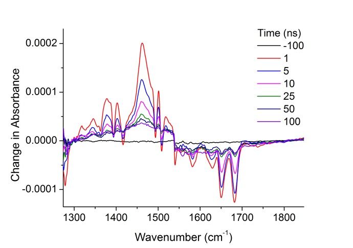
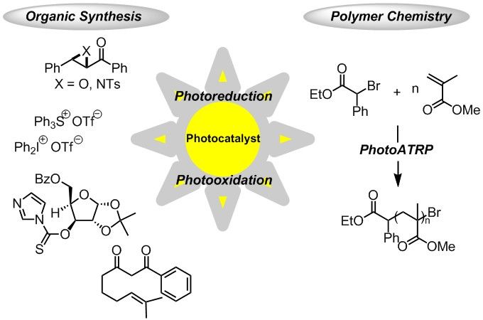
Recent Advances in Visible-Light Photoredox Catalysis
From Organic Synthesis to Polymer Chemistry
Cyril Ollivier
Institut Parisien de Chimie Moléculaire (UMR CNRS 8232), Sorbonne Universités UPMC Univ Paris 06,
4 Place Jussieu, C. 229, 75005 Paris, France; e-mail: cyril.ollivier@upmc.fr
Nowdays, visible-light photoredox catalysis has emerged as a valuable and efficient tool for
the generation of radicals by single electron transfer reactions from an appropriate
photocatalyst that absorbs light in the visible region in a greener way.1 Since the pioneering
studies of Kellogg and Deronzier, important contributions have been reported for synthetic
purposes. In this context, we investigated various radical transformations involving
photoreduction of ketoepoxides, ketoaziridines,2 onium salts3-4 and O-thiocarbamates5 and
photooxidation of 1,3-dicarbonyl compounds.6
The use of visible-light photoredox catalysis had a tremendous impact not only in organic
chemistry, but also in polymer chemistry. Quite recently, reactive systems exploiting the
redox properties of copper and iridium catalysts in the presence of light have been developed.
In this field, we report here the first gold-catalyzed photoATRP process of methacrylates and
acrylates.7
References
[1] For general reviews on photoredox catalysis in organic synthesis, see: (a) Narayanam, M. R.; Stephenson, C.
R. J. Chem. Soc. Rev. 2011, 40, 102. (b) Teplý, F. Collect. Czech. Chem. Commun. 2011, 76, 859. (c) Tucker,
J. W.; Stephenson, C. R. J. J. Org. Chem. 2012, 77, 1617. (d) Xuan, J.; Xiao, W.-J. Angew. Chem. Int. Ed.
2012, 51, 6828. (e) Prier, C.K.; Rankic, D. A.; MacMillan D. W. C. Chem. Rev. 2013, 113, 5322.
[2] Larraufie, M.-L.; Pellet, R.; Fensterbank, L.; Goddard, J.-P.; Lacôte, E.; Malacria, M.; Ollivier, C. Angew.
Chem. Int. Ed. 2011, 50, 4463.
[3] Donck, S.; Baroudi, A.; Fensterbank, L.; Goddard, J.-P.; Ollivier, C. Adv. Synth. Catal. 2013, 355, 1477.
[4] Baralle, A.; Fensterbank, L.; Goddard, J.-P.; Ollivier, C. Chem. Eur. J. 2013, 19, 10809.
[5] Chenneberg, L.; Baralle, A.; Daniel, M.; Fensterbank, L.; Goddard, J.-P.; Ollivier, C. Adv. Synth. Catal.
2014, 356, 2756.
[6] Daniel, M.; Fensterbank, L.; Goddard, J.-P.; Ollivier, C. Org. Chem. Front. 2014, 1, 551.
[7] Nzulu, F.; Telitel, S.; Stoffelbach, F.; Graff, B.; Morlet-Savary, F.; Lalevée, J.; Fensterbank, L.; Goddard, J.-
P.; Ollivier, C. Polym. Chem. 2015, DOI: 10.1039/C5PY00435G.
20
CM1201
WG2/WG4
Diastereomeric 5′,8-cyclo-2′-deoxypurines: brief overview of
synthetic strategies, modeling and in vitro biological activity
Annalisa Masia and Chryssostomos Chatgilialoglua, b
a) ISOF, Consiglio Nazionale delle Ricerche, Via P. Gobetti 101, 40129 Bologna, Italy; b) Institute of
Nanoscience and Nanotechnology, N.C.S.R. “Demokritos”, 15341 Agia Paraskevi, Athens, Greece;
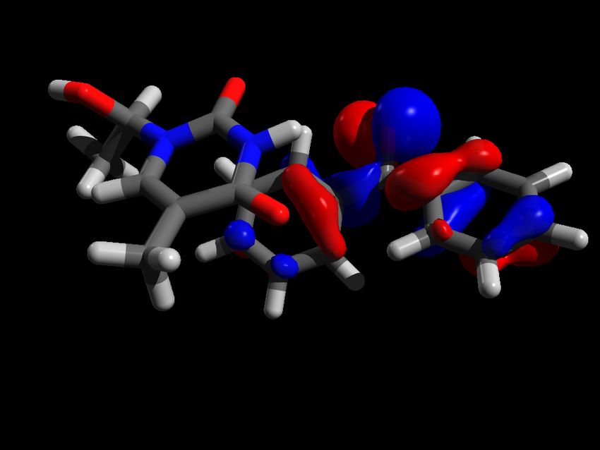
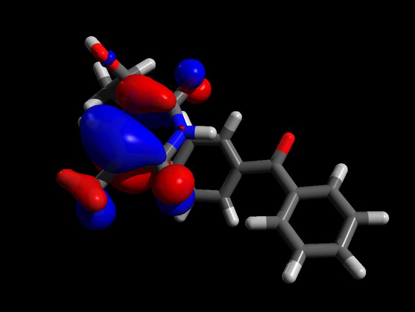
e-mail: annalisa.masi@isof.cnr.it
5′,8-cyclo-2′-deoxypurines (cdPus) are typical DNA lesions resulting from endogenous and
environmental free radical stress. The interest in these lesions is connected with the
mechanism of their formation due to the HO• attack at the H5′ atom of the 2-deoxyribose
moiety, followed by intramolecular cyclization between C5′-C8 bond and subsequent
oxidation of the resulting N7-radical.1,2 Two diastereomeric cdPus are formed in the 5′R and
5′S forms (Fig.1).
The two diastereomeric forms are repaired by nucleotide excision repair (NER) with different
efficiency, the 5′R isomer being 2 times more efficiently repaired than the 5′S isomer.
Molecular dynamics simulation elucidated that 5′R diastereoisomeric forms cause greater
DNA backbone distortions than the 5′S diastereomers, thus theoretically supporting a different
efficiency of NER3 mechanism. We recently discovered that DNA polymerase β (pol β) has
different behavior with 5′R-cdA lesion (efficiently bypassed) than 5′S-cdA (inefficiently
bypassed) during DNA replication and base excision repair (BER),4,5 highlighting that the
nature of the DNA lesion can play a crucial role in biological processes.
The diastereoisomeric 5′S- and 5′R-cdPus lesions are discussed in terms of differences in:
i. Synthetic strategy and automated synthesis efficiency.
ii. Physical-chemical properties (MD simulations, NMR, Melting Temperature)
iii. Biological Activity in vitro
References
[1] Chatgilialoglu, C.;Ferreri, C.; Terzidis, M.A. Chem.Soc.Rev. 2011, 40, 1153.
[2] Boussicault, F.; Kaloudis, P.; Caminal, C.; Mulazzani, Q. G.; Chatgilialoglu C. J. Am Chem. Soc. 2008, 130,
8377.
[3] Kropachev, K.; Ding, S.; Terzidis, M.A.; Masi, A.; Liu, Z.; Cai, Y.; Kolbanovskiy, M.; Chatgilialoglu, C.;
Broyde, S.; Nicholas E. Geacintov, N.E.; Shafirovich, V. Nucleic Acids Research, 2014, 42, 5020.
[4] Xu, M.; Lai, Y.; Jiang, Z.; Terzidis, M.A.; Masi, A.; Chatgilialoglu, C.; Liu, Y. Nucleic Acids Research,
2014. 42,13749
[5] Jiang, Z.; Xu, M.; Lai, Y.; Laverde, E.E.; Terzidis, M.A.; Masi, A.; Chatgilialoglu, C.; Liu, Y. DNA Repair,
2015, 33, 24.
21
You can also read